Embed Size (px)
Citation preview
Trematolobelia: Seed Dispersal; Anatomy of Fruit and Seeds
SHERWIN CARLQUIST1
THE ENDEMIC HAWAIIAN GENUS Trematolobelia (Lobeliaceae, or Campanulaceae, subfamilyLobelioideae) was erected on the basis of itsdistinctive fruit. This fruit has a seed-dispersalmechanism unique in the family. Assertionshave been made by some workers that holes inthe fruit wall are the work of insects, and are notrelated to the dispersal mechanism. This contention has been adequately disproved by otherinvestigators, but, in fact, the precise nature ofthe dispersal mechanism and the anatomicalstructure responsible for its action have neverbeen adequately described. In addition, the present study reveals the potential taxonomic useof capsular anatomy, a feature of importancebecause various authors recognize one, two, orthree species in the genus. These species arebased largely on floral features or foliar characteristics, and not on those of the fruit. Unusuallygood material of Trematolohelia collected bythe writer during the summer of 1958 providesa sufficient basis for presenting the featuresmentioned above in some detail.
MATERIALS AND METHODS
The following specimens were utilized forthis study: Trematolobelia macrostachys var.macrostachys Zahlbruckner: Carlquist 563 (PuuKukui, Maui); Carlquist 612 (Huumulu Rd.,Hawaii); Flavious Peter April 21, 1958 (Molokai). T. macrostachys ~ar. kauaiensis Rock:Carlquist 508 (Pihea, Kauai). T. macrostach'ysvar. grandifolia Rock: Carlquist 612A (Cultivated at Volcano, Hawaii; plant brought fromKehena Ditch Trail, Kohala Mts., Hawaii, byMrs. Ella Stephens).
All of these specimens were collected 10
the field; portions of each were preserved 10
1 Claremont Graduate School, Rancho Santa AnaBotanic Garden, Claremont, California. Manusctipt received February 24, 1961.
formalin-propiono-alcohol (Johansen, 1940).Other portions were dried. Herbarium specimens of all of these collections are located inthe Rancho Santa Ana Botanic Garden Herbanum.
The flowers and fruits which were studied(see figure legends for specimens used) wereembedded in paraffin according to the tertiarybutyl alcohol technique of Johansen 0940:130-131), sectioned, and stained with a safraninfast green combination corresponding to Northen's modification of Foster's tannic acid-ferricchloride method (Johansen, 1940: 92-93).
ANATOMICAL DESCRIPTIONS
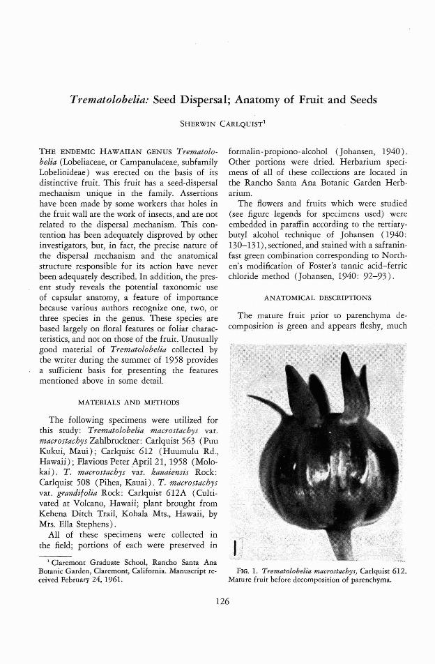
The mature fruit prior to parenchyma decomposition is green and appears fleshy, much
FIG. 1. Trematolobelia macrostachys, Carlquist 612.Mature fruit before decomposition of parenchyma.
126
Trematolobelia-CARLQUIST
'8 110:
127
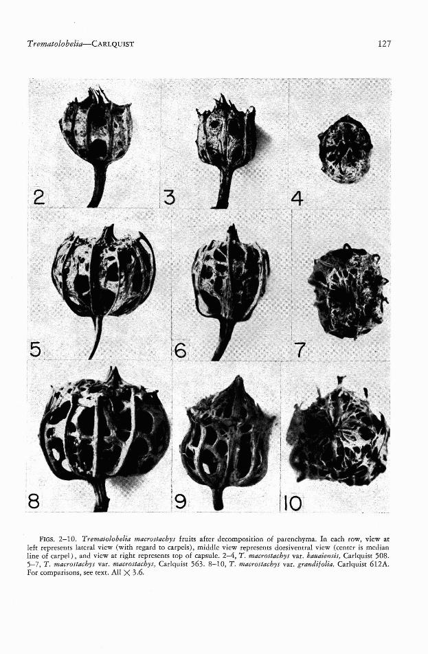
FIGS. 2-10. Trematolobelia macrostachys fruits after decomposition of parenchyma. In each row, view atleft represents lateral view (with regard to carpels), middle view represents dorsivenrral view (center is medianline of carpel), and view at right represents top of capsule. 2-4, T. macrostachys var. kauaiemis, Carlquist 508.5-7, T. macrostachys var. macrostachys, Carlquist 563. 8-10, T. macrostachys var. grandi/olia. Carlquist 612A.For comparisons, see text. All X 3.6.

128
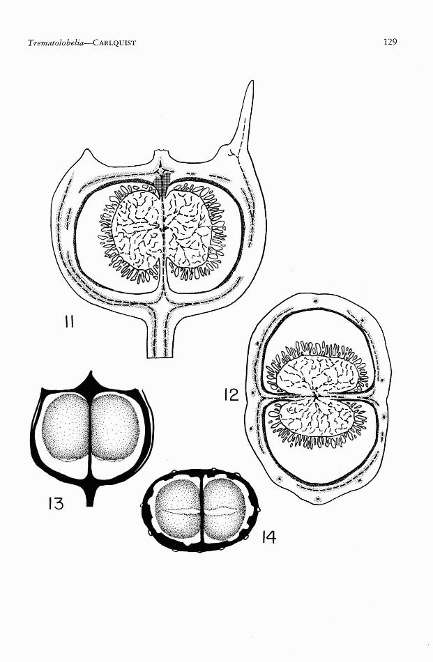
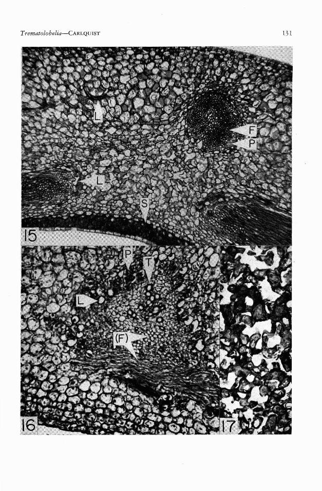
like the baccate fruits of the other endemic Hawaiian lobeliads, such as Cyanea and Clermontia(Fig. 1). Sections of this fruit (Figs. 11, 12, 15)reveal three distinctive tissues: ground-tissueparenchyma, endocarp sclerenchyma, and fibrousvascular bundles. The ground tissue of the fruitwall is composed of thin-walled parenchymacells which are large in diameter (decreasing insize toward interior and exterior of the fruitwall). These parenchyma cells are rich in chloroplasts. Although the inner epidermis of thefruit wall is thin-walled, there are two to fourlayers of thick-walled sclereids internal to theepidermis (Fig. 15). As shown in Figures 11and 12, this endocarp sclerenchyma is presentaround the inner surface of the carpels with theexception of the basal portion of the carpels andthe portion lying between the two placentas.The apical portion of the carpels is not coatedwith endocarp sclerenchyma, but possesses instead a spongy sclerenchyma (Fig. 11; Fig. 17)which connects endocarp sclerenchyma with thevascular bundles which form a pointed terminus, as seen in a dry fruit (e.g., Fig. 5, above) .The vascular tissue is composed of two sorts ofbundles: the 10 main bundles which extendupwardly into the calyx, corolla, and stamens ofthe flower, and the carpellary bundles, internalto the 10 main bundles. The carpellary bundlesform a dense mesh, in which large pores arepresent. The distinctions between the two sortsof bundles can be seen especially well in Figs.5-7, where they are adnate only to a limitedextent. The drawing in Fig. 11 shows the carpellary bundles united to the 10 vertical bundlesonly in the basal portion of the fruit. Thus, theyappear largely separate in Fig. 12. The 10 mainbundles represent the fusion of bundles fromthe three outermost whorls of the flower. Theyseparate into calyx, corolla, and stamen tracesonly at the top of the ovary (Fig. 11, upperright). The carpellary bundles form a network-
PAOFIC SCIENCE, Vol. XVI, January 1962
like system which encloses the carpels. Thisnetwork, seen in Figs. 2-10, is composed ofbundles which run in all directions, and are absent where pores are formed. These pores arepreformed in the fruit (e.g., the space betweenbundles at left and right, below, in Fig. 15, willbe such a pore). The carpellary bundles notonly form a network on the outside of the carpels, but between them (Fig. 12) as well. Thesecentral carpellary bundles supply the two placentas. In their upward extent, carpellary bundlessupply the style.
The vascular bundles, both inner carpellaryand outer calyx-corolla-stamen bundles, show apeculiar feature of construction. The prominence and persistence of these bundles, as seenfollowing the decomposition of the parenchymatous portion of the fruit, is due to abundanceof fibers present 'in these bundles. Only thebasal portion of calyx-traces and style-traces possess such fibers, and entire traces are thus absentin the dry fruit. Such thick-walled fibers may beseen in Fig. 15. One might suppose that thesefibers have the same origin as the fibers in mostbundles, that is to say, from a bundle-cap, orprotophloem region. This is, however, not thecase. Sections of the ovary wall from flowersat anthesis (Fig. 16) reveal clearly that thebundles are amphicribral in construction. Theperiphery of the bundle consists of phloem, inwhich many articulated non-anastomosing ladcifers are embedded. These laticifers, althoughpresent with particular prominence at the periphery of the bundles, also extend into the groundtissue of the ovary wall (Fig. 15, above left).This close association between laticifers andphloem is frequent in dicotyledons. The centerof .the bundle, as shown in Fig. 16, containsmature tracheary elements. Longitudinal sections of bundles reveal that these are mostlyvessel elements. Between the phloem and thetracheary elements is a zone which consists
FIGS. 11-14. Trematolobelia macrostachys var. macrostachys. 11, 12, sections of fruits from the collectionCarlquist 612. 11, Median longitudinal (sagittal) section of mature fruit before decomposition of parenchyma.12, Transverse section. Conventions as follows: broken lines=tracheary elements of vascular bundles; stippled= xylary fibers; cross-hatched = spongy sclerenchyma (see Fig. 17); spatter pattern = endocatp sclerenchyma;phloem, laticifers, parenchyma, and seeds are left white. 13, 14, Fruits, following decomposition of parenchyma,dissected to show the endocarp sclerenchyma sacs; sclerenchymatous vascular tissue black, endocarp sacs stippled; from the collection Carlquist 563. 13, Lateral view, showing sacs shrunken away from base, slits below.14, View from below, vascular tissue cut away; note slits in sacs, which permit escape of seeds. 11-12, X 5.4.13-14, X 4.
Trematolobelia-CARLQUIsT 129
130
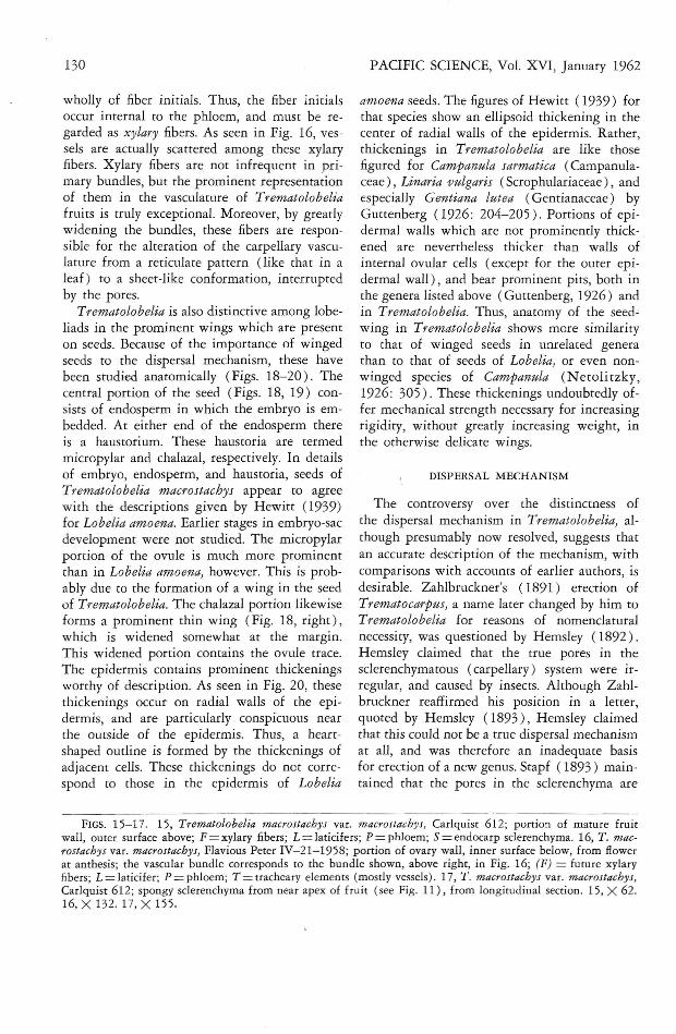
wholly ef fiber initials. Thus, the fiber initialsoccur internal to the phloem, and must be regarded as xylary fibers. As seen in Fig. 16, vessels are actually scattered among these xylaryfibers. Xylary fibers are not infrequent in primary bundles, but the prominent representationof them in the vasculature of Trematolobeliafruits is truly exceptional. Moreover, by greatlywidening the bundles, these fibers are responsible for the alteration of the carpellary vasculature from a reticulate pattern (like that in aleaf) to a sheet-like conformation, interruptedby the pores.
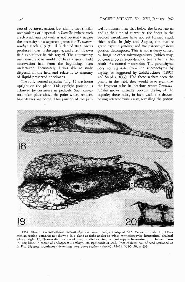
Trematolobelia is also distinctive among lobeliads in the prominent wings which are presenton seeds. Because of the importance of wingedseeds to the dispersal mechanism, these havebeen studied anatomically (Figs. 18-20). Thecentral portion of the seed (Figs. 18, 19) consists of endosperm in which the embryo is embedded. At either end of the endosperm thereis a haustorium. These haustoria are termedmicropylar and chalazal, respectively. In detailsof embryo, endosperm, and haustoria, seeds ofTrematolobelia macrostachys appear to agreewith the descriptions given by Hewitt (19'39)for Lobelia amoena. Earlier stages in embryo-sacdevelopment were not studied. The micropylarportion of the ovule is much more prominentthan in Lobelia amoena, however. This is probably due to the formation of a wing in the seedof Trematolobelia. The chalazal portion likewiseforms a prominent thin wing (Fig. 18, right),which is widened somewhat at the margin.This widened portion contains the ovule trace.The epidermis contains prominent thickeningsworthy of description. As seen in Fig. 20, thesethickenings occur on radial walls of the epidermis, and are particularly conspicuous nearthe outside of the epidermis. Thus, a heartshaped outline is formed by the thickenings ofadjacent cells. These thickenings do not correspond to those in the epidermis of Lobelia
PACIFIC SCIENCE, Vol. XVI, January 1962
amoena seeds. The figures of Hewitt (1939) forthat species show an ellipsoid thickening in thecenter of radial walls of the epidermis. Rather,thickenings in Trematolobelia are like thosefigured for Campanula sarmatica (Campanulaceae), Linaria vulgaris (Scrophulariaceae), andespecially Gentiana lutea (Gentianaceae) byGuttenberg (1926: 204-205). Portions of epidermal walls which are not prominently thickened are nevertheless thicker than walls ofinternal ovular cells (except for the outer epidermal wall), and bear prominent pits, both inthe genera listed above (Guttenberg, 1926) andin Trematolobelia. Thus, anatomy of the seed"wing in Trematolobelia shows more similarityto that of winged seeds in unrelated generathan to that of seeds of Lobelia, or even nonwinged species of Campanula (Netolitzky,1926: 305). These thickenings undoubtedly offer mechanical strength necessary for increasingrigidity, without greatly increasing weight, inthe otherwise delicate wings.
DISPERSAL MECHANISM
The controversy over the distinctness ofthe dispersal mechanism in Trematolobelia, although presumably now resolved, suggests thatan accurate description of the mechanism, withcomparisons with accounts of earlier authors, isdesirable. Zahlbruckner's (1891) erection ofTrematocarpus, a name later changed by him toTrematolobelia for reasons of nomenclaturalnecessity, was questioned by Hemsley (1892).Hemsley claimed that the true pores in thesclerenchymatous (carpellary) system were irregular, and caused by insects. Although Zahlbruckner reaffirmed his position in a letter,quoted by Hemsley (1893), Hemsley claimedthat this could not be a true dispersal mechanismat all, and was therefore an inadequate basisfor erection of a new genus. Stapf (1893) maintained that the pores in the sclerenchyma are
FIGS. 15-17. 15, Trematolobelia macrostachys var. macrostachys, Carlquist 612; portion of mature fruitwall, outer surface above; F=xylary fibers; L=laticifers; P=phloem; S=endocarp sclerenchyma. 16, T. macrostachys var. macrostachys, Flavious Peter IV-21-1958; portion of ovary wall, inner surface below, from flowerat anthesis; the vascular bundle corresponds to the bundle shown, above right, in Fig. 16; (F) = future xylaryfibers; L=laticifer; P=phloem; T=tracheary elements (mostly vessels). 17, T. macrostachys var. macrostachys,Carlquist 612; spongy Sclerenchyma from near apex of fruit (see Fig. 11), from longitudinal section. 15, X 62.16, X 132. 17, X 155.
Trematolobelia-CARLQUIsT 131
132
caused by insect action, but claims that similarmechanisms of dispersal in Lobelia (where sucha sclerenchyma network is not present) negatethe necessity of a separate genus for T. macrostachys. Rock (l919: 141) denied that insectsproduced holes in the capsule, and cited his ownfield experience in this regard. The controversymentioned above would not have arisen if fieldobservation had, from the beginning, beenundertaken. Fortunately, I was able to studydispersal in the field and relate it to anatomyof liquid-preserved specimens.
The fully-formed capsules (Fig. 1) are borneupright on the plant. This upright position isachieved by curvature in pedicels. Such curvature takes place above the point where reducedbract-leaves are borne. This portion of the ped-
PACIFIC SCIENCE, Vol. XVI, January 1962
icel is thinner than that below the bract leaves,and at the time of curvature, the fibers in thepedicel vasculature have not yet formed rigid,thick walls. In July and August, the maturegreen capsule yellows, and the parenchymatousportion decomposes. This is not a decay causedby fungi or other microorganisms (which may,of course, occur secondarily), but rather is theresult of a natural maceration. The parenchymadoes not separate from the sclerenchyma bydrying, as suggested by Zahlbruckner (1891)and Stapf (1893). Had these writers seen theplants in the field, they would have seen thatthe frequent rains in locations where Trematolobelia grows virtually prevent drying of thecapsule; these rains, in fact, wash the decomposing sclerenchyma away, revealing the porous
FIGS. 18-20. Trematolobelia macrostachys var. macrostachys, Carlquist 612. Views of seeds. 18, Nearmedian section (embryo not shown) in a plane at right angles to wing; m = micropylar haustorium; chalazaledge at right. 19, Near-median section of seed, parallel to wing; m = micropylar haustorium; c = chalazal haustorium; black in cenrer of endosperm = embryo. 20, Epidermis of seed, from chalazal end of seed sectioned asin Fig. 18; note prominenr thickenings near outer surface (above). 18-19, X 90. 20, X 633.
Trematolobelia-CARLQUIST
sclerenchyma. Pores are not formed during disappearance of the parenchyma, as claimed byZahlbruckner (1891). Rather, they are preformed, and loss of the parenchyma exocarpmerely exposes these patterns. Views of thesclerenchyma network are shown in Figs. 2-10for three collections of Trematolobelia.
The porous sclerenchyma varies in numberand size of pores. The collection shown in Figs.2-4 has fewer, smaller pores than those of thecollection shown in Figs. 5-7; the largest pores,however, are shown by capsules of the collection illustrated in Figs. 8-10. The apex of thecapsule may be composed of tooth-like structures, separate at their tips, as shown in Figs.2-4, or as illustrated in T. macrostachys var.kaalae by Degener (1936). Other collections(Figs. 5-10), however, show that the apicalportion of the capsule consists of a closed network of bundles. The pores are smaller thanthose in the lateral portions of the capsule.
Within the sclerenchymatous network, parenchyma around the endocarp sclerenchyma decomposes at the same time as that external tothe network. The endocarp thus exposed doesnot have slits or pores at its apex, which is connected with the network above by the persistentspongy sclerenchyma. During occasional dryperiods in the rain forest where Trematolobeliagrows, the thin, papery endocarp can dry. Drying of the endocarp results in shrinkage, so thatthe sacs shrink upward from the base of thecapsule (Fig. 13), and the splits in the basaland placental regions (Fig. 14) become prominent. Through these slits, the winged seedsescape. This escape is not rapid, and a few seedsmay be found in capsules which are a year old.The endocarp, which does not have splits above,is apparently functional in preventing wettingof the undispersed seeds. Alternate wetting anddrying can result in successive dispersals ofseeds over a longer period of time, so that theentire contents of the capsule may be slowlylost. Because capsules are borne upright, andbecause splits occur in the basal and centralportions of the endocarp, seeds are probablyscattered mostly through the most basal pores inthe sclerenchyma network, and the upper holesdo not function appreciably in the dispersalprocess. I was able to demonstrate this experi-
133
mentally with the capsules illustrated in Figs.8-10, which were full of seeds when collected.Presumably the sclerenchyma network can, ordoes, slow seed dispersal somewhat, especiallyif the pores are relatively small. Undoubtedlythe shaking afforded by winds, as actually observed in the field, does promote escape of seedsthrough the pores.
The winged nature of seeds undoubtedly isprobably effective in permitting more widespread distribution (presumably by wind) ofseeds. Thus, of the Hawaiian species of lobeliadsstudied by Rock (1919), only one, Trematolobelia macrostachys (the only species of thegenus in Rock's treatment), occurs on all majorislands. Within each island, Trematolobelia ispresent in many wetter areas of the rain forest,and although it is never abundant in a particular locality, few suitable areas seem to lack italtogether.
TAXONOMIC CONCLUSIONS
Rock (1919: 141-148) recognizes one species of Trematolobelia with three varieties. Degener (1934, 1936) recognizes three species,one of which is considered to have two varieties.
'Wimmer (1953) reduces one of these speciesto a variety, but otherwise follows Degener'streatment. More information obviously is neededbefore a clear-cut designation can be made asto the rank of entities involved. For this reason,the conservative treatment of Rock (1919') isused here. However, morphology of the dry capsules seems to be singularly neglected. Capsulesare, in fact, not often collected. The taxa whichhave been named depend for their recognitionprimarily on floral characteristics, and secondarily on those of leaves. The three collections forwhich capsules are illustrated here represent thethree major taxa: T. macrostachys var. kauaiensis (Figs. 2-4), T. macrostachys var. macrostachys (Figs. 5-7), and T. macrostachys var.grandifolia (Figs. 8-10). Distinctions amongthese include formation of an apical network(Figs. 7, 10) or separate teeth (Figs. 2-4),comparative size and frequency of holes in thesclerenchymatous network, total size of capsules,and relative union of the carpellary network tothe 10 vertical bundles. These latter bundles are
134
united with the network in var. kattaiensis (Figs.2-4 ), are largely separate in var. mact'ostachJ1s(Figs. 5-7), and are united, but with prominentfree tips in var. grandi/olia (Figs. 8-10). Noneof the differences mentioned may, when morecollections have been made, prove to be entirelyconstant, but the fact that such prominent differences occur suggests that features potentiallyvaluable to the taxonomy of this genus deservefurther investigation.
Detailed anatOmical studies on capsules ofother genera of Lobeliaceae may also benefit systematics by demonstrating the relationships ofTrematolobelia. Such studies can probably aidin assessing the relative merit of various hypotheses, such as that of Stapf (1893) that Trematolobelia is close to Lobelia, or that of Wimmer(1953: 754) that Trematolobelia should begrouped with Sclerotheca.
ACKNOWLEDGMENTS
Without aid of several individuals, the writerwould have been unable to locate plants andobtain materials. Mrs. Ella Stephens of Volcano,Hawaii, was very helpful in locating livingplants. Mr. Bruce Fleming, of Honokawai, Maui,aided investigations on Puu Kukui. The BishopMuseum supplied the liquid-preserved flowerscollected by Mr. Peter. Appreciation is expressedto these individuals, and to Claremont College,which aided research with a research and publication funds grant.
REFERENCES
DEGENER, Orro. 1934. Trematolobelia. In: TheNew Illustrated Flora of the Hawaiian Islands, Book 2. Privately published by theauthor.
PACIFIC SCIENCE, Vol. XVI, January 1962
--- 1936. Trematolobelia sandwicensis var.kaalae Degener. In: The New IllustratedFlora of the Hawaiian Islands, Book 3. Privately published by the author.
GUTTENBERG, HERMANN VON. 1926. Die Bewegungsgewebe. In: K. Linsbauer, ed., Handbuch der Pflanzenanatomie 5 (1): 1-289.Gebriider Borntraeger, Berlin.
HEMSLEY, WILLIAM B. 1892. Trematocarptts.Ann. Bot. 6: 154.
--- 189'3. The genus Trematocarpus. Ann.Bot. 7: 289-290.
HEWITT, W. C. 1939. Seed development ofLobelia amoena. Jour. Elisha Mitchell Sci. Soc.55: 63-82.
JOHANSEN, DONALD A. 1940. Plant Microtechnique. McGraw Hill, New York. xi +523 pp.
NETOLITZKY, FRITZ. 1926. Anatomie der Angiospermen-Samen. In: K. Linsbauer, ed.,Handbuch der Pflanzenanatomie 10( 1): 1364.
ROCK, JOSEPH F. 1919. A monographic studyof the Hawaiian species of the tribe Lobelioideae, family Campanulaceae. Mem. BerniceP. Bishop Mus. 7 (2): 1-394.
STAPF, Orro. 1893. The genus Trematocarpus.Ann. Bot. 7: 396-398.
WIMMER, F. E. 1953. Campanulaceae-Lobelioideae, II Teil. In: R. Mansfeld, ed., DasPflanzenreich 107(2): 261-813. AkademieVerlag, Berlin.
ZAHLBRUCKNER, ALEXANDER. 1891. Uebereinige Lobeliaceen des Wiener Herbariums.Ann. Naturh. Hofmus. Wien 6: 430-445.