Embed Size (px)
Citation preview
THE HUMAN prepro THYROTROPIN-RELEASING HORMONE(TRH) GENE; CLONING, CHARACTERIZATION, HORMONAL
REGULATION, AND GENE LOCALIZATION*
JOHN F. WILBER, and (by invitation) MASANOBU YAMADA, UN J. KIM,PEI FENG, N. ERIC CARNELL
BALTIMORE
Our laboratory has demonstrated recently that thyrotropin (TSH)secretion is regulated in part by thyroid hormone inhibition of TSH-releasing hormone (TRH) synthesis and secretion, both in vitro and invivo. Minute amounts of triiodothyronine (T3) can suppress TSH secre-tion when administered intracerebraventricularly, whereas identicalquantities of T3 administered by a peripheral route do not lower circu-lating TSH concentrations (1). In addition, TRH secretion is augmentedin vitro from experimentally hypothyroid rats and secretion is belownormal from thyrotoxic rat hypothalami, and T3 (10-9-10-1 M) intro-duced into incubational media can prevent ouabain-activated TRH se-cretion in vitro (2). Most recently, we have demonstrated that there isaugmented TRH messenger RNA in experimentally hypothyroid ratsand reduced messenger RNA in the hypothalamic paraventricular nu-cleus in thyrotoxic rats, whereas no changes were seen in either groupwhen whole hypothalamic RNA was hybridized (1). On the basis of theseconsiderations, we wanted to determine whether thyroid hormones wereinvolved also in the inhibitory regulation of the human TRH gene. TwoX 106 plaques of a human lung fibroblast Lambda Fix genomic librarywere screened with a 32P-cDNA rat preproTRH (ppTRH) gene probe,which included the last three TRH coding sequences of the third exon.Under conditions of moderate stringency, an initial 15 kb DNA fragmentwas identified, which included the complete ppTRH genomic sequenceof 3.3 kb, containing three exons separated by two introns of 1,050 and650 base pairs respectively Figure 1 (3). The inferred cDNA sequencewas confirmed by PCR amplification of hypothalamic cDNA derivedfrom freshly prepared human hypothalamic RNA. Human hypothalamicppTRH cDNA contained 6 identical repeats encoding TRH in the thirdexon, with 7 intervening cryptic peptides which can be processed fromthe prohormonal peptide to subserve functions potentially as neuromod-ulators and/or neurotransmitters. The rat ppTRH gene, in contrast,contains 5 sequences encoding TRH. Of interest is that the third exon
* From The Division of Endocrinology and Metabolism University of Maryland MedicalSchool, Baltimore, MD. 21201
111
c DNA
Exon Exon 2 Exon 3 20
Genomic DNAFIG. 1. Structure of Human preproTRH cDNA and Gene and the Sequencing Strategy.
The top line shows a schematic diagram ofhuman preproTRH cDNA. Solid boxes representTRH coding sequences. Representation of the human preproTRH gene is schematizedbelow. Exons are represented by open boxes. Vertical connecting lines indicate relationshipsbetween structural regions of the cDNA and the genomic DNA. The horizontal arrowsbeneath the genomic DNA indicate the direction and extent of sequencing.
open reading frame of 700 base pairs of the human gene, shares 52%homology at the DNA level with the chromogranin A gene, a commonendocrine-associated gene present in the hypothalamus, pituitary, para-thyroid, and adrenal medulla. Homology of the human ppTRH gene withthe rat ppTRH gene is 73.3% and 59.5% at the nucleic acid and aminoacid levels respectively (3).The 5' flanking region of the human ppTRH gene contains four
features of particular interest (3). First, there is a TATA box at -25 to-30 bp from the start site of transcription. Second, there is a potentialCREB binding site at -53 to -60 bp. Third, two GC rich inverted repeatsfurther upstream likely represent SP-1 binding sites. Fourth and mostimportantly, 2 octameric sequences were identified at -158 and -165 bpand at -183 and -190 bp that could serve as thyroid hormone recognitionelements (TREs). The sequence between -183 and -190 interestingly isidentical to that in the human ppTSH fl-chain gene, which is regulatednegatively by L-T3 (1).To investigate whether or not L-T3 regulates the human ppTRH gene,
chimeric plasmid constructs were generated with a SV-40 virus cassette
112 JOHN F. WILBER
THYROTROPIN-RELEASING HORMONE
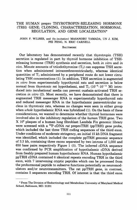
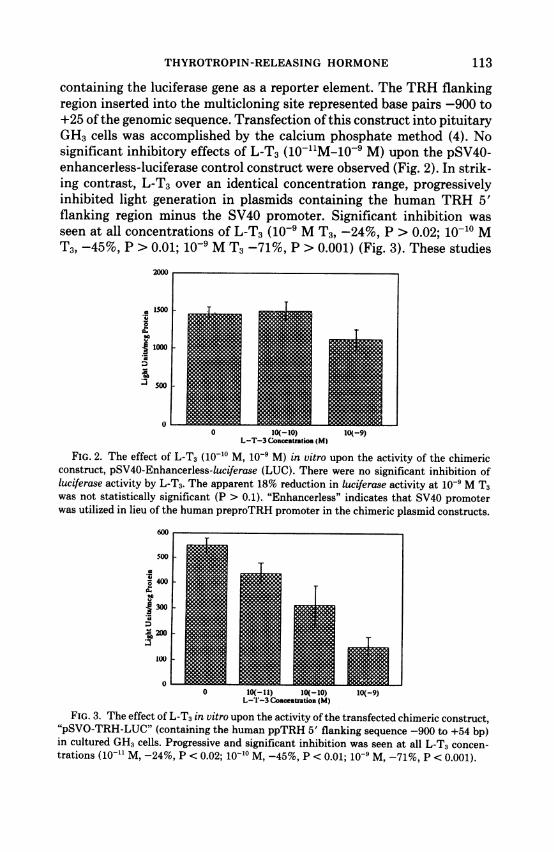
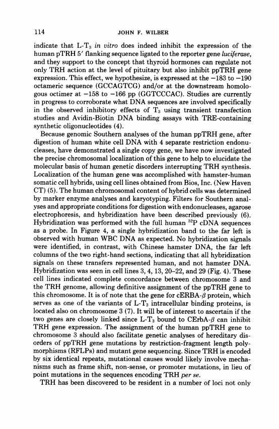
containing the luciferase gene as a reporter element. The TRH flankingregion inserted into the multicloning site represented base pairs -900 to+25 of the genomic sequence. Transfection of this construct into pituitaryGH3 cells was accomplished by the calcium phosphate method (4). Nosignificant inhibitory effects of L-T3 (10-11M-10-9 M) upon the pSV40-enhancerless-luciferase control construct were observed (Fig. 2). In strik-ing contrast, L-T3 over an identical concentration range, progressivelyinhibited light generation in plasmids containing the human TRH 5'flanking region minus the SV40 promoter. Significant inhibition wasseen at all concentrations of L-T3 (10-' M T3, -24%, P > 0.02; 10-10 MT3, -45%, P > 0.01; 10-9 M T3 -71%, P > 0.001) (Fig. 3). These studies
.0
,I
0.00
L-T-3 Concentration (M)
FIG. 2. The effect of L-T3 (10-1o M, 10- M) in vitro upon the activity of the chimericconstruct, pSV40-Enhancerless-luciferase (LUC). There were no significant inhibition ofluciferase activity by L-T3. The apparent 18% reduction in luciferase activity at 10' M T3was not statistically significant (P > 0.1). "Enhancerless" indicates that SV40 promoterwas utilized in lieu of the human preproTRH promoter in the chimeric plasmid constructs.
A,
.000
A.
3
0 10(-11) IO(-10) 10(-9)L-'-3 Concenlration (M)
FIG. 3. The effect of L-T3 in vitro upon the activity of the transfected chimeric construct,"pSVO-TRH-LUC" (containing the human ppTRH 5' flanking sequence -900 to +54 bp)in cultured GH3 cells. Progressive and significant inhibition was seen at all L-T3 concen-trations (10-1" M, -24%, P < 0.02; 10-10 M, -45%, P < 0.01; 10-9 M, -71%, P < 0.001).
113
JOHN F. WILBER
indicate that L-T3 in vitro does indeed inhibit the expression of thehuman pTRH 5' flanking sequence ligated to the reporter gene luciferase,and they support to the concept that thyroid hormones can regulate notonly TRH action at the level of pituitary but also inhibit ppTRH geneexpression. This effect, we hypothesize, is expressed at the -183 to -190octameric sequence (GCCAGTCG) and/or at the downstream homolo-gous octimer at -158 to -166 pp (GGTCCCAC). Studies are currentlyin progress to corroborate what DNA sequences are involved specificallyin the observed inhibitory effects of T3 using transient transfectionstudies and Avidin-Biotin DNA binding assays with TRE-containingsynthetic oligonucleotides (4).Because genomic Southern analyses of the human ppTRH gene, after
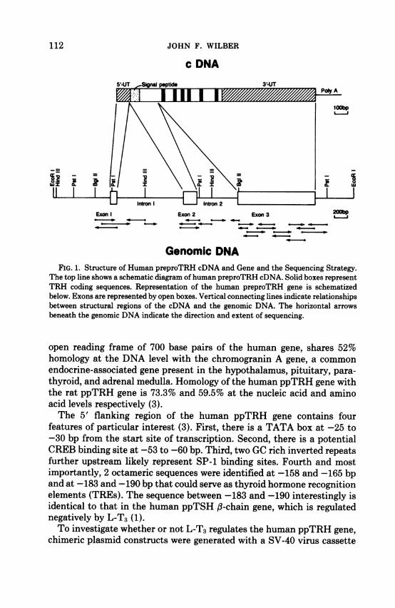
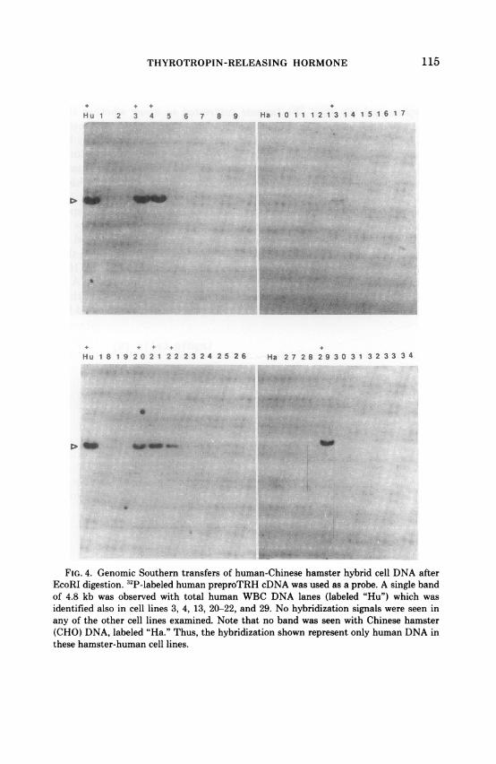
digestion of human white cell DNA with 4 separate restriction endonu-cleases, have demonstrated a single copy gene, we have now investigatedthe precise chromosomal localization of this gene to help to elucidate themolecular basis of human genetic disorders interrupting TRH synthesis.Localization of the human gene was accomplished with hamster-humansomatic cell hybrids, using cell lines obtained from Bios, Inc. (New HavenCT) (5). The human chromosomal content of hybrid cells was determinedby marker enzyme analyses and karyotyping. Filters for Southern anal-yses and appropriate conditions for digestion with endonucleases, agaroseelectrophoresis, and hybridization have been described previously (6).Hybridization was performed with the full human 32p cDNA sequencesas a probe. In Figure 4, a single hybridization band to the far left isobserved with human WBC DNA as expected. No hybridization signalswere identified, in contrast, with Chinese hamster DNA, the far leftcolumns of the two right-hand sections, indicating that all hybridizationsignals on these transfers represented human, and not hamster DNA.Hybridization was seen in cell lines 3, 4, 13, 20-22, and 29 (Fig. 4). Thesecell lines indicated complete concordance between chromosome 3 andthe TRH genome, allowing definitive assignment of the ppTRH gene tothis chromosome. It is of note that the gene for cERBA-f protein, whichserves as one of the variants of L-T3 intracellular binding proteins, islocated also on chromosome 3 (7). It will be of interest to ascertain if thetwo genes are closely linked since L-T3 bound to CErbA-f can inhibitTRH gene expression. The assignment of the human ppTRH gene tochromosome 3 should also facilitate genetic analyses of hereditary dis-orders of ppTRH gene mutations by restriction-fragment length poly-morphisms (RFLPs) and mutant gene sequencing. Since TRH is encodedby six identical repeats, mutational causes would likely involve mecha-nisms such as frame shift, non-sense, or promoter mutations, in lieu ofpoint mutations in the sequences encoding TRH per se.TRH has been discovered to be resident in a number of loci not only
114
THYROTROPIN-RELEASING HORMONE
Hu 1 2 3 4 5 6 7 8 9 HA 1 0 1 1 1 2 1 3 1 4 1 5 1 6 1 7
Hu IO 1 9 20 2 1 2 2 2 3 24 25 26 Ha 27 28 29 30 3 1 3 2 33 3 4
FIG. 4. Genomic Southern transfers of human-Chinese hamster hybrid cell DNA afterEcoRI digestion. 32P-labeled human preproTRH cDNA was used as a probe. A single bandof 4.8 kb was observed with total human WBC DNA lanes (labeled "Hu") which wasidentified also in cell lines 3, 4, 13, 20-22, and 29. No hybridization signals were seen inany of the other cell lines examined. Note that no band was seen with Chinese hamster(CHO) DNA, labeled "Ha." Thus, the hybridization shown represent only human DNA inthese hamster-human cell lines.
115
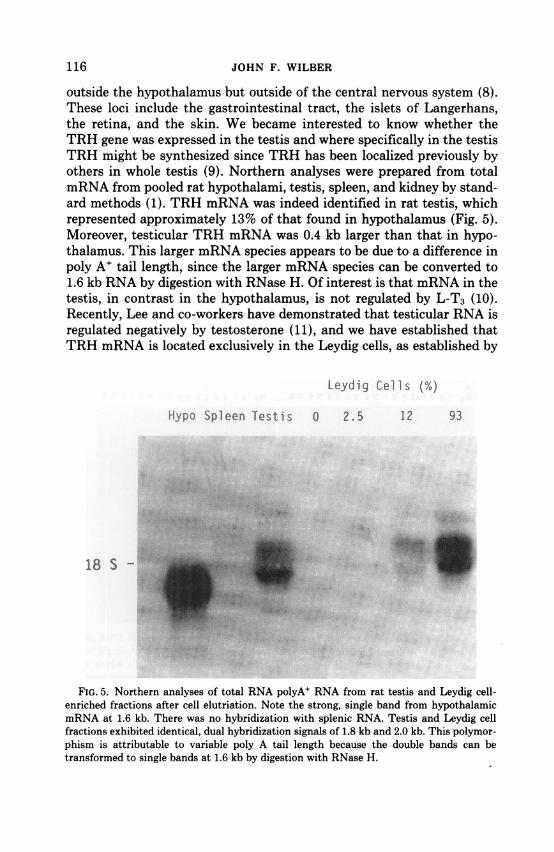
outside the hypothalamus but outside of the central nervous system (8).These loci include the gastrointestinal tract, the islets of Langerhans,the retina, and the skin. We became interested to know whether theTRH gene was expressed in the testis and where specifically in the testisTRH might be synthesized since TRH has been localized previously byothers in whole testis (9). Northern analyses were prepared from totalmRNA from pooled rat hypothalami, testis, spleen, and kidney by stand-ard methods (1). TRH mRNA was indeed identified in rat testis, whichrepresented approximately 13% of that found in hypothalamus (Fig. 5).Moreover, testicular TRH mRNA was 0.4 kb larger than that in hypo-thalamus. This larger mRNA species appears to be due to a difference inpoly A' tail length, since the larger mRNA species can be converted to1.6 kb RNA by digestion with RNase H. Of interest is that mRNA in thetestis, in contrast in the hypothalamus, is not regulated by L-T3 (10).Recently, Lee and co-workers have demonstrated that testicular RNA isregulated negatively by testosterone (11), and we have established thatTRH mRNA is located exclusively in the Leydig cells, as established by
Leydig Cells (%)
Hypo Spleen Testis 0 2.5 12 93
18 S -
FIG. 5. Northern analyses of total RNA polyA' RNA from rat testis and Leydig cell-enriched fractions after cell elutriation. Note the strong, single band from hypothalamicmRNA at 1.6 kb. There was no hybridization with splenic RNA. Testis and Leydig cellfractions exhibited identical, dual hybridization signals of 1.8 kb and 2.0 kb. This polymor-phism is attributable to variable poly A tail length because the double bands can betransformed to single bands at 1.6 kb by digestion with RNase H.
116 JOHN F. WILBER
THYROTROPIN-RELEASING HORMONE
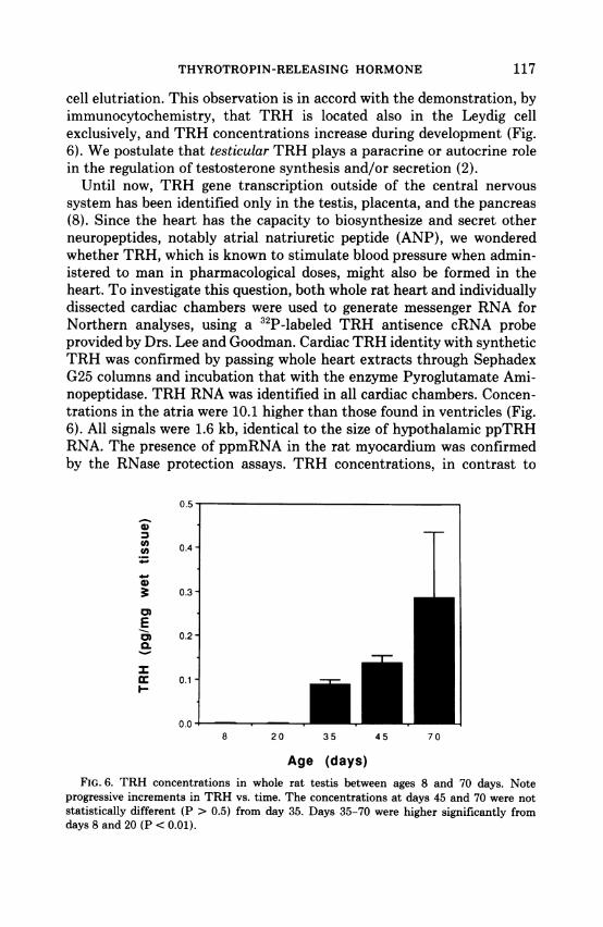
cell elutriation. This observation is in accord with the demonstration, byimmunocytochemistry, that TRH is located also in the Leydig cellexclusively, and TRH concentrations increase during development (Fig.6). We postulate that testicular TRH plays a paracrine or autocrine rolein the regulation of testosterone synthesis and/or secretion (2).
Until now, TRH gene transcription outside of the central nervoussystem has been identified only in the testis, placenta, and the pancreas(8). Since the heart has the capacity to biosynthesize and secret otherneuropeptides, notably atrial natriuretic peptide (ANP), we wonderedwhether TRH, which is known to stimulate blood pressure when admin-istered to man in pharmacological doses, might also be formed in theheart. To investigate this question, both whole rat heart and individuallydissected cardiac chambers were used to generate messenger RNA forNorthern analyses, using a 32P-labeled TRH antisence cRNA probeprovided by Drs. Lee and Goodman. Cardiac TRH identity with syntheticTRH was confirmed by passing whole heart extracts through SephadexG25 columns and incubation that with the enzyme Pyroglutamate Ami-nopeptidase. TRH RNA was identified in all cardiac chambers. Concen-trations in the atria were 10.1 higher than those found in ventricles (Fig.6). All signals were 1.6 kb, identical to the size of hypothalamic ppTRHRNA. The presence of ppmRNA in the rat myocardium was confirmedby the RNase protection assays. TRH concentrations, in contrast to
U.Z) I
C')(A 0.4co
.-0
3. 0.3-
EIm 0.2-C
0
C 0.10-
0.0o8 20 35 45 70
Age (days)FIG. 6. TRH concentrations in whole rat testis between ages 8 and 70 days. Note
progressive increments in TRH vs. time. The concentrations at days 45 and 70 were notstatistically different (P > 0.5) from day 35. Days 35-70 were higher significantly fromdays 8 and 20 (P < 0.01).
117
Ir
118 JOHN F. WILBER
1 2 3 4 5 6 7
2.37
1.36
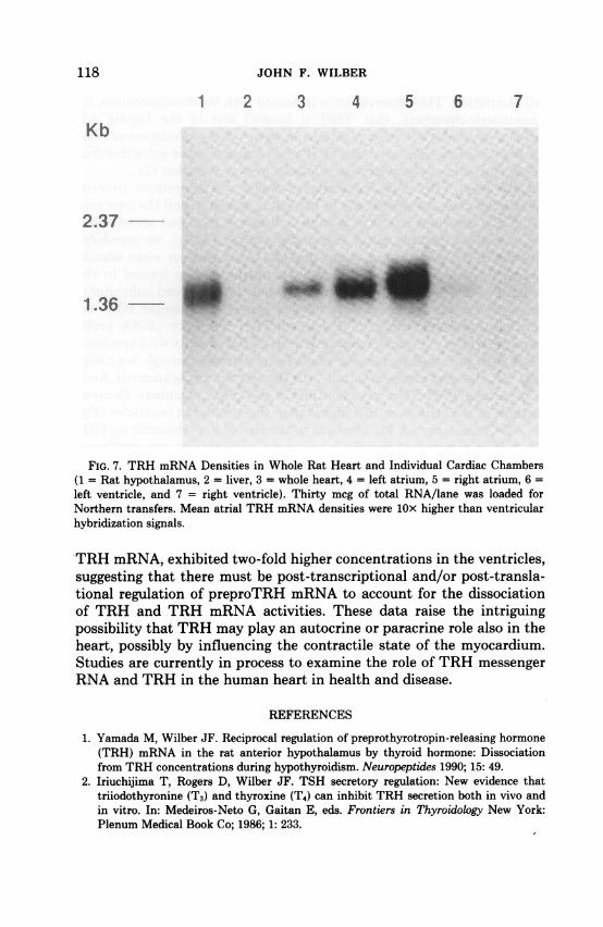
FIG. 7. TRH mRNA Densities in Whole Rat Heart and Individual Cardiac Chambers(1 = Rat hypothalamus, 2 = liver, 3 = whole heart, 4 = left atrium, 5 = right atrium, 6 =left ventricle, and 7 = right ventricle). Thirty mcg of total RNA/lane was loaded forNorthern transfers. Mean atrial TRH mRNA densities were lOx higher than ventricularhybridization signals.
TRH mRNA, exhibited two-fold higher concentrations in the ventricles,suggesting that there must be post-transcriptional and/or post-transla-tional regulation of preproTRH mRNA to account for the dissociationof TRH and TRH mRNA activities. These data raise the intriguingpossibility that TRH may play an autocrine or paracrine role also in theheart, possibly by influencing the contractile state of the myocardium.Studies are currently in process to examine the role of TRH messengerRNA and TRH in the human heart in health and disease.
REFERENCES
1. Yamada M, Wilber JF. Reciprocal regulation of preprothyrotropin-releasing hormone(TRH) mRNA in the rat anterior hypothalamus by thyroid hormone: Dissociationfrom TRH concentrations during hypothyroidism. Neuropeptides 1990; 15: 49.
2. Iriuchijima T, Rogers D, Wilber JF. TSH secretory regulation: New evidence thattriiodothyronine (T3) and thyroxine (T4) can inhibit TRH secretion both in vivo andin vitro. In: Medeiros-Neto G, Gaitan E, eds. Frontiers in Thyroidology New York:Plenum Medical Book Co; 1986; 1: 233.
THYROTROPIN-RELEASING HORMONE 119
3. Yamada M, Radovick S, Wondisford FE, et al. Cloning and structure ofhuman genomicDNA and hypothalamic cDNA encoding human prepro thyrotropin-releasing hormone.Mol Endocrinol 1990; 4: 551.
4. Yamada M, Radovick S, Wondisford FE, et al. The human preproTRH (ppTRH) gene:Inhibition of expression in GH3 cells of plasmid chimeric luciferase constructs by L-triiodothyronine (L-T3). Proceedings of The 10th Annual International Thyroid Con-gress, The Hague, The Netherlands, 1991.
5. Yamada M, Wondisford FE, Radovick S, et al. Assignment of human prepro thyrotro-pin-releasing hormone (TRH) gene to chromosome 3. Somat Cell Mol Genet 1991; 17:97.
6. Carlock LR, Smith D, Wasmuth JJ. Somat Cell Mol Genet 1986; 12: 163.7. Weinberger C, Thompson CC, Ong ES, Lebo R, Groul DJ, Evans RM. Nature 1986;
324: 641.8. Wilber JF, Yamada M. Thyrotropin-releasing hormone: Current concepts. In: Greer
MA, ed. The Thyroid Gland. New York: Raven Press, Ltd.; 1990: 127.9. Pekary AE, Bhasin S, Smith V, et al. Thyroid hormone modulation of thyrotropin-
releasing hormone (TRH) and TRH-Gly levels in the male rat reproductive system. JEndocr 1987; 114: 271.
10. Feng P, Kim U, Yamada M, et al. Rat preproTRH gene expression: Selective alterationsby thyroid status in the paraventricular nucleus (PVN) but not in testis, a new extra-CNS locus for ppTRH gene expression. Proceedings of The 10th Annual InternationalThyroid Congress, The Hague, The Netherlands, Feb. 1991.
11. Yang IM, Krieg RJ, Lee SL. Hormonal regulation of thyrotropin releasing hormone(TRH) gene expression in rat testes. Thyroid 1991; 1: S-81.