Embed Size (px)
Citation preview
(CANCER RESEARCH 53. 15.V157. January I. I993|
Tumor-secreted Vascular Permeability Factor/Vascular Endothelial Growth FactorInfluences Photosensitizer Uptake1
W. Gregory Roberts2 and Tayyaba Hasan3
\Vi'llnian laboratories of Pholomedicine. Department of Dermatology. Massachusetts General Hospital and Han-aril Medical School. Boston. Massachusetts 02114
ABSTRACT
The role of vascular permeability in the preferential accumulation ofphotosensitize» in tumor tissue was investigated. Two murine tumors[experimental mammary tumor carcinoma (EMT-6) and methylcholan-threne-induced rhabdomyosarcoma (MIS)] and a human bladder carci
noma (EJ) were grown s.c. on the flank in athymic nude mice and analyzedfor in vivo vessel permeability, vascular permeability factor (VPF) secretion, and accumulation of the photosensitizer, chloroaluminum sulfon-
ated phthalocyanine. In vivo tumor vessel permeability and vascularvolume were quantitated by measuring Evans blue extravasation andaccumulation of a high molecular weight fluoresceinated dextran, respectively. VPF was isolated from serum-free tumor cell conditioned mediumusing heparin-Sepharose affinity chromatography. Dot and Western blotsstained with anti-VPF antiserum positively identified VPF in samplesfrom each tumor. Chloroaluminum sulfonated phthalocyanine pharmaco-kinetics in tumor-bearing mice were measured using a Tiber-based spectro-
ftuorometer.In vivo vessel permeability was found to be greatest in MIS tumors, next
in EMT-6 tumors and finally in EJ tumors. Consistent with in vivo data,MIS and EMT-6 tumor cells in culture secrete significantly more VPF
than EJ tumor cells. Chloroaluminum sulfonated phthalocyanine accumulation was approximately 2 times greater in MIS and EMT-6 tumors
compared to EJ tumors. Our data present evidence that photosensitizeraccumulation can be correlated to in vivo tumor vessel permeability andVPF secretion of that tumor. Taken together, the data support the hypothesis that vascular permeability differences among tumors play a significant role in the uptake and retention of photodynamic agents.
INTRODUCTION
Selective photosensitizer retention in tumors is the hallmark ofphotodynamic therapy with many porphyrins, chlorins. and phthalo-
cyanines. Although it is believed that photodynamic destruction oftumors is largely due to effects on tumor vasculature (1, 2), themechanisms responsible for photosensitizer retention in tumors areless clear. It is well documented that tumonmuscle ratios of a givenphotosensitizer vary with respect to the tumor type (3); however, thereason for this variability has yet to be elucidated. Tumor propertiessuch as poor lymphatic drainage (4), variability in vessel density (5,6), incomplete subendothelial barrier (7, 8), unique microvascularendothelium (9), and increased vessel permeability (10, 11) may allplay a role in the selective retention of photosensitizers. The relativecontribution of each one of these tumor characteristics may determinethe variation in photosensitizer retention among different tumor types.
VPF4 (also known as vascular endothelial growth factor) is a highly
conserved heparin-binding protein (12) which has recently been pu-
Received 11/2/91 : accepted 10/20/92.The costs of publication of this article were defrayed in part by the payment of page
charges. This article must therefore be hereby marked advertisement in accordance with18 U.S.C. Section 1734 solely to indicale this fact.
1This work was supported in pan by ONR Contracts N00014-86-K01I7 and R29
AR389I8. W. G. R. received support from the Ford Foundation Postdoctoral Fellowshipand NIH Postdoctoral Fellowship (CM 13763).
2 Present address: Center for Cellular and Molecular Medicine-0669. University ofCalifornia-San Diego. 9500 Oilman Drive. La Jolla, CA 92093-0669.
•¿�'To whom requests for reprints should be addressed.4 The abbreviations used are: VPF. vascular permeability factor; CASPc. chloroalu
minum sulfonated phthalocyanine; EMT-6. experimental mammary tumor; EJ. humanbladder carcinoma; MIS. methylcholanthrene-induced rhabdomyosarcoma; PBS. phosphate-buffered saline; SDS. sodium dodecyl sulfate: BSA, bovine serum albumin: i.d..intradermally.
rified and sequenced (13. 14). This tumor-secreted factor has a molecular weight of 34,000-43.000 (15) and is specific and mitogenic for
endothelium in vitro (16) and angiogenic in vivo (17). A variety oftumor cells secrete this growth factor which is responsible for increased vessel permeability characteristic of solid tumors (14, 15, 18,19). This increased permeability results in greater extravasation ofmacromolecules. such as low density lipoprotein (20) and albumin(21). Once injected into an animal, most photosensitizers bind lowdensity lipoprotein or albumin, both of which then act as carriers(22, 23). Therefore, any differences in vascular permeability amongtumor types may critically influence the ability of a tumor to retain thephotosensitizer.
We have recently demonstrated that increased vessel permeabilitywithout the presence of neovascular endothelium is not sufficient toprovide selective retention of a variety of photosensitizers; however,permeable vessels resulted in increased uptake of all photosensitizers(24). We hypothesized that increased vessel permeability may be anadditive factor in photosensitizer retention providing that neovascu-
lature is present. In this article, we test this hypothesis by comparingthe amount of photosensitizer uptake and retention among three tumors, the amount of vascular permeability activity secreted by therespective tumor cells in vitro, and the relative permeability of eachtumor in vivo. We clearly demonstrate a positive correlation betweenphotosensitizer retention, VPF secretion, and in vivo permeabilityamong EMT-6, MIS, and EJ tumors.
MATERIALS AND METHODS
Sensitizers
CASPc (M, 835) was a gift from Ciba-Geigy (Basel, Switzerland) and wasstored in the dark at -20°C and diluted in phosphate-buffered saline (Gibco.
Grand Island. NY) to a concentration of I mg/ml before use (25).
Cells
EMT-6 carcinoma cells were grown in tissue culture flasks with RPMI 1640.MIS cells were grown in Dulbecco's modified Eagle's medium. EJ cells weregrown in McCoy's 5A medium. All media were purchased from Gibco (Grand
Island, NY) and contained 100 units/ml penicillin, UK) ug/ml streptomycin,and 5<7rfetal calf serum. Cells were incubated at 37°Cunder 5% COy For
tumor inoculation, cells were trypsinized and resuspended in medium at aconcentration of 1 x IO7 cells/ml.
Anti-VPF Antiserum
The antiserum was raised in rabbits against a synthetic amino-terminal
guinea pig VPF sequence ( 13). The antiserum was kindly provided by Dr. DonSenger.
Animals
Mice. For all murine experiments, male athymic nude mice (4-5 weeks
old) were used. All protocols were approved by an independent animal usecommittee.
Guinea Pigs. Male Hartley albino guinea pigs (300-450 g) were used for
the Miles assay to quantitate the vascular permeability activity secreted fromeach tumor cell type (26).
153
on June 7, 2018. © 1993 American Association for Cancer Research. cancerres.aacrjournals.org Downloaded from
[NHF.RKNT VPF INCREASES PHTHALOCYANINE UPTAKE
Tumor Inoculation
Mice received a s.c. inoculation of 1 x IO6 cells on the right flank. Within
10-14 days (20-30 days for EJ tumors), the tumors were well vascularized and5-8 mm in diameter. Tumors averaged 1.15 cm' at the time of measurements.
In Vivo Spectroscopy
Animals received an i.p. injection of CASPc at a concentration of 10 mg/kgbody weight. CASPc distribution at 30 min and I. 2. 3. 6. 9. and 24 h wasdetermined spectrofluorometrically in the tumor and contralateral flank (normal) using a SPEX Fluorolog 2 speclrofluorometer (Spex. Edison. NJ) coupledto a bifurcated quart/, fiberoptic assembly (24). The Y-shaped fiberoptic as
sembly consists of numerous quartz fibers, one half of which transmits lightfrom the excitation source to the sample (e.g.. lumor). while the other halftransmits emitted fluorescence from the sample to an emission monochrometerand photomultiplier tube. All measurements were completed in the dark withthe fiber in contact with the tissue. CASPc was excited at 610 nm and emissionwas read from 630 to 800 nm. The fluoresceinated dextran was measured byexciting at 492 nm and monitoring emission from 500 to 550 nm. Datarepresent the average ±SD of four animals. Statistical significance wasdetermined using Student's t test.
Tumor Vessel Permeability
Tumor-bearing mice received a 0.1-ml i.v. injection of Evans blue dye (M,
961: 0.1% in saline; Baker Chemical Co.. Phillipsburg. NJ). Twenty min laterthe animals were sacrificed and Evans blue was extracted from tumors andnormal muscle as described under Miles vascular permeability assay (13. 27).To determine tumor vascular volume, a high molecular weight fluoresceinateddextran (M, 2.000.000) (Sigma Chemical Co.. St. Louis. MO) was continuously monitored after i.v injection (10 mg/kg) for 20 min. The peak fluorescence was used to calculate relative vascular volume among the tumor types.Data listed in Table 2 have been corrected for the vascular volume of eachtumor. Data represent the average ±SD of 10-15 animals. Statistical significance was determined using Student's t test.
Vascular Permeability Factor Isolation
Tumor cells were incubated as above with the following modifications.Cells were plated in T750 flasks and allowed to reach 80% confluency. Cellswere then washed three times with PBS. and fresh serum-free medium was
added. The conditioned medium was harvested 24 h later. Conditioned medium(500 ml) was filtered and passed over a heparin-Sepharose column, washed
with PBS. and eluted with 2 bed volumes of 2 MNaCl. This procedure recoversthe maximum tumor-secreted permeability-increasing activity. It is an abbre
viated method described previously for the purification of VPF (13). NaCleluants were concentrated using a Centricon 10 (Amicon. Danvers. MA) mi-
croconcentralor prior to electrophoresis. immunoblotting. and the Miles assay(described below). SDS-polyacrylamide (12.5%) gel electrophoresis was performed to identify the presence of vascular permeability factor and silver-stained with a commercially available kit (Bio-Rad Laboratories. Richmond.CA) in accordance with the manufacturer's instructions. Bands were identified
visually and using a densitometer. The gel was also transferred to nitrocelluloseand stained with antiserum as described below. Concentrated VPF isolates(20 ul) were blotted onto a nitrocellulose strip and blocked with 3% BSA-PBSfor 3 h. Anti-VPF antiserum was diluted 200-fold with 1% BSA-PBS and
incubated with the nitrocellulose strip at room temperature for 1 h. The stripwas rinsed briefly with double-distilled water (ddH:O) and washed twice withPBS for IO min. It was then covered with goat anti-rabbit IgG horseradishperoxidase. diluted 1:3000 with 1% BSA-PBS, and allowed to incubate
at room temperature for I h. After a washing with PBS. diaminobenzidine(Sigma) was added and allowed to develop for 5 min. Western blots werestained as above.
Miles Vascular Permeability Assay
In order to quantitate the extent of permeability caused by each VPF sample,shaved and depilated guinea pigs were anesthetized prior to receiving a 0.2-ml
i.v. injection of Evans blue dye. After 2 h. 0.05 ml of concentrated VPF wasinjected i.d. Twenty min later, the animals were euthanized and the injectionspot ( 1 cm in diameter) was surgically removed. Tissues were dissolved in 1 mlof tissue solubilizer (Soluene 350: Packard Industries. Downers Grove, IL) at37°Covernight and then at 57°Cfor I h. The solution was allowed to cool
before the addition of 2 ml of ethyl acetate, the mixture was vortexed. and2 ml of l s HCI were added, vortexed again, and cemrifuged at 2500 rpm for15 min. Absorption of the upper phase was read at 626 nm on a diode arrayspectrophotometer (Model 8451 A: Hewlett Packard. Palo Alto, CA). Concentrations were determined from a standard curve of Evans blue dye. Datarepresent the average ±SD of eight injections and are corrected for the amountof total protein injected. Statistical significance was determined using Student's / test for all experiments.
RESULTS
CASPc distribution in EMT-6. MIS, and EJ tumors is shown in
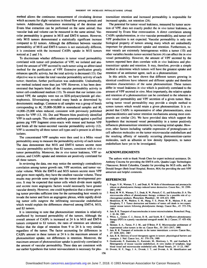
Table 1. CASPc concentration is significantly greater at 2, 3, 6, and24 h postinjection in MIS tumors compared to EJ tumors (P < 0.08).Photosensitizer amount in EMT-6 tumors is greater at 3, 9. and 24 h
compared to the EJ tumors (P < 0.07). There is also a slight increasein CASPc concentration in MIS tumors versus EMT-6 tumors at 2 and
3 h but this increase is not statistically different. Differential retentionof CASPc in MIS. EMT-6. and EJ tumors, grown in the same mouse
strain, led to experiments measuring tumor vascular volume and permeability.
Evans blue was extracted from tumors as a measure of tumor vesselleakiness. Differences in vascular volume among the tumor types wereaccounted for when determining tumor vascular permeability. To measure vascular volume, tumor-bearing animals were monitored spec-
trofluorimetrically after injection with a large M, fluoresceinated dextran. Peak fluorescence always occurred between 5 and 10 minpostinjection, regardless of tumor type. The peak fluorescence signalwas used to determine relative vascular volumes among tumor types.
Table I Relative CASPc tumor plmrmacokineticsChloroaluminum sulfonated phthalocyanine pharmacokinetics in MIS. EMT-6, or EJ tumors. Athymic nude mice received a 10-mg/kg ¡.p.injeclion and sensitizer amount was
delected using a fiber-based speclrofluorometer. Excitation wavelength was 610 nm while the emission was scanned from 630 to 800 nm. CASPc concentration is significantly higherin M l S tumors versus EJ tumors at 2. 3. 6, and 24 h whereas EMT-6 tumors have greater amounts of the photosensitizer at 3. 9. and 24 h. Values are normalized to the maximum CASPcuptake and are therefore relative lo each olher. Data represent the mean ±SD of four animals. Statistical significance was determined using Student's l test.
Time(postinjection)30
min1h2
h3h6h9h24hMIS0.46
+0.160.79+0.191.0+0.21'1.0+0.17«0.94+0.18'0.91
+0.230.75+ 0.08'EMT-60.42
+0.220.68+0.200.78+0.210.84+0.15''0.85
+0.130.89+0.07''0.76+ 0.14''EJ0.34
+0.120.51+0.170.63
+0.230.63+0.170.72+0.200.71+0.190.47
+ 0.12P"
(MIS. EMT-6 vv.EJ)NSÄ.
NSNS.
NS0.02'.NS0.004'.
0.07''0.08'.NSNS.
0.06''0.005'. 0.003''
" P values compare CASPc amount in MIS or EMT-6 tumors to amount in EJ tumors.'' NS. not significant.'CASPc pharmacokinelics in MIS tumors.'' CASPc pharmacokinetics in EMT-6 tumors.
154
on June 7, 2018. © 1993 American Association for Cancer Research. cancerres.aacrjournals.org Downloaded from
INHERENT VPF INCREASES PHTHALOCYANINE UPTAKE
Vascular leak is greatest in MIS and EMT-6 tumors and least in EJ
tumors (Table 2). Since tumor vessel permeability is due, in part, tothe tumor-secreted protein, VPF, we decided to analyze tumors for the
production of this factor.The heparin-binding fraction of serum-free conditioned medium
from each tumor cell was analyzed for the presence of VPF. Silver-stained SDS-polyacrylamide gels revealed a broad band corresponding to M, 35,000-50,000 in nonreduced samples and Mr 14,000-
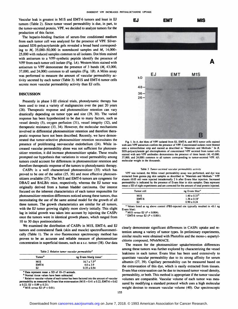
25,000 with reduced samples common to all isolates. Dot blots stainedwith antiserum to a VPF-synthetic peptide identify the presence of
VPF from each tumor cell isolate (Fig. \A). Western blots stained withantiserum to VPF demonstrate the presence of 3 bands (M, 43,000,37,000, and 24,000) common to all samples (Fig. Iß).A Miles assaywas performed to measure the amount of vascular permeability activity secreted by each tumor (Table 3). MIS and EMT-6 tumor cells
secrete more vascular permeability activity than EJ cells.
DISCUSSION
Presently in phase I-III clinical trials, photodynamic therapy has
been used to treat a variety of malignancies over the past 20 years(28). Therapeutic response and photosensitizer retention can varydrastically depending on tumor type and size (29, 30). The variedresponse has been hypothesized to be due to many factors, such asvessel density (5), oxygen perfusion (31), vessel integrity (32), andtherapeutic resistance (33, 34). However, the molecular mechanismsinvolved in differential photosensitizer retention and therefore therapeutic response have not been described. Recently, we have demonstrated that tumor-selective photosensitizer retention necessitates the
presence of proliferating neovascular endothelium (24). While increased vascular permeability alone was not sufficient for photosensitizer retention, it did increase photosensitizer uptake. These resultsprompted our hypothesis that variations in vessel permeability amongtumors could account for differences in photosensitizer retention andtherefore therapeutic response of the tumors to photodynamic therapy.
CASPc is a well characterized photosensitizer (35) which hasproved to be one of the safest (25, 36) and most effective photosen-sitizers available (37). The MIS and EMT-6 tumors are syngeneic for
DBA/2 and BALB/c mice, respectively, whereas the EJ tumor wasoriginally derived from a human bladder carcinoma. Our interestfocused on the inherent characteristics of each tumor responsible forphotosensitizer retention differences noticed among these tumors, thusnecessitating the use of the same animal model for the growth of allthree tumors. The growth characteristics are similar for all tumors,with the EJ tumor growing slightly more slowly initially. This slightlag in initial growth was taken into account by injecting the CASPconce the tumors were in identical growth phases, which ranged from10 to 30 days postinoculation.
We examined the distribution of CASPc in MIS, EMT-6, and EJtumors and contralateral flank (skin and muscle) spectrofluorometri-
cally (Table 1). The in vivo fluorescence spectroscopy method hasproven to be an accurate and reliable measure of photosensitizerconcentration in superficial tissues, such as a s.c. tumor (38). Our data
Table 2 Relative tumor vascular permeability"
EJ EMT MIS
Tumor'' ng Evans blue/g tissue'
MISEMT-6
EJ
16.7 ±1.5d
4.0 ±3.90.35 ±0.54
" Data represent mean ±SD of 10-15 animals.'' Normal tissue values have been subtracted.' Relative vascular volume of each tumor has been factored into the amount of vascular
permeability as measured by Evans blue extravasation (MIS = 0.41 ±0.22; EMT-6 = 0.62±0.22; EJ = 0.89 ±0.21).
''MIS vpâ„¢jEJ</><O.OI).
EMT M1S
Fig. I. In A. dot blots of VPF isolated from FJ. EMT-6. and M l S tumor cells stainedwith anli-VPF antiserum confirm the presence of VPF. Concentrated isolates were blottedonto a nitrocellulose strip and stained as described in "Materials and Methods." In B.
SDS-polyacrylamide gel electrophoresis of concentrated VPF secreted from tumor cellsstained with anti-VPF antibodies demonstrates the presence of three bands (Mr 43.(HK).37.ÕKM).and 24.000) common to all tumors corresponding to tumor-secreted VPF. kl).molecular weight in the thousands.
Table 3 Tumor-secreted vascular permeability activity
VPF was isolated, the Miles vessel permeability assay was performed, and dye wasextracted from guinea pig skin samples as described in "Materials and Methods." VPF
eluants (0.05 ml) were injected intradermally 2 h after Evans blue injection. Increasedpermeability is indicated by the presence of Evans blue in skin samples. Data representmean ±SD of eight experiments and are corrected for the amount of total protein injected.
Tumor cell ng Evans blue"
MISEMT-6
EJ
1.40 ±0.33"1.36±0.18'
0.90 ±0.24" Values listed as ng above control (PBS-injected site typically resulted in <(). I ng
Evans blue)."MIS versus El (P < 0.004).' EMT-6 versus EJ (P < 0.001).
clearly demonstrate significant differences in CASPc uptake and retention among a variety of tumor types. In preliminary experiments,similar results were obtained with Photofrin and the mono-L-aspartyl-
chlorin compound, NPe6/MACE.The reason for the photosensitizer uptake/retention differences
among these tumors was further explored by characterizing the vesselleakiness in each tumor. Evans blue has been used extensively toquantitate vascular permeability due to its strong affinity for serumalbumin (27, 39). Capillary permeability can be measured based onthe extravasation of this dye, which is easily extracted from tissues.Evans blue extravasation can be due to increased tumor vessel density,permeability, or both. This method is appropriate if the tumor vascularvolumes are comparable. Vascular volume of each tumor was measured by modifying a standard protocol which uses a high molecularweight dextran to measure vascular volume (40). Our spectroscopic
155
on June 7, 2018. © 1993 American Association for Cancer Research. cancerres.aacrjournals.org Downloaded from
INHERENT VPF INCREASES PHTHAI.OCYANINE UPTAKE
method allows the continuous measurement of circulating dextranwhich accounts tor slight variations in blood How among animals andtumors. Additionally, fluorescence monitoring of the dextran andEvans blue extraction can be performed on the same animal; thusvascular leak and volume can be measured in the same animal. Vascular permeability is greatest in MIS and EMT-6 tumors. However,
only MIS tumors demonstrated a statistically significant increasecompared to EJ tumors. Although the difference between vascularpermeability of MIS and EMT-6 tumors is not statistically different,
it is consistent with the increased CASPc uptake in MIS tumorsnoticed at 2 and 3 h.
To discriminate whether differences in tumor vessel permeabilitycorrelated with tumor cell production of VPF, we isolated and analyzed the amount of VPF secreted by each tumor using an abbreviatedmethod for the purification of VPF. Extensive purification greatlyenhances specific activity, but the total activity is decreased (13). Ourobjective was to isolate the total vascular permeability activity of eachtumor; therefore, further purification was undesirable. Undoubtedly,our method isolated more than just VPF. However, it has been demonstrated that heparin binds all the vascular permeability activity intumor cell-conditioned medium ( 13). To ensure that our isolates contained VPF, the samples were run on SDS-polyacrylamide gel elec-
trophoresis. Silverstaining revealed many bands as determined bydensitometric readings. Common to all samples was a group of bandscorresponding to M, 35,000-50,000 in nonreduced samples and Mr14,000-25,000 when reduced, consistent with previously published
reports for VPF (13, 18). Dot and Western blots positively identifiedVPF in each sample. This rabbit antibody generated against a purifiedguinea pig VPF fragment cross-reacts with mouse and human VPFand binds all the tumor-secreted vascular permeability activity (12).
VPF is secreted by all three tumor cell types and is present in all threeisolates.
The concentrated VPF samples were then used in a Miles vesselpermeability assay to measure the permeability activity of each tumor.The data demonstrate that MIS and EMT-6 tumors secrete more
vascular permeability activity than EJ tumors, consistent with in vivotumor permeability. Moreover, the in vivo tumor leakiness, VPF secretion, and CASPc uptake and retention are positively correlated forall three tumors.
In reviewing the data, one may notice the seemingly contradictorycorrelation among tumor growth rate, VPF secretion, and tumor vascular volume. While the EMT-6 and MIS tumors secrete more VPF
and grow more rapidly, they have the smallest vascular volume. Theseresults may provide some insight into the tumor developmental process. It may be expected that tumor cells which divide more rapidlyand secrete more angiogenic factors would necessarily have greatervascular density. However, one could hypothesize that a slower growing tumor provides sufficient time for the surrounding host vessels toinvade the tumor and form an angiogenic plexus, while rapidly dividing tumor cells outgrow the infiltrating neovascular endotheliumwhich would explain the differences observed among EMT-6. MIS.
and EJ tumors.It is interesting to note that photosensitizer retention is relatively
unaffected by increased permeability of the tumors. Although theoverall amount of CASPc is increased at 24 h in MIS and EMT-6
tumors compared to EJ tumors, the rates of retention are identical.Notice that the slope of retention from 9 to 24 h is very similarregardless of the tumor. The factor accounting for differences inCASPc amount in these tumors at 24 h is the maximum amount ofphotosensitizer uptake, not differences in retention. Moreover, themaximum amount of photosensitizer uptake is positively correlated tothe amount of vascular permeability. These data are consistent withour earlier hypothesis that tumor neovasculature is necessary for pho
tosensitizer retention and increased permeability is responsible forincreased uptake, not retention (24).
The potential for tumor vessel leakiness, measured by tumor secretion of VPF, does not exactly predict the in vivo tumor leakiness, asmeasured by Evans blue extravasation. A direct correlation amongCASPc uptake/retention, in vivo vascular permeability, and tumor cellVPF production is not expected. Vascular permeability is only onebiological property of tumors among many which are undoubtedlyimportant for photosensitizer uptake and retention. Furthermore, tumor vessels are extremely heterogeneous within a tumor (10) andother variables besides tumor-secreted factors (4) can affect the in vivo
vessel permeability. However, the amount of VPF secreted by thetumors reported here does correlate with in vivo leakiness and photosensitizer uptake and retention. It may. therefore, provide a simplemethod to determine which tumors will have the greatest uptake andretention of an antitumor agent, such as a photosensitizer.
In this article, we have shown that different tumors growing inidentical conditions have inherent and different uptake and thereforeretention characteristics of the same photosensitizer. These tumorsdiffer in vessel leakiness in vivo which is positively correlated to theamount of VPF secreted in vitro. Most importantly, the relative uptakeand retention of a photosensitizer can be correlated to the inherent invivo vessel permeability and VPF secretion of a given tumor. Measuring tumor vessel permeability may provide a simple method toscreen tumors which would retain a given photosensitizer. It is expected that CASPc is representative of other photosensitizers, sincepharmacokinetics and tumor retention mechanisms among most compounds are similar (24). We have provided data which support thehypothesis that increased vessel permeability in a tumor positivelyinfluences photosensitizer retention by increasing initial uptake. However, other factors including variable expression of proteoglycans orcell adhesion molecules on the tumor microvascular endothelium andthe resulting affinity of naturally occurring photosensitizer-carrier
molecules, such as albumin or low density lipoprotein, to tumorendothelium have yet to be investigated.
ACKNOWLEDGMENTS
The authors wish to thank Norah Chen for expert technical assistance. Dr.Anthony Cincotta tor providing the EMT-6 cells. Quadra Logic Technologies
(Vancouver. British Columbia. Canada) tor providing the MIS cells, and Dr.Donald Senger (Beth Israel Hospital. Boston. MA) for providing the anti-VPF
antiserum and helpful comments.
REFERENCES
1. Pingar. V. H.. Wieman. T. J.. and Doak. K. W. Role of thromboxane and prostacyclinrelease on pholodynamic therapy-induced tumor destruction. Cancer Res., 50: 2599-
2603, 1990.2. Reed. M. W. R.. Wieman. T. J., Doak. K. W.. Pietsch, C. G.. and Schuschke. D. A. The
microvascular effects of photodynamic therapy: evidence for a possible role of cy-clooxygenase products. Photochem. Photobiol., 50: 419-423, 1989.
3. Henderson, B. W.. Waldow. S. M., Mang. T. S., Roller, W. R., Malone, P. B., andDougherty, T. J. Tumor destruction and kinetics of tumor cell death in two experimental mouse tumors following photodynamic therapy. Cancer Res.. 45: 572-576.
1985.4. Jain. R. K. Transpon of macromolecules in tumor microcirculation. Biolechnol. Prog..
/.•81-94, 1985.
5. White. L., Gomer. C. J.. Doiron. D. R., and Szirth. B. C. Ineffective photodynamictherapy (PDT) in a poorly vascularized xenograft model. Br. J. Cancer. 57: 455^58.1988.
6. Skinner, S. A.. Tutton. P. J. M., and O'Brien. P. E. Microvascular architecture of
experimental colon tumors in the rat. Cancer Res., 50: 2411-2417, 1990.7. Jain. R. K. Transpon of molecules in the lumor interstitium: a review. Cancer Res.,
47: 3039-3051, 1987.
8. Jain. R. K. Delivery of novel therapeutic agents in tumors: physiological barriers andstrategies. J. Nati. Cancer Insu fil: 570-576, 1989.
9. Gumkowski. F.. Kaminska. G-, Kaminski, M., Morrissey. L. W., and Auerbach. R.Heterogeneity of mouse vascular endothelium. In vitni studies of lymphatic, largeblood vessel and microvascular endothelial cells. Blood Vessels, 24: 11-23. 1987.
10. Dvorak. H. F.. Nagy, J. A.. Dvorak. J. T. and Dvorak, A. M. Identification and
156
on June 7, 2018. © 1993 American Association for Cancer Research. cancerres.aacrjournals.org Downloaded from
INHERENT VPF INCREASES PHTHALOCYANINE UPTAKE
characterization of the blood vessels of solid tumors that are leaky to circulatingmacromolecules. Am. J. Palhol., 133: 95-109. 1988.
11. Sands, H., Jones. P. L., Shah, S. A., Palme, D., Vessella, R. L., and Gallagher, B. M.Correlation of vascular permeability and blood flow with monoclonal antibody uptakeby human Clouser and renal cell xenographs. Cancer Res., 48: 188-193, 1988.
12. Senger. D. R., Peruzzi, C. A., Feder, J.. and Dvorak. H. F. A highly conserved vascularpermeability factor secreted by a variety of human and rodent tumor cell lines. CancerRes., 46: 5629-5632, 1986.
13. Senger, D. R., Connolly, D. T.. Van De Water, L.. Feder, J., and Dvorak, H. F.Purification and NH^-terminal amino acid sequence of guinea pig tumor secretedvascular permeability factor. Cancer Res., 50: 1774-1778, 1990.
14. Keck, P. J., Hauser. S. D., Krivi, G., et al. Vascular permeability factor, an endothelialcell mitogen related to PDGF. Science (Washington DC), 246: 1309-1312. 1989.
15. Senger, D. R., Galli. S. J.. Dvorak. A. M., Perruzzi, C. A., Harvey, V. S., and Dvorak,H. F. Tumor cells secrete a vascular permeability factor that promotes accumulationof ascites fluid. Science (Washington DC), 219: 983-985, 1983.
16. Ferrara. N., Houck, K.. Jakeman, L., and Leung, D. W. Molecular and biologicalproperties of the vascular endothelial growth factor family of proteins. Endocr. Rev.,13: 18-32, 1992.
17. Connolly, D. T., Heuvelman. D. M.. Nelson, R., ft al. Tumor vascular permeabilityfactor stimulates endothelial cell growth and angiogenesis. J. Clin. Invest.. 84: 1470-
1478, 1989.18. discuoio, G. R.. Merrill. M. J.. and Oldfield. E. H. Further characterization of
malignant glioma-derived vascular permeability factor. J. Neurosurg., 69: 254-262,
1988.19. Rosemhal, R. A., Megyesi, J. F., Henzel, W. J., Ferrara, N., and Folkman. J. Condi
tioned medium from mouse sarcoma 180 cells contains vascular endothelial growthfactor. Growth Factors. 4: 53-59, 1990.
20. Rutledge, J. C., Curry, F-R. E., Lenz, J. F.. and Davis, P. A. Low density lipoprotein
transport across a microvascular endothelial barrier after permeability is increased.Circ. Res., 66: 486-495, 1990.
21. Lynch, J. J.. Ferro. T. J.. Blumenstock, F. A.. Brockenauer, A. M.. and Malik, A. B.Increased endothelial albumin permeability mediated by protein kinase C activation.J. Clin. Invest., 85: 1991-1998. 1990.
22. Kongshaug, M. Distribution of tetrapyrrole photosensitizers among human plasmaproteins. Int. J. Biochem., 24: 1239-1265, 1992.
23. Barel. A., Jori. G.. Perin, A., Romandini, P., Pagnan, A., and Biffanti, S. Role of high,low and very low-density lipoproteins in the transport and tumor-delivery of hemato-porphyrin in viva. Cancer Lett., 32: 145-150. 1986.
24. Roberts, W. G., and Hasan. T. Role of neovasculature and vascular permeability in theretention of photodynamic agents. Cancer Res.. 52: 924-930. 1992.
25. Roberts, W. G., Smith, K. M.. McCullough. J. L., and Berns, M. W. Skin pholosen-sitivity and photodestruction of several potential photodynamic sensitizers. Photo-chem. Photobiol., 49: 431-438, 1989.
26. Miles, A.A., and Miles, E. M. Vascular reactions to histamine, histamine-liberator andleukotaxine in the skin of guinea-pigs. J. Physiol. (Lond.). US: 228-257, 1952.
27. Udaka, K.. Takeuchi, Y., and Movat. H. Z. Simple method for quantitation of enhanced vascular permeability. Proc. Soc. Exp. Biol. Med., 133: 1384-1387, 1970.
28. Dougherty, T. J., Potter, W. R., and Bellnier. D. Photodynamic therapy for thetrealment of cancer: current status and advances. In: D. Kessel (éd.),PhotodynamicTherapy of Neoplastic Disease, pp. 1-20. Boca Raton. FL: CRC Press. 1991.
29. Nelson. J. S., Wright, W. H., and Berns. M. W. Histopathological comparison of theeffects of hematoporphyrin derivative on two different murine tumors using computer-enhanced digital video fluorescence microscopy. Cancer Res., 45: 5781-5786,
1985.30. Gluckman, J. L. Hematoporphyrin photodynamic therapy: Is there truly a future in
head and neck oncology? Reflections on a Five year experience. Laryngoscope, 101:36-41, 1991.
31. Henderson, B. W.. and Pingar. V. H. Relationship of tumor hypoxia and response tophotodynamic treatment in an experimental mouse tumor. Cancer Res.. 47: 3110-
3114, 1987.32. Nelson, J. S., Liaw, L-H.. Orenslein. A., Roberts. W. G., and Berns, M. W. Mechanism
of tumor destruction following photodynamic therapy with hematoporphyrin derivative, chlorin, and phthalocyanine. J. Nati. Cancer Inst., 80: 1599-1605, 1988.
33. Gomer, C. J., Rucker, N., and Wong, S. Porphyrin photosensitivity in cell linesexpressing a heat-resistant phenotype. Cancer Res., 50: 5365-5368. 1990.
34. Singh. G., Wilson. B. C., Sharkey, S. M.. Browman, G. P., and Deschamps, P.Resistance to photodynamic therapy in radiation induced fibrosarcoma-1 and Chinesehamster ovary-multi-drug resistant cells />i vitro. Photochem. Photobiol., 54: 307-
312, 1991.35. Rosenthal, I. Phthalocyanines as photodynamic sensitizers. Photochem. Photobiol.,
53: 859-870, 1991.
36. Tralau, C. J., Young. A. R.. Walker, N. P. J.. el til. Mouse skin photosensitivity withdihaematoporphyrin ether (DHE) and aluminum sulphonated phthalocyanine (Al-SPc): a comparative study. Photochem. Photobiol.. 49: 305-312. 1989.
37. Roberts, W. G., Klein, M. K., Loomis, M., Weldy, S.. and Berns. M. W. Photodynamictherapy of spontaneous cancers in felines, canines, and snakes with chloroaluminumsulfonated phthalocyanine. J. Nati. Cancer Inst., 83: 18-23, 1991.
38. Biolo, R., Jori. G., Kennedy. J. C., et al. A comparison of fluorescence methods usedin the pharmacokinetic studies of Zn(II) phthalocyaninc in mice. Photochem. Photobiol., 53: 113-118, 1991.
39. Green, T. P., Johnson. D. E., Marchessault, R. P., and Gatto, C. W. Transvascular fluxand tissue accrual of Evans blue: effects of endotoxin and histamine. J. Lab. Clin.Med.. ///: 173-183. 1988.
40. Shockley, T. R., Lin, K.. Nagy. J. A., Tompkins, R. G.. Yarmush, M. L., Dvorak, H.F. Spatial distribution of tumor-specific monoclonal antibodies in human melanomaxenografts. Cancer Res., 52: 367-376, 1992.
157
on June 7, 2018. © 1993 American Association for Cancer Research. cancerres.aacrjournals.org Downloaded from
1993;53:153-157. Cancer Res W. Gregory Roberts and Tayyaba Hasan Endothelial Growth Factor Influences Photosensitizer UptakeTumor-secreted Vascular Permeability Factor/Vascular
Updated version
http://cancerres.aacrjournals.org/content/53/1/153
Access the most recent version of this article at:
E-mail alerts related to this article or journal.Sign up to receive free email-alerts
Subscriptions
Reprints and
To order reprints of this article or to subscribe to the journal, contact the AACR Publications
Permissions
Rightslink site. Click on "Request Permissions" which will take you to the Copyright Clearance Center's (CCC)
.http://cancerres.aacrjournals.org/content/53/1/153To request permission to re-use all or part of this article, use this link
on June 7, 2018. © 1993 American Association for Cancer Research. cancerres.aacrjournals.org Downloaded from
![Growth Inhibition of Human Tumor Cells in Athymic Mice by ...[CANCER RESEARCH 44, 1002-1007, March 1984] Growth Inhibition of Human Tumor Cells in Athymic Mice by Anti-Epidermal Growth](https://img.pdfslide.net/doc/110x75/5e7bcebb508ec15dc92ee12e/growth-inhibition-of-human-tumor-cells-in-athymic-mice-by-cancer-research-44.jpg)