Embed Size (px)
Citation preview
TWO CASES OF OESOPHAGEAL STRICTURE INCHILDHOOD REQUIRING RESECTION
BY
R. H. FRANKLIN and I. D. HENDERSONFrom the Department of Surgery, Postgraduate Medical School ofLondon
(RECEIVED FOR PUBLICATION OCTOBER 18, 1954)
Reflux oesophagitis appears to affect principallytwo great groups of patients, infants on the one handand patients of late middle age on the other. Theintervening years seem to be comparatively freefrom the condition and it is relatively uncommon forsymptoms to occur for the first time in late childhoodor early adult life. An exception to this generaliza-tion is the patient who develops oesophagitis inassociation with excessive vomiting of pregnancy.Most of the affected infants recover from their earlyoesophagitis either spontaneously or as the result oftreatment, a few die, and in the remainder symptomsof oesophagitis persist.One of the patients described below suffered from
persistent symptoms starting in infancy and in theother the condition appears to have shown itself forthe first time at the age of 13 years. In bothpatients resection was carried out and neither childhas had any further symptoms.
Case ReportsCase 1. K.H., a girl aged 14 years, was referred from
the Hammersmith Chest Clinic on August 10, 1951, withthe complaint of recurrent attacks of 'vomiting' aftermeals.
She had been a normal healthy baby, breast fed tillI year old. Subsequently there had not been any illnessof note until September, 1950, when the present com-plaint began. The 'vomiting' occurred immediatelyafter taking food and was associated with a feeling ofsticking in the midsternal region. Pain was absent.The remittent attacks were precipitated by solids and wereassociated on some occasions with difficulty in swallowingliquids.
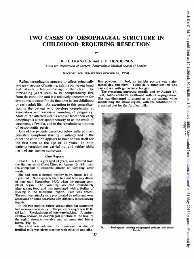
In the two months before consultation her symptomshad increased in severity. The patient's weight was 86 lb.(39 kg.). Physical signs of note were lacking. A bariumswallow showed an oesophageal stricture at the level ofthe eighth thoracic vertebra and an associated hiatushernia (Fig. 1).The child was admitted for treatment. A diet of
fortified milk was given together with olive oil and alka-55
line powders. In bed, an upright posture was main-tained day and night. Twice daily autodilatation wascarried out with gum-elastic bougies.The symptoms improved steadily and by August 27,
1951, solids could be swallowed without regurgitation.She was discharged to attend as an out-patient, whilemaintaining the above regime, with the substitution ofa normal diet for the fortified milk.
FIG. I.-Radiograph showing oesophageal stricture and hiatushernia.
copyright. on 31 M
ay 2018 by guest. Protected by
http://adc.bmj.com
/A
rch Dis C
hild: first published as 10.1136/adc.30.149.55 on 1 February 1955. D
ownloaded from
ARCHIVES OF DISEASE IN CHILDHOOD
.. ...../;:;
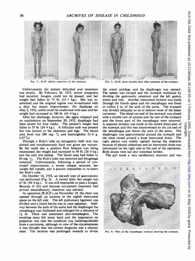
.before resection of the str
Unfortunately the patient defaulted and treatmentwas erratic. By February 26, 1952, severe symptomshad recurred, bougies could not be passed, and herweight had fallen to 73 lb. (33-1 kg.). She was re-admitted and the original regime was re-instituted witha slow but steady improvement. On discharge onMay 2, 1952, solids could be swallowed with ease and herweight had increased to 100 lb. (45 4 kg.).
After her discharge, however, she again relapsed andon readmission on September 20, 1952, dysphagia hadbeen severe for four weeks. The patient's weight hadfallen to 55 lb. (24-9 kg.). A follicular rash was presentbut was limited to the abdomen and legs. The bloodurea level was 206 mg. % and haemoglobin 21 -6 g.(147 %).Through a Ryle's tube an intragastric milk drip was
started and simultaneously fluid was given per rectum.By the tenth day a positive fluid balance was beingmaintained, her weight had increased to 58 lb. (26- 4 kg.)and the rash was fading. The blood urea had fallen to48 mg. %. The Ryle's tube was removed and bouginagerestarted. Unfortunately, following a period of con-tinued improvement, a severe relapse recurred, herweight fell rapidly, and it proved impossible to re-insertthe Ryle's tube.On October 14, 1952, an inkwell type of gastrostomy
was performed (Fig. 2). A month later, her weight was67 lb. (30 * 4 kg.). It was still impossible to pass a bougie.Because of this and because out-patient treatment hadproved unsatisfactory, resection was advised.At operation (R.H.F.) on November 18, the chest was
opened through an incision in the eighth intercostalspace on the left side. The left pulmonary ligament wasdivided and a hiatal hernia was at once apparent. Half-way between the arch of the aorta and the diaphragm theoesophagus was thickened and enlarged for a distance of1 1 in. There was associated peri-oesophagitis. Theresulting mass felt stony hard and the impression onpalpation was that the condition was indistinguishablefrom a carcinoma, although in view of the circumstancesit was thought that the correct diagnosis was a chroniculcer. The incision was prolonged inwards to divide
Fio. 3.-K.H. three months later after resection of the stricture.
FIG. 3.-K.H. three months later after resection of the stricture.
the costal cartilage and the diaphragm was opened.The spleen was excised and the stomach mobilized bydividing the gastrocolic omentum and the left gastricartery and vein. Another intercostal incision was madethrough the fourth space and the oesophagus was freedto within i in. of the arch of the aorta. The stomachwas divided obliquely so as to remove most of the lessercurvature. The distal cut end of the stomach was closedwith a double row of sutures and the rest of the stomachand the lower part of the oesophagus were removed.A separate incision was made in the closed distal part ofthe stomach and this was anastomosed to the cut end ofthe oesophagus just below the arch of the aorta. Thediaphragm was approximated around the stomach andthe chest closed around a large intercostal drain. Theright pleura was widely opened during the resectionbecause of pleural adhesions and an intercostal drain wasintroduced on the right side at the end of the operation.Both drains were led into waterseal bottles.The girl made a very satisfactory recovery and was
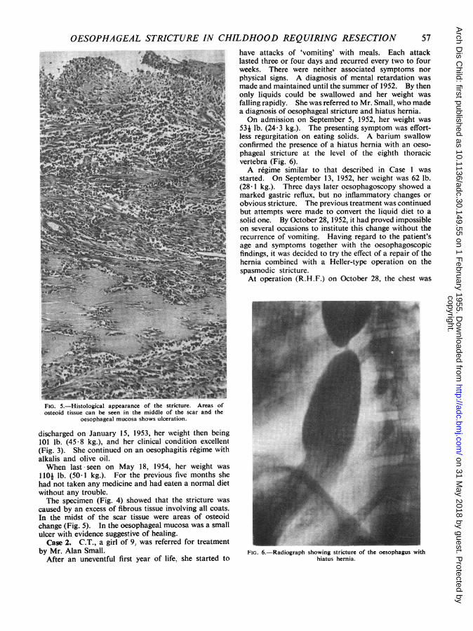
w~ ii...4~ 67 8,9 IjO.^"#.t. 2. ^ | -4^ .5 ~~~~~~~~~~~~~~~~~~~~~~~~~~~~................ 9 1Q l
FiG. 4.-Part of the oesophagus resected showing the stricture.
56
copyright. on 31 M
ay 2018 by guest. Protected by
http://adc.bmj.com
/A
rch Dis C
hild: first published as 10.1136/adc.30.149.55 on 1 February 1955. D
ownloaded from
OESOPHAGEAL STRICTURE IN CHILDHOOD REQUIRING RESECTIONhave attacks of 'vomiting' with meals. Each attacklasted three or four days and recurred every two to fourweeks. There were neither associated symptoms norphysical signs. A diagnosis of mental retardation wasmade and maintained until the summer of 1952. By thenonly liquids could be swallowed and her weight was
falling rapidly. She was referred to Mr. Small, who madea diagnosis of oesophageal stricture and hiatus hernia.On admission on September 5, 1952, her weight was
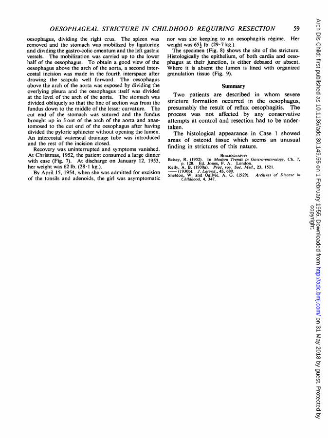
^ 53j lb. (24-3 kg.). The presenting symptom was effort-*_ j less regurgitation on eating solids. A barium swallow
confirmed the presence of a hiatus hernia with an oeso-phageal stricture at the level of the eighth thoracicvertebra (Fig. 6).A r6gime similar to that described in Case 1 was
started. On September 13, 1952, her weight was 62 lb.(28- 1 kg.). Three days later oesophagoscopy showed amarked gastric reflux, but no inflammatory changes orobvious stricture. The previous treatment was continuedbut attempts were made to convert the liquid diet to asolid one. By October 28, 1952, it had proved impossible
3 on several occasions to institute this change without therecurrence of vomiting. Having regard to the patient'sage and symptoms together with the oesophagoscopicfindings, it was decided to try the effect of a repair of thehernia combined with a Heller-type operation on thespasmodic stricture.
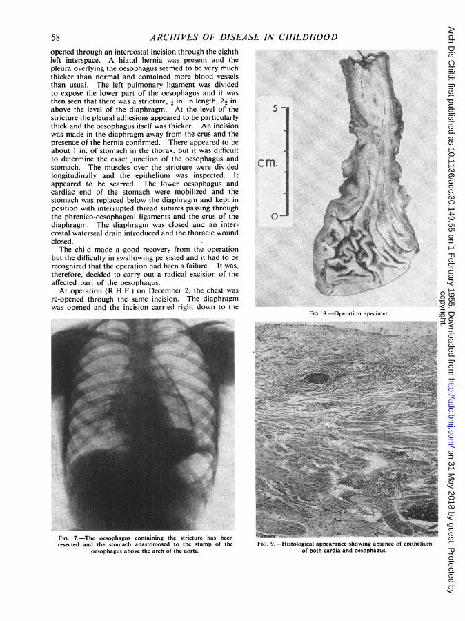
- At operation (R.H.F.) on October 28, the chest was2~~ ~ ~~ ~~~~iLF- .
~~ ~ ~NFIG. 5.-Histological appearance of the stricture. Areas ofosteoid tissue can be seen in the middle of the scar and the
oesophageal mucosa shows ulceration.
discharged on January 15, 1953, her weight then being101 lb. (45-8 kg.), and her clinical condition excellent(Fig. 3). She continued on an oesophagitis regime withalkalis and olive oil.When last seen on May 18, 1954, her weight was ...
1101 lb. (50-1 kg.). For the previous five months she U-had not taken any medicine and had eaten a normal dietwithout any trouble.The specimen (Fig. 4) showed that the stricture was
caused by an excess of fibrous tissue involving all coats.In the midst of the scar tissue were areas of osteoidchange (Fig. 5). In the oesophageal mucosa was a smallulcer with evidence suggestive of healing.
Case 2. C.T., a girl of 9, was referred for treatmentby Mr. Alan Small. FIG. 6.-Radiograph showing stricture of the oesophagus with
After an uneventful first year of life, she started to hiatus hernia.
57
copyright. on 31 M
ay 2018 by guest. Protected by
http://adc.bmj.com
/A
rch Dis C
hild: first published as 10.1136/adc.30.149.55 on 1 February 1955. D
ownloaded from
ARCHIVES OF DISEASE IN CHILDHOOD
opened through an intercostal incision through the eighthleft interspace. A hiatal hernia was present and thepleura overlying the oesophagus seemed to be very muchthicker than normal and contained more blood vesselsthan usual. The left pulmonary ligament was dividedto expose the lower part of the oesophagus and it wasthen seen that there was a stricture, in. in length, 21 in.above the level of the diaphragm. At the level of thestricture the pleural adhesions appeared to be particularlythick and the oesophagus itself was thicker. An incisionwas made in the diaphragm away from the crus and thepresence of the hernia confirmed. There appeared to beabout I in. of stomach in the thorax, but it was difficultto determine the exact junction of the oesophagus andstomach. The muscles over the stricture were dividedlongitudinally and the epithelium was inspected. Itappeared to be scarred. The lower oesophagus andcardiac end of the stomach were mobilized and thestomach was replaced below the diaphragm and kept inposition with interrupted thread sutures passing throughthe phrenico-oesophageal ligaments and the crus of thediaphragm. The diaphragm was closed and an inter-costal waterseal drain introduced and the thoracic woundclosed.The child made a good recovery from the operation
but the difficulty in swallowing persisted and it had to berecognized that the operation had been a failure. It was,therefore, decided to carry out a radical excision of theaffected part of the oesophagus.At operation (R.H.F.) on December 2, the chest was
re-opened through the same incision. The diaphragmwas opened and the incision carried right down to the
FIG. 7.-The oesophagus containing the stricture has beenresected and the stomach anastomosed to the stump of the
oesophagus above the arch of the aorta.
FIG. 8.-Operation specimen.
.-R _9 <x 1
FIG. 9.-Histological appearance showing absence of epitheliumof both cardia and oesophagus.
58
copyright. on 31 M
ay 2018 by guest. Protected by
http://adc.bmj.com
/A
rch Dis C
hild: first published as 10.1136/adc.30.149.55 on 1 February 1955. D
ownloaded from
OESOPHAGEAL STRICTURE IN CHILDHOOD REQUIRING RESECTION 59
oesophagus, dividing the right crus. The spleen wasremoved and the stomach was mobilized by ligaturingand dividing the gastro-colic omentum and the left gastricvessels. The mobilization was carried up to the lowerhalf of the oesophagus. To obtain a good view of theoesophagus above the arch of the aorta, a second inter-costal incision was made in the fourth interspace afterdrawing the scapula well forward. The oesophagusabove the arch of the aorta was exposed by dividing theoverlying pleura and the oesophagus itself was dividedat the level of the arch of the aorta. The stomach wasdivided obliquely so that the line of section was from thefundus down to the middle of the lesser curvature. Thecut end of the stomach was sutured and the fundusbrought up in front of the arch of the aorta and anas-tomosed to the cut end of the oesophagus after havingdivided the pyloric sphincter without opening the lumen.An intercostal waterseal drainage tube was introducedand the rest of the incision closed.
Recovery was uninterrupted and symptoms vanished.At Christmas, 1952, the patient consumed a large dinnerwith ease (Fig. 7). At discharge on January 12, 1953,her weight was 62 lb. (28-1 kg.).By April 15, 1954, when she was admitted for excision
of the tonsils and adenoids, the girl was asymptomatic
nor was she keeping to an oesophagitis regime. Herweight was 65j lb. (29.7 kg.).The specimen (Fig. 8) shows the site of the stricture.
Histologically the epithelium, of both cardia and oeso-phagus at their junction, is either debased or absent.Where it is absent the lumen is lined with organizedgranulation tissue (Fig. 9).
SummaryTwo patients are described in whom severe
stricture formation occurred in the oesophagus,presumably the result of reflux oesophagitis. Theprocess was not affected by any conservativeattempts at control and resection had to be under-taken.The histological appearance in Case 1 showed
areas of osteoid tissue which seems an unusualfinding in strictures of this nature.
BIBLIOGRAPHYBelsey, R. (1952). In Modern Trends in Gasiro-enterology, Ch. 7,
p. 128. Ed. Jones, F. A. London.Kelly, A. B. (1930a). Proc. roy. Soc. Med., 23, 1521.
(1930b). J. Laryng., 45, 680.Sheldon, W. and Ogilvie, A. G. (1929). Archives of Disease int
Childhood, 4, 347.
copyright. on 31 M
ay 2018 by guest. Protected by
http://adc.bmj.com
/A
rch Dis C
hild: first published as 10.1136/adc.30.149.55 on 1 February 1955. D
ownloaded from