Embed Size (px)
Citation preview
Type-I IFN drives the differentiation of short-livedeffector CD81 T cells in vivo
Melanie Wiesel�1, Josh Crouse�1, Gregor Bedenikovic1,
Andrew Sutherland2, Nicole Joller2 and Annette Oxenius1
1 Institute for Microbiology, ETH Zurich, Zurich, Switzerland2 Department of Neurology, Center for Neurologic Diseases, Brigham and Women’s Hospital
and Harvard Medical School, Boston, MA, USA
Two subsets of CD81 T cells are generated early during an immune response; one of these
subsets forms the memory pool, known as memory precursor effector cells (MPECs),
identified by high expression of CD127 and low expression of KLRG1, whereas the other
subset forms short-lived effector cells (SLECs) identified by low expression of CD127 and
high expression of KLRG1. Here, we studied in vivo the role of type-I IFN in this fate
decision. We found that under priming conditions dominated by type-I IFN, as observed in
lymphocytic choriomeningitis virus (LCMV) infection, type-I IFN signaling directly
impacted the regulation of T-bet and thus the early fate decision of CD81 T cells. In the
absence of type-I IFN signaling, CD81 T cells failed to form SLECs but could form MPECs
that give rise to functional memory CD81 T cells. Together, these findings identify type-I
IFN as an important factor driving SLEC differentiation and thus instructing the early
division between the effector and memory precursor CD81 T-cell pool.
Key words: CD81 T cells . Differentiation . Type I interferon . Viral infection
Supporting Information available online
Introduction
In response to an acute infection CD81 T cells rapidly expand to
form a pool of effector cells with cytolytic and cytokine secretion
activity. The pool of early effector cells can be divided into two
main subsets according to their ability to form terminally
differentiated effector cells or long-lived memory cells; referred
to as short-lived effector cells (SLECs), CD127low and KLRG1high,
and as memory precursor effector cells (MPECs), CD127high and
KLRG1low, respectively [1, 2]. There is strong evidence that
inflammatory cytokines present during CD81 T-cell priming play
a key role in the effector and memory fate decision process [3–5]. In
support of this notion it has been shown that IL-12 signaling is
mandatory for driving activated CD81 T cells toward an SLEC
phenotype upon infection with Listeria monocytogenes but not
vesicular stomatitis virus (VSV), vaccinia virus (VV) or lymphocytic
choriomeningitis virus (LCMV) [5]. Similar to IL-12, type-I IFN
signaling has been shown to support the proliferation and
development of cytolytic activity of CD81 T cells in vitro [6–10]
and can act as an adjuvant in vivo to a variety of stimuli [3, 11–14].
IL-12 and type-I IFN were shown to support programming of
memory CD81 T cells in response to L. monocytogenes and VV
infection [10]. Moreover, it was recently shown that prolonged
�The authors contributed equally to this work.Correspondence: Prof. Annette Oxeniuse-mail: [email protected]
& 2011 WILEY-VCH Verlag GmbH & Co. KGaA, Weinheim www.eji-journal.eu
DOI 10.1002/eji.201142091 Eur. J. Immunol. 2012. 42: 320–329Melanie Wiesel et al.320
IL-2 signaling on CD81 T cells during the priming with LCMV
promotes SLEC differentiation [15, 16]. Thus, depending on the
nature of the infection, the associated cytokine milieu critically
regulates effector and memory CD81 T-cell development.
Although there are several in vivo studies focusing on the role
of IL-12 in this fate decision process [3–5], a direct role of type-I
IFN in the instruction of SLEC versus MPEC differentiation has so
far not been studied in vivo.
Here, we have addressed the requirement of type-I IFN
signaling on the early fate decision of CD81 T cells in a type-I IFN
biased cytokine milieu as found in LCMV infection. We provide
evidence that direct type-I IFN signaling in CD81 T cells
augments the level of the transcription factor T-bet and thereby
instructs the transition of CD81 T cells toward an SLEC pheno-
type. CD81 T cells lacking the type-I IFN receptor fail to form
SLECs but instead preferentially give rise to MPECs. Although the
primary expansion of these cells is strongly reduced, they have
the capacity to develop into functional memory cells. In
summary, the data presented here demonstrate that during
infections associated with abundant levels of type-I IFN, the early
lineage choice toward the differentiation of SLECs is mediated by
direct type-I IFN signaling on CD81 T cells, identifying type-I IFN
as an important factor instructing the early division between the
effector and memory CD81 T-cell pool.
Results
Impaired expansion of CD81 T cells in the absence ofdirect type-I IFN signaling
To investigate the role of direct type-I IFN signaling on the SLEC
versus MPEC fate decision of CD81 T cells, we used an
established LCMV8.7 and vaccinia virus expressing the LCMV
glycoprotein (VVG2) co-infection model [17] combined with
adoptive transfer of LCMV gp33-specific TCR-transgenic CD81
T cells (P14) which are either sufficient or deficient for type-I IFN
signaling (hereafter: WT P14 and interferon-alpha receptor
(IFNAR)�/� P14 respectively). Using this system we were able
to generate a type-I IFN-dominated inflammatory environment
induced by LCMV8.7 infection in face of antigen presentation
exclusively derived from a recombinant VVG2, as P14 cells only
recognize their cognate epitope expressed by VVG2 but do not
recognize the mutant gp33 (V35L) epitope expressed by
LCMV8.7. We chose this co-infection system as it avoids the
LCMV-inherent abundant antigen presentation and hence puts
more emphasis on the role of the cytokine milieu in CD81 T-cell
priming and differentiation. Consistent with previous findings
upon single infection with WT LCMV [17–19], WT and IFNAR�/�
P14 cells underwent substantial expansion during the first three
days after LCMV8.7 and VVG2 co-infection. However, IFNAR�/�
P14 cells were already five-fold reduced in frequency by day 3,
and by day 6 post-infection the expansion of IFNAR�/� P14 cells
was found to be drastically reduced in frequency and total
numbers in the spleen compared with the frequency and number
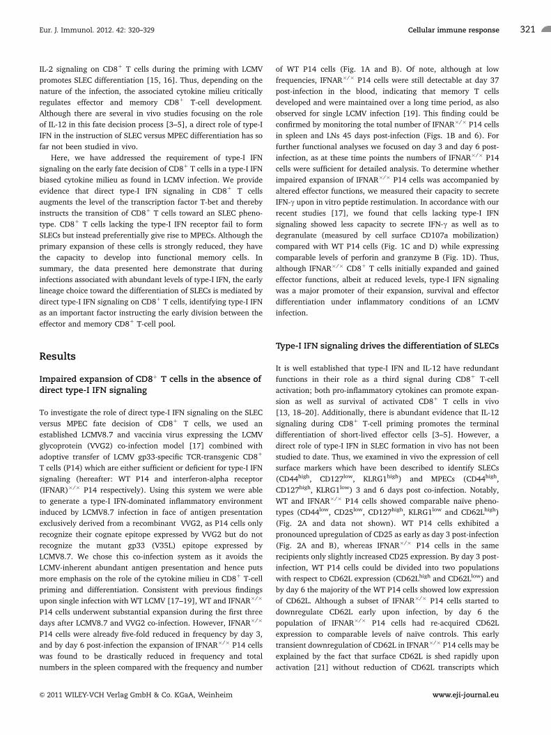
of WT P14 cells (Fig. 1A and B). Of note, although at low
frequencies, IFNAR�/� P14 cells were still detectable at day 37
post-infection in the blood, indicating that memory T cells
developed and were maintained over a long time period, as also
observed for single LCMV infection [19]. This finding could be
confirmed by monitoring the total number of IFNAR�/� P14 cells
in spleen and LNs 45 days post-infection (Figs. 1B and 6). For
further functional analyses we focused on day 3 and day 6 post-
infection, as at these time points the numbers of IFNAR�/� P14
cells were sufficient for detailed analysis. To determine whether
impaired expansion of IFNAR�/� P14 cells was accompanied by
altered effector functions, we measured their capacity to secrete
IFN-g upon in vitro peptide restimulation. In accordance with our
recent studies [17], we found that cells lacking type-I IFN
signaling showed less capacity to secrete IFN-g as well as to
degranulate (measured by cell surface CD107a mobilization)
compared with WT P14 cells (Fig. 1C and D) while expressing
comparable levels of perforin and granzyme B (Fig. 1D). Thus,
although IFNAR�/� CD81 T cells initially expanded and gained
effector functions, albeit at reduced levels, type-I IFN signaling
was a major promoter of their expansion, survival and effector
differentiation under inflammatory conditions of an LCMV
infection.
Type-I IFN signaling drives the differentiation of SLECs
It is well established that type-I IFN and IL-12 have redundant
functions in their role as a third signal during CD81 T-cell
activation; both pro-inflammatory cytokines can promote expan-
sion as well as survival of activated CD81 T cells in vivo
[13, 18–20]. Additionally, there is abundant evidence that IL-12
signaling during CD81 T-cell priming promotes the terminal
differentiation of short-lived effector cells [3–5]. However, a
direct role of type-I IFN in SLEC formation in vivo has not been
studied to date. Thus, we examined in vivo the expression of cell
surface markers which have been described to identify SLECs
(CD44high, CD127low, KLRG1high) and MPECs (CD44high,
CD127high, KLRG1low) 3 and 6 days post co-infection. Notably,
WT and IFNAR�/� P14 cells showed comparable naıve pheno-
types (CD44low, CD25low, CD127high, KLRG1low and CD62Lhigh)
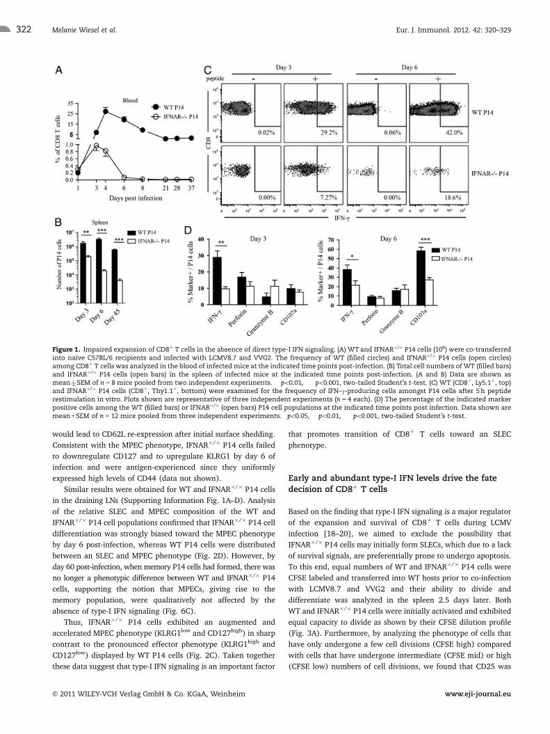
(Fig. 2A and data not shown). WT P14 cells exhibited a
pronounced upregulation of CD25 as early as day 3 post-infection
(Fig. 2A and B), whereas IFNAR�/� P14 cells in the same
recipients only slightly increased CD25 expression. By day 3 post-
infection, WT P14 cells could be divided into two populations
with respect to CD62L expression (CD62Lhigh and CD62Llow) and
by day 6 the majority of the WT P14 cells showed low expression
of CD62L. Although a subset of IFNAR�/� P14 cells started to
downregulate CD62L early upon infection, by day 6 the
population of IFNAR�/� P14 cells had re-acquired CD62L
expression to comparable levels of naıve controls. This early
transient downregulation of CD62L in IFNAR�/� P14 cells may be
explained by the fact that surface CD62L is shed rapidly upon
activation [21] without reduction of CD62L transcripts which
Eur. J. Immunol. 2012. 42: 320–329 Cellular immune response 321
& 2011 WILEY-VCH Verlag GmbH & Co. KGaA, Weinheim www.eji-journal.eu
would lead to CD62L re-expression after initial surface shedding.
Consistent with the MPEC phenotype, IFNAR�/� P14 cells failed
to downregulate CD127 and to upregulate KLRG1 by day 6 of
infection and were antigen-experienced since they uniformly
expressed high levels of CD44 (data not shown).
Similar results were obtained for WT and IFNAR�/� P14 cells
in the draining LNs (Supporting Information Fig. 1A–D). Analysis
of the relative SLEC and MPEC composition of the WT and
IFNAR�/� P14 cell populations confirmed that IFNAR�/� P14 cell
differentiation was strongly biased toward the MPEC phenotype
by day 6 post-infection, whereas WT P14 cells were distributed
between an SLEC and MPEC phenotype (Fig. 2D). However, by
day 60 post-infection, when memory P14 cells had formed, there was
no longer a phenotypic difference between WT and IFNAR�/� P14
cells, supporting the notion that MPECs, giving rise to the
memory population, were qualitatively not affected by the
absence of type-I IFN signaling (Fig. 6C).
Thus, IFNAR�/� P14 cells exhibited an augmented and
accelerated MPEC phenotype (KLRG1low and CD127high) in sharp
contrast to the pronounced effector phenotype (KLRG1high and
CD127low) displayed by WT P14 cells (Fig. 2C). Taken together
these data suggest that type-I IFN signaling is an important factor
that promotes transition of CD81 T cells toward an SLEC
phenotype.
Early and abundant type-I IFN levels drive the fatedecision of CD81 T cells
Based on the finding that type-I IFN signaling is a major regulator
of the expansion and survival of CD81 T cells during LCMV
infection [18–20], we aimed to exclude the possibility that
IFNAR�/� P14 cells may initially form SLECs, which due to a lack
of survival signals, are preferentially prone to undergo apoptosis.
To this end, equal numbers of WT and IFNAR�/� P14 cells were
CFSE labeled and transferred into WT hosts prior to co-infection
with LCMV8.7 and VVG2 and their ability to divide and
differentiate was analyzed in the spleen 2.5 days later. Both
WT and IFNAR�/� P14 cells were initially activated and exhibited
equal capacity to divide as shown by their CFSE dilution profile
(Fig. 3A). Furthermore, by analyzing the phenotype of cells that
have only undergone a few cell divisions (CFSE high) compared
with cells that have undergone intermediate (CFSE mid) or high
(CFSE low) numbers of cell divisions, we found that CD25 was
Figure 1. Impaired expansion of CD81 T cells in the absence of direct type-I IFN signaling. (A) WT and IFNAR�/� P14 cells (106) were co-transferredinto naıve C57BL/6 recipients and infected with LCMV8.7 and VVG2. The frequency of WT (filled circles) and IFNAR�/� P14 cells (open circles)among CD81 T cells was analyzed in the blood of infected mice at the indicated time points post-infection. (B) Total cell numbers of WT (filled bars)and IFNAR�/� P14 cells (open bars) in the spleen of infected mice at the indicated time points post-infection. (A and B) Data are shown asmean7SEM of n 5 8 mice pooled from two independent experiments. ��po0.01, ���po0.001, two-tailed Student’s t-test. (C) WT (CD81, Ly5.11, top)and IFNAR�/� P14 cells (CD81, Thy1.11, bottom) were examined for the frequency of IFN-g-producing cells amongst P14 cells after 5 h peptiderestimulation in vitro. Plots shown are representative of three independent experiments (n 5 4 each). (D) The percentage of the indicated markerpositive cells among the WT (filled bars) or IFNAR�/� (open bars) P14 cell populations at the indicated time points post infection. Data shown aremean1SEM of n 5 12 mice pooled from three independent experiments. �po0.05, ��po0.01, ���po0.001, two-tailed Student’s t-test.
Eur. J. Immunol. 2012. 42: 320–329Melanie Wiesel et al.322
& 2011 WILEY-VCH Verlag GmbH & Co. KGaA, Weinheim www.eji-journal.eu
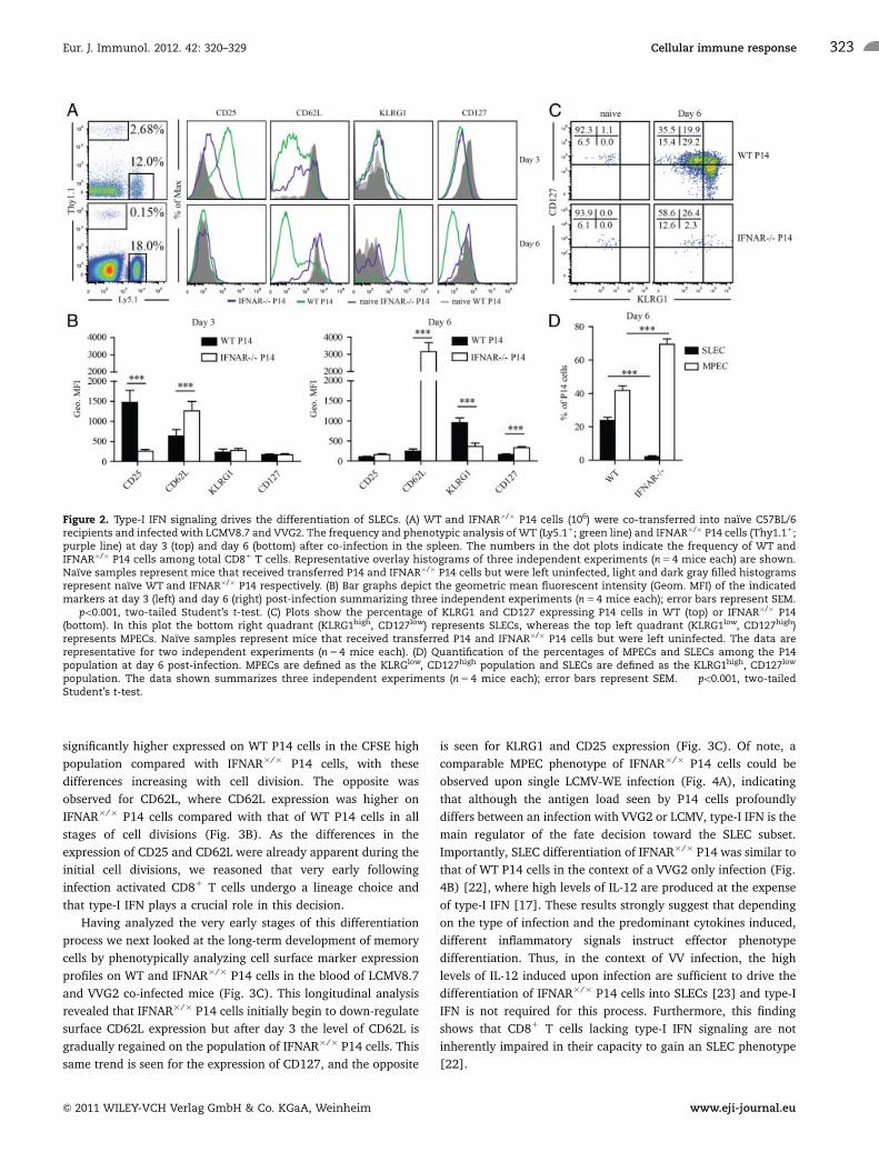
significantly higher expressed on WT P14 cells in the CFSE high
population compared with IFNAR�/� P14 cells, with these
differences increasing with cell division. The opposite was
observed for CD62L, where CD62L expression was higher on
IFNAR�/� P14 cells compared with that of WT P14 cells in all
stages of cell divisions (Fig. 3B). As the differences in the
expression of CD25 and CD62L were already apparent during the
initial cell divisions, we reasoned that very early following
infection activated CD81 T cells undergo a lineage choice and
that type-I IFN plays a crucial role in this decision.
Having analyzed the very early stages of this differentiation
process we next looked at the long-term development of memory
cells by phenotypically analyzing cell surface marker expression
profiles on WT and IFNAR�/� P14 cells in the blood of LCMV8.7
and VVG2 co-infected mice (Fig. 3C). This longitudinal analysis
revealed that IFNAR�/� P14 cells initially begin to down-regulate
surface CD62L expression but after day 3 the level of CD62L is
gradually regained on the population of IFNAR�/� P14 cells. This
same trend is seen for the expression of CD127, and the opposite
is seen for KLRG1 and CD25 expression (Fig. 3C). Of note, a
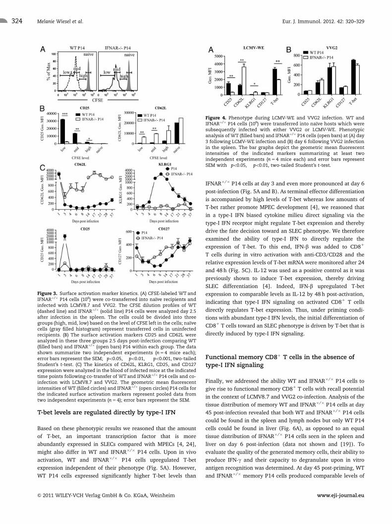
comparable MPEC phenotype of IFNAR�/� P14 cells could be
observed upon single LCMV-WE infection (Fig. 4A), indicating
that although the antigen load seen by P14 cells profoundly
differs between an infection with VVG2 or LCMV, type-I IFN is the
main regulator of the fate decision toward the SLEC subset.
Importantly, SLEC differentiation of IFNAR�/� P14 was similar to
that of WT P14 cells in the context of a VVG2 only infection (Fig.
4B) [22], where high levels of IL-12 are produced at the expense
of type-I IFN [17]. These results strongly suggest that depending
on the type of infection and the predominant cytokines induced,
different inflammatory signals instruct effector phenotype
differentiation. Thus, in the context of VV infection, the high
levels of IL-12 induced upon infection are sufficient to drive the
differentiation of IFNAR�/� P14 cells into SLECs [23] and type-I
IFN is not required for this process. Furthermore, this finding
shows that CD81 T cells lacking type-I IFN signaling are not
inherently impaired in their capacity to gain an SLEC phenotype
[22].
Figure 2. Type-I IFN signaling drives the differentiation of SLECs. (A) WT and IFNAR�/� P14 cells (106) were co-transferred into naıve C57BL/6recipients and infected with LCMV8.7 and VVG2. The frequency and phenotypic analysis of WT (Ly5.11; green line) and IFNAR�/� P14 cells (Thy1.11;purple line) at day 3 (top) and day 6 (bottom) after co-infection in the spleen. The numbers in the dot plots indicate the frequency of WT andIFNAR�/� P14 cells among total CD81 T cells. Representative overlay histograms of three independent experiments (n 5 4 mice each) are shown.Naıve samples represent mice that received transferred P14 and IFNAR�/� P14 cells but were left uninfected, light and dark gray filled histogramsrepresent naıve WT and IFNAR�/� P14 respectively. (B) Bar graphs depict the geometric mean fluorescent intensity (Geom. MFI) of the indicatedmarkers at day 3 (left) and day 6 (right) post-infection summarizing three independent experiments (n 5 4 mice each); error bars represent SEM.���po0.001, two-tailed Student’s t-test. (C) Plots show the percentage of KLRG1 and CD127 expressing P14 cells in WT (top) or IFNAR�/� P14(bottom). In this plot the bottom right quadrant (KLRG1high, CD127low) represents SLECs, whereas the top left quadrant (KLRG1low, CD127high)represents MPECs. Naıve samples represent mice that received transferred P14 and IFNAR�/� P14 cells but were left uninfected. The data arerepresentative for two independent experiments (n 5 4 mice each). (D) Quantification of the percentages of MPECs and SLECs among the P14population at day 6 post-infection. MPECs are defined as the KLRGlow, CD127high population and SLECs are defined as the KLRG1high, CD127low
population. The data shown summarizes three independent experiments (n 5 4 mice each); error bars represent SEM. ���po0.001, two-tailedStudent’s t-test.
Eur. J. Immunol. 2012. 42: 320–329 Cellular immune response 323
& 2011 WILEY-VCH Verlag GmbH & Co. KGaA, Weinheim www.eji-journal.eu
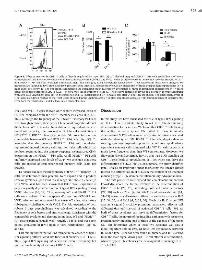
T-bet levels are regulated directly by type-I IFN
Based on these phenotypic results we reasoned that the amount
of T-bet, an important transcription factor that is more
abundantly expressed in SLECs compared with MPECs [4, 24],
might also differ in WT and IFNAR�/� P14 cells. Upon in vivo
activation, WT and IFNAR�/� P14 cells upregulated T-bet
expression independent of their phenotype (Fig. 5A). However,
WT P14 cells expressed significantly higher T-bet levels than
IFNAR�/� P14 cells at day 3 and even more pronounced at day 6
post-infection (Fig. 5A and B). As terminal effector differentiation
is accompanied by high levels of T-bet whereas low amounts of
T-bet rather promote MPEC development [4], we reasoned that
in a type-I IFN biased cytokine milieu direct signaling via the
type-I IFN receptor might regulate T-bet expression and thereby
drive the fate decision toward an SLEC phenotype. We therefore
examined the ability of type-I IFN to directly regulate the
expression of T-bet. To this end, IFN-b was added to CD81
T cells during in vitro activation with anti-CD3/CD28 and the
relative expression levels of T-bet mRNA were monitored after 24
and 48 h (Fig. 5C). IL-12 was used as a positive control as it was
previously shown to induce T-bet expression, thereby driving
SLEC differentiation [4]. Indeed, IFN-b upregulated T-bet
expression to comparable levels as IL-12 by 48 h post-activation,
indicating that type-I IFN signaling on activated CD81 T cells
directly regulates T-bet expression. Thus, under priming condi-
tions with abundant type-I IFN levels, the initial differentiation of
CD81 T cells toward an SLEC phenotype is driven by T-bet that is
directly induced by type-I IFN signaling.
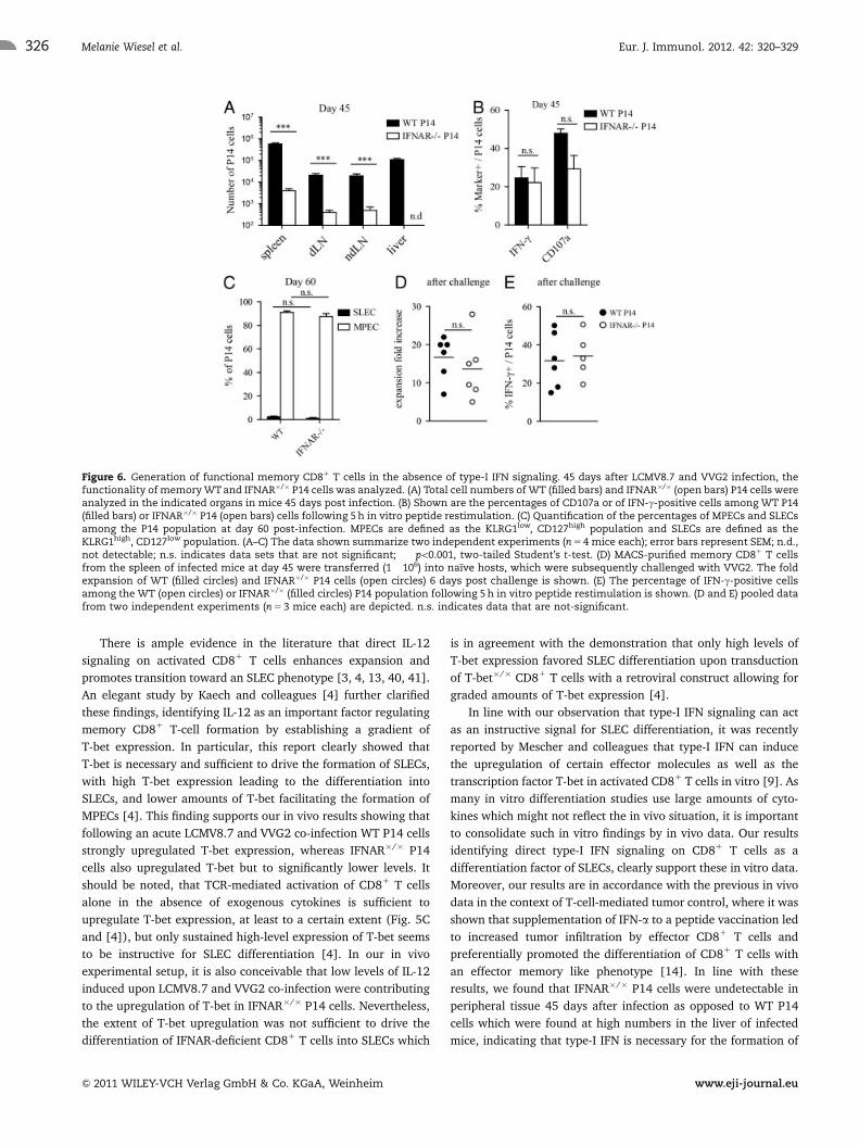
Functional memory CD81 T cells in the absence oftype-I IFN signaling
Finally, we addressed the ability WT and IFNAR�/� P14 cells to
give rise to functional memory CD81 T cells with recall potential
in the context of LCMV8.7 and VVG2 co-infection. Analysis of the
tissue distribution of memory WT and IFNAR�/� P14 cells at day
45 post-infection revealed that both WT and IFNAR�/� P14 cells
could be found in the spleen and lymph nodes but only WT P14
cells could be found in liver (Fig. 6A), as opposed to an equal
tissue distribution of IFNAR�/� P14 cells seen in the spleen and
liver on day 6 post-infection (data not shown and [19]). To
evaluate the quality of the generated memory cells, their ability to
produce IFN-g and their capacity to degranulate upon in vitro
antigen recognition was determined. At day 45 post-priming, WT
and IFNAR�/� memory P14 cells produced comparable levels of
Figure 3. Surface activation marker kinetics. (A) CFSE-labeled WT andIFNAR�/� P14 cells (106) were co-transferred into naıve recipients andinfected with LCMV8.7 and VVG2. The CFSE dilution profiles of WT(dashed line) and IFNAR�/� (solid line) P14 cells were analyzed day 2.5after infection in the spleen. The cells could be divided into threegroups (high, mid, low) based on the level of CFSE left in the cells; naıvecells (gray filled histogram) represent transferred cells in uninfectedrecipients. (B) The surface activation markers CD25 and CD62L wereanalyzed in these three groups 2.5 days post-infection comparing WT(filled bars) and IFNAR�/� (open bars) P14 within each group. The datashown summarize two independent experiments (n 5 4 mice each);error bars represent the SEM; �po0.05, ��po0.01, ���po0.001, two-tailedStudent’s t-test. (C) The kinetics of CD62L, KLRG1, CD25, and CD127expression were analyzed in the blood of infected mice at the indicatedtime points following co-transfer of WT and IFNAR�/� P14 cells and co-infection with LCMV8.7 and VVG2. The geometric mean fluorescentintensities of WT (filled circles) and IFNAR�/� (open circles) P14 cells forthe indicated surface activation markers represent pooled data fromtwo independent experiments (n 5 4); error bars represent the SEM.
Figure 4. Phenotype during LCMV-WE and VVG2 infection. WT andIFNAR�/� P14 cells (106) were transferred into naıve hosts which weresubsequently infected with either VVG2 or LCMV-WE. Phenotypicanalysis of WT (filled bars) and IFNAR�/� P14 cells (open bars) at (A) day3 following LCMV-WE infection and (B) day 6 following VVG2 infectionin the spleen. The bar graphs depict the geometric mean fluorescentintensities of the indicated markers summarizing at least twoindependent experiments (n 5 4 mice each) and error bars representSEM with �po0.05, ��po0.01, two-tailed Student’s t-test.
Eur. J. Immunol. 2012. 42: 320–329Melanie Wiesel et al.324
& 2011 WILEY-VCH Verlag GmbH & Co. KGaA, Weinheim www.eji-journal.eu
IFN-g and WT P14 cells showed only slightly increased levels of
CD107a compared with IFNAR�/� memory P14 cells (Fig. 6B).
Thus, although the frequency of the IFNAR�/� memory P14 cells
was strongly reduced, their per-cell functional properties did not
differ from WT P14 cells. In addition to equivalent ex vivo
functional capacity, the proportion of P14 cells exhibiting a
CD127high KLRG1low phenotype at day 60 post-infection was
comparable between WT and IFNAR�/� P14 cells (Fig. 6C). To
ascertain that the memory IFNAR�/� P14 cell population
represented indeed memory cells and not naıve cells which had
not been recruited into the primary response, we measured CD44
expression on the IFNAR�/� P14 cells. As all IFNAR�/� P14 cells
uniformly expressed high levels of CD44, we conclude that these
cells are indeed antigen-experienced memory cells (data not
shown).
To further validate the functionality of IFNAR�/�memory P14
cells, we determined their potential to re-expand and to produce
effector cytokines upon viral re-challenge. We chose a challenge
with VVG2 as it has been shown that CD81 T-cell expansion is
only marginally dependent on direct type-I IFN signaling during
VVG2 infection [10, 17]. Thus, memory WT and IFNAR�/� P14
cells were isolated from the spleen 45 days post-LCMV8.7 and
VVG2 infection and transferred into naıve WT mice, which were
subsequently challenged with VVG2. The fold expansion of both
subsets 6 days post-challenge was calculated according to the
frequency of cells before and after challenge. Consistent with the
comparable cytokine and degranulation data, WT and IFNAR�/�
P14 cells expanded equally well (roughly 15-fold) and were both
potent producers of IFN-g upon in vitro restimulation (Fig. 6D
and E).
This finding shows that MPECs formed in the absence of type-I
IFN signaling differentiated into functional memory CD81 T cells.
Thus, type-I IFN signaling influences the overall frequency but
not the functionality of memory CD81 T cells.
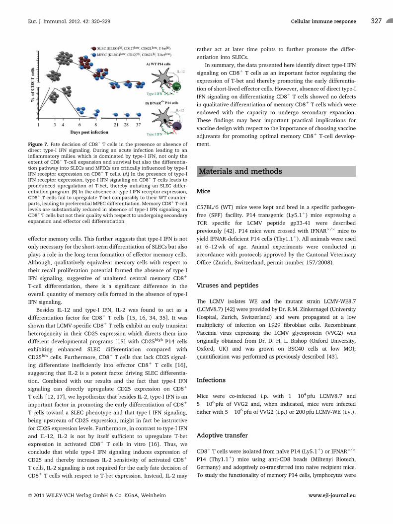
Discussion
In this study, we have elucidated the role of type-I IFN signaling
on CD81 T cells and its ability to act as a fate-determining
differentiation factor in vivo. We found that CD81 T cells lacking
the ability to sense type-I IFN failed to form terminally
differentiated SLECs following an acute viral infection associated
with abundant type-I IFN. IFNAR�/� P14 cells, despite demon-
strating a reduced expansion potential, could form qualitatively
equivalent memory cells compared with WT P14 cells, albeit at a
much lower frequency than their WT counterparts. Moreover, we
showed in vivo and confirmed in vitro that type-I IFN signaling on
CD81 T cells leads to upregulation of T-bet which can drive the
differentiation of SLECs (Fig. 7). In summary, this study identifies
type-I IFN as an important factor instructing the lineage choice
toward the differentiation of SLECs in the context of an infection
inducing a type-I IFN-dominated inflammatory cytokine milieu.
The data presented here expand and complement our current
knowledge about the factors involved in the differentiation of
CD81 T cells [25, 26], including both cell intrinsic factors
[27, 28] such as T-bet [4, 24, 28–31] and eomesodermin [24,
31–33] as well as cell extrinsic differentiation factors, such as IL-2
[15, 34, 35] and IL-12 [4, 5, 28, 30]. Much like IL-12, type-I IFN
acts as a signal 3 cytokine promoting expansion, effector cell
differentiation and survival of activated CD81 T cells [36]. As
both of these cytokines can serve as differentiation factors for
CD81 T cells, the nature of the invading pathogen with respect to
predominantly inducing one of those at the expense of the other
[37, 38] determines which of these two cytokines will play a
more important role in vivo. Of note, less redundancy between
IL-12 and type-I IFN has been found in humans and IL-12 seems
to be the main signal driving CD81 T-cell effector differentiation,
whereas type-I IFN enhances the development of memory CD81
T cells [39].
Figure 5. T-bet expression in CD81 T cells is directly regulated by type-I IFN. (A) WT (dashed line) and IFNAR�/� P14 cells (solid line) (106) wereco-transferred into naıve mice which were then co-infected with LCMV8.7 and VVG2. Naıve samples represent mice that received transferred WTand IFNAR�/� P14 cells but were left uninfected (light and dark gray filled histograms respectively). T-bet expression levels were analyzed byintracellular staining at day 3 (top) and day 6 (bottom) post-infection. Representative overlay histograms of three independent experiments (n 5 4mice each) are shown (B) The bar graph summarizes the geometric mean fluorescent intensities of three independent experiments (n 5 4 miceeach); error bars represent SEM. �po0.05, ��po0.01, two-tailed Student’s t-test. (C) The relative expression levels of T-bet upon in vitro activationwith anti-CD3/CD28 (light gray bar) in the presence of IL-12 (black bar) and IFN-b (white bar) after 24 and 48 h are shown. The expression levels ofT-bet were calculated relative to the T-bet levels obtained in the unstimulated 0 h control sample. Data pooled from four independent experiments,error bars represent SEM. �po0.05, two-tailed Student’s t-test.
Eur. J. Immunol. 2012. 42: 320–329 Cellular immune response 325
& 2011 WILEY-VCH Verlag GmbH & Co. KGaA, Weinheim www.eji-journal.eu
There is ample evidence in the literature that direct IL-12
signaling on activated CD81 T cells enhances expansion and
promotes transition toward an SLEC phenotype [3, 4, 13, 40, 41].
An elegant study by Kaech and colleagues [4] further clarified
these findings, identifying IL-12 as an important factor regulating
memory CD81 T-cell formation by establishing a gradient of
T-bet expression. In particular, this report clearly showed that
T-bet is necessary and sufficient to drive the formation of SLECs,
with high T-bet expression leading to the differentiation into
SLECs, and lower amounts of T-bet facilitating the formation of
MPECs [4]. This finding supports our in vivo results showing that
following an acute LCMV8.7 and VVG2 co-infection WT P14 cells
strongly upregulated T-bet expression, whereas IFNAR�/� P14
cells also upregulated T-bet but to significantly lower levels. It
should be noted, that TCR-mediated activation of CD81 T cells
alone in the absence of exogenous cytokines is sufficient to
upregulate T-bet expression, at least to a certain extent (Fig. 5C
and [4]), but only sustained high-level expression of T-bet seems
to be instructive for SLEC differentiation [4]. In our in vivo
experimental setup, it is also conceivable that low levels of IL-12
induced upon LCMV8.7 and VVG2 co-infection were contributing
to the upregulation of T-bet in IFNAR�/� P14 cells. Nevertheless,
the extent of T-bet upregulation was not sufficient to drive the
differentiation of IFNAR-deficient CD81 T cells into SLECs which
is in agreement with the demonstration that only high levels of
T-bet expression favored SLEC differentiation upon transduction
of T-bet�/� CD81 T cells with a retroviral construct allowing for
graded amounts of T-bet expression [4].
In line with our observation that type-I IFN signaling can act
as an instructive signal for SLEC differentiation, it was recently
reported by Mescher and colleagues that type-I IFN can induce
the upregulation of certain effector molecules as well as the
transcription factor T-bet in activated CD81 T cells in vitro [9]. As
many in vitro differentiation studies use large amounts of cyto-
kines which might not reflect the in vivo situation, it is important
to consolidate such in vitro findings by in vivo data. Our results
identifying direct type-I IFN signaling on CD81 T cells as a
differentiation factor of SLECs, clearly support these in vitro data.
Moreover, our results are in accordance with the previous in vivo
data in the context of T-cell-mediated tumor control, where it was
shown that supplementation of IFN-a to a peptide vaccination led
to increased tumor infiltration by effector CD81 T cells and
preferentially promoted the differentiation of CD81 T cells with
an effector memory like phenotype [14]. In line with these
results, we found that IFNAR�/� P14 cells were undetectable in
peripheral tissue 45 days after infection as opposed to WT P14
cells which were found at high numbers in the liver of infected
mice, indicating that type-I IFN is necessary for the formation of
Figure 6. Generation of functional memory CD81 T cells in the absence of type-I IFN signaling. 45 days after LCMV8.7 and VVG2 infection, thefunctionality of memory WT and IFNAR�/� P14 cells was analyzed. (A) Total cell numbers of WT (filled bars) and IFNAR�/� (open bars) P14 cells wereanalyzed in the indicated organs in mice 45 days post infection. (B) Shown are the percentages of CD107a or of IFN-g-positive cells among WT P14(filled bars) or IFNAR�/� P14 (open bars) cells following 5 h in vitro peptide restimulation. (C) Quantification of the percentages of MPECs and SLECsamong the P14 population at day 60 post-infection. MPECs are defined as the KLRG1low, CD127high population and SLECs are defined as theKLRG1high, CD127low population. (A–C) The data shown summarize two independent experiments (n 5 4 mice each); error bars represent SEM; n.d.,not detectable; n.s. indicates data sets that are not significant; ���po0.001, two-tailed Student’s t-test. (D) MACS-purified memory CD81 T cellsfrom the spleen of infected mice at day 45 were transferred (1� 106) into naıve hosts, which were subsequently challenged with VVG2. The foldexpansion of WT (filled circles) and IFNAR�/� P14 cells (open circles) 6 days post challenge is shown. (E) The percentage of IFN-g-positive cellsamong the WT (open circles) or IFNAR�/� (filled circles) P14 population following 5 h in vitro peptide restimulation is shown. (D and E) pooled datafrom two independent experiments (n 5 3 mice each) are depicted. n.s. indicates data that are not-significant.
Eur. J. Immunol. 2012. 42: 320–329Melanie Wiesel et al.326
& 2011 WILEY-VCH Verlag GmbH & Co. KGaA, Weinheim www.eji-journal.eu
effector memory cells. This further suggests that type-I IFN is not
only necessary for the short-term differentiation of SLECs but also
plays a role in the long-term formation of effector memory cells.
Although, qualitatively equivalent memory cells with respect to
their recall proliferation potential formed the absence of type-I
IFN signaling, suggestive of unaltered central memory CD81
T-cell differentiation, there is a significant difference in the
overall quantity of memory cells formed in the absence of type-I
IFN signaling.
Besides IL-12 and type-I IFN, IL-2 was found to act as a
differentiation factor for CD81 T cells [15, 16, 34, 35]. It was
shown that LCMV-specific CD81 T cells exhibit an early transient
heterogeneity in their CD25 expression which directs them into
different developmental programs [15] with CD25high P14 cells
exhibiting enhanced SLEC differentiation compared with
CD25low cells. Furthermore, CD81 T cells that lack CD25 signal-
ing differentiate inefficiently into effector CD81 T cells [16],
suggesting that IL-2 is a potent factor driving SLEC differentia-
tion. Combined with our results and the fact that type-I IFN
signaling can directly upregulate CD25 expression on CD81
T cells [12, 17], we hypothesize that besides IL-2, type-I IFN is an
important factor in promoting the early differentiation of CD81
T cells toward a SLEC phenotype and that type-I IFN signaling,
being upstream of CD25 expression, might in fact be instructive
for CD25 expression levels. Furthermore, in contrast to type-I IFN
and IL-12, IL-2 is not by itself sufficient to upregulate T-bet
expression in activated CD81 T cells in vitro [16]. Thus, we
conclude that while type-I IFN signaling induces expression of
CD25 and thereby increases IL-2 sensitivity of activated CD81
T cells, IL-2 signaling is not required for the early fate decision of
CD81 T cells with respect to T-bet expression. Instead, IL-2 may
rather act at later time points to further promote the differ-
entiation into SLECs.
In summary, the data presented here identify direct type-I IFN
signaling on CD81 T cells as an important factor regulating the
expression of T-bet and thereby promoting the early differentia-
tion of short-lived effector cells. However, absence of direct type-I
IFN signaling on differentiating CD81 T cells showed no defects
in qualitative differentiation of memory CD81 T cells which were
endowed with the capacity to undergo secondary expansion.
These findings may bear important practical implications for
vaccine design with respect to the importance of choosing vaccine
adjuvants for promoting optimal memory CD81 T-cell develop-
ment.
Materials and methods
Mice
C57BL/6 (WT) mice were kept and bred in a specific pathogen-
free (SPF) facility. P14 transgenic (Ly5.11) mice expressing a
TCR specific for LCMV peptide gp33-41 were described
previously [42]. P14 mice were crossed with IFNAR�/� mice to
yield IFNAR-deficient P14 cells (Thy1.11). All animals were used
at 6–12 wk of age. Animal experiments were conducted in
accordance with protocols approved by the Cantonal Veterinary
Office (Zurich, Switzerland, permit number 157/2008).
Viruses and peptides
The LCMV isolates WE and the mutant strain LCMV-WE8.7
(LCMV8.7) [42] were provided by Dr. R.M. Zinkernagel (University
Hospital, Zurich, Switzerland) and were propagated at a low
multiplicity of infection on L929 fibroblast cells. Recombinant
Vaccinia virus expressing the LCMV glycoprotein (VVG2) was
originally obtained from Dr. D. H. L. Bishop (Oxford University,
Oxford, UK) and was grown on BSC40 cells at low MOI;
quantification was performed as previously described [43].
Infections
Mice were co-infected i.p. with 1� 104 pfu LCMV8.7 and
5� 106 pfu of VVG2 and, when indicated, mice were infected
either with 5� 106 pfu of VVG2 (i.p.) or 200 pfu LCMV-WE (i.v.).
Adoptive transfer
CD81 T cells were isolated from naıve P14 (Ly5.11) or IFNAR�/�
P14 (Thy1.11) mice using anti-CD8 beads (Miltenyi Biotech,
Germany) and adoptively co-transferred into naive recipient mice.
To study the functionality of memory P14 cells, lymphocytes were
Figure 7. Fate decision of CD81 T cells in the presence or absence ofdirect type-I IFN signaling. During an acute infection leading to aninflammatory milieu which is dominated by type-I IFN, not only theextent of CD81 T-cell expansion and survival but also the differentia-tion pathway into SLECs and MPECs are critically influenced by type-IIFN receptor expression on CD81 T cells. (A) In the presence of type-IIFN receptor expression, type-I IFN signaling on CD81 T cells leads topronounced upregulation of T-bet, thereby initiating an SLEC differ-entiation program. (B) In the absence of type-I IFN receptor expression,CD81 T cells fail to upregulate T-bet comparably to their WT counter-parts, leading to preferential MPEC differentiation. Memory CD81 T-celllevels are substantially reduced in absence of type-I IFN signaling onCD81 T cells but not their quality with respect to undergoing secondaryexpansion and effector cell differentiation.
Eur. J. Immunol. 2012. 42: 320–329 Cellular immune response 327
& 2011 WILEY-VCH Verlag GmbH & Co. KGaA, Weinheim www.eji-journal.eu
isolated 45 days after LCMV8.7 and VVG2 infection from the
spleen. A total of 106 CD81 T cells were purified by anti-CD8 beads
and transferred into naıve recipient mice. Prior to transfer, the
frequency of memory WT and IFNAR�/� P14 cells among total CD81 T cells was determined by flow cytometry.
Immunofluorescence staining and analysis
All surface and intracellular stainings were performed as
described previously [23]. The following antibodies were
purchased from Biolegend (San Diego, CA, USA): anti-CD8
(53-6.7), anti-CD45.1 (A20), anti-CD127 (SB/199), anti-CD25
(3C7), anti-T-bet (4B10) and anti-CD107a (1D4B). Anti-CD62L
(MEC-14) and anti-IFN-g (XMG1.2) were purchased from BD
Biosciences (Switzerland). Anti-Granzyme B (16G6), anti-
Perforin (eBioOMAK-D), anti-Thy1.1 (HIS51) and anti-KLRG-1
(2F1) were purchased from eBioscience (San Diego, CA, USA).
Intracellular T-bet staining was performed using the Foxp3
staining kit according to the manufacturer’s protocol eBioscience
(San Diego, CA, USA). Data were acquired on a LSRIITM flow
cytometer (BD Bioscience, Switzerland) and analyzed using
Flowjo software (Treestar, Ashland, OR, USA).
Quantitative real-time PCR
For in vitro T-cell activation, CD81 T cells were isolated using
anti-CD8 beads (Miltenyi Biotech) and stimulated with plate
bound anti-CD3 (145-2C11, 2 mg/mL) and anti-CD28 (PV-1,
2 mg/mL) in the presence of 1000 U/mL IFN-b or 25 ng/mL IL-12
(both R&D Systems, Abingdon, UK). RNA was extracted with
RNAeasy Mini kits (Qiagen, Valencia, CA, USA) and was analyzed
by real-time PCR according to the manufacturer’s instructions
(Applied Biosystems, Carlsbad, CA, USA). Primers–probe mixtures
were: T-bet (Mm00450960_m1) and b-actin (Mm00446968_m1).
CFSE labeling
The labeling was performed as recently described [23].
Statistical analysis
Statistical significance was determined by a two-tailed unpaired
t test using GraphPad Prism (La Jolla, CA, USA).
Acknowledgements: We thank Peter Aichele (University of
Freiburg) for providing the P14 IFNAR�/� mice. We are grateful
to Roman Sporri and Wolfgang Kratky for helpful discussions. This
work was supported by the ETH and the Swiss National Science
Foundation (Grant No. 310030-113947 to AO)
Conflict of interest: The authors declare no financial or
commercial conflict of interest.
References
1 Kaech, S. M., Tan, J. T., Wherry, E. J., Konieczny, B. T., Surh, C. D. and
Ahmed, R., Selective expression of the interleukin 7 receptor identifies
effector CD8T cells that give rise to long-lived memory cells. Nat. Immunol.
2003. 4: 1191–1198.
2 Sallusto, F., Lenig, D., Forster, R., Lipp, M. and Lanzavecchia, A., Two
subsets of memory T lymphocytes with distinct homing potentials and
effector functions. Nature 1999. 401: 708–712.
3 Cui, W., Joshi, N. S., Jiang, A. and Kaech, S. M., Effects of Signal 3 during
CD8T cell priming: bystander production of IL-12 enhances effector T cell
expansion but promotes terminal differentiation. Vaccine 2009. 27:
2177–2187.
4 Joshi, N. S., Cui, W., Chandele, A., Lee, H. K., Urso, D. R., Hagman, J.,
Gapin, L. and Kaech, S. M., Inflammation directs memory precursor and
short-lived effector CD8(1) T cell fates via the graded expression of T-bet
transcription factor. Immunity 2007. 27: 281–295.
5 Keppler, S. J., Theil, K., Vucikuja, S. and Aichele, P., Effector T-cell
differentiation during viral and bacterial infections: role of direct IL-12
signals for cell fate decision of CD8(1) T cells. Eur. J. Immunol. 2009. 39:
1774–1783.
6 Schmidt, C. S. and Mescher, M. F., Peptide antigen priming of naive, but
not memory, CD8 T cells requires a third signal that can be provided by
IL-12. J. Immunol. 2002. 168: 5521–5529.
7 Curtsinger, J. M., Johnson, C. M. and Mescher, M. F., CD8T cell clonal
expansion and development of effector function require prolonged
exposure to antigen, costimulation, and signal 3 cytokine. J. Immunol.
2003. 171: 5165–5171.
8 Curtsinger, J. M., Lins, D. C. and Mescher, M. F., Signal 3 determines
tolerance versus full activation of naive CD8T cells: dissociating
proliferation and development of effector function. J. Exp. Med. 2003.
197: 1141–1151.
9 Agarwal, P., Raghavan, A., Nandiwada, S. L., Curtsinger, J. M., Bohjanen,
P. R., Mueller, D. L. and Mescher, M. F., Gene regulation and chromatin
remodeling by IL-12 and type I IFN in programming for CD8T cell effector
function and memory. J. Immunol. 2009. 183: 1695–1704.
10 Xiao, Z., Casey, K. A., Jameson, S. C., Curtsinger, J. M. and Mescher, M. F.,
Programming for CD8 T cell memory development requires IL-12 or type I
IFN. J. Immunol. 2009. 182: 2786–2794.
11 Curtsinger, J. M., Schmidt, C. S., Mondino, A., Lins, D. C., Kedl, R. M.,
Jenkins, M. K. and Mescher, M. F., Inflammatory cytokines provide a third
signal for activation of naive CD41 and CD81T cells. J. Immunol. 1999. 162:
3256–3262.
12 Le Bon, A., Etchart, N., Rossmann, C., Ashton, M., Hou, S., Gewert, D.,
Borrow, P. and Tough, D. F., Cross-priming of CD81T cells stimulated by
virus-induced type I interferon. Nat. Immunol. 2003. 4: 1009–1015.
13 Pearce, E. L. and Shen, H., Generation of CD8T cell memory is regulated
by IL-12. J. Immunol. 2007. 179: 2074–2081.
14 Sikora, A. G., Jaffarzad, N., Hailemichael, Y., Gelbard, A., Stonier, S. W.,
Schluns, K. S., Frasca, L. et al., IFN-alpha enhances peptide vaccine-
Eur. J. Immunol. 2012. 42: 320–329Melanie Wiesel et al.328
& 2011 WILEY-VCH Verlag GmbH & Co. KGaA, Weinheim www.eji-journal.eu
induced CD81T cell numbers, effector function, and antitumor activity.
J. Immunol. 2009. 182: 7398–7407.
15 Kalia, V., Sarkar, S., Subramaniam, S., Haining, W. N., Smith, K. A. and
Ahmed, R., Prolonged interleukin-2Ralpha expression on virus-specific
CD81T cells favors terminal-effector differentiation in vivo. Immunity
2010. 32: 91–103.
16 Pipkin, M. E., Sacks, J. A., Cruz-Guilloty, F., Lichtenheld, M. G., Bevan, M. J.
and Rao, A., Interleukin-2 and inflammation induce distinct transcrip-
tional programs that promote the differentiation of effector cytolytic
T cells. Immunity 2010. 32: 79–90.
17 Wiesel, M., Kratky, W. and Oxenius, A., Type I IFN substitutes for T cell
help during viral infections. J. Immunol. 2011. 186: 754–763.
18 Aichele, P., Unsoeld, H., Koschella, M., Schweier, O., Kalinke, U. and
Vucikuja, S., CD8T cells specific for lymphocytic choriomeningitis virus
require type I IFN receptor for clonal expansion. J. Immunol. 2006. 176:
4525–4529.
19 Kolumam, G. A., Thomas, S., Thompson, L. J., Sprent, J. and Murali-
Krishna, K., Type I interferons act directly on CD8T cells to allow clonal
expansion and memory formation in response to viral infection. J. Exp.
Med. 2005. 202: 637–650.
20 Thompson, L. J., Kolumam, G. A., Thomas, S. and Murali-Krishna, K.,
Innate inflammatory signals induced by various pathogens differentially
dictate the IFN-I dependence of CD8T cells for clonal expansion and
memory formation. J. Immunol. 2006. 177: 1746–1754.
21 Kahn, J., Walcheck, B., Migaki, G. I., Jutila, M. A. and Kishimoto, T. K.,
Calmodulin regulates L-selectin adhesion molecule expression and
function through a protease-dependent mechanism. Cell 1998. 92:
809–818.
22 Keppler, S. J. and Aichele, P., Signal 3 requirement for memory CD8(1)
T-cell activation is determined by the infectious pathogen. Eur. J. Immunol.
2011. 41: 3176–3186.
23 Wiesel, M., Joller, N., Ehlert, A. K., Crouse, J., Sporri, R., Bachmann, M. F.
and Oxenius, A., Th cells act via two synergistic pathways to promote
antiviral CD81T cell responses. J. Immunol. 2010. 185: 5188–5197.
24 Intlekofer, A. M., Takemoto, N., Wherry, E. J., Longworth, S. A., Northrup,
J. T., Palanivel, V. R., Mullen, A. C. et al., Effector and memory CD81T cell
fate coupled by T-bet and eomesodermin. Nat. Immunol. 2005. 6: 1236–1244.
25 Lefrancois, L. and Obar, J. J., Once a killer, always a killer: from cytotoxic
T cell to memory cell. Immunol. Rev. 2010. 235: 206–218.
26 Parish, I. A. and Kaech, S. M., Diversity in CD8(1) T cell differentiation.
Curr. Opin. Immunol. 2009. 21: 291–297.
27 Rutishauser, R. L. and Kaech, S. M., Generating diversity: transcriptional
regulation of effector and memory CD8 T-cell differentiation. Immunol.
Rev. 2010. 235: 219–233.
28 Rao, R. R., Li, Q., Odunsi, K. and Shrikant, P. A., The mTOR kinase
determines effector versus memory CD81T cell fate by regulating the
expression of transcription factors T-bet and Eomesodermin. Immunity
2010. 32: 67–78.
29 Yeo, C. J. and Fearon, D. T., T-bet-mediated differentiation of the activated
CD81T cell. Eur. J. Immunol. 2011. 41: 60–66.
30 Decaluwe, H., Taillardet, M., Corcuff, E., Munitic, I., Law, H. K., Rocha, B.,
Riviere, Y. and Di Santo, J. P., Gamma(c) deficiency precludes CD81T cell
memory despite formation of potent T cell effectors. Proc. Natl. Acad. Sci.
USA 2010. 107: 9311–9316.
31 Cruz-Guilloty, F., Pipkin, M. E., Djuretic, I. M., Levanon, D., Lotem, J.,
Lichtenheld, M. G., Groner, Y. and Rao, A., Runx3 and T-box proteins
cooperate to establish the transcriptional program of effector CTLs. J. Exp.
Med. 2009. 206: 51–59.
32 Banerjee, A., Gordon, S. M., Intlekofer, A. M., Paley, M. A., Mooney, E. C.,
Lindsten, T., Wherry, E. J. and Reiner, S. L., Cutting edge: the transcription
factor eomesodermin enables CD81T cells to compete for the memory
cell niche. J. Immunol. 2010. 185: 4988–4992.
33 Pearce, E. L., Mullen, A. C., Martins, G. A., Krawczyk, C. M., Hutchins, A. S.,
Zediak, V. P., Banica, M. et al., Control of effector CD81T cell function by
the transcription factor Eomesodermin. Science 2003. 302: 1041–1043.
34 Obar, J. J., Molloy, M. J., Jellison, E. R., Stoklasek, T. A., Zhang, W.,
Usherwood, E. J. and Lefrancois, L., CD41T cell regulation of CD25
expression controls development of short-lived effector CD81T cells in
primary and secondary responses. Proc. Natl. Acad. Sci. USA 2010. 107: 193–198.
35 Obar, J. J. and Lefrancois, L., Early signals during CD8T cell priming
regulate the generation of central memory cells. J. Immunol. 2010. 185:
263–272.
36 Curtsinger, J. M., Valenzuela, J. O., Agarwal, P., Lins, D. and
Mescher, M. F., Type I IFNs provide a third signal to CD8T cells to
stimulate clonal expansion and differentiation. J. Immunol. 2005. 174:
4465–4469.
37 Cousens, L. P., Peterson, R., Hsu, S., Dorner, A., Altman, J. D., Ahmed, R.
and Biron, C. A., Two roads diverged: interferon alpha/beta- and
interleukin 12-mediated pathways in promoting T cell interferon gamma
responses during viral infection. J. Exp. Med. 1999. 189: 1315–1328.
38 Dalod, M., Salazar-Mather, T. P., Malmgaard, L., Lewis, C., Asselin-
Paturel, C., Briere, F., Trinchieri, G. and Biron, C. A., Interferon
alpha/beta and interleukin 12 responses to viral infections: pathways
regulating dendritic cell cytokine expression in vivo. J. Exp. Med. 2002. 195:
517–528.
39 Huber, J. P. and Farrar, J. D., Regulation of effector and memory T-cell
functions by type I interferon. Immunology 2011. 132: 466–474.
40 Wilson, D. C., Matthews, S. and Yap, G. S., IL-12 signaling drives CD81
T cell IFN-gamma production and differentiation of KLRG11effector
subpopulations during Toxoplasma gondii infection. J. Immunol. 2008. 180:
5935–5945.
41 Takemoto, N., Intlekofer, A. M., Northrup, J. T., Wherry, E. J. and Reiner,
S. L., Cutting edge: IL-12 inversely regulates T-bet and eomesodermin
expression during pathogen-induced CD81T cell differentiation.
J. Immunol. 2006. 177: 7515–7519.
42 Pircher, H., Moskophidis, D., Rohrer, U., Burki, K., Hengartner, H. and
Zinkernagel, R. M., Viral escape by selection of cytotoxic T cell-resistant
virus variants in vivo. Nature 1990. 346: 629–633.
43 Oxenius, A., Bachmann, M. F., Zinkernagel, R. M. and Hengartner, H.,
Virus-specific MHC-class II-restricted TCR-transgenic mice: effects on
humoral and cellular immune responses after viral infection. Eur.
J. Immunol. 1998. 28: 390–400.
Abbreviations: IFNAR: interferon-alpha receptor � LCMV: lymphocytic
choriomeningitis virus � MPEC: memory precursor effector cell � SLEC:
short-lived effector cell � VV: Vaccinia virus � VVG2: Vaccinia virus
expressing the LCMV glycoprotein
Full correspondence: Prof. Annette Oxenius, Institute for Microbiology,
ETH Zurich, Wolfgang-Pauli-Str. 10, 8093 Zurich, Switzerland
Fax: 141–44-632–10-98
e-mail: [email protected]
Received: 7/9/2011
Revised: 17/10/2011
Accepted: 9/11/2011
Accepted article online: 21/11/2011
Eur. J. Immunol. 2012. 42: 320–329 Cellular immune response 329
& 2011 WILEY-VCH Verlag GmbH & Co. KGaA, Weinheim www.eji-journal.eu


![· Web viewThe primary goal of DC-based immunotherapy is to induce an antigen-specific immune response [7,8]. The effector arm of anti-tumor immunity is comprised of CD4+ and CD8+](https://img.pdfslide.net/doc/110x75/5f03ceb47e708231d40ade7f/web-view-the-primary-goal-of-dc-based-immunotherapy-is-to-induce-an-antigen-specific.jpg)