Embed Size (px)
Citation preview
Ultrasoundof the Right LateralIntercostal Space
Erin L. Brinkman-Ferguson, DVMa,*, David S. Biller, DVMb
KEYWORDS
� Ultrasound � Liver � Porta hepatis � Pancreas� Kidney � Adrenal
Ultrasound is a widely used, safe, noninvasive diagnostic tool in veterinary medicine.Over time, ultrasound equipment has become more sophisticated, yet more afford-able, for many practitioners. However, the quality of an ultrasonographic examinationdepends on the skill and experience of the individual performing the study. Manysonographers perform an entire abdominal examination from a ventral approach,confining the scan to a subcostal window. Although a subcostal approach may beadequate for some dogs, it may be inadequate for evaluation of the structures ofthe right cranial abdomen in others. These structures include the right side of the liver,porta hepatis (caudal vena cava, portal vein, and common bile duct), right limb andbody of the pancreas, duodenum, right kidney, right adrenal gland, and hepatic lymphnodes. These structures are especially difficult to evaluate via a ventral approach indogs that are large, deep-chested, have microhepatica, have a large amount ofgastrointestinal gas, or have a large volume of peritoneal effusion. For the instancesdescribed here, a right lateral intercostal approach is indicated.1 The technique ofthe right lateral intercostal approach, normal ultrasonographic anatomy, and clinicalindications of this approach are described.
TECHNIQUE AND NORMAL ANATOMY
Very little patient preparation is required for the right lateral intercostal approach. Thistechnique may be easily performed during a standard examination. As with anyabdominal ultrasound study, the hair should be adequately clipped. The hair shouldbe clipped dorsally to the level of the epaxial muscles, caudally to the pelvis, and
a Department of Clinical Sciences, College of Veterinary Medicine, Mississippi State University,Box 6100, Mississippi State, MS 39762, USAb Department of Clinical Sciences, Kansas State University, Veterinary Medical TeachingHospital, 1800 Denison Avenue, Manhattan, KS 66506, USA* Corresponding author.E-mail address: [email protected] (E.L. Brinkman-Ferguson).
Vet Clin Small Anim 39 (2009) 761–781doi:10.1016/j.cvsm.2009.04.007 vetsmall.theclinics.com0195-5616/09/$ – see front matter ª 2009 Elsevier Inc. All rights reserved.
Brinkman-Ferguson & Biller762
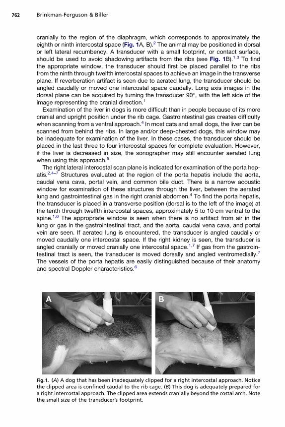
cranially to the region of the diaphragm, which corresponds to approximately theeighth or ninth intercostal space (Fig. 1A, B).2 The animal may be positioned in dorsalor left lateral recumbency. A transducer with a small footprint, or contact surface,should be used to avoid shadowing artifacts from the ribs (see Fig. 1B).1,3 To findthe appropriate window, the transducer should first be placed parallel to the ribsfrom the ninth through twelfth intercostal spaces to achieve an image in the transverseplane. If reverberation artifact is seen due to aerated lung, the transducer should beangled caudally or moved one intercostal space caudally. Long axis images in thedorsal plane can be acquired by turning the transducer 90�, with the left side of theimage representing the cranial direction.1
Examination of the liver in dogs is more difficult than in people because of its morecranial and upright position under the rib cage. Gastrointestinal gas creates difficultywhen scanning from a ventral approach.4 In most cats and small dogs, the liver can bescanned from behind the ribs. In large and/or deep-chested dogs, this window maybe inadequate for examination of the liver. In these cases, the transducer should beplaced in the last three to four intercostal spaces for complete evaluation. However,if the liver is decreased in size, the sonographer may still encounter aerated lungwhen using this approach.5
The right lateral intercostal scan plane is indicated for examination of the porta hep-atis.2,4–7 Structures evaluated at the region of the porta hepatis include the aorta,caudal vena cava, portal vein, and common bile duct. There is a narrow acousticwindow for examination of these structures through the liver, between the aeratedlung and gastrointestinal gas in the right cranial abdomen.4 To find the porta hepatis,the transducer is placed in a transverse position (dorsal is to the left of the image) atthe tenth through twelfth intercostal spaces, approximately 5 to 10 cm ventral to thespine.1,6 The appropriate window is seen when there is no artifact from air in thelung or gas in the gastrointestinal tract, and the aorta, caudal vena cava, and portalvein are seen. If aerated lung is encountered, the transducer is angled caudally ormoved caudally one intercostal space. If the right kidney is seen, the transducer isangled cranially or moved cranially one intercostal space.1,7 If gas from the gastroin-testinal tract is seen, the transducer is moved dorsally and angled ventromedially.7
The vessels of the porta hepatis are easily distinguished because of their anatomyand spectral Doppler characteristics.6
Fig.1. (A) A dog that has been inadequately clipped for a right intercostal approach. Noticethe clipped area is confined caudal to the rib cage. (B) This dog is adequately prepared fora right intercostal approach. The clipped area extends cranially beyond the costal arch. Notethe small size of the transducer’s footprint.
Ultrasound of the Right Lateral Intercostal Space 763
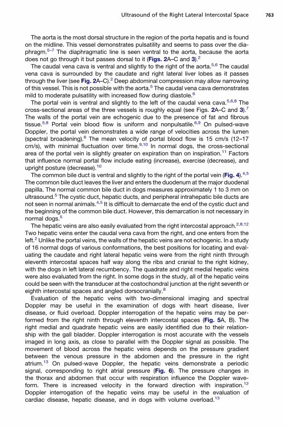
The aorta is the most dorsal structure in the region of the porta hepatis and is foundon the midline. This vessel demonstrates pulsatility and seems to pass over the dia-phragm.5–7 The diaphragmatic line is seen ventral to the aorta, because the aortadoes not go through it but passes dorsal to it (Figs. 2A–C and 3).2
The caudal vena cava is ventral and slightly to the right of the aorta.5,6 The caudalvena cava is surrounded by the caudate and right lateral liver lobes as it passesthrough the liver (see Fig. 2A–C).2 Deep abdominal compression may allow narrowingof this vessel. This is not possible with the aorta.5 The caudal vena cava demonstratesmild to moderate pulsatility with increased flow during diastole.6
The portal vein is ventral and slightly to the left of the caudal vena cava.5,6,8 Thecross-sectional areas of the three vessels is roughly equal (see Figs. 2A–C and 3).7
The walls of the portal vein are echogenic due to the presence of fat and fibroustissue.5,8 Portal vein blood flow is uniform and nonpulsatile.6,9 On pulsed-waveDoppler, the portal vein demonstrates a wide range of velocities across the lumen(spectral broadening).6 The mean velocity of portal blood flow is 15 cm/s (12–17cm/s), with minimal fluctuation over time.9,10 In normal dogs, the cross-sectionalarea of the portal vein is slightly greater on expiration than on inspiration.11 Factorsthat influence normal portal flow include eating (increase), exercise (decrease), andupright posture (decrease).10
The common bile duct is ventral and slightly to the right of the portal vein (Fig. 4).4,5
The common bile duct leaves the liver and enters the duodenum at the major duodenalpapilla. The normal common bile duct in dogs measures approximately 1 to 3 mm onultrasound.5 The cystic duct, hepatic ducts, and peripheral intrahepatic bile ducts arenot seen in normal animals.4,5 It is difficult to demarcate the end of the cystic duct andthe beginning of the common bile duct. However, this demarcation is not necessary innormal dogs.5
The hepatic veins are also easily evaluated from the right intercostal approach.2,8,12
Two hepatic veins enter the caudal vena cava from the right, and one enters from theleft.2 Unlike the portal veins, the walls of the hepatic veins are not echogenic. In a studyof 16 normal dogs of various conformations, the best positions for locating and eval-uating the caudate and right lateral hepatic veins were from the right ninth througheleventh intercostal spaces half way along the ribs and cranial to the right kidney,with the dogs in left lateral recumbency. The quadrate and right medial hepatic veinswere also evaluated from the right. In some dogs in the study, all of the hepatic veinscould be seen with the transducer at the costochondral junction at the right seventh oreighth intercostal spaces and angled dorsocranially.8
Evaluation of the hepatic veins with two-dimensional imaging and spectralDoppler may be useful in the examination of dogs with heart disease, liverdisease, or fluid overload. Doppler interrogation of the hepatic veins may be per-formed from the right ninth through eleventh intercostal spaces (Fig. 5A, B). Theright medial and quadrate hepatic veins are easily identified due to their relation-ship with the gall bladder. Doppler interrogation is most accurate with the vesselsimaged in long axis, as close to parallel with the Doppler signal as possible. Themovement of blood across the hepatic veins depends on the pressure gradientbetween the venous pressure in the abdomen and the pressure in the rightatrium.13 On pulsed-wave Doppler, the hepatic veins demonstrate a periodicsignal, corresponding to right atrial pressure (Fig. 6). The pressure changes inthe thorax and abdomen that occur with respiration influence the Doppler wave-form. There is increased velocity in the forward direction with inspiration.12
Doppler interrogation of the hepatic veins may be useful in the evaluation ofcardiac disease, hepatic disease, and in dogs with volume overload.13
Brinkman-Ferguson & Biller764
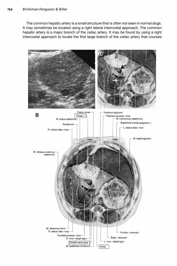
The common hepatic artery is a small structure that is often not seen in normal dogs.It may sometimes be located using a right lateral intercostal approach. The commonhepatic artery is a major branch of the celiac artery. It may be found by using a rightintercostal approach to locate the first large branch of the celiac artery that courses
Ultrasound of the Right Lateral Intercostal Space 765
to the porta hepatis (Fig. 7). The celiac artery is easily identified because of its closeassociation with the cranial mesenteric artery. The common hepatic artery was inter-rogated with pulsed-wave Doppler in 10 normal adult beagles, 20 normal puppies, and7 dogs with hepatic disease. In the normal adult beagles, mean peak systolic velocitywas 1.5 m/s (1.1–2.3 m/s), with a resistive index of 0.68 (0.62–0.74). In the normalpuppies, the mean peak systolic velocity was lower at 1.0 m/s (0.8–1.3 m/s) witha lower resistive index of 0.59 (0.46–0.65). There were no differences in values ob-tained after fasting and postprandially. Two dogs with congenital arterioportal fistulaedemonstrated higher peak systolic velocity and lower mean resistive index thannormal puppies. There were no differences in the normal adult beagles and the fiveadult dogs with acquired hepatoportal disease.14 Intrahepatic arteries are not seenin normal animals.5
In the past, the normal pancreas was difficult, if not impossible, to evaluate withultrasound. With improvements in equipment, the normal pancreas is not the elusivestructure it once was. However, proper technique is required to image this organ.Complete evaluation is often impossible from a subcostal approach. The right limband body may be examined with a right lateral intercostal approach. This approachis especially helpful in deep-chested dogs and dogs with pain in the right cranialabdomen.3 The pancreas consists of a left lobe, body, and right lobe. If seen, theleft lobe is typically imaged from a subcostal approach, whereas the body and rightlimb often require a right intercostal approach (Fig. 8A–E). Several structures serveas landmarks for the pancreas. The pancreatic body unites the right and left lobesand can be found caudal to the pylorus, ventral to the portal vein, and craniomedialto the right kidney and caudate process of the caudate lobe of the liver. The rightlobe lies in the mesoduodenum, dorsal or dorsomedial to the descending duodenum,ventral to the right kidney, and ventrolateral to the portal vein.3,15 To make sure that theentire right lobe has been imaged, the descending duodenum should be followedcaudally to its caudal flexure.3 The normal pancreas is isoechoic or slightly hypere-choic to the liver.3,15
The only visible veins in the pancreas are those that drain the right lobe. The cranialand caudal parts of the pancreaticoduodenal vein lie in the right lobe and run parallelto the descending duodenum. The descending duodenum is identified by its straightcourse and prominent walls.3 The cranial pancreaticoduodenal vein becomes thegastroduodenal vein, which drains into the portal vein near the porta hepatis.3,16
The caudal pancreaticoduodenal vein meets with the cranial mesenteric vein.3
The right kidney is often more difficult than the left to evaluate from a ventral or sub-costal approach because of its dorsocranial position in the renal fossa of the caudatelobe of the liver and because it is dorsal to the duodenum and proximal portion of the
Fig. 2. (A) Transverse right lateral intercostal ultrasonographic image of the porta hepatis ina normal dog. The aorta (Ao) is the most dorsal of the three vascular structures. The caudalvena cava (CVC) is ventral and slightly to the right of the aorta. The portal vein (PV) is ventralandslightly to the left of the caudal vena cava. D, dorsal; R, right; V, ventral; L, left. (B) Cross sectionof a canine cadaver at the level of the twelfth thoracic vertebra. Note the location of the portalvein, caudal vena cava, and aorta. (Adapted from Feeney DA, Fletcher TF, Hardy RM. Atlas ofcorrelative imaginganatomyof thenormaldog:ultrasoundandcomputedtomography.Philadel-phia: WB Saunders; 1991. p. 246; with permission.) (C) Same image as Fig.3B magnified to demon-strate the three major blood vessels of the porta hepatis. The portal vein (black arrow), caudalvena cava (solid white arrow), and aorta (open white arrow) are identified. (Adapted from FeeneyDA, Fletcher TF, Hardy RM. Atlas of correlative imaging anatomy of the normal dog: ultrasoundand computed tomography. Philadelphia: WB Saunders; 1991. p. 246; with permission.)
:
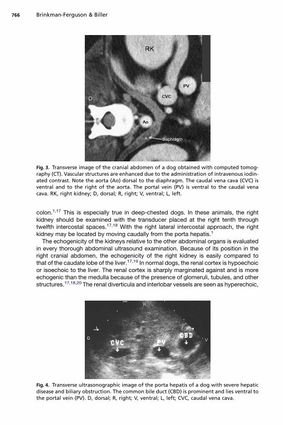
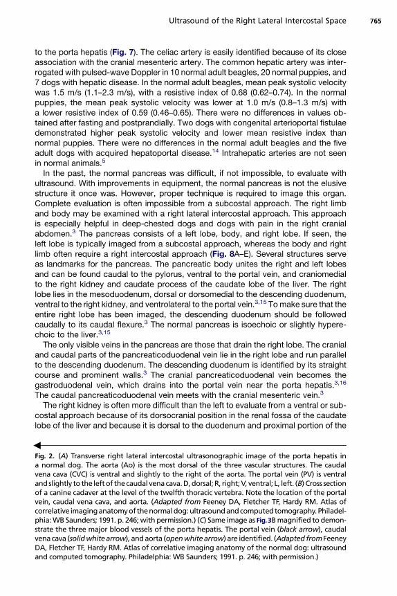
Fig. 3. Transverse image of the cranial abdomen of a dog obtained with computed tomog-raphy (CT). Vascular structures are enhanced due to the administration of intravenous iodin-ated contrast. Note the aorta (Ao) dorsal to the diaphragm. The caudal vena cava (CVC) isventral and to the right of the aorta. The portal vein (PV) is ventral to the caudal venacava. RK, right kidney; D, dorsal; R, right; V, ventral; L, left.
Brinkman-Ferguson & Biller766
colon.1,17 This is especially true in deep-chested dogs. In these animals, the rightkidney should be examined with the transducer placed at the right tenth throughtwelfth intercostal spaces.17,18 With the right lateral intercostal approach, the rightkidney may be located by moving caudally from the porta hepatis.1
The echogenicity of the kidneys relative to the other abdominal organs is evaluatedin every thorough abdominal ultrasound examination. Because of its position in theright cranial abdomen, the echogenicity of the right kidney is easily compared tothat of the caudate lobe of the liver.17,19 In normal dogs, the renal cortex is hypoechoicor isoechoic to the liver. The renal cortex is sharply marginated against and is moreechogenic than the medulla because of the presence of glomeruli, tubules, and otherstructures.17,18,20 The renal diverticula and interlobar vessels are seen as hyperechoic,
Fig. 4. Transverse ultrasonographic image of the porta hepatis of a dog with severe hepaticdisease and biliary obstruction. The common bile duct (CBD) is prominent and lies ventral tothe portal vein (PV). D, dorsal; R, right; V, ventral; L, left; CVC, caudal vena cava.
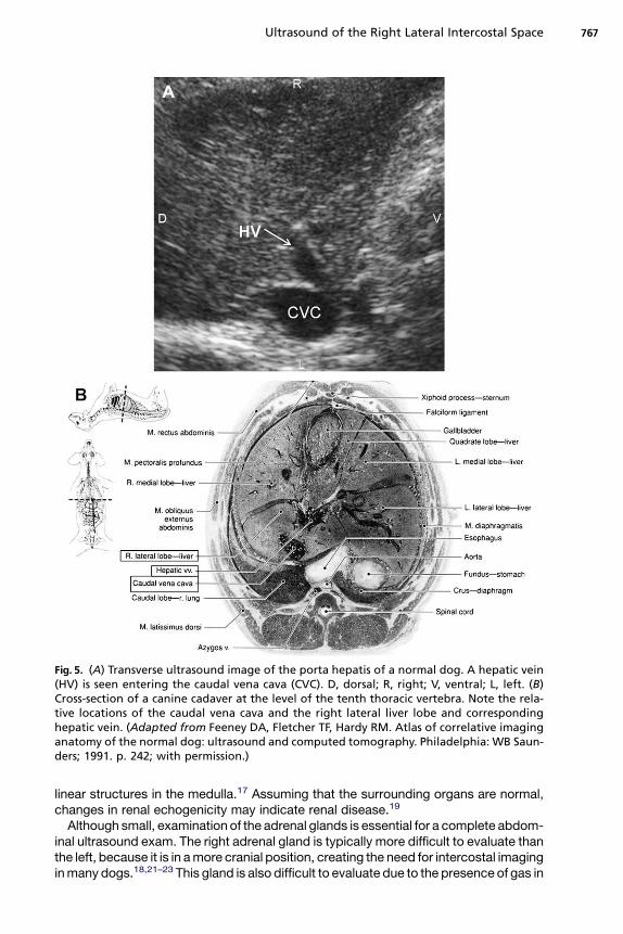
Fig. 5. (A) Transverse ultrasound image of the porta hepatis of a normal dog. A hepatic vein(HV) is seen entering the caudal vena cava (CVC). D, dorsal; R, right; V, ventral; L, left. (B)Cross-section of a canine cadaver at the level of the tenth thoracic vertebra. Note the rela-tive locations of the caudal vena cava and the right lateral liver lobe and correspondinghepatic vein. (Adapted from Feeney DA, Fletcher TF, Hardy RM. Atlas of correlative imaginganatomy of the normal dog: ultrasound and computed tomography. Philadelphia: WB Saun-ders; 1991. p. 242; with permission.)
Ultrasound of the Right Lateral Intercostal Space 767
linear structures in the medulla.17 Assuming that the surrounding organs are normal,changes in renal echogenicity may indicate renal disease.19
Although small, examination of the adrenal glands is essential for a complete abdom-inal ultrasound exam. The right adrenal gland is typically more difficult to evaluate thanthe left, because it is in a more cranial position, creating the need for intercostal imagingin many dogs.18,21–23 This gland is also difficult to evaluate due to the presence of gas in
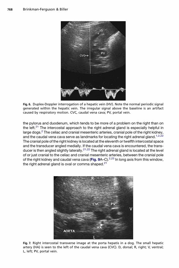
Fig. 6. Duplex-Doppler interrogation of a hepatic vein (HV). Note the normal periodic signalgenerated within the hepatic vein. The irregular signal above the baseline is an artifactcaused by respiratory motion. CVC, caudal vena cava; PV, portal vein.
Brinkman-Ferguson & Biller768
the pylorus and duodenum, which tends to be more of a problem on the right than onthe left.21 The intercostal approach to the right adrenal gland is especially helpful inlarge dogs.2 The celiac and cranial mesenteric arteries, cranial pole of the right kidney,and the caudal vena cava serve as landmarks for locating the right adrenal gland.1,2,22
The cranial pole of the right kidney is located at the eleventh or twelfth intercostal spaceand the transducer angled medially. If the caudal vena cava is encountered, the trans-ducer is then angled slightly laterally.21,22 The right adrenal gland is located at the levelof or just cranial to the celiac and cranial mesenteric arteries, between the cranial poleof the right kidney and caudal vena cava (Fig. 9A–C).2,23 In long axis from this window,the right adrenal gland is oval or comma shaped.21
Fig. 7. Right intercostal transverse image at the porta hepatis in a dog. The small hepaticartery (HA) is seen to the left of the caudal vena cava (CVC). D, dorsal; R, right; V, ventral;L, left; PV, portal vein.
Ultrasound of the Right Lateral Intercostal Space 769
Although typically not seen in a normal dog, multiple lymph nodes may be examinedvia a right intercostal approach.1 Normal lymph nodes are usually isoechoic tosurrounding tissues. Blood vessels or other organs are used as landmarks for locatinglymph nodes on ultrasound. The hepatic lymph nodes lie on both sides of the portalvein, approximately 1 to 2 cm caudal to the porta hepatis (Fig. 10). The right nodesvary in number from one to five, are adjacent to the body of the pancreas, and aresmaller than those on the left. The left is larger at 1 to 6 cm in length and is found inthe lesser omentum dorsal to the common bile duct. The hepatic lymph nodes drainthe stomach, duodenum, pancreas, and liver. The gastric lymph nodes are inconsis-tently found in the lesser omentum near the pylorus and right gastric artery and drainthe stomach, esophagus, diaphragm, liver, mediastinum, and peritoneum. The pan-creaticoduodenal lymph nodes are also inconsistent and may be found in the meso-duodenum and greater omentum. They drain the duodenum, pylorus, and right limb ofthe pancreas. The lymph nodes described here are part of the celiac lymphocenter ofthe visceral abdominal lymph nodes.24
CLINICAL INDICATIONS
The right lateral intercostal ultrasound scan plane is indicated in some dogs for eval-uation of diseases involving the right lateral, right medial, and caudate lobes of theliver, especially in large and deep-chested dogs and in cases of microhepatica or largevolumes of peritoneal effusion. In large or deep-chested dogs, mass or nodular lesionsof the right aspect of the liver may be missed if only a subcostal approach is used(Fig. 11).1 If mass lesions are detected in other abdominal organs, it is important tothoroughly examine the liver. The liver is commonly the first organ where metastasisis seen, because many abdominal organs are drained by the portal vein.25
When the liver is small, there may be a very small window of visible hepatic tissuebetween the aerated lung and gas in the stomach.5 Conditions that may cause micro-hepatica include cirrhosis, congenital portosystemic shunts, or other chronic diseasesof the liver.1
The ‘‘classic’’ combination of ultrasonographic findings in hepatic cirrhosis includesa small, irregularly marginated, hyperechoic liver with nodules and peritoneal effusion.However, in a study of 55 dogs and two cats with a histopathologic diagnosis ofhepatic cirrhosis, this classic appearance was seen only in 5% of the cases. Fourdogs (7%) had a normal study. The most common finding in this study was peritonealeffusion in 62%, followed by irregular liver margination in 53%, and hepatic nodules in51%. More livers were normal in size (55%) and echogenicity (51%) than were small(34%) and hyperechoic (38%).26 The ‘‘classic’’ form of cirrhosis may be detectedonly late in the disease process.27 However, the right lateral intercostal view is stillindicated due to the presence of effusion.
Cirrhosis is the most common cause of portal hypertension in dogs.27 In a study of10 normal dogs and 10 dogs with surgically induced hepatic cirrhosis, the portal veinwas interrogated with pulsed-wave Doppler. The transducer was placed at the righteleventh or twelfth intercostal space for these examinations. Mean portal flow andmean portal flow velocity were decreased in dogs with cirrhosis. The portal veindiameter was unchanged.11
Congenital portosystemic shunts are abnormal vascular connections between theportal venous system and the systemic venous system. Most congenital, single extra-hepatic portosystemic shunts connect a major tributary of the portal vein and thecaudal vena cava, cranial to the phrenicoabdominal veins. In dogs, the shunt vesselusually arises from the main portal vein, splenic vein, or left gastric vein.16 These
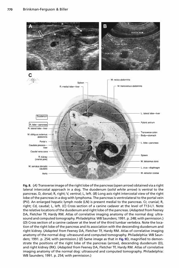
Fig. 8. (A) Transverse image of the right lobe of the pancreas (open arrow) obtained via a rightlateral intercostal approach in a dog. The duodenum (solid white arrow) is ventral to thepancreas. D, dorsal; R, right; V, ventral; L, left. (B) Long axis right intercostal view of the rightlobe of the pancreas in a dog with lymphoma. The pancreas is ventrolateral to the portal vein(PV). An enlarged hepatic lymph node (LN) is present medial to the pancreas. Cr, cranial; R,right; Cd, caudal; L, left. (C) Cross section of a canine cadaver at the level of T13-L1. Notethe relative locations of the duodenum and right lobe of the pancreas. (Adapted from FeeneyDA, Fletcher TF, Hardy RM. Atlas of correlative imaging anatomy of the normal dog: ultra-sound and computed tomography. Philadelphia: WB Saunders; 1991. p. 248; with permission.)(D) Cross section of a canine cadaver at the level of the third lumbar vertebra. Note the loca-tion of the right lobe of the pancreas and its association with the descending duodenum andright kidney. (Adapted from Feeney DA, Fletcher TF, Hardy RM. Atlas of correlative imaginganatomy of the normal dog: ultrasound and computed tomography. Philadelphia: WB Saun-ders; 1991. p. 254; with permission.) (E) Same image as that in Fig. 9D, magnified to demon-strate the positions of the right lobe of the pancreas (arrow), descending duodenum (D),and right kidney (RK). (Adapted from Feeney DA, Fletcher TF, Hardy RM. Atlas of correlativeimaging anatomy of the normal dog: ultrasound and computed tomography. Philadelphia:WB Saunders; 1991. p. 254; with permission.)
Brinkman-Ferguson & Biller770
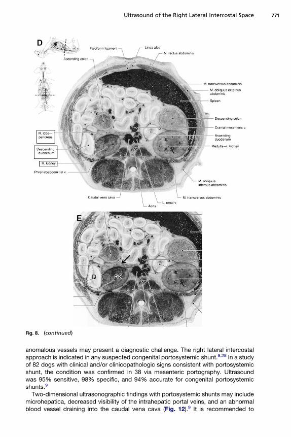
Fig. 8. (continued)
Ultrasound of the Right Lateral Intercostal Space 771
anomalous vessels may present a diagnostic challenge. The right lateral intercostalapproach is indicated in any suspected congenital portosystemic shunt.9,28 In a studyof 82 dogs with clinical and/or clinicopathologic signs consistent with portosystemicshunt, the condition was confirmed in 38 via mesenteric portography. Ultrasoundwas 95% sensitive, 98% specific, and 94% accurate for congenital portosystemicshunts.9
Two-dimensional ultrasonographic findings with portosystemic shunts may includemicrohepatica, decreased visibility of the intrahepatic portal veins, and an abnormalblood vessel draining into the caudal vena cava (Fig. 12).9 It is recommended to
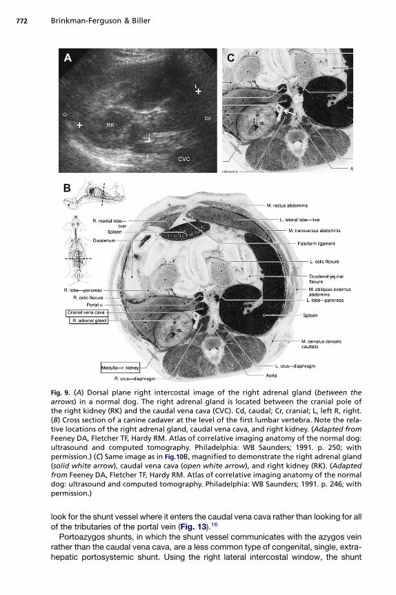
Fig. 9. (A) Dorsal plane right intercostal image of the right adrenal gland (between thearrows) in a normal dog. The right adrenal gland is located between the cranial pole ofthe right kidney (RK) and the caudal vena cava (CVC). Cd, caudal; Cr, cranial; L, left R, right.(B) Cross section of a canine cadaver at the level of the first lumbar vertebra. Note the rela-tive locations of the right adrenal gland, caudal vena cava, and right kidney. (Adapted fromFeeney DA, Fletcher TF, Hardy RM. Atlas of correlative imaging anatomy of the normal dog:ultrasound and computed tomography. Philadelphia: WB Saunders; 1991. p. 250; withpermission.) (C) Same image as in Fig.10B, magnified to demonstrate the right adrenal gland(solid white arrow), caudal vena cava (open white arrow), and right kidney (RK). (Adaptedfrom Feeney DA, Fletcher TF, Hardy RM. Atlas of correlative imaging anatomy of the normaldog: ultrasound and computed tomography. Philadelphia: WB Saunders; 1991. p. 246; withpermission.)
Brinkman-Ferguson & Biller772
look for the shunt vessel where it enters the caudal vena cava rather than looking for allof the tributaries of the portal vein (Fig. 13).16
Portoazygos shunts, in which the shunt vessel communicates with the azygos veinrather than the caudal vena cava, are a less common type of congenital, single, extra-hepatic portosystemic shunt. Using the right lateral intercostal window, the shunt
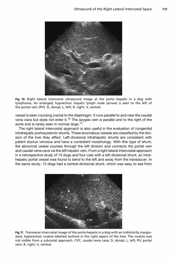
Fig. 10. Right lateral transverse ultrasound image at the porta hepatis in a dog withlymphoma. An enlarged, hypoechoic hepatic lymph node (arrow) is seen to the left ofthe portal vein (PV). D, dorsal; L, left; R, right; V, ventral.
Ultrasound of the Right Lateral Intercostal Space 773
vessel is seen coursing cranial to the diaphragm. It runs parallel to and near the caudalvena cava but does not enter it.16 The azygos vein is parallel and to the right of theaorta and is rarely seen in normal dogs.12
The right lateral intercostal approach is also useful in the evaluation of congenitalintrahepatic portosystemic shunts. These anomalous vessels are classified by the divi-sion of the liver they affect. Left-divisional intrahepatic shunts are consistent withpatent ductus venosus and have a consistent morphology. With this type of shunt,the abnormal vessel courses through the left division and connects the portal veinand caudal vena cava via the left hepatic vein. From a right lateral intercostal approachin a retrospective study of 13 dogs and four cats with a left-divisional shunt, an intra-hepatic portal vessel was found to bend to the left and away from the transducer. Inthe same study, 13 dogs had a central-divisional shunt, which was easy to see from
Fig.11. Transverse intercostal image of the porta hepatis in a dog with an indistinctly margin-ated, hyperechoic nodule (dashed outline) in the right aspect of the liver. The nodule wasnot visible from a subcostal approach. CVC, caudal vena cava; D, dorsal; L, left; PV, portalvein; R, right; V, ventral.
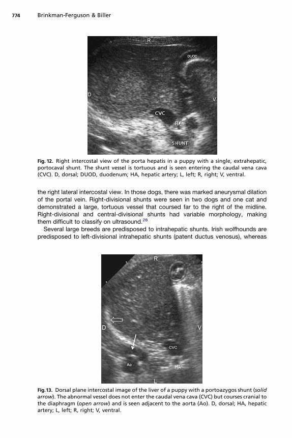
Fig. 12. Right intercostal view of the porta hepatis in a puppy with a single, extrahepatic,portocaval shunt. The shunt vessel is tortuous and is seen entering the caudal vena cava(CVC). D, dorsal; DUOD, duodenum; HA, hepatic artery; L, left; R, right; V, ventral.
Brinkman-Ferguson & Biller774
the right lateral intercostal view. In those dogs, there was marked aneurysmal dilationof the portal vein. Right-divisional shunts were seen in two dogs and one cat anddemonstrated a large, tortuous vessel that coursed far to the right of the midline.Right-divisional and central-divisional shunts had variable morphology, makingthem difficult to classify on ultrasound.28
Several large breeds are predisposed to intrahepatic shunts. Irish wolfhounds arepredisposed to left-divisional intrahepatic shunts (patent ductus venosus), whereas
Fig.13. Dorsal plane intercostal image of the liver of a puppy with a portoazygos shunt (solidarrow). The abnormal vessel does not enter the caudal vena cava (CVC) but courses cranial tothe diaphragm (open arrow) and is seen adjacent to the aorta (Ao). D, dorsal; HA, hepaticartery; L, left; R, right; V, ventral.
Ultrasound of the Right Lateral Intercostal Space 775
Old English sheepdogs are predisposed to central-divisional shunts. Australian cattledogs are predisposed to right-sided and central-divisional shunts. Retrievers arepredisposed to multiple types of intrahepatic shunt morphologies.28
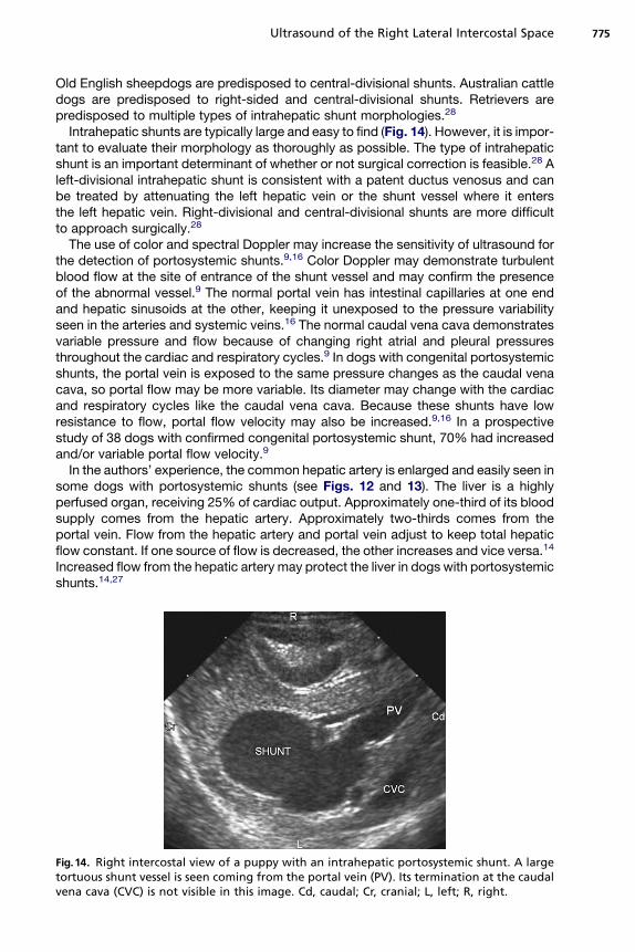
Intrahepatic shunts are typically large and easy to find (Fig. 14). However, it is impor-tant to evaluate their morphology as thoroughly as possible. The type of intrahepaticshunt is an important determinant of whether or not surgical correction is feasible.28 Aleft-divisional intrahepatic shunt is consistent with a patent ductus venosus and canbe treated by attenuating the left hepatic vein or the shunt vessel where it entersthe left hepatic vein. Right-divisional and central-divisional shunts are more difficultto approach surgically.28
The use of color and spectral Doppler may increase the sensitivity of ultrasound forthe detection of portosystemic shunts.9,16 Color Doppler may demonstrate turbulentblood flow at the site of entrance of the shunt vessel and may confirm the presenceof the abnormal vessel.9 The normal portal vein has intestinal capillaries at one endand hepatic sinusoids at the other, keeping it unexposed to the pressure variabilityseen in the arteries and systemic veins.16 The normal caudal vena cava demonstratesvariable pressure and flow because of changing right atrial and pleural pressuresthroughout the cardiac and respiratory cycles.9 In dogs with congenital portosystemicshunts, the portal vein is exposed to the same pressure changes as the caudal venacava, so portal flow may be more variable. Its diameter may change with the cardiacand respiratory cycles like the caudal vena cava. Because these shunts have lowresistance to flow, portal flow velocity may also be increased.9,16 In a prospectivestudy of 38 dogs with confirmed congenital portosystemic shunt, 70% had increasedand/or variable portal flow velocity.9
In the authors’ experience, the common hepatic artery is enlarged and easily seen insome dogs with portosystemic shunts (see Figs. 12 and 13). The liver is a highlyperfused organ, receiving 25% of cardiac output. Approximately one-third of its bloodsupply comes from the hepatic artery. Approximately two-thirds comes from theportal vein. Flow from the hepatic artery and portal vein adjust to keep total hepaticflow constant. If one source of flow is decreased, the other increases and vice versa.14
Increased flow from the hepatic artery may protect the liver in dogs with portosystemicshunts.14,27
Fig.14. Right intercostal view of a puppy with an intrahepatic portosystemic shunt. A largetortuous shunt vessel is seen coming from the portal vein (PV). Its termination at the caudalvena cava (CVC) is not visible in this image. Cd, caudal; Cr, cranial; L, left; R, right.
Brinkman-Ferguson & Biller776
Congenital hepatic arterioportal fistulas may cause portosystemic shunting. Thiscondition may be difficult to diagnose, because it demonstrates features of bothcongenital and acquired portosystemic shunts on ultrasound.16 These conditionsmay all form a complex pattern of dilated vessels in the liver.29 However, arterioportalfistulas demonstrate reversed, pulsatile portal flow and signs of portal hypertension,such as peritoneal effusion and hepatofugal blood flow.16,29
Portal vein thrombosis is an uncommon complication of portosystemic shunt liga-tion in dogs.30–32 However, ante mortem diagnosis of spontaneously occurring portalvein thrombosis is rare.32 Conditions appropriate for the development of thromboem-boli include a hypercoagulable state, vascular stasis, and vascular endothelialdamage.33 Reported causes of spontaneous portal vein thrombosis in dogs includeehrlichiosis, pancreatitis, autoimmune disease, renal amyloidosis, sepsis, peritonitis,and retrograde growth of hepatic tumors.32,33 In many cases, the cause is never deter-mined. Neoplasia can cause portal vein thrombosis by direct invasion into the vessellumen (tumor thrombus) or by distorting the vessel wall and causing a blood clot.32
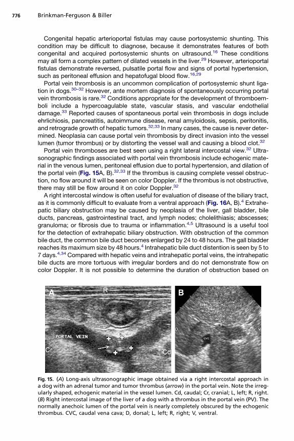
Portal vein thromboses are best seen using a right lateral intercostal view.32 Ultra-sonographic findings associated with portal vein thrombosis include echogenic mate-rial in the venous lumen, peritoneal effusion due to portal hypertension, and dilation ofthe portal vein (Fig. 15A, B).32,33 If the thrombus is causing complete vessel obstruc-tion, no flow around it will be seen on color Doppler. If the thrombus is not obstructive,there may still be flow around it on color Doppler.32
A right intercostal window is often useful for evaluation of disease of the biliary tract,as it is commonly difficult to evaluate from a ventral approach (Fig. 16A, B).4 Extrahe-patic biliary obstruction may be caused by neoplasia of the liver, gall bladder, bileducts, pancreas, gastrointestinal tract, and lymph nodes; cholelithiasis; abscesses;granuloma; or fibrosis due to trauma or inflammation.4,5 Ultrasound is a useful toolfor the detection of extrahepatic biliary obstruction. With obstruction of the commonbile duct, the common bile duct becomes enlarged by 24 to 48 hours. The gall bladderreaches its maximum size by 48 hours.4 Intrahepatic bile duct distention is seen by 5 to7 days.4,34 Compared with hepatic veins and intrahepatic portal veins, the intrahepaticbile ducts are more tortuous with irregular borders and do not demonstrate flow oncolor Doppler. It is not possible to determine the duration of obstruction based on
Fig. 15. (A) Long-axis ultrasonographic image obtained via a right intercostal approach ina dog with an adrenal tumor and tumor thrombus (arrow) in the portal vein. Note the irreg-ularly shaped, echogenic material in the vessel lumen. Cd, caudal; Cr, cranial; L, left; R, right.(B) Right intercostal image of the liver of a dog with a thrombus in the portal vein (PV). Thenormally anechoic lumen of the portal vein is nearly completely obscured by the echogenicthrombus. CVC, caudal vena cava; D, dorsal; L, left; R, right; V, ventral.
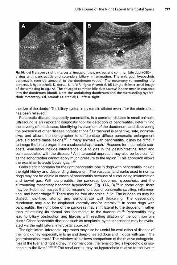
Fig.16. (A) Transverse right intercostal image of the pancreas and common bile duct (CBD) ina dog with pancreatitis and secondary biliary inflammation. The enlarged, hypoechoicpancreas is seen dorsomedial to the duodenum (duod). The mesentery surrounding thepancreas is hyperechoic. D, dorsal; L, left; R, right; V, ventral. (B) Long-axis intercostal imageof the same dog in Fig.17A. The enlarged common bile duct (arrow) is seen near its entranceinto the duodenum (duod). Note the undulating duodenum and the surrounding hypere-choic mesentery. Cd, caudal; Cr, cranial; L, left; R, right.
Ultrasound of the Right Lateral Intercostal Space 777
the size of the ducts.4 The biliary system may remain dilated even after the obstructionhas been relieved.5
Pancreatic disease, especially pancreatitis, is a common disease in small animals.Ultrasound is an important diagnostic tool for detection of pancreatitis, determiningthe severity of the disease, identifying involvement of the duodenum, and discoveringthe presence of other disease complications.3 Ultrasound is sensitive, safe, noninva-sive, and allows the sonographer to differentiate diffuse pancreatic enlargementversus discrete mass lesions.35 In many animals with pancreatitis, it may be difficultto image the entire organ from a subcostal approach.1 Reasons for incomplete sub-costal evaluation include interference due to gas in the gastrointestinal tract andpain associated with the disease.3 An intercostal approach may also be less painful,as the sonographer cannot apply much pressure to the region.1 This approach allowsthe examiner to avoid bowel gas.1,15
Consistent landmarks for the right pancreatic lobe in dogs with pancreatitis includethe right kidney and descending duodenum. The vascular landmarks used in normaldogs may not be visible in cases of pancreatitis because of surrounding inflammationand bowel gas. With pancreatitis, the pancreas becomes hypoechoic, and thesurrounding mesentery becomes hyperechoic (Fig. 17A, B).15 In some dogs, theremay be ill-defined masses that correspond to areas of pancreatic swelling, inflamma-tion, and hemorrhage.35 There may be free abdominal fluid. The duodenum may bedilated, fluid-filled, atonic, and demonstrate wall thickening. The descendingduodenum may also be displaced ventrally and/or laterally.15 In some dogs withpancreatitis, the right lobe of the pancreas may shift lateral to the duodenum ratherthan maintaining its normal position medial to the duodenum.35 Pancreatitis maylead to biliary obstruction and fibrosis with resulting dilation of the common bileduct.3 Other pancreatic diseases such as neoplasia, cysts, or abscess may be evalu-ated via the right lateral intercostal approach.1
The right lateral intercostal approach may also be useful for evaluation of disease ofthe right kidney, especially in large and deep-chested dogs and in dogs with gas in thegastrointestinal tract.1 This window also allows comparison of the relative echogenic-ities of the liver and right kidney. In normal dogs, the renal cortex is hypoechoic or iso-echoic to the liver.17,18,20 The renal cortex may be hyperechoic relative to the liver in
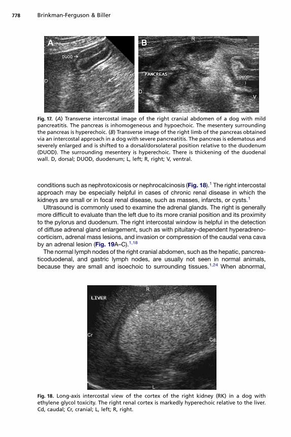
Fig. 17. (A) Transverse intercostal image of the right cranial abdomen of a dog with mildpancreatitis. The pancreas is inhomogeneous and hypoechoic. The mesentery surroundingthe pancreas is hyperechoic. (B) Transverse image of the right limb of the pancreas obtainedvia an intercostal approach in a dog with severe pancreatitis. The pancreas is edematous andseverely enlarged and is shifted to a dorsal/dorsolateral position relative to the duodenum(DUOD). The surrounding mesentery is hyperechoic. There is thickening of the duodenalwall. D, dorsal; DUOD, duodenum; L, left; R, right; V, ventral.
Brinkman-Ferguson & Biller778
conditions such as nephrotoxicosis or nephrocalcinosis (Fig. 18).1 The right intercostalapproach may be especially helpful in cases of chronic renal disease in which thekidneys are small or in focal renal disease, such as masses, infarcts, or cysts.1
Ultrasound is commonly used to examine the adrenal glands. The right is generallymore difficult to evaluate than the left due to its more cranial position and its proximityto the pylorus and duodenum. The right intercostal window is helpful in the detectionof diffuse adrenal gland enlargement, such as with pituitary-dependent hyperadreno-corticism, adrenal mass lesions, and invasion or compression of the caudal vena cavaby an adrenal lesion (Fig. 19A–C).1,18
The normal lymph nodes of the right cranial abdomen, such as the hepatic, pancrea-ticoduodenal, and gastric lymph nodes, are usually not seen in normal animals,because they are small and isoechoic to surrounding tissues.1,24 When abnormal,
Fig. 18. Long-axis intercostal view of the cortex of the right kidney (RK) in a dog withethylene glycol toxicity. The right renal cortex is markedly hyperechoic relative to the liver.Cd, caudal; Cr, cranial; L, left; R, right.
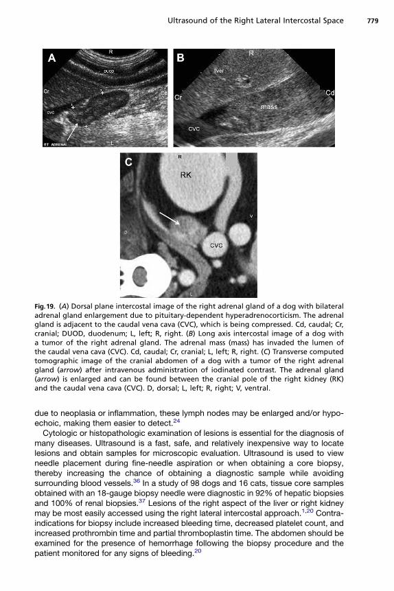
Fig.19. (A) Dorsal plane intercostal image of the right adrenal gland of a dog with bilateraladrenal gland enlargement due to pituitary-dependent hyperadrenocorticism. The adrenalgland is adjacent to the caudal vena cava (CVC), which is being compressed. Cd, caudal; Cr,cranial; DUOD, duodenum; L, left; R, right. (B) Long axis intercostal image of a dog witha tumor of the right adrenal gland. The adrenal mass (mass) has invaded the lumen ofthe caudal vena cava (CVC). Cd, caudal; Cr, cranial; L, left; R, right. (C) Transverse computedtomographic image of the cranial abdomen of a dog with a tumor of the right adrenalgland (arrow) after intravenous administration of iodinated contrast. The adrenal gland(arrow) is enlarged and can be found between the cranial pole of the right kidney (RK)and the caudal vena cava (CVC). D, dorsal; L, left; R, right; V, ventral.
Ultrasound of the Right Lateral Intercostal Space 779
due to neoplasia or inflammation, these lymph nodes may be enlarged and/or hypo-echoic, making them easier to detect.24
Cytologic or histopathologic examination of lesions is essential for the diagnosis ofmany diseases. Ultrasound is a fast, safe, and relatively inexpensive way to locatelesions and obtain samples for microscopic evaluation. Ultrasound is used to viewneedle placement during fine-needle aspiration or when obtaining a core biopsy,thereby increasing the chance of obtaining a diagnostic sample while avoidingsurrounding blood vessels.36 In a study of 98 dogs and 16 cats, tissue core samplesobtained with an 18-gauge biopsy needle were diagnostic in 92% of hepatic biopsiesand 100% of renal biopsies.37 Lesions of the right aspect of the liver or right kidneymay be most easily accessed using the right lateral intercostal approach.1,20 Contra-indications for biopsy include increased bleeding time, decreased platelet count, andincreased prothrombin time and partial thromboplastin time. The abdomen should beexamined for the presence of hemorrhage following the biopsy procedure and thepatient monitored for any signs of bleeding.20
Brinkman-Ferguson & Biller780
SUMMARY
When performing an abdominal ultrasound, a ventral or subcostal approach may beinadequate for a thorough examination. A right lateral intercostal window may benecessary for complete evaluation of the right cranial abdomen. Structures evaluatedwith this intercostal approach include the right aspect of the liver, porta hepatis,pancreas, proximal duodenum, right kidney, right adrenal gland, and several lymphnodes. Dogs for which this window may be most useful include large and deep-chested dogs, dogs with large volumes of peritoneal effusion or gas in the gastrointes-tinal tract, and cases of microhepatica and abdominal pain. The right intercostalapproach is simple and requires little patient preparation.
REFERENCES
1. Brinkman EL, Biller DS, Armbrust LJ, et al. The clinical utility of the right lateralintercostal scan technique in dogs. J Am Anim Hosp Assoc 2007;43:179–86.
2. Spaulding KA. A review of sonographic identification of abdominal blood vesselsand juxtavascular organs. Vet Radiol Ultrasound 1997;38(1):4–23.
3. Saunders HM. Ultrasonography of the pancreas. Probl Vet Med 1991;3(4):583–603.
4. Nyland TG, Gillett NA. Sonographic evaluation of experimental bile duct ligation inthe dog. Vet Radiol 1982;23(6):252–60.
5. Partington BP, Biller DS. Hepatic imaging with radiology and ultrasound. Vet ClinNorth Am Small Anim Pract 1995;25(2):305–35.
6. Kantrowitz BM, Nyland TG, Fisher P. Estimation of portal blood flow using duplexreal-time and pulsed Doppler ultrasound imaging in the dog. Vet Radiol 1989;30(5):222–6.
7. Szatm�ari V, Rothizen J, Voorhout G. Standard planes for ultrasonographic exam-ination of the portal system in dogs. J Am Vet Med Assoc 2004;224(5):713–6.
8. Wu JX, Carlisle CH. Ultrasonographic examination of the canine liver based onrecognition of hepatic and portal veins. Vet Radiol Ultrasound 1995;36(3):234–9.
9. Lamb CR. Ultrasonographic diagnosis of congenital portosystemic shunts indogs: results of a prospective study. Vet Radiol Ultrasound 1996;37(4):281–8.
10. Lamb CR, Mahoney PN. Comparison of three methods for calculating portalblood flow velocity in dogs using duplex-Doppler ultrasonography. Vet RadiolUltrasound 1994;35(3):190–4.
11. Nyland TG, Fisher PE. Evaluation of experimentally induced canine hepaticcirrhosis using duplex Doppler ultrasound. Vet Radiol 1990;31(4):189–94.
12. Szatzm�ari V, S�otonyi P, Voros K. Normal duplex Doppler waveforms of majorabdominal blood vessels in dogs: a review. Vet Radiol Ultrasound 2001;42(2):93–107.
13. Smithenson BT, Mattoon JS, Bonagura JD, et al. Pulsed-wave Doppler ultrasono-graphic evaluation of hepatic veins during variable hemodynamic states inhealthy anesthetized dogs. Am J Vet Res 2004;65(6):734–40.
14. Lamb CR, Burton CA, Carlisle CH. Doppler measurement of hepatic arterial flowin dogs: technique and preliminary findings. Vet Radiol Ultrasound 1999;40(1):77–81.
15. Nyland TG, Mulvany MH, Strombeck DR. Ultrasonic features of experimentallyinduced, acute pancreatitis in the dog. Vet Radiol 1983;24(6):260–6.
16. Lamb CR. Ultrasonography of portosystemic shunts in dogs and cats. Vet ClinNorth Am Small Anim Pract 1998;28(4):725–53.
Ultrasound of the Right Lateral Intercostal Space 781
17. Widmer WR, Biller DS, Adams LG. Ultrasonography of the urinary tract in smallanimals. J Am Vet Med Assoc 2004;225(1):46–54.
18. Lamb CR. Abdominal ultrasonography in small animals: intestinal tract andmesentery, kidneys, adrenal glands, uterus, and prostate. J Small Anim Pract1990;31:295–304.
19. Hartzband LE, Tidwell AS, Lamb CR. Relative Echogenicity of the renal cortexand liver in normal dogs [abstract]. Br J Radiol 1991;64:654.
20. Smith S. Ultrasound-guided biopsy. Vet Clin North Am Small Anim Pract 1985;15(6):1249–62.
21. Grooters AM, Biller DS, Miyabayashi T, et al. Evaluation of routine abdominalultrasonography as a technique for imaging the canine adrenal glands. J AmAnim Hosp Assoc 1994;30:457–62.
22. Tidwell AS, Pennick DG, Besso JG. Imaging of adrenal gland disorders. Vet ClinNorth Am Small Anim Pract 1997;27(2):237–54.
23. Kantrowitz BM, Nyland TG, Feldman EC. Adrenal ultrasonography in the dog:detection of tumors and hyperplasia in hyperadrenocorticism. Vet Radiol 1986;27(3):91–6.
24. Pugh CR. Ultrasonographic examination of abdominal lymph nodes in the dog.Vet Radiol Ultrasound 1994;35(2):110–5.
25. Nyman HT, Kristensen AT, Flagstad A, et al. A review of the sonographic assess-ment of tumor metastasis in liver and superficial lymph nodes. Vet Radiol Ultra-sound 2004;45(5):438–48.
26. O’Brien RT. A retrospective study on the sonographic and clinicopathologicfeatures of cirrhosis in dogs and cats [abstract]. Vet Radiol Ultrasound 1999;40(6):659.
27. Salwei RM, O’Brien RT, Matheson JS. Use of contrast harmonic ultrasound for thediagnosis of congenital portosystemic shunts in three dogs. Vet Radiol Ultra-sound 2003;44(3):301–5.
28. Lamb CR, White RN. Morphology of congenital intrahepatic portocaval shunts indogs and cats. Vet Rec 1998;142:55–60.
29. d’Anjou MA. Huneault L. Imaging Diagnosis—complex intrahepatic portosyste-mic shunt in a dog. Vet Radiol Ultrasound 2008;49(1):51–5.
30. Mathews K, Gofton N. Congenital extrahepatic portosystemic shunt occlusion inthe dog: gross observations during surgical correction. J Am Anim Hosp Assoc1988;24:387–94.
31. Roy RG, Post GS, Waters DJ, et al. Portal vein thrombosis as a complication ofportosystemic shunt ligation in two dogs. J Am Anim Hosp Assoc 1992;28:53–8.
32. Lamb CR, Wrigley RH, Simpson KW, et al. Ultrasonographic diagnosis of portalvein thrombosis in four dogs. Vet Radiol Ultrasound 1996;37(2):121–9.
33. Bressler C, Himes LC, Moreau RE. Portal vein and aortic thromboses in a Siberianhusky with ehrlichiosis and hypothyroidism. J Small Anim Pract 2003;44:408–10.
34. Zeman RK, Taylor KJW, Rosenfield AT, et al. Acute experimental biliary obstruc-tion in the dog: sonographic findings and clinical implications. AJR Am J Roent-genol 1981;136:965–7.
35. Murtaugh RJ, Herring DS, Jacobs RM, et al. Pancreatic ultrasonography in dogswith experimentally induced pancreatitis. Vet Radiol 1985;26(1):27–32.
36. Burkhard MJ, Meyer DJ. Invasive cytology of internal organs: cytology of thethorax and abdomen. Vet Clin North Am Small Anim Pract 1996;26(5):1203–22.
37. Barr F. Percutaneous biopsy of abdominal organs under ultrasound guidance.J Small Anim Pract 1995;36:105–13.