Embed Size (px)
Citation preview
SCIENTIA MARINA 70S2October 2006, 23-40, Barcelona (Spain)
ISSN: 0214-8358
Ultrastructural studies of the morphological variationsof the egg surface and envelopes of the African catfishClarias gariepinus (Burchell, 1822) before and after
fertilisation, with a discussion of the fertilisation mechanism
IMAM A.A. MEKKAWY 1 and ALAA G.M. OSMAN 2
1 Department of Zoology, Faculty of Science, Assiut University, 71516 Assiut, Egypt. E-mail: [email protected]
2 Department of Biology and Ecology of Fishes, Leibniz Institute of Freshwater Ecology and Inland Fisheries,Müggelseedamm 310, D-12587 Berlin, Germany.
SUMMARY: Much of the existing knowledge of the mechanisms involved in teleost fertilisation is based on a few small modelspecies that have no commercial value. Research is therefore urgently required to address mechanisms involved in fertilisationin species of great commercial value. In this study, the ultrastructural morphological variations in the surface of the egg ofClarias gariepinus were recorded before and after fertilisation by using electron microscopy. The outer surface of the unfer-tilised egg was smooth, whereas the fertilised egg acquired a network of projections on the vegetal hemisphere. Moreover, dif-ferent patterns of ornamentation on the egg surface were evident. This pattern of ornamentation varied with the progress ofembryonic development. The micropyle of the C. gariepinus egg consisted of a funnel-shaped vestibule, from the bottom ofwhich a cylindrical micropylar canal extended. The micropylar canal decreased in diameter after completion of fertilisation,forming a micropylar disc. The sperm behaviour on the egg surface was oriented towards any depression on the chorion sur-face. The chorion of ovulated eggs consisted of one layer. After fertilisation the chorion was differentiated into three layers: thedouble-layered coat, the zona radiata externa and the zona radiata interna. Four protein subunits of the chorion of C. gariepinuswere identified by SDS-PAGE. IR-spectra obtained from C. gariepinus chorion revealed that the vibration of chorion proteinsexhibited different weak activities in the IR-spectra with minor difference between pre- and post-fertilisation chorion proteins.
Keywords: ultrastructure, egg envelops, micropyle, African catfish, chorion proteins.
RESUMEN: ESTUDIOS ULTRAESTRUCTURALES DE LAS VARIACIONES MORFOLÓGICAS DE LA SUPERFICIE Y ENVOLTURAS DEL HUEVODEL PEZ-GATO AFRICANO CLARIAS GARIEPINUS (BURCHELL, 1822) ANTES Y DESPUÉS DE LA FERTILIZACIÓN, CON UNA DISCUSIÓN DELOS MECANISMOS DE FERTILIZACIÓN. – Gran parte del conocimiento existente sobre los mecanismos de fertilización en teleós-teos se basa en unas pocas especies modelo, que no tienen valor comercial. Por ello, se requiere investigación dirigida a cono-cer los mecanismos involucrados en la fertilización de especies de gran valor comercial. En el presente estudio, mediante eluso del Microscopio Electrónico, se obtuvieron imagines de las variaciones en la morfología ultraestructural de la superficiede los huevos de Clarias gariepinus antes y después de la fertilización. La superficie externa de los huevos infertilizados eralisa, mientras que la de los huevos fertilizados adquiría una red de proyecciones en el hemisferio vegetativo. Además, fueronevidentes diferentes patrones de ornamentación de la superficie del huevo. Este patrón de ornamentación varió a lo largo deldesarrollo embrionario. El micropilo de los huevos de C. gariepinus consistió en un vestíbulo con forma de embudo desdela base del cual se extendía un canal micropilar cilíndrico. El diámetro de dicho canal disminuía tras la finalización de la fer-tilización, formándose el disco micropilar. El comportamiento del esperma en la superficie de los huevos se orientaba haciacualquier depresión de la superficie del corion. El corion de los ovocitos consistió en una sola capa. Tras la fertilización, elcorion se diferenció en tres capas: la capa double-acodada, la zona radiada externa and y la zona radiada interna. MedianteSDS-PAGE se identificaron cuatro subunidades de proteínas en el corion de los huevos de C. gariepinus. Los espectros IRobtenidos del corion revelaron que la vibración de las proteínas del corion exhibía diversas débiles actividades en el espec-tro IR con menores diferencias entre pre- y post-fertilización proteínas del corion.
Palabras clave: ultraestructura, envuelta del huevo, micropilo, pez-gato africano, corion, proteínas.
RECENT ADVANCES IN THE STUDY OFFISH EGGS AND LARVAEM.P. Olivar and J.J. Govoni (eds.)
INTRODUCTION
Teleosts include more than half the vertebratespecies (Baldacci et al., 2001). A key feature ofteleost evolutionary success is their reproductivesystem, which must be functional in all aquatic envi-ronmental conditions. Much of the existing knowl-edge of the mechanisms involved in teleost fertilisa-tion is based on a few small model species such aszebrafish, medaka and bitterling, which have no realcommercial value (Coward et al., 2002). Research isrequired to address mechanisms involved in fertili-sation in species of commercial value, which repre-sent a cornerstone of the field of aquaculture and theassociated biotechnology, genetic engineering andbiodiversity fields.
Interspecific differences in the microstructure ofthe chorion (egg envelope) of teleost fishes havebeen recognised for almost 30 years (Merrett andBarnes, 1996). Such differences have been used notonly to identify eggs in plankton samples (Merrettand Barnes, 1996), but also as potentially useful tax-onomic characters (Gill and Mooi, 1993; Johnsonand Brothers, 1993; Britz et al., 1995; Chen et al.,1999; Britz and Breining, 2000). Chen et al. (1999)concluded that the ultrastructural features of the eggenvelope were found to be helpful in species identi-fication of the distantly related species, but not ofclosely related ones. Merrett and Barnes (1996) clar-ified Marshall’s (1973) suggestion that egg envelopeornamentation is a family characteristic.
The chorion structure and its chemical con-stituents is the end product of different evolutionarytrends, adaptation processes and environmental fac-tors (Yamagami et al., 1992; Celius and Walther,1998; Quagio-Grassiotto and Guimuraes, 2003).Hyllner et al. (2001) concluded that the chorion pro-teins from the vertebrate groups of fishes and theiramphibian, avian and mammalian counterpartsshare a common ancestry and form a unique groupof structural proteins. Such proteins exhibit dramat-ic changes during chorion hardening.
Several important questions arise with regard tofertilisation. Where do the instructions for theprocess of teleost fertilisation lie? What is the lan-guage of these instructions? Are these instructionsand their language influenced by genetic programsof both parents? To answer these questions, the fer-tilisation process of a fish must be described in thelight of reorganisation of the chorion at fertilisation,its physiological roles, the dynamic changes of the
egg cortex, the secretory functions of cortical gran-ules, mechanisms of sperm-egg interactions, andmechanical blocking of polyspermy (Lönning andHagstrom, 1975; Hart, 1990; Yamagami et al., 1992;Griffin et al., 1996; Merrett and Barnes, 1996;Linhart and Kudo, 1997; Chen et al., 1999; Britzand Breining, 2000; Coward et al., 2002).
Literature dealing with Clarias gariepinus(Burchell, 1822) chorion and fertilisation and theassociated mechanisms is very rare. Only two arti-cles (Riehl and Appelbaum, 1991; Wenbiao et al.,1991) are at hand. The present study aimed to eluci-date the morphological variations of the chorion ofC. gariepinus eggs before and after fertilisation. Italso aimed to emphasize the process of chorionhardening and its biochemical composition, in addi-tion to the structure and behaviour of spermatozoaon the chorion surface, using transmission and scan-ning electron microscopy. The micropyle shape andits closure, the micropyle-like depressions, the fold-ed chorion and the polyspermy prevention and fer-tilisation mechanisms were also considered and dis-cussed in the light of the available information.
MATERIALS AND METHODS
Gamete collection
Mature African catfish, C. gariepinus (weight of900-1500 g) were collected from the River Nile atAssuit, Egypt and transported to the Fish Lab,Zoology Department, Assuit University. The catfishwere kept in 100 l glass tanks to be acclimatised fora two-week period at 24-26ºC. The photoperiod wasa 12 hour light to 12 hour dark cycle and the catfishwere fed on a commercial pellet diet (3% of thebody weight/day).
For collection of semen, male fish were anaes-thetised with 200 mg/l MS-222 and one of the testeswas removed surgically. Alternatively, the fish werekilled and whole gonads were removed. Testes werecleaned from the blood by surgical towels. Spermfrom the testes was pressed through a mesh fabricinto a sterile dry petri dish and used directly for dryfertilisation. For collection of eggs, ovulation wasartificially induced. Females were injected intra-peritoneally with pellets (gonadotropin-releasinghormone analogue, GnRHa, D-Ala6, Pro9 Net) con-taining 2.5-3.0 mg of water-soluble dopamine antag-onist metoclopramide (Interfish Ltd, Hungary) dis-
SCI. MAR., 70S2, October 2006, 23-40. ISSN: 0214-8358
24 • I.A.A. MEKKAWY and A.G.M. OSMAN
solved in 0.65% NaCl. One pellet was used per kgbody weight. Eleven to 12 h after injection, the fishwere stripped and the eggs were collected in cleandry plastic containers.
Light microscope
Pieces of the ripe ovary of C. gariepinus werefixed in 10% neutral buffered formalin (pH 7.5),dehydrated in ascending series of ethyl alcohol, andcleared in methyl benzoate. Embedded tissues weresectioned at 3 µm and stained with hematoxylin andeosin (H&E) (Bancroft and Stevens, 1982).
Transmission electron microscope (TEM)
Pieces of the eggs and the testis were immediate-ly fixed by immersion in 2.5% glutaraldehyde in 0.1M cacodylate buffer for 24 h at 4ºC. The specimenswere washed in 0.1 M cacodylate buffer (pH 7.2)for 1-3 h and then post-fixed in 1% osmium teraox-ide for 2 h. The tissue pieces were placed in propy-lene oxide for 60 min, then in pure Epon 812.
Tissues were sectioned at 1 µm and stained withtoluidine blue. Sections were examined by lightmicroscope to identify different representativeregions to be sectioned. Ultrathin sections weremounted in copper grids, stained with uranyl acetateand lead citrate (Bancroft and Stevens 1982), andexamined with a TEM (JEOL 100, CXII) operated at80 kv.
Scanning electron microscope (SEM)
Eggs before insemination and after fertilisationwere fixed with 5% glutaraldehyde in 100 mMphosphate buffer (pH 7.4, 4ºC) for 24 h. They werepost fixed with 1.5 osmium tetraoxide for 2 h andwashed four times with 100 mM phosphate buffer(pH 7.4). Some eggs were cut into halves with a finerazor. After slow dehydration with an ethanol series,the eggs were dried at 30-40ºC, glued to stubs coat-ed with 20 nm of gold, and viewed with SEM(GAOL, GSMS 400 LV) at 15 kv.
Gel electrophoresis of chorion proteins
The isolated chorion of the fertilised and unfer-tilised eggs of C. gariepinus were dissolved inbuffer containing 150 mM NaCl, 20 mM Tris,10 mM EDTA, and 1% SDS with boiling at 100ºC
for 5 min and centrifugation to remove undissolvedremnants. The chorion was also dissolved in thesame buffer with 8 M urea for another run becauseone-dimensional SDS-PAGE using 10% acrylamidewas performed according to the procedure ofLaemmli (1970) and according to Swank andMunkres (1971) for SDS-Urea-PAGE. GradientSDS-PAGE (5-20%) was prepared according toHames (1981). The low molecular weight standards(Pierce, USA) were run concurrently and the proteinmolecular mass was determined using Gel-ProAnalyser package (Media Cybernetics, 1998).
Infrared spectroscopy analysis
The chorion of fertilised and unfertilised eggs ofC. gariepinus were isolated under dissecting micro-scope and washed thoroughly several times with dis-tilled water in a sonicator to remove the newlyformed embryo and other remnants. The chorionwas air-dried. Infrared spectra were recorded on theinfrared spectroscopy SHIMADZU (IR-470)according to Iconomidou et al. (2000) with modifi-cation.
Terminology of the chorion
There is considerable variation in the nomencla-ture used to describe the external membrane ofteleost eggs. Commonly used terms for this outercovering include zona radiata, zona pellucida, chori-on, radiate membrane, egg membrane, primarymembrane, vitelline membrane, vitelline envelope,egg envelope, egg shell and egg capsule. In the pres-ent work, the term chorion is used according toYamagami et al. (1992).
RESULTS AND DISCUSSION
The ripe testes and sperm structure
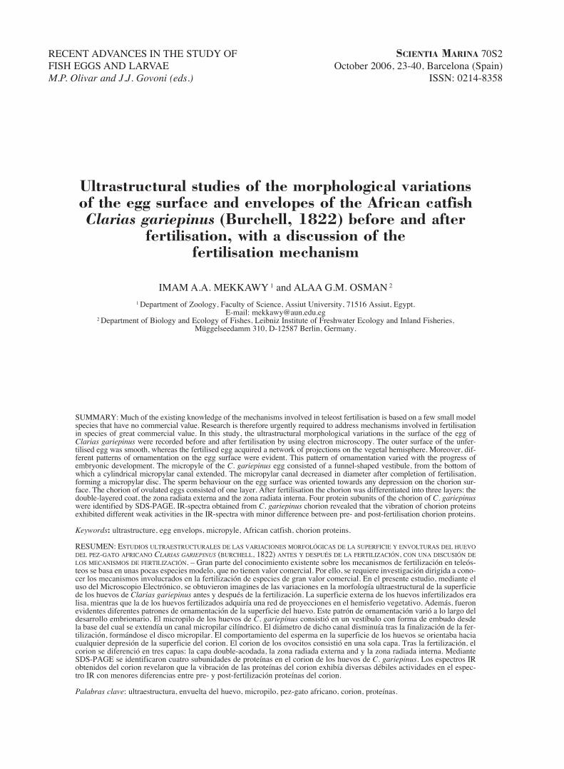
The testes of C. gariepinus in a ripe stage dis-played various stages of active spermatogenesis.The spermatozoa of C. gariepinus were tightlypacked in the lumen of the testes lobules (Fig. 1A).Spermatozoa consisted of a head, midpiece and verylong tail (Fig. 1B). The head region contained thenucleus, which consisted of variable electron densegranular chromatin materials. The head region wasrelatively triangular or rectangular. The midpiece
SCI. MAR., 70S2, October, 2006, 23-40. ISSN: 0214-8358
ULTRASTRUCTURE OF CLARIAS GARIEPINUS EGGS • 25
had an inverted conical shape forming an ovalshaped structure with the head. The head-midpiecesurface showed irregularity (surface with irregularfolding, Fig. 1D). The irregular inverted conicalshape of the midpiece reflected an increased numberof mitochondria. The midpiece had a reticular struc-ture around the flagellum (Fig. 1C), in which themitochondria were distributed separately. Thesefindings were in contrast with those reported byMansour et al. (2002), who stated that several singlemitochondria were fused with each other andformed a complex chondriosome. It was difficult topostulate such a chondriosome structure with thereticulate structure of the midpiece of C. gariepinus.Figure 1 (E, F) showed other related structures,especially the flagellum structure in cross and longi-tudinal sections. The topography of spermatozoa ofC. gariepinus seemed to be adjusted to the diameterof the inner aperture of the micropylar canal. Thiscondition was indicated by Linhart and Kudo (1997)
as contributing to the prevention of polyspermy.Also, the topography of the head-midpiece regionprovided binding facets for the attachment of thesperm on the chorion surface by a ligand-receptormechanism.
The ripe ovary and the oocyte
The ripe period of C. gariepinus gonad was char-acterised chiefly by migration of the nucleus towardthe animal pole (Zaki et al., 1986). The ovarian ripeoocyte of C. gariepinus had four distinct layers: anoutermost follicular layer (outer theca + inner gran-ulosa layer), a median zona radiata (the chorion),and an inner oolemma or oocyte plasma membrane(Fig. 2A, B). Zaki et al. (1986) confused the termi-nology of the layers surrounding the oocyte sincethey referred to the outer layer (zona granulose) aschorion and the inner layer as zona radiata (withclearly discernible pores). Rizkalla (1960) referred
SCI. MAR., 70S2, October 2006, 23-40. ISSN: 0214-8358
26 • I.A.A. MEKKAWY and A.G.M. OSMAN
FIG. 1. – Spermatozoa in the lobules of the rip testis of Clarias gariepinus (A: ×10000). The structures of the spermatozoa as revealed by SEM(B, C) in semen (×3500) and TEM (×20000). The irregularity of sperm head and midpiece were evident with a very long tail (B) and a
midpiece process (D: ×20000). E, F represent transverse and longitudinal sections of the flagellum (×20000).
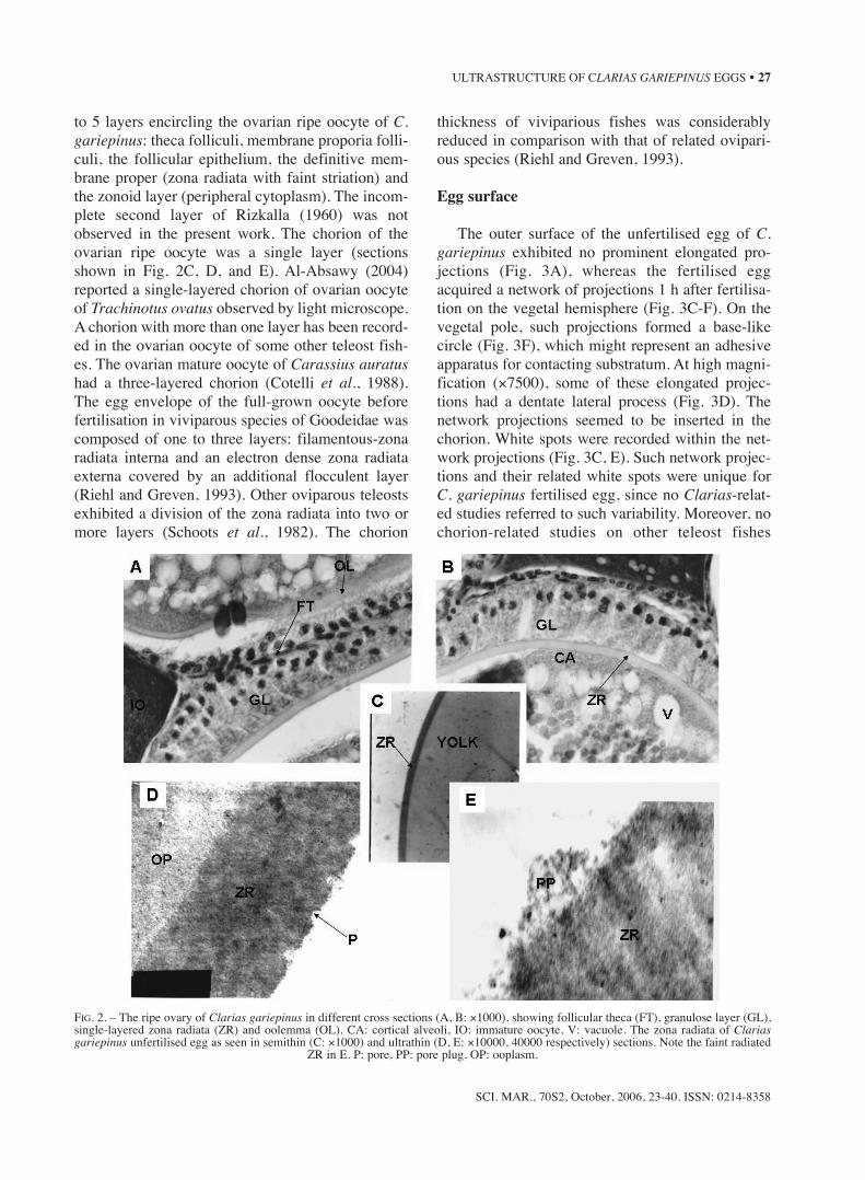
to 5 layers encircling the ovarian ripe oocyte of C.gariepinus: theca folliculi, membrane proporia folli-culi, the follicular epithelium, the definitive mem-brane proper (zona radiata with faint striation) andthe zonoid layer (peripheral cytoplasm). The incom-plete second layer of Rizkalla (1960) was notobserved in the present work. The chorion of theovarian ripe oocyte was a single layer (sectionsshown in Fig. 2C, D, and E). Al-Absawy (2004)reported a single-layered chorion of ovarian oocyteof Trachinotus ovatus observed by light microscope.A chorion with more than one layer has been record-ed in the ovarian oocyte of some other teleost fish-es. The ovarian mature oocyte of Carassius auratushad a three-layered chorion (Cotelli et al., 1988).The egg envelope of the full-grown oocyte beforefertilisation in viviparous species of Goodeidae wascomposed of one to three layers: filamentous-zonaradiata interna and an electron dense zona radiataexterna covered by an additional flocculent layer(Riehl and Greven, 1993). Other oviparous teleostsexhibited a division of the zona radiata into two ormore layers (Schoots et al., 1982). The chorion
thickness of viviparious fishes was considerablyreduced in comparison with that of related ovipari-ous species (Riehl and Greven, 1993).
Egg surface
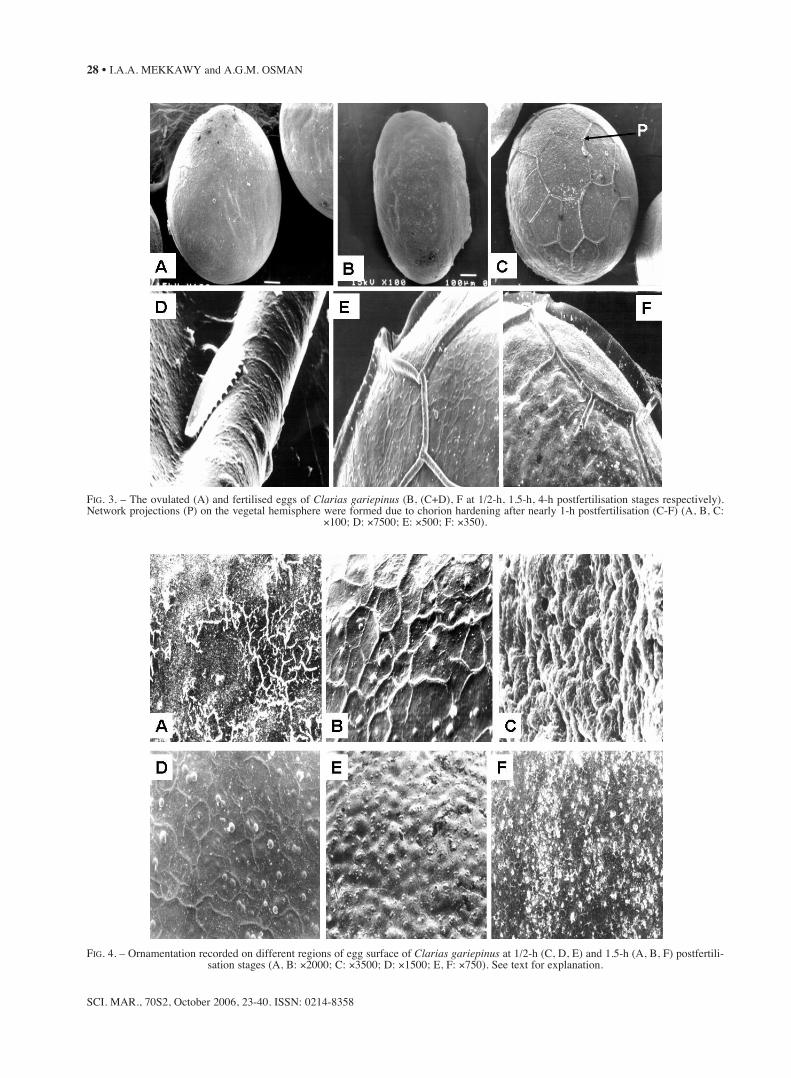
The outer surface of the unfertilised egg of C.gariepinus exhibited no prominent elongated pro-jections (Fig. 3A), whereas the fertilised eggacquired a network of projections 1 h after fertilisa-tion on the vegetal hemisphere (Fig. 3C-F). On thevegetal pole, such projections formed a base-likecircle (Fig. 3F), which might represent an adhesiveapparatus for contacting substratum. At high magni-fication (×7500), some of these elongated projec-tions had a dentate lateral process (Fig. 3D). Thenetwork projections seemed to be inserted in thechorion. White spots were recorded within the net-work projections (Fig. 3C, E). Such network projec-tions and their related white spots were unique forC. gariepinus fertilised egg, since no Clarias-relat-ed studies referred to such variability. Moreover, nochorion-related studies on other teleost fishes
SCI. MAR., 70S2, October, 2006, 23-40. ISSN: 0214-8358
ULTRASTRUCTURE OF CLARIAS GARIEPINUS EGGS • 27
FIG. 2. – The ripe ovary of Clarias gariepinus in different cross sections (A, B: ×1000), showing follicular theca (FT), granulose layer (GL),single-layered zona radiata (ZR) and oolemma (OL). CA: cortical alveoli, IO: immature oocyte, V: vacuole. The zona radiata of Clariasgariepinus unfertilised egg as seen in semithin (C: ×1000) and ultrathin (D, E: ×10000, 40000 respectively) sections. Note the faint radiated
ZR in E. P: pore, PP: pore plug, OP: ooplasm.
SCI. MAR., 70S2, October 2006, 23-40. ISSN: 0214-8358
28 • I.A.A. MEKKAWY and A.G.M. OSMAN
FIG. 3. – The ovulated (A) and fertilised eggs of Clarias gariepinus (B, (C+D), F at 1/2-h, 1.5-h, 4-h postfertilisation stages respectively).Network projections (P) on the vegetal hemisphere were formed due to chorion hardening after nearly 1-h postfertilisation (C-F) (A, B, C:
×100; D: ×7500; E: ×500; F: ×350).
FIG. 4. – Ornamentation recorded on different regions of egg surface of Clarias gariepinus at 1/2-h (C, D, E) and 1.5-h (A, B, F) postfertili-sation stages (A, B: ×2000; C: ×3500; D: ×1500; E, F: ×750). See text for explanation.

referred to such projections. Different patterns ofornamentation on the egg surface of C. gariepinuswere evident (Fig. 4). Such ornamentation has beenrepresented as tubercle and/or reticular (Fig. 4A),debris-like dots and batches (Fig. 4B,D), irregularlylobulated ornamentations (Fig. 4C) and partiallyreticulated debris (Fig. 4F). These patterns wererecorded in different regions and at different post-fertilisation times. Irregularly distributed porebulges were recorded 1/2 h after postfertilisation(Fig. 4E). The debris-like dots and the partiallyreticulated debris might represent the poorly pre-served remains of the diffuse mucus layer (Lönningand Hagstrom, 1975; Johnson and Werner, 1986).
The ornamentation varied from the germinal discregion to the vegetal hemisphere (Fig. 5). Moreover,the pattern of ornamentation varied with theprogress in embryonic development since hairs anddepressions appeared on the animal hemisphere butnot on the vegetal one (Fig. 5D). Further changeswere recorded at the 30 h stage (Fig.5E). Similarornamentation patterns have been recorded by many
authors working on different teleost species belong-ing to different taxonomic groups (Johnson andWerner, 1986; Cotelli et al., 1988; Merrett andBarnes, 1996; Chen et al., 1999; Rizzo et al., 2002;Chiou et al., 2004). Some of these authors consid-ered the egg surface ornamentation as taxonomiccharacters at the specific level. The variability inornamentation of C. gariepinus makes their use inspecies identification difficult owing to their associ-ation with the fertilisation and development process.Chen et al. (1999) referred to the importance of theouter surface of the chorion in egg identification andphylogenetic study. However, they added that theouter surface of the chorion did not show remark-able differences in microstructure among species ina genus or a family.
The micropyle
The micropyle of C. gariepinus egg consisted ofa funnel-shaped vestibule from the bottom of whicha cylindrical micropylar canal extended (Fig. 6A,
SCI. MAR., 70S2, October, 2006, 23-40. ISSN: 0214-8358
ULTRASTRUCTURE OF CLARIAS GARIEPINUS EGGS • 29
FIG. 5. – Ornamentation on the micropylar disc (A, at 1/2-h postfertilisation), the separation line between animal and vegetal hemispheres (B,C, at 1-h postfertilisation; D, at 9-h postfertilisation) and the region of Clarias gariepinus embryo attachment (E, at 30-h postfertilisation)
(A: ×350; B, E: ×200; C, D: ×750).
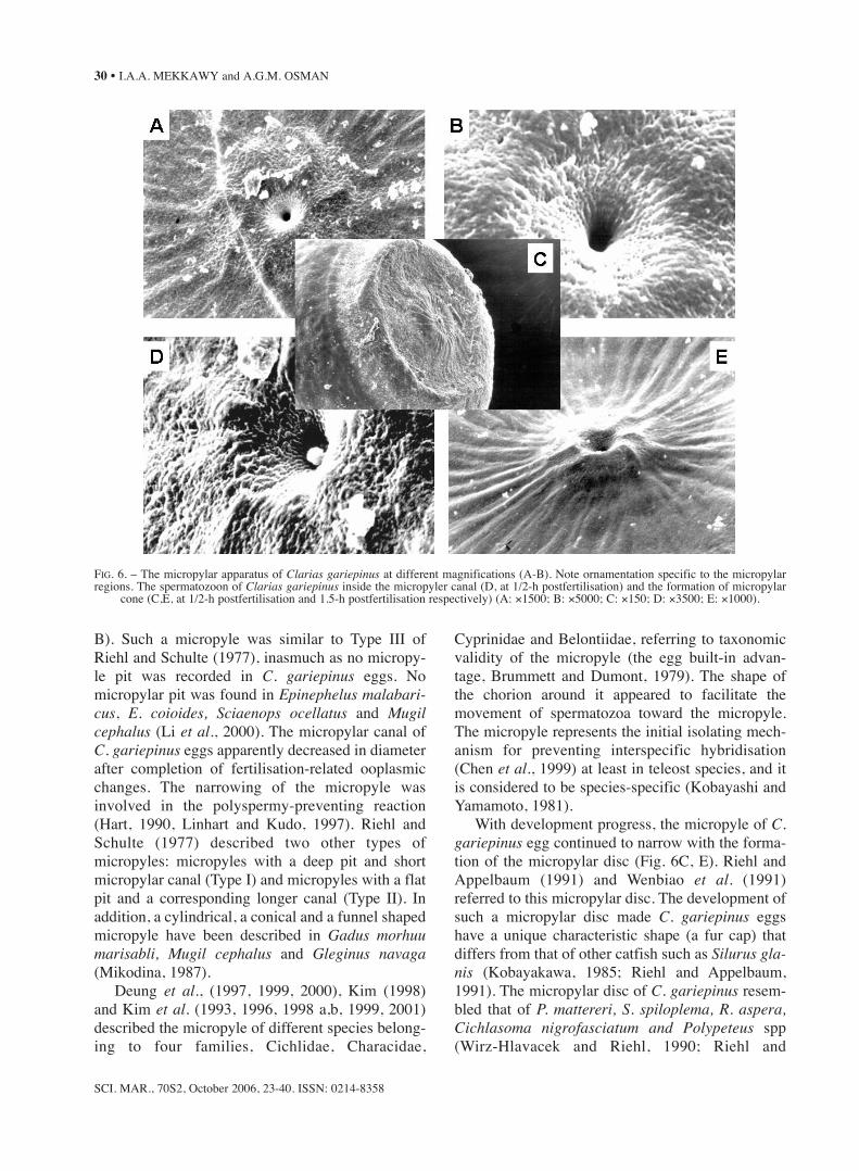
B). Such a micropyle was similar to Type III ofRiehl and Schulte (1977), inasmuch as no micropy-le pit was recorded in C. gariepinus eggs. Nomicropylar pit was found in Epinephelus malabari-cus, E. coioides, Sciaenops ocellatus and Mugilcephalus (Li et al., 2000). The micropylar canal ofC. gariepinus eggs apparently decreased in diameterafter completion of fertilisation-related ooplasmicchanges. The narrowing of the micropyle wasinvolved in the polyspermy-preventing reaction(Hart, 1990, Linhart and Kudo, 1997). Riehl andSchulte (1977) described two other types ofmicropyles: micropyles with a deep pit and shortmicropylar canal (Type I) and micropyles with a flatpit and a corresponding longer canal (Type II). Inaddition, a cylindrical, a conical and a funnel shapedmicropyle have been described in Gadus morhuumarisabli, Mugil cephalus and Gleginus navaga(Mikodina, 1987).
Deung et al., (1997, 1999, 2000), Kim (1998)and Kim et al. (1993, 1996, 1998 a,b, 1999, 2001)described the micropyle of different species belong-ing to four families, Cichlidae, Characidae,
Cyprinidae and Belontiidae, referring to taxonomicvalidity of the micropyle (the egg built-in advan-tage, Brummett and Dumont, 1979). The shape ofthe chorion around it appeared to facilitate themovement of spermatozoa toward the micropyle.The micropyle represents the initial isolating mech-anism for preventing interspecific hybridisation(Chen et al., 1999) at least in teleost species, and itis considered to be species-specific (Kobayashi andYamamoto, 1981).
With development progress, the micropyle of C.gariepinus egg continued to narrow with the forma-tion of the micropylar disc (Fig. 6C, E). Riehl andAppelbaum (1991) and Wenbiao et al. (1991)referred to this micropylar disc. The development ofsuch a micropylar disc made C. gariepinus eggshave a unique characteristic shape (a fur cap) thatdiffers from that of other catfish such as Silurus gla-nis (Kobayakawa, 1985; Riehl and Appelbaum,1991). The micropylar disc of C. gariepinus resem-bled that of P. mattereri, S. spiloplema, R. aspera,Cichlasoma nigrofasciatum and Polypeteus spp(Wirz-Hlavacek and Riehl, 1990; Riehl and
SCI. MAR., 70S2, October 2006, 23-40. ISSN: 0214-8358
30 • I.A.A. MEKKAWY and A.G.M. OSMAN
FIG. 6. – The micropylar apparatus of Clarias gariepinus at different magnifications (A-B). Note ornamentation specific to the micropylarregions. The spermatozoon of Clarias gariepinus inside the micropyler canal (D, at 1/2-h postfertilisation) and the formation of micropylar
cone (C,E, at 1/2-h postfertilisation and 1.5-h postfertilisation respectively) (A: ×1500; B: ×5000; C: ×150; D: ×3500; E: ×1000).
Appelbaum, 1991; Bartsch and Britz, 1997; Rizzo etal., 2002). In the absence of data that elucidate themechanism by which eggs of these species adhere tosubstratum, Riehl and Appelbaum (1991) concludedthat the micropylar disc may play a role in theiradhesiveness. Similarly, Wenbiao et al. (1991)termed the micropylar disc as the attachment disc.As such, the eggs should be fertilised before theirattachment to the substratum (Wirz-Hlavacek andRiehl, 1990). The network of projections on the veg-etal hemisphere founding C. gariepinus leads to theconclusion that these projections might representanother attachment mechanism. In fertilisationexperiments of C. gariepinus, the animal pole wasusually directed upward. Most catfish, including C.gariepinus, possess demersal eggs, which becomesticky after encountering water. Catfish eggs adhereto substratum with several other methods (Riehl andAppelbaum, 1991). In Silurns glanis and twoJapanese Silurus species (S. asotus and S. biwaen-sis), the eggs adhered with a voluminous layer ofjelly (Kobayakawa, 1985; Hilge et al., 1987; Riehl
and Appelbaum, 1991), whereas the eggs ofJapanese Silurus lithophilus were not adhesive. Thejelly layer coat was also present in the adhesive eggsof other siluriformes, including Ictalurus spp andChrysichthys spp, in addition to Silurus spp. (Sato,1999; Rizzo et al., 2002) and in non-adhesive eggsof siluriformes including Paulicea luetkeni,Pimelodus maculatus and Conorhynchus conirostris(Sato, 1999; Rizzo et al., 2002). The adhesive appa-ratus of C. gariepinus was formed of a large numberof tiny attaching filaments, which were embedded ina certain cement substance (Riehl and Appelbaum,1991). Such tiny structures were observed in thepresent work as microvilli extending from the outersurface of the chorion in embryonic stages beforehatching (Fig. 5E).
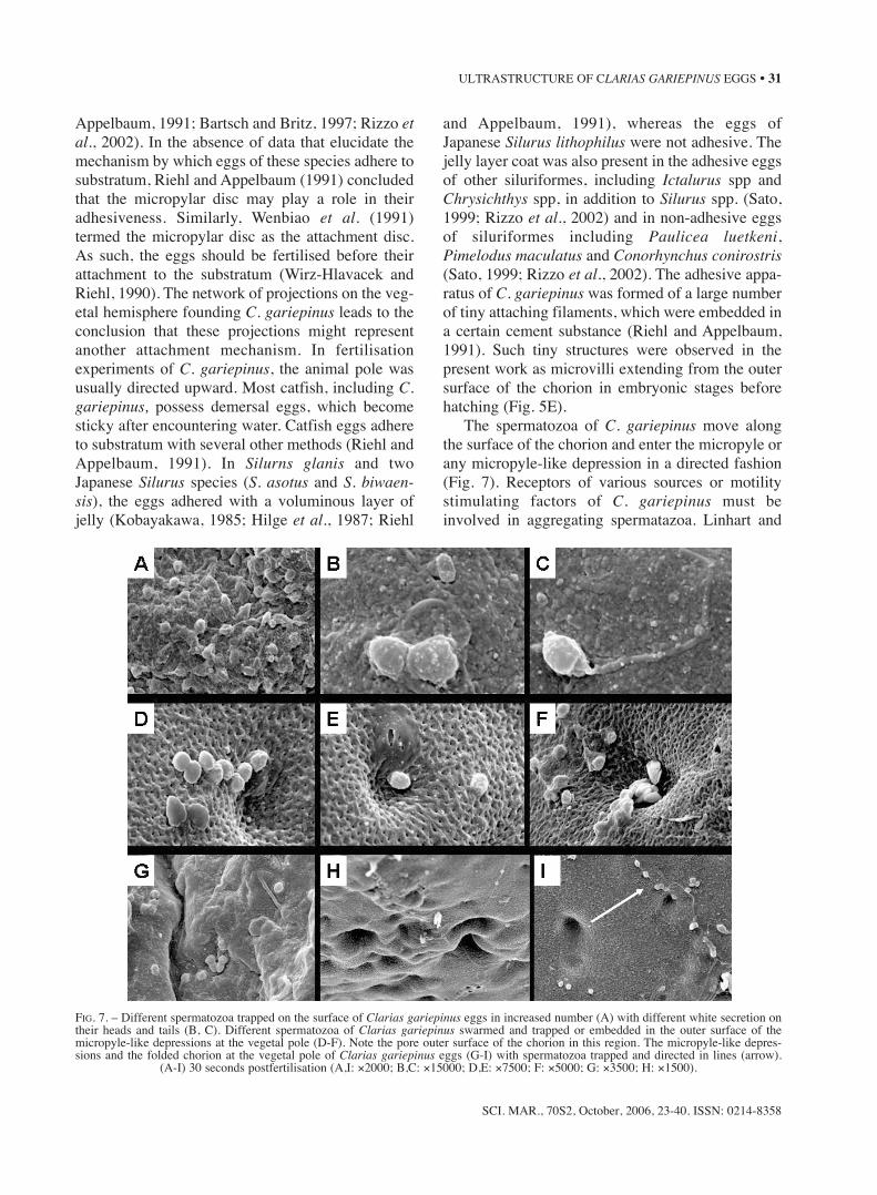
The spermatozoa of C. gariepinus move alongthe surface of the chorion and enter the micropyle orany micropyle-like depression in a directed fashion(Fig. 7). Receptors of various sources or motilitystimulating factors of C. gariepinus must beinvolved in aggregating spermatazoa. Linhart and
SCI. MAR., 70S2, October, 2006, 23-40. ISSN: 0214-8358
ULTRASTRUCTURE OF CLARIAS GARIEPINUS EGGS • 31
FIG. 7. – Different spermatozoa trapped on the surface of Clarias gariepinus eggs in increased number (A) with different white secretion ontheir heads and tails (B, C). Different spermatozoa of Clarias gariepinus swarmed and trapped or embedded in the outer surface of themicropyle-like depressions at the vegetal pole (D-F). Note the pore outer surface of the chorion in this region. The micropyle-like depres-sions and the folded chorion at the vegetal pole of Clarias gariepinus eggs (G-I) with spermatozoa trapped and directed in lines (arrow).
(A-I) 30 seconds postfertilisation (A,I: ×2000; B,C: ×15000; D,E: ×7500; F: ×5000; G: ×3500; H: ×1500).
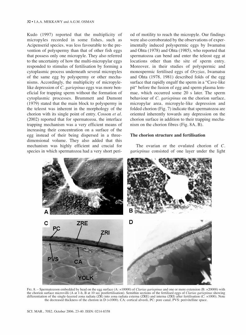
Kudo (1997) reported that the multiplicity ofmicropyles recorded in some fishes, such asAcipenserid species, was less favourable to the pre-vention of polyspermy than that of other fish eggsthat possess only one micropyle. They also referredto the uncertainty of how the multi-micropylar eggsresponded to stimulus of fertilisation by forming acytoplasmic process underneath several micropylesof the same egg by polyspermy or other mecha-nisms. Accordingly, the multiplicity of micropyle-like depression of C. gariepinus eggs was more ben-eficial for trapping sperm without the formation ofcytoplasmic processes. Brummett and Dumont(1979) stated that the main block to polyspermy inthe teleost was inherent in the morphology of thechorion with its single point of entry. Cosson et al.(2002) reported that for spermatozoa, the interfacetrapping mechanism was a very efficient means ofincreasing their concentration on a surface of theegg instead of their being dispersed in a three-dimensional volume. They also added that thismechanism was highly efficient and crucial forspecies in which spermatozoa had a very short peri-
od of motility to reach the micropyle. Our findingswere also corroborated by the observations of exper-imentally induced polyspermic eggs by Iwamatsuand Ohta (1978) and Ohta (1985), who reported thatspermatozoa can bend and enter the teleost egg atlocations other than the site of sperm entry.Moreover, in their studies of polyspermic andmonospermic fertilised eggs of Oryzias, Iwamatsuand Ohta (1978, 1981) described folds of the eggsurface that rapidly engulf the sperm in a “Cave-likepit“ before the fusion of egg and sperm plasma lem-mae, which occurred some 20 s later. The spermbehaviour of C. gariepinus on the chorion surface,micropylar area, micropyle-like depression andfolded chorion (Fig. 7) indicate that spermatozoa areoriented inherently towards any depression on thechorion surface in addition to their trapping mecha-nism on the chorion fibres (Fig. 8A, B).
The chorion structure and fertilisation
The ovarian or the ovulated chorion of C.gariepinus consisted of one layer under the light
SCI. MAR., 70S2, October 2006, 23-40. ISSN: 0214-8358
32 • I.A.A. MEKKAWY and A.G.M. OSMAN
FIG. 8. – Spermatozoon embedded by head on the egg surface (A: ×10000) of Clarias gariepinus and one or more extension (B: ×20000) withthe chorion surface microvilli (A at 1-h, B at 10 sec postfertilisation). Semithin sections of the fertilised eggs of Clarias gariepinus showingdifferentiation of the single-layered zona radiata (ZR) into zona radiata externa (ZRE) and interna (ZRI) after fertilisation (C: ×1000). Note
the decreased thickness of the chorion in D (×1000). CA: cortical alveoli, PC: pore canal, PVS: perivitelline space.
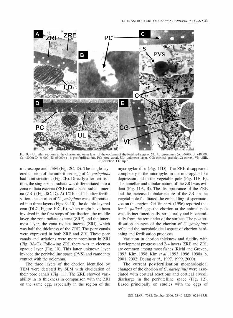
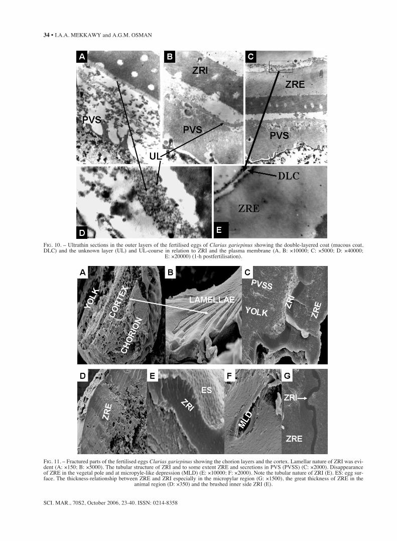
microscope and TEM (Fig. 2C, D). The single-lay-ered chorion of the unfertilised egg of C. gariepinushad faint striations (Fig. 2E). Directly after fertilisa-tion, the single zona radiata was differentiated into azona radiata externa (ZRE) and a zona radiata inter-na (ZRI) (Fig. 8C, D). At 1/2 h and 1 h after fertili-sation, the chorion of C. gariepinus was differentiat-ed into three layers (Figs. 9, 10), the double-layeredcoat (DLC, Figure 10C, E), which might have beeninvolved in the first steps of fertilisation, the middlelayer, the zona radiata externa (ZRE) and the inner-most layer, the zona radiata interna (ZRI), whichwas half the thickness of the ZRE. The pore canalswere expressed in both ZRE and ZRI. These porecanals and striations were more prominent in ZRI(Fig. 9A-C). Following ZRI, there was an electronopaque layer (Fig. 10). This latter unknown layerinvaded the perivitelline space (PVS) and came intocontact with the oolemma.
The three layers of the chorion identified byTEM were detected by SEM with elucidation oftheir pore canals (Fig. 11). The ZRE showed vari-ability in its thickness in comparison with the ZRIon the same egg, especially in the region of the
mycropylar disc (Fig. 11D). The ZRE disappearedcompletely in the micropyle, in the micropylar-likedepression and in the vegetable pole (Fig. 11E, F).The lamellar and tubular nature of the ZRI was evi-dent (Fig. 11A, B). The disappearance of the ZREand the increased tubular nature of the ZRI in thevegetal pole facilitated the embedding of spermato-zoa on this region. Griffin et al. (1996) reported thatfor C. pallasi eggs the chorion at the animal polewas distinct functionally, structurally and biochemi-cally from the remainder of the surface. The postfer-tilisation changes of the chorion of C. gariepinusreflected the morphological aspect of chorion hard-ening and fertilisation processes.
Variation in chorion thickness and rigidity withdevelopment progress and 2-4 layers, ZRE and ZRI,are common among most fishes (Riehl and Greven,1993; Kim, 1998; Kim et al., 1993, 1996, 1998a, b,2001, 2002; Deung et al., 1997, 1999, 2000).
The current postfertilisation morphologicalchanges of the chorion of C. gariepinus were asso-ciated with cortical reactions and cortical alveolidischarge in the perivitelline space (Fig. 12).Based principally on studies with the eggs of
SCI. MAR., 70S2, October, 2006, 23-40. ISSN: 0214-8358
ULTRASTRUCTURE OF CLARIAS GARIEPINUS EGGS • 33
FIG. 9. – Ultrathin sections in the chorion and outer layer of the ooplasm of the fertilised eggs of Clarias gariepinus (A: ×6700; B: ×40000;C: ×8000; D: ×4000; E: ×5000) (1-h postfertilisation). PC: pore canal, UL: unknown layer, CG: cortical granule, C: cortex, VI: villii,
S: secretion, LD: lipid.
SCI. MAR., 70S2, October 2006, 23-40. ISSN: 0214-8358
34 • I.A.A. MEKKAWY and A.G.M. OSMAN
FIG. 10. – Ultrathin sections in the outer layers of the fertilised eggs of Clarias gariepinus showing the double-layered coat (mucous coat,DLC) and the unknown layer (UL) and UL-course in relation to ZRI and the plasma membrane (A, B: ×10000; C: ×5000; D: ×40000;
E: ×20000) (1-h postfertilisation).
FIG. 11. – Fractured parts of the fertilised eggs Clarias gariepinus showing the chorion layers and the cortex. Lamellar nature of ZRI was evi-dent (A: ×150; B: ×5000). The tubular structure of ZRI and to some extent ZRE and secretions in PVS (PVSS) (C: ×2000). Disappearanceof ZRE in the vegetal pole and at micropyle-like depression (MLD) (E: ×10000; F: ×2000). Note the tubular nature of ZRI (E). ES: egg sur-face. The thickness-relationship between ZRE and ZRI especially in the micropylar region (G: ×1500), the great thickness of ZRE in the
animal region (D: ×350) and the brushed inner side ZRI (E).
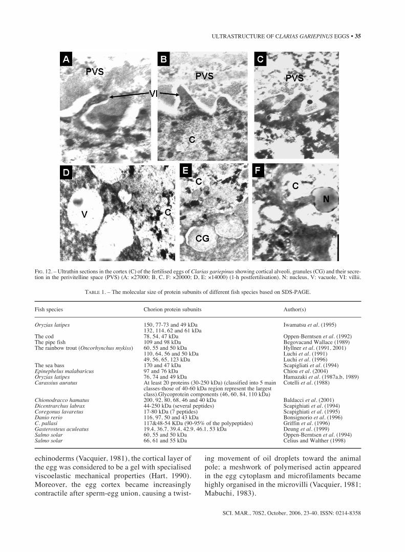
echinoderms (Vacquier, 1981), the cortical layer ofthe egg was considered to be a gel with specialisedviscoelastic mechanical properties (Hart, 1990).Moreover, the egg cortex became increasinglycontractile after sperm-egg union, causing a twist-
ing movement of oil droplets toward the animalpole; a meshwork of polymerised actin appearedin the egg cytoplasm and microfilaments becamehighly organised in the microvilli (Vacquier, 1981;Mabuchi, 1983).
SCI. MAR., 70S2, October, 2006, 23-40. ISSN: 0214-8358
ULTRASTRUCTURE OF CLARIAS GARIEPINUS EGGS • 35
FIG. 12. – Ultrathin sections in the cortex (C) of the fertilised eggs of Clarias gariepinus showing cortical alveoli, granules (CG) and their secre-tion in the perivitelline space (PVS) (A: ×27000; B, C, F: ×20000; D, E: ×14000) (1-h postfertilisation). N: nucleus, V: vacuole, VI: villii.
TABLE 1. – The molecular size of protein subunits of different fish species based on SDS-PAGE.
Fish species Chorion protein subunits Author(s)
Oryzias latipes 150, 77-73 and 49 kDa Iwamatsu et al. (1995)132, 114, 62 and 61 kDa
The cod 78, 54, 47 kDa Oppen-Berntsen et al. (1992)The pipe fish 109 and 98 kDa Begovacand Wallace (1989)The rainbow trout (Oncorhynchus mykiss) 60, 55 and 50 kDa Hyllner et al. (1991, 2001)
110, 64, 56 and 50 kDa Luchi et al. (1991)49, 56, 65, 123 kDa Luchi et al. (1996)
The sea bass 170 and 47 kDa Scapigliati et al. (1994)Epinephelus malabaricus 97 and 76 kDa Chiou et al. (2004)Oryzias latipes 76, 74 and 49 kDa Hamazaki et al. (1987a,b, 1989)Carassius auratus At least 20 proteins (30-250 kDa) (classified into 5 main Cotelli et al. (1988)
classes-those of 40-60 kDa region represent the largest class).Glycoprotein components (46, 60, 84, 110 kDa)
Chionodracco hamatus 200, 92, 80, 68, 46 and 40 kDa Baldacci et al. (2001)Dicentrarchus labrax 44-250 kDa (several peptides) Scapighiati et al. (1994)Coregonus lavaretus 17-80 kDa (7 peptides) Scapighiati et al. (1995)Danio rerio 116, 97, 50 and 43 kDa Bonsignorio et al. (1996)C. pallasi 117&48-54 KDa (90-95% of the polypeptides) Griffin et al. (1996)Gasterosteus aculeatus 19.4, 36.7, 39.4, 42.9, 46.1, 53 kDa Deung et al. (1999)Salmo solar 60, 55 and 50 kDa Oppen-Berntsen et al. (1994)Salmo solar 66, 61 and 55 kDa Celius and Walther (1998)

Actin and actin-containing filaments have beendescribed in the cortical layer of Brachydanio(Wolenski and Hart, 1988) and Oncorhynchus(Kobayashi, 1985) eggs. These lectins, as majorcomponents in vertebrate cortical granules(Krajhanzl, 1990), are involved in the formation ofthe egg envelope and in turn in its polyspermy-blocking functions (Quill and Hedrick, 1996). Donget al. (2004) identified a C-type lectin from oocyteof a freshwater fish species Carassius auratusgibelio. TEM showed that the cortical cytoplasm ofthe eggs of C. gariepinus contained membrane-lim-ited cortical granules with an internal matrix ofvarying electron density (Fig. 12). The determina-tion of the chemical and molecular composition of
cortical granules is essential to understanding therole of these organelles in fertilisation and earlydevelopment (Hart, 1990). Cortical reaction seemsto be a prerequisite for the chorion hardeningprocess, which has been considered by some authorsto be independent of fertilisation (Lönning et al.,1984; Davenport et al., 1986). There was a preciserelationship between sperm behaviour, chorionhardening and cortical reaction of C. gariepinus.
Chorion hardening is a process of initiated chainpolymerisation of substances within the membraneitself (Hart, 1990) to form insoluble proteins ofhigher molecular weight (Yamagami et al., 1992).The solubility of chorion of unfertilised egg is arequirement for the hardening system or machinery
SCI. MAR., 70S2, October 2006, 23-40. ISSN: 0214-8358
36 • I.A.A. MEKKAWY and A.G.M. OSMAN
TABLE 2. – The chorion protein subunits (bands) identified by SDS-PAGE in the ovulated (OE), hardened OE and post-fertilised (1-minute, 1-hour and 2-hour FE) eggs of Clarias gariepinus.
MW 1 2 3 4 5Lanes: MW-marker Hardened OE OE 1-minute FE 1-hour FE 2-hour FEBands MW MW % MW % MW % MW % MW %
r1 223.0r2 133.6 43.1 132.6 47.0 129.7 29.8r3 110.0 108.9 38.6 108.9 47.8r4 81.6r5 46.8r6 36.4 31.5 35.6 24.6 35.9 35.1r7 31.8 31.5 32.0 33.7 26.8r8 28.3 25.4 28.8 12.0 29.2 15.0r9 24.8 25.3 16.0 26.9 12.6r10 16.5 16.0 13.4 17.0 12.8 17.0 16.4 17.0 20.1
TABLE 3. – The chorion protein subunits (bands) identified by SDS-UREA-PAGE in the ovulated (OE), hardened OE and post-fertilised (2- and 25-minute, 1-, 2- and 3-hour FE) eggs of Clarias gariepinus in addition to its semen and sperm proteins.
Lanes: 1 2 3 4 5 6 7 8 9 MWBands Semen Sperm 3-HFE 2-HFE 1-HFE 25-MFE 2-MFE Hardened OE OE Marker
Molecular weight
r1 248.5r2 235.3 230.2 231.0 223.0r3 199.1 188.0 192.8 199.1 200.7 195.9r4 183.2 175.3 168.9r5 153.0 156.2 159.3 164.1 166.9r6 137.1 146.4 133.9 135.9r7 100.0r8 101.8 100.3 95.8 98.8r9 92.8 89.8 89.1 89.1r10 85.3 86.8 80.3 81.6r11 74.8 79.3 79.0r12 65.5 63.9 65.5 65.0 66.9 65.5 66.7 68.2r13 54.6 54.3 54.3r14 49.4 48.5r15 45.8 46.0 46.2 46.2 46.6 46.6 46.6 46.8r16 44.9 43.7 43.7 45.1 44.3 44.3 44.5r17 43.0 41.6 41.6r18 39.9 39.7 40.3 40.8 41.3 41.0 41.3r19 35.1 35.1r20 31.0 30.8 31.8 31.4 31.8r21 30.4 29.9 29.9 30.4 29.5r22 27.3 26.6r23 25.9 25.6 25.4 25.2 25.6 25.6 25.7 25.3 25.2 24.8
SCI. MAR., 70S2, October, 2006, 23-40. ISSN: 0214-8358
ULTRASTRUCTURE OF CLARIAS GARIEPINUS EGGS • 37
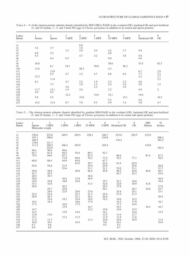
TABLE 4. – % of the chorion protein subunits (bands) identified by SDS-UREA-PAGE in the ovulated (OE), hardened OE and post-fertilised(2- and 25-minute, 1-, 2- and 3-hour FE) eggs of Clarias gariepinus in addition to its semen and sperm proteins.
Lanes: 1 2 3 4 5 6 7 8 9Bands Semen Sperm 3-HFE 2-HFE 1-HFE 25-MFE 2-MFE Hardened OE OE
r1 0.8r2 3.2 3.7 1.8r3 3.2 2.9 3.0 4.2 3.7 5.0r4 6.9 3.5 4.0r5 4.3 4.7 4.2 3.8 5.6r6 6.4 6.5 9.0 4.4r7r8 16.8 38.6 33.5 62.3r9 8.1 54.1 59.0 56.3r10 13.4 58.2 4.3r11 13.0 6.7 2.9r12 6.5 4.7 3.2 4.7 8.8 6.5 7.7 3.2r13 12.3 2.3 1.7r14 11.0 2.2r15 8.1 4.7 2.2 1.9 2.3 2.2 4.0r16 7.5 1.0 1.3 2.4 1.6 2.8 1.5r17 3.6 2.2 3.4r18 11.7 12.1 1.4 2.6 2.2 4.9 2.r19 3.0 9.2r20 12.2 14.6 14.1 14.6r21 5.9 5.5 15.0 15.1 19.3r22 5.3 5.6r23 14.2 13.4 9.7 7.8 9.3 4.9 7.4 3.0 4.7
TABLE 5. – The chorion protein subunits (bands) identified by gradient SDS-PAGE in the ovulated (OE), hardened OE and post-fertilised (2- and 25-minute, 1-, 2- and 3-hour FE) eggs of Clarias gariepinus in addition to its semen and sperm proteins.
Lanes: 1 2 3 4 5 6 7 8 MW 10Bands Sperm 3-HFE 2-HFE 1-HFE 25-MFE 2-MFE Hardened OE OE Marker Semen
Molecular weight
r1 230.6 223.0 249.5 249.5 228.1 246.7 223.0 220.5 223.0r2 197.1 190.6 210.8 206.2r3 168.5 176.2 182.2r4 134.6 121.7r5 111.2 108.5 106.4 103.9 105.4 110.0r6 102.9 102.5r7 99.1 96.8 99.6r8 92.7 91.4 94.5 93.6 89.3 92.7r9 79.5 84.0 87.2 83.2 84.0 81.6 87.2r10 73.9 69.8 76.3 77.5 78.3 77.1 70.2r11 68.0 68.4 64.9 64.6 67.7r12 61.0 59.7 61.6 61.6 62.9r13 55.0 55.6 57.9 55.0 55.8 55.0 58.8r14 53.6 51.1 51.1 53.0 51.1 51.7r15 49.0 49.8 49.8 46.4 45.8 48.5 45.8 46.8 46.7r16 43.7 45.5 44.7 42.3 43.9r17 40.5 39.7 38.8r18 38.0 38.2 37.8 36.9 36.9r19 34.9 35.8 34.8 34.4 35.7 35.1 34.6 34.4r20 33.1 32.0 33.3 32.6 32.0 30.9 31.8r21 28.8 30.2 28.3 27.8 27.8r22 25.7 25.8 26.1 26.7 24.8 25.7r23 24.0 22.9 24.6 23.6 24.4 24.1 23.1r24 22.4 22.4 22.3 21.3 22.6 21.9 21.3r25 20.9 20.3 21.1 20.1 20.4 20.4r26 19.4 19.2 19.4 18.9 19.2 19.6 19.2r27 18.2 18.2 18.6 17.9 18.3 17.9 18.1r28 17.3 16.9 17.5 17.3 16.9r29 15.9 16.7 16.1 16.5 16.7r30 14.7 15.2 15.8 15.0 15.5r31 14.3 13.9 14.4 14.1 14.4 13.5r32 12.8 13.0 12.5 13.4 12.7r33 12.1 12.3 11.5 11.9 11.9 11.6r34 11.2 11.3 11.3 10.8 10.6 10.9 11.4r35 10.4 10.1 10.5 9.8 10.1 10.4r36 9.7 9.6 9.3 9.3r37 8.9 8.5 8.7
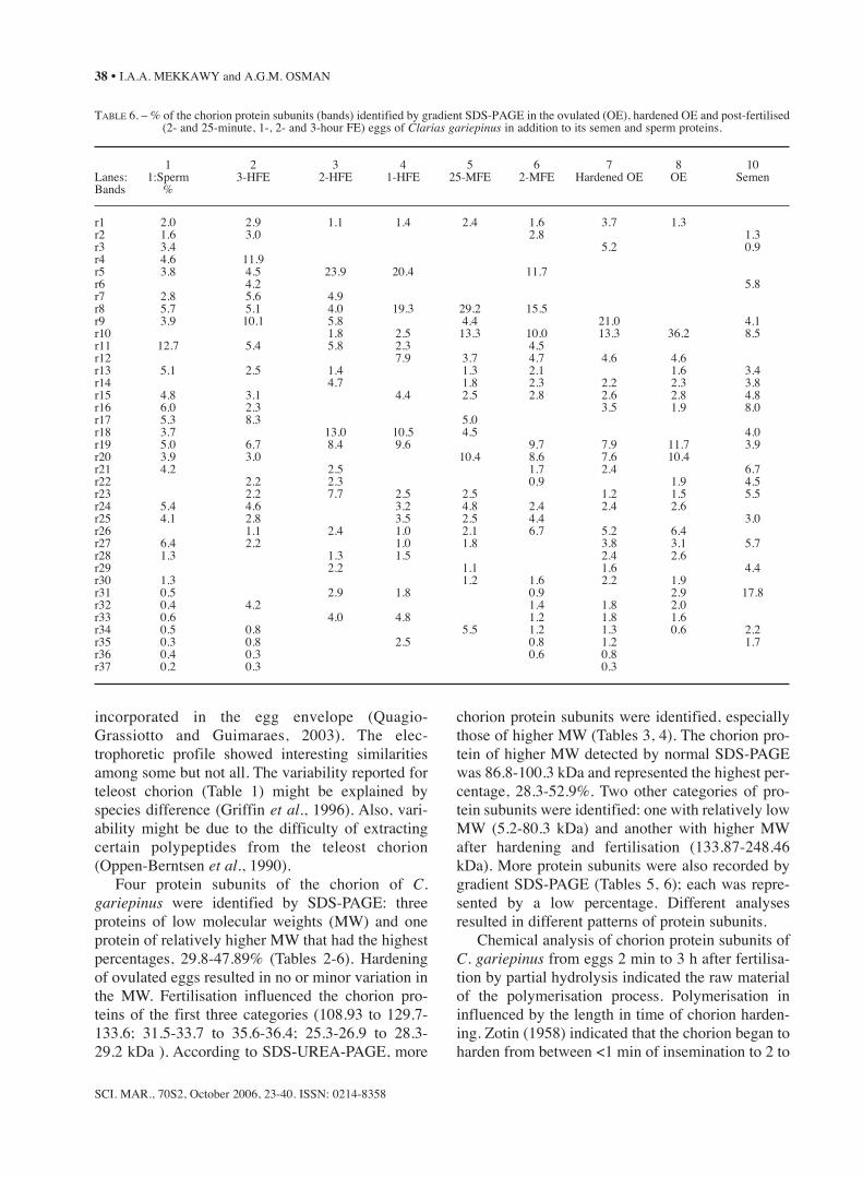
incorporated in the egg envelope (Quagio-Grassiotto and Guimaraes, 2003). The elec-trophoretic profile showed interesting similaritiesamong some but not all. The variability reported forteleost chorion (Table 1) might be explained byspecies difference (Griffin et al., 1996). Also, vari-ability might be due to the difficulty of extractingcertain polypeptides from the teleost chorion(Oppen-Berntsen et al., 1990).
Four protein subunits of the chorion of C.gariepinus were identified by SDS-PAGE: threeproteins of low molecular weights (MW) and oneprotein of relatively higher MW that had the highestpercentages, 29.8-47.89% (Tables 2-6). Hardeningof ovulated eggs resulted in no or minor variation inthe MW. Fertilisation influenced the chorion pro-teins of the first three categories (108.93 to 129.7-133.6; 31.5-33.7 to 35.6-36.4; 25.3-26.9 to 28.3-29.2 kDa ). According to SDS-UREA-PAGE, more
chorion protein subunits were identified, especiallythose of higher MW (Tables 3, 4). The chorion pro-tein of higher MW detected by normal SDS-PAGEwas 86.8-100.3 kDa and represented the highest per-centage, 28.3-52.9%. Two other categories of pro-tein subunits were identified: one with relatively lowMW (5.2-80.3 kDa) and another with higher MWafter hardening and fertilisation (133.87-248.46kDa). More protein subunits were also recorded bygradient SDS-PAGE (Tables 5, 6); each was repre-sented by a low percentage. Different analysesresulted in different patterns of protein subunits.
Chemical analysis of chorion protein subunits ofC. gariepinus from eggs 2 min to 3 h after fertilisa-tion by partial hydrolysis indicated the raw materialof the polymerisation process. Polymerisation ininfluenced by the length in time of chorion harden-ing. Zotin (1958) indicated that the chorion began toharden from between <1 min of insemination to 2 to
SCI. MAR., 70S2, October 2006, 23-40. ISSN: 0214-8358
38 • I.A.A. MEKKAWY and A.G.M. OSMAN
TABLE 6. – % of the chorion protein subunits (bands) identified by gradient SDS-PAGE in the ovulated (OE), hardened OE and post-fertilised (2- and 25-minute, 1-, 2- and 3-hour FE) eggs of Clarias gariepinus in addition to its semen and sperm proteins.
1 2 3 4 5 6 7 8 10Lanes: 1:Sperm 3-HFE 2-HFE 1-HFE 25-MFE 2-MFE Hardened OE OE SemenBands %
r1 2.0 2.9 1.1 1.4 2.4 1.6 3.7 1.3r2 1.6 3.0 2.8 1.3r3 3.4 5.2 0.9r4 4.6 11.9r5 3.8 4.5 23.9 20.4 11.7r6 4.2 5.8r7 2.8 5.6 4.9r8 5.7 5.1 4.0 19.3 29.2 15.5r9 3.9 10.1 5.8 4.4 21.0 4.1r10 1.8 2.5 13.3 10.0 13.3 36.2 8.5r11 12.7 5.4 5.8 2.3 4.5r12 7.9 3.7 4.7 4.6 4.6r13 5.1 2.5 1.4 1.3 2.1 1.6 3.4r14 4.7 1.8 2.3 2.2 2.3 3.8r15 4.8 3.1 4.4 2.5 2.8 2.6 2.8 4.8r16 6.0 2.3 3.5 1.9 8.0r17 5.3 8.3 5.0r18 3.7 13.0 10.5 4.5 4.0r19 5.0 6.7 8.4 9.6 9.7 7.9 11.7 3.9r20 3.9 3.0 10.4 8.6 7.6 10.4r21 4.2 2.5 1.7 2.4 6.7r22 2.2 2.3 0.9 1.9 4.5r23 2.2 7.7 2.5 2.5 1.2 1.5 5.5r24 5.4 4.6 3.2 4.8 2.4 2.4 2.6r25 4.1 2.8 3.5 2.5 4.4 3.0r26 1.1 2.4 1.0 2.1 6.7 5.2 6.4r27 6.4 2.2 1.0 1.8 3.8 3.1 5.7r28 1.3 1.3 1.5 2.4 2.6r29 2.2 1.1 1.6 4.4r30 1.3 1.2 1.6 2.2 1.9r31 0.5 2.9 1.8 0.9 2.9 17.8r32 0.4 4.2 1.4 1.8 2.0r33 0.6 4.0 4.8 1.2 1.8 1.6r34 0.5 0.8 5.5 1.2 1.3 0.6 2.2r35 0.3 0.8 2.5 0.8 1.2 1.7r36 0.4 0.3 0.6 0.8r37 0.2 0.3 0.3
4 h in salmonid and coregonid eggs, whereas Hart(1990) found that maximum hardening of the chori-on was reached within 3-7 days in the trout and 1-2days in the whitefish. For lump sucker (Cyclopteurs)and cod (Gadus), chorion hardening started shortlyafter exposure to sea water and reached a maximumresistance by about 24 h (Lönning et al., 1984).Hardening in these two salt water species did notrequire fertilisation (Hart, 1990).
Iconomidou et al. (2000) and Hamodrakas et al.(1987) indicated that the β-pleated sheet was themolecular conformation of protein macromoleculesthat constitute the fibrils and of fibrils of the egg shellwith a helicoidal architecture. These studies did notdeal with the interaction between chorion hardening,fertilisation and progress in development. Furtherspectroscopic studies must be done to demonstrate arelationship between chorion hardening and the timeof development based on different amide ranges.
ACKNOWLEDGEMENTS
The second author was indebted to Prof. FrankKirschbaum, Head of Department of Biology andEcology of Fishes, Leibniz-Institute of FreshwaterEcology and Inland Fisheries, Müggelseedamm 310,D-12587 Berlin, Germany, for his help in obtainingfinancial support to participate in 29th Annual LarvalFish Conference held in Barcelona, Spain, on 11-14July, 2005. The authors would like to thank Dr. J.J.Govoni and Dr. M.P. Olivar for their invaluable com-ments and suggestions on the manuscript.
REFERENCES
Al-Absawy, M.A.G. – 2004. Reproduction and UltrastructureStudies on Gonads of Trachinotus ovatus (Family: Characidae)from the Egyptian Mediterranean Waters. PhD. thesis, ZagazigUniv., Egypt.
Bancroft, J.D. and A. Stevens. – 1982. Theory and Practice ofHistologica Techniques. Churchill Livingstone, London.
Baldacci, A., A.R. Taddei, M. Mazzini, A.M. Fausto, F. Buonocoreand G. Scapigliati. – 2001. Ultrastructure and proteins of theegg chorion of the Antarctic fish Chionodraco hamates(Teleostei, Notothenioidei). Polar. Biol., 24: 417-421.
Bartsch, P. and R. Britz. – 1997. A single micropyle in the eggs of themost basal living actinopterygian fish, Polypterus (Actinopterygii,Polypteriformes). J. Zool. (London), 241: 589-592.
Britz, R. and T. Breining. – 2000. Egg surface structure of threeclingfish species, using scanning electron microscopy. J. FishBiol., 56: 1129-1137.
Britz, R., M. Kokoscha and R. Riel. – 1995. The anabantoid generaCtenops, Luciocephalus, Parasphaerichthys, and Sphaerichthys(Teleostei: Perciformes) as a monophyletic group: evidencefrom egg surface structure and reproductive behaviour. Jpn. J.Ichthyol., 42: 71-79.
Brummett, A.R. and J.N. Dumont. – 1979. Initial stages of spermpenetration into the egg of Fundulus heteroclitus. J. Exp. Zool.,210: 417-432.
Celius, T. and B.T. Walther. – 1998. Differential sensitivity of zon-agenesis and vitellogenesis in Atlantic salmon (Salmo solar L.)to DDT pesticides. J. Exp. Zool., 281: 346-353.
Chen, K.C., K.T. Shao and J.S. Yang. – 1999. Using micropylarUltrastructure for species identification and phylogenetic infer-ence among four species of Sparidae. J. Fish Biol., 55: 288-300.
Chiou, L.M., M.C. Chung, P.G. Tung, T. Hsu and J. S. Yang. –2004. The use of egg chorion glycoprotein of Epinephelus mal-abaricus for egg identification. J. Fish Biol., 65: 1614-1621.
Cosson, J., P. Huitorel and C. Gagnon. – 2002. How spermatozoacome to be confined to surfaces. Cell Motil. Cytoskel., 54: 56-63.
Cotelli, F., F. Andronico, M. Brivio and C.L. Lora lamia. – 1988.Structure and composition of the fish egg chorion (Carassiusauratus). J. Ultrastruct. Mol.Struct. Res., 99: 70-78.
Coward, K., N.R. Bromage, O. Hibbitt and J. Parrington. – 2002.Gamete physiology, fertilization and egg activation in teleostfish. Rev. Fish Biol. Fisher., 12: 33-58.
Davenport, J., S. Lonning and E. Kjorsvik. – 1986. Some mechani-cal and morphological properties of the chorions of marineTeleost eggs. J. Fish Biol., 29: 289-301.
Deung, Y.K., D.H. Kim and D.S. Reu. – 1999. Ultrastructure ofGametes in the Three-spine stickleback, Gasterosteus aculea-tus aculeatus. Korean J. Electron Microscopy, 29: 177-187.
Deung, Y.K., D.H. Kim and D.S. Reu. – 2000. Ultrastructure of thefertilized egg envelope from Pale Chub, Cyprinidae, Teleost.Korean J. Electron Microscopy, 30: 321-326.
Deung, Y., D.S. Reu and D. H. Kim. – 1997. Comparative ultra-structures of the fertilized egg envelopes in golden severum,convic cichlid and discus, Cichlidae, teleost. Korean J.Electron Microscopy, 27: 417-432.
Dong, C., S. Yang, Z. Yang, L. Zhang and J. Gui. – 2004. A C-typelectin associated and translocated with cortical granules duringOocyte maturation and egg fertilization and egg fertilization infish. Dev. Biol., 265: 341-354.
Gill, A.C. and R.D. Mooi. – 1993. Monophyly of the Grammatidaeand of the Notograptidae with evidence for their phylogeneticpositions among perciforms. Bull. Mar. Sci., 52: 327-354.
Griffin, F.J., C.A. Vines, M. C. Pillia, R. Yanagimachi and G.N.Cherr. – 1996. Sperm motility initiation factor is a minor com-ponent of the pacific herring egg chorion. Dev. Growth Differ.,38: 193-202.
Hames, B.D. – 1981. One-dimensional polyacrylamide gel elec-trophoresis. In: B. D. Hames, and D. Rickwood (eds.), GelElectrophoresis of Proteins. A practical approach, pp. 1-174.IRL press, Oxford and Washington D.C.
Hamodrakas, S.J., E.I. Kamitsos and P.G. Papadopoulou. – 1987.Laser-Raman and infrared spectroscopic studies of protein con-formation in the eggshell of the fish Salmo gairdneri. Biochim.Biophys. Acta, 913: 163-169.
Hart, N.H. – 1990. Fertilization in teleost fishes: mechanisms ofsperm-egg interactions. Int. Rev. Cytol., 121: 1-66.
Hilge, V., M. Abraham and R. Riehl. – 1987. The Jelly coat of theOocyts in the Eurobian Catfish. Proceedings of the ThirdInternational Symposium on the Reproductive Physiology ofFish, August 2-7. Memorial University of Newfoundland, St.John’s, Canada.
Hyllner, S.J., L. Westerlund, P. Olsson and A. Schopen. – 2001.Cloning of rainbow trout egg envelope proteins: Members of aunique group of structural proteins. Biol. Reprod., 64: 805-811.
Iconomidou, V.A., D.G. Chryssikos, V. Gionis, M.A. Pavlidis, A.Paipetis. and S.J. Hamodrakas. – 2000. Secondary structure ofchorion proteins of the teleostean fish Dentex dentex by ATR FT-IR and FT-Raman spectroscopy. J. Struct. Biol., 132: 112-122.
Iwamatsu, T. and T. Ohta. – 1978. Electron microscopic observa-tion on sperm penetration and pronuclear formation in the fishegg. J . Exp. Zool., 205: 157-180.
Iwamatsu, T. and T. Ohta. – 1981. Scanning electron microscopicobservations on sperm penetration in teleostean fish. J. Exp.Zool., 218: 261-277.
Johnson, E.Z. and R.G. Werner. – 1986. Scanning electronmicroscopy of the chorion of selected freshwater fishes. J. FishBiol., 29: 257-265.
Johnson, G.D. and E.B. Brothers. – 1993. Schindleria- A paedomor-phic goby (Teleostei: Gobiodei). Bull. Mar. Sci., 52: 441-471.
SCI. MAR., 70S2, October, 2006, 23-40. ISSN: 0214-8358
ULTRASTRUCTURE OF CLARIAS GARIEPINUS EGGS • 39
Kim, D.H. – 1998. Comparative Ultrastructure of Fertilized EggEnvelops in Teleost. Department of Biology, Graduate School,Chongju University Press.
Kim, D.H., K. Deung, W. J. Kim, D.S. Reu and S.J. Kang. – 2001.Ultrastructure of the fertilized envelope from long nose barbel,Cyprinidae, Teleost. Korean J. Electron Microscopy, 31: 85-90.
Kim, D.H., Y.K. Deung, W. J. Kim, D.S. Reu and S.J. Kang. –1999. Comparative Ultrastructure of the fertilized eggsenvelopes from three–spot gourami, Pearl gourami and Marblegourami, Belontiidae, Teleost. Korean J. Electron Microscopy,29: 343-351.
Kim, D.H., D.S. Reu and Y. K. Deung. – 1996. A Comparativestudy on the Ultrastructures of the egg envelope in fertilizedeggs of fishes, Characidae, three species. Korean J. ElectronMicroscopy, 26: 277-291.
Kim, D.H., D.S. Reu and Y.K. Deung. – 1998a. ComparativeUltrastructure of the fertilized egg envelope in three species,Cyprinidae, Teleost. Korean J. Electron Microscopy, 28: 237-253.
Kim, D.H., D.S. Reu and Y.K. Deung. – 1998b. Ultrastructure offertilized egg envelope in the tomato clown Anemonfish,Amphiprion freatus (Pomacentridae: Marine Teleostei). KoreanJ. Electron Microscopy, 28: 273-282.
Kim, D.H., D.S. Reu and Y.K. Deung. – 2002. Ultrastructure of thefertilized egg envelope from dark sleeper, Eleotrididae, Teleost.Korean J. Electron Microscopy, 32: 39-44.
Kim, D.H., D.S. Reu, W.J. Kim and Y.K. Deung. – 1993. AComparative study on the Ultrastructures of the egg envelope infertilized eggs of the Angelfish (Pterophyllum eimekei) andZebrafish (Brachydanio rerio). Korean J. Electron Microscopy,23: 115-128.
Kobayakawa, M. – 1985. External characteristics of the eggs ofJapanese catfishes (Silurus). Jpn. J. Ichthyol., 32: 104-106.
Kobayashi, W. – 1985. Electron microscopic observation of thebreakdown of cortical vesicles in the chum salmon egg. J. Fac.Sci. Hokaido Univ., 624: 87-102.
Kobayashi, W. and T.S. Yamamoto. – 1981- Fine structure of themicropylar apparatus of the chum salmon egg, with a discus-sion of the mechanism for blocking polyspermy. J. Exp. Zool.,217: 265-275.
Krajhanzl, K. – 1990. Egg lectins of invertebrates and lower verte-brates: Properties and biological function. Adv. Lectin Res., 3:83-131.
Laemmli, U.K. – 1970. Cleavage of structural proteins during assem-bly of the head of bacteriophage T4. Nature, 227: 680-685.
Li, Y.H., C.C. Wu and J.S. Yang. – 2000. Comparative ultrastruc-tural Studies of marine fish eggs in three genera in Perciformes.J. Fish Biol., 56: 615-621.
Linhart, O. and S. Kudo. – 1997. Surface structure of paddlefisheggs before and after fertilization. J. Fish Biol., 51: 573-582.
Lönning, S. and B.E. Hagstrom. – 1975. Scanning electron micro-scope studies of the surface of the fish egg. Astarte, 8: 17-22.
Lönning, S., E. Kjorsvik and J. Davemport. – 1984. The hardeningprocess of the egg chorion of the cod, Gadus morhua L. andlumpsucker, Cyclopterus lumpus L. J. Fish Biol., 24: 505-522.
Mabuchi, I. – 1983. An actin-depolymerized protein (Depactin)from starfish Oocytes: Properties and interaction with actin. J.Cell Biol., 97: 1612-1621.
Mansour, N., F. Lahnsteiner and R.A. Patzner. – 2002. The sper-matozoa of the African catfish: fine structure, motility, viabili-ty and its behaviour in seminal vesicle secretion. J. Fish Biol.,60: 545-560.
Marshall, N.B. – 1973. Family Macrouridae. In: D.M. Cohen (eds.),Fishes of the Western North Atlantic, PP. 496-537. MemoirSears Foundation for Marine Research.
Media-Cybernetics – 1998. Gel-Pro Analyser software packageV.3.1. Media-Cybernetics, L.P., Georgia.
Merrett, N.R. and S.H. Barnes. – 1996. Preliminary survey of egg
envelope morphology in the Macrouridae and the possibleimplications of its ornamentation. J. Fish Biol., 48: 101-119.
Mikodina, E.V. – 1987. Surface structure of the egg of teleosteanfishes. Voprosy Ikhtiologii, 1: 106-113.
Ohta, T. – 1985. Electron microscopic observations on sperm entryand pronuclear formation in naked eggs of the Rose Bitterlingin polyspermic fertilization. J. Exp. Zool., 234: 273-281.
Oppen-Berntsen, D. O., J.V. Helvik and B. T. Walther. – 1990. Themajor structural proteins of code (Gadus morhua) egg shellsand protein crosslinking during teleost egg hardening. Dev.Biol., 137: 258-265.
Quagio-Grassiotto, I. and A. C.D. Guimaraes. – 2003. Follicularepithelium, theca and egg envelope formation in Serrasalmusspilopleura (Teleostei, Characiformes, Characidae). Acta Zool.-Stochkolm, 84: 121-129.
Quill, T.A. and J.L. Hedrick. – 1996. The fertilization layer mediat-ed block to polyspermy in Xenopus laevis: Isolation of cortical-granule lectin ligand. Arch. Biochem. Biophys., 2: 326-332.
Riehl, R. and S. Appelbaum. – 1991. A Unique adhesion apparatuson the eggs of the catfish Clarias gariepinus (Teleostei,Clariidae). Jpn. J. Ichthyol., 38: 191-197.
Riehl, R. and H. Greven. – 1993. Fine structure of egg envelops insome viviparous goodeid fishes, with comments on the relationof envelope thinness to viviparity. Can. J. Zool., 71: 91-97.
Riehl, R. and E. Schulte. – 1977. Vergleichende rasterelektronen-mikroskopische Untersuchungen an der Mikropylen ausgewählterSüßwasser- Teleosteer. Arch. Fischerei wiss., 28: 95-107.
Rizkalla, W. – 1960. The Endocrine Glands of Clarias lazera. PhD.thesis, Univ. Ain Shams, Egypt.
Rizzo, E., Y. Sato, B.P. Barreto and H.P. Godinho. – 2002.Adhesiveness and surface patterns of eggs in neotropical fresh-water teleost. J. Fish Biol., 61: 615-632.
Sato, Y. – 1999. Reproduction of the São Francisco river basin fish-es: induction and characterization of patterns. PhD. thesis,Fedral University of São Carlos, SP, Brazil.
Schoots, A.F.M., J.J.M. Stikkelbroeck, J.F. Bekhuis and J.M.Denuce. – 1982. Hatching in teleostean fishes: fine structuralchanges in the egg envelope during enzymatic breakdown invivo and vitro. J. Ultrastruct. Res., 80: 185-196.
Swank, R.T. and K.D. Munkres. – 1971. Molecular weight analysisof oligopeptides by electrophoresis in polyacrylamide gel withsodium dodecyl sulfate. Anal. Biochem., 39: 462-477.
Vacquier, V.D. – 1981. Review, Dynamic changes of the egg cor-tex. Dev. Biol., 84: 1-26.
Wenbiao, Z.,P. Jionghua, A. Dong and L. Wensheng. – 1991.Observation on the process of fertilization of Clarias lazera byscanning electron microscopy. Zool. Res., 12: 111-115.
Wirz-Hlavacek, G. and R. Riehl. – 1990. Reproductive behaviorand egg structure of the piranha Serrassalmus nattererri (Kner,1860). Acta Biol. Benrodis, 2: 19-38.
Wolenski, J.S. and N.H. Hart. – 1988. Effects of Cytochalasins Band D on the fertilization of Zebrafish (Brachydanio) eggs. J.Exp. Zool., 246: 202-215.
Yamagami, K., T.S. Hamazaki, S. Yasumasu, K. Masuda and I.Luchi. – 1992. Molecular and cellular basis of formation, hard-ening, and breakdown of the egg envelope in fish. Int. Rev.Cytol., 136: 51-92.
Zaki, M.I., M.N. Dowidar and A. Abdala. – 1986. Reproductivebiology of Clarias gariepinus (Syn. Lazera) Burchell(Claridae) in Lake Manzala, Egypt. I. Structure of Ovaries.Folia Morphologica, 34: 301-306.
Zotin, A.I. – 1958. the mechanism of hardening of the salmonid eggmembrane after fertilization or spontaneous activation. J.Embryol. Exp. Morph., 6: 546-568.
Received August 29, 2005. Accepted April 10, 2006.Published online September 26, 2006.
SCI. MAR., 70S2, October 2006, 23-40. ISSN: 0214-8358
40 • I.A.A. MEKKAWY and A.G.M. OSMAN