Embed Size (px)
Citation preview

Folia biologica (Kraków), vol. 57 (2009), No 3-4doi:10.3409/fb57_3-4.131-137
Ultrastructure and Transovarial Transmission of Endosymbiotic
Microorganisms in Conomelus anceps and Metcalfa pruinosa (Insecta,
Hemiptera, Fulgoromorpha)*
Anna MICHALIK, W³adys³awa JANKOWSKA and Teresa SZKLARZEWICZ
Accepted April 20, 2009
MICHALIK A., JANKOWSKA W., SZKLARZEWICZ T. 2009. Ultrastructure and transovarialtransmission of endosymbiotic microorganisms inConomelus anceps andMetcalfa pruinosa(Insecta, Hemiptera, Fulgoromorpha). Folia biol. (Kraków) 57: 131-137.Endosymbiotic microorganisms commonly occur in fulgoromorphans, as in other plantsap-sucking hemipterans. Large syncytial organs termed mycetomes are present in the bodycavities of Conomelus anceps (Delphacidae) and Metcalfa pruinosa (Flatidae), in the closevicinity of the ovaries. The mycetomes are surrounded by a one-layered epithelium. Themycetome cytoplasm is filled with yeast-like symbiotic microorganisms (YLSs). The YLSsare transovarially transmitted to the next generation. The endosymbionts are released fromthe mycetomes and migrate towards the ovarioles containing vitellogenic oocytes. The YLSspass through the cells of the ovariole stalk (pedicel) and enter the perivitelline space. Then, adeep depression is formed at the posterior pole of the oocyte. The YLSs accumulate in theoocyte depression and form a characteristic �symbiont ball�. The mycetome cytoplasm ofMetcalfa pruinosa as well as epithelial cells surrounding the mycetome contain small,rod-shaped bacteria.Key words: Endosymbiotic microorganisms, transovarial transmission, ovariole,Fulgoromorpha, planthoppers.AnnaMICHALIK, W³adys³awa JANKOWSKA, Teresa SZKLARZEWICZ, Department of SystematicZoology and Zoogeography, Institute of Zoology, Jagiellonian University, R. Ingardena 6,30-060 Kraków, Poland.E-mail: [email protected]
Fulgoromorphans, like other plant sap-suckinghemipterans, harbor obligate, intracellular symbi-otic microorganisms (see BUCHNER 1965; HOUK& GRIFFITHS 1980; BAUMANN 2005, 2006, forfurther details). Endosymbiotic microorganismsare housed in large cells termed mycetocytes(=bacteriocytes). Mycetocytes are usually inte-grated into discrete organs termed mycetomes(=bacteriomes) that in some hemipterans may besurrounded by a one-layered epithelium. BUCH-NER (1965) on the basis of comparative studies ofall hemipteran groups distinguished two catego-ries of endosymbiotic microorganisms: primaryendosymbionts (currently called P-symbionts) andaccessory endosymbionts (currently called facul-tative, secondary or S-symbionts). Primary endo-symbionts are always present in all the specimens,whereas secondary endosymbionts occur in somepopulations only. More recent studies have shownthat the occurrence of primary endosymbionts in
the insect body is related to a restricted diet, defi-cient in some essential nutrients (see BAUMANN2005, 2006, for full review). Since hemipteransfeed on phloem sap devoid of amino acids, theirprimary endosymbionts are responsible for aminoacid synthesis and delivery to the host insect (e.g.SASAKI & ISHIKAWA 1995; WILKINSON & ISHI-KAWA 2001). Thus, the occurrence of primary en-dosymbionts is necessary for the survival andreproduction of the host insects. In contrast to pri-mary endosymbionts, the role of the secondary en-dosymbionts for their host insects remains unclear.Recent studies revealed that aphids harboring S-symbionts may better survive heat stress (MONTL-LOR et al. 2002) as well as attacks by parasitic hy-menopterans (OLIVER et al. 2003) and fungalpathogens (SCARBOROUGH et al. 2005) than ster-ile specimens.
The symbiotic associations between primary endo-symbionts and insects are the results of ancient in-
_______________________________________*Supported by Research Grant DS/IZ/ZS/2008.
fections with free living bacteria (BAUMANN 2006),whereas associations between secondary endosym-bionts and insects are much younger and are due tomultiple, independent infections (THAO et al. 2000).
Most hemipterans have prokaryotic endosymbi-onts, however, eukaryotic microorganisms histori-cally termed “yeast-like symbionts” (YLSs) occurin some aphids, scale insects, leafhoppers and ful-goromorphans (see BUCHNER 1965; ISHIKAWA2003, for further details).
Both bacterial and yeast-like endosymbionts arematernally inherited by transovarial transmission.Studies on transovarial transmission of bacterialendosymbionts have shown that variable modes ofovary infection by bacteria exist in insects (¯ELA-ZOWSKA & BILIÑSKI 1999; SZKLARZEWICZ &MOSKAL 2001; SZKLARZEWICZ et al. 2006). Theendosymbiotic microorganisms may invade younggerm cells (=cystocytes) or oocytes in the olderstage of development (i.e. vitellogenic or chorio-genic). The ovaries may be invaded by bacteria orby intact mycetocytes. The endosymbionts maymigrate through the neighboring follicular cells(i.e. cells surrounding oocytes) or may enter thecytoplasm of follicular cells. In contrast to bacte-rial endosymbionts, the transovarial transmissionof YLSs is poorly known, and there are only threepublished observations on this process (NODA1977; CHENG & HOU 2001; SACCHI et al. 2008),two of which consider members of the family Del-phacidae.
The present study was undertaken to provide in-formation about the mode of transmission of YLSsin Metcalfa pruinosa, a member of the Flatidaefamily, and in another member of the family Del-phacidae, Conomelus anceps.
Material and Methods
Adult specimens of Conomelus anceps (Germar,1821) were collected in July near Nowy Targ(southern Poland). Adult specimens of Metcalfapruinosa (Say, 1830) were collected in July inGrabels (southern France). The abdomens and dis-sected ovaries of individuals of each species werefixed in 2.5% glautaraldehyde in 0.1 phosphatebuffer (pH 7.4) for three months. The material wasthen rinsed in 0.1 M phosphate buffer (pH 7.4)with addition of 5.8% sucrose, postfixed in 1% os-mium tetroxidae, dehydrated in a series of alcoholand acetone and embedded in epoxy resin Epox812 (Fullam Inc., Latham, N.Y., USA). Semithinsections were stained with 1% methylene blue in1% borax and photographed in a Jenalumar (ZeissJena) microscope. Ultrathin sections were stainedwith lead citrate and uranyl acetate and examinedusing a JEM 100 SX electron microscope at 80 kV.
Results
Gross morphology of the ovaries
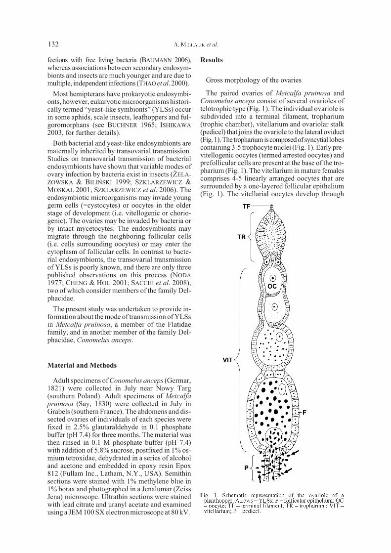
The paired ovaries of Metcalfa pruinosa andConomelus anceps consist of several ovarioles oftelotrophic type (Fig. 1). The individual ovariole issubdivided into a terminal filament, tropharium(trophic chamber), vitellarium and ovariolar stalk(pedicel) that joins the ovariole to the lateral oviduct(Fig.1).The trophariumiscomposedofsyncytial lobescontaining 3-5 trophocyte nuclei (Fig. 1). Early pre-vitellogenic oocytes (termed arrested oocytes) andprefollicular cells are present at the base of the tro-pharium (Fig. 1). The vitellarium in mature femalescomprises 4-5 linearly arranged oocytes that aresurrounded by a one-layered follicular epithelium(Fig. 1). The vitellarial oocytes develop through
A. MICHALIK et al.132
Fig. 1. Schematic representation of the ovariole of aplanthopper. Arrows � YLSs; F � follicular epithelium; OC� oocyte; TF � terminal filament; TR � tropharium; VIT �vitellarium; P � pedicel.

Endosymbiotic Microorganisms in Planthoppers 133
Figs 2-8. Fig. 2.M. pruinosa. The mycetome filled with YLSs (arrowhead) in the body of the male. T � testis. Methylene blue, H570. Fig. 3. M. pruinosa. Fragment of the epithelial cell surrounding the mycetome. Arrows � rod-shaped bacteria; EN �nucleus of the epithelial cell; L � lipid droplet. TEM, H 18 100. Fig. 4. C. anceps. The mycetome filled with YLSs(arrowheads) in the body of the female. Arrows � mycetome nuclei embedded in the common cytoplasm; EP � epithelial cellssurrounding the mycetome; L � lipid droplets. Methylene blue, H 530. Fig. 5.M. pruinosa. Cross section through the cell of theYLS. Arrow � cell wall composed of two distinct layers; YN � nucleus; NU � nucleolus. TEM, H 16 500. Fig. 6. M. pruinosa.Fragment of the mycetome cytoplasm. Arrows � rod-shaped bacteria; MN � mycetome nuclei embedded in the commoncytoplasm. TEM, H 17 500. Fig. 7. M. pruinosa. Fragment of the cell of the YLS. Arrow � cell wall; L � lipid droplet; M �mitochondria; YN � nucleus. TEM, H 15 100. Fig. 8. C. anceps. The YLS reproducing by budding. TEM, H 9 300.

three stages: previtellogenesis (i.e. synthesis andaccumulation of RNA’s), vitellogenesis (i.e. syn-thesis and accumulation of reserve substances) andchoriogenesis (i.e. synthesis and secretion of pre-cursors of eggshells and their deposition on the oo-cyte surface) (for a detailed description of ovariesof planthoppers see SZKLARZEWICZ et al. 2007).
Ultrastructure, distribution and transovarialtransmission of yeast-like endosymbiotic micro-organisms
Both in males and females of C. anceps and M.pruinosa, the spaces between internal organs arefilled with large structures termed mycetomes(Figs 2, 4). The mycetomes have a syncytial char-acter, i.e. they have numerous nuclei embedded in
a common cytoplasm (Figs 4, 6). The mycetomesare surrounded by a one-layered epithelium (Fig. 4).In young specimens epithelial cells are small, izo-diametric and closely adhere to each other (notshown), while in the older specimens they becomeirregular and voluminous with large lipid dropletsin the cytoplasm (Fig. 4). Numerous rod-shapedbacteria occur in epithelial cells (Fig. 3) as well asin mycetome cytoplasm (Fig. 6) of all examinedspecimens of M. pruinosa. The bacteria measure1.2-1.6 Fm in length and 0.3-0.5 Fm in diameter.Both in M. pruinosa and C. anceps, the mycotomecytoplasm contains an enormous number ofyeast-like symbionts (YLSs) (Figs 2, 4). The sizeof YLSs is 8-10 Fm in length and 3,0-3,5 Fm in di-ameter. They are surrounded by a thick cell wallcomposed of two distinct layers (Figs 5, 7, 8). The
Figs 9-13. Figs 9, 10. Longitudinal section through the posterior end of the ovariole, pedicel (P) and lateral oviduct (LOV)during infection by the YLSs (arrows). Methylene blue, H 1 000. Fig. 9. C. anceps. Fig. 10. M. pruinosa. Asterisk � deepdepression of the oolemma; F � follicular cells; OC � oocyte. Fig. 11. C. anceps. Fragment of the follicular epithelium (F)during migration of YLSs. FN � follicular cell nucleus. TEM, H 6 500. Figs 12, 13. C. anceps. Cross section through theposterior pole of the ovariole containing a �symbiont ball� filled with YLSs (encircled). TEM, H 500. Fig. 12. Oocyte duringlate vitellogenesis stage; Fig. 13. Full-grown oocyte. F � follicular epithelium; OC � oocyte.
A. MICHALIK et al.134
outer layer is electron dense and has a thickness of30 nm. The inner layer has lower electron densityand is 140 nm thick. In the central part of the yeastcell a large, spherical nucleus with a single nucleo-lus is present (Fig. 5). The remaining cytoplasm isfilled with ribosomes, mitochondria and large lipiddroplets (Fig. 7). The YLSs reproduce by budding(Fig. 8). Sexual reproduction of YLSs was not ob-served. In older females (i.e. containing terminaloocytes in the stage of late vitellogenesis), theYLSs leave the mycetome cytoplasm. They be-come released into a haemolymph and migrate to-wards the terminal oocytes (Fig. 9). The endosym-bionts pass through the cells of the ovariole stalk(pedicel) (Fig. 9) as well as follicular cells surround-ing the posterior pole of the oocyte (Fig. 10, 11).Subsequently, they enter the perivitelline space(Figs 9, 10). At the same time a deep depression isformed at the posterior pole of the oocyte. TheYLSs accumulate in the oocyte depression andform a characteristic “symbiont ball” (Fig. 12). Atthe end of oocyte growth, the “symbiont ball” istightly packed with YLSs (Fig. 13). Until the endof oocyte growth the YLSs are isolated from theooplasm by oolemma and do not enter the oo-plasm. The endosymbionts gathered in the depres-sion of the oocytes, like those harbored in themycetome, undergo budding (not shown).
Discussion
Our observations revealed that both adult males andfemales of M. pruinosa (Flatidae) as well as C. anceps(Delphacidae) harbor a large number of intracellu-lar YLSs. This observation shows that these endo-symbionts are essential for both sexes of examinedplanthoppers. In recent years, the metabolic signifi-cance of YLSs for growth and reproduction of thehost insects has been extensively studied using therice brown planthopper, Nilaparvata lugens (SASAKIet al. 1996; HONGOH & ISHIKAWA 1997,2000;WILK-INSON & ISHIKAWA 2001). These studies revealedthat planthopper YLSs are involved in nitrogen re-cycling using uric acid as a nitrogenous resource.Planthoppers, in addition to producing uric acid asa nitrogenous waste product, also synthesize it as astorage product during nitrogen deficiency. Uricacid is stored in mycetomes and converted by uri-case, secreted by YLSs, into compounds of nutri-tional value. It should be noted that CHENG andHOU (2005) demonstrated that YLSs are also en-gaged in synthesis of yolk precursors in females ofthe rice brown planthopper, Nilaparvata lugens.
Our studies showed that YLSs in M. pruinosaand C. anceps are transmitted from the mother tothe progeny by a similar route, i.e. via cytoplasm ofcells of the pedicel as well as follicular cells sur-
rounding the posterior pole of the oocyte. Since thesame situation has been observed in other mem-bers of the family Delphacidae, i.e. Laodelphaxstriatellus (NODA 1977) and Nilaparvata lugens(CHENG & HOU 2001) as well as in the leafhopper,Scaphoideus titanus (SACCHI et al. 2008), it seemsprobable that all hemipterans have developed thesame mode of transmission of YLSs to the nextgeneration. Thus, the transmission of YLSs in in-sects is more uniform than the transmission of bac-teria (see Introduction). Moreover, molecular stud-ies revealed that YLSs in hemipterans are not onlysimilarly inherited but are also phylogeneticallyclosely related to each other. The analysis of 18Sribosomal DNA sequences of YLSs in delphacids(NODA et al. 1995; XET-MULL et al. 2004), aphids(FUKATSU & ISHIKAWA 1996) and leafhoppers (SAC-CHI et al. 2008) indicated that they belong to theclass Pyrenomycetes in the phylum Ascomytina.The YLSs in the examined rice delphacids are char-acterized by a high degree of similarity of 18S ribo-somal DNA sequences (NODA et al. 1995; XET-MULL et al. 2004). This finding strongly suggeststhat YLSs of delphacids are closely related (i.e.constitute a monophyletic group). This implies, inturn, that the symbiosis of YLSs and delphacids isthe result of a single infection of the common an-cestor of present delphacids. The YLSs harboredin members of the Flatidae family have so far notbeen examined by molecular methods, thereforetheir systematic position remains unknown. Sinceflatids and delphacids are phylogenetically distantwithin planthoppers (i.e. do not represent a mono-pyletic taxon) (BOURGOIN et al. 1997; URBAN &CRYAN 2007), it is unlikely that their endosymbi-onts have been acquired through a common ancestor.In this light, it may be assumed that YLSs were hori-zontally transferred between flatid and delphacidlineages. It is noteworthy that HONGOH and ISHI-KAWA (2000), on the basis of an analysis of uricasegene sequences of YLSs in aphids and delphacids,provided evidence of a close relationship betweenYLSs in these phylogenetically distant groups ofinsects as a result of the horizontal transfer of micro-organismsfrom the aphid to the planthopper lineage.
Both in M. pruinosa and C. anceps, the migrationof YLSs is correlated with ovary development (theYLSs infect vitellogenic oocytes). Thus, this ob-servation strongly supports the hypothesis that themovement of microorganisms is stimulated by anunknown factor released by ovaries (EBERLE &MC LEAN 1982; ¯ELAZOWSKA & BILIÑSKI 1999;SZKLARZEWICZ & MOSKAL 2001; SZKLARZEWICZet al. 2006).
We observed that apart from YLSs, rod-shapedbacteria are present in the body of all specimens ofM. pruinosa. In contrast to YLSs, bacteria havenever been found in the ovaries of M. pruinosa.
Endosymbiotic Microorganisms in Planthoppers 135
The absence of these bacteria in the ovaries indi-cates that they may be horizontally transmitted be-tween specimens. The large number of bacteria bothin the mycetome cytoplasm as well as in its epithe-lium suggests that they may have a significant(positive or negative) influence on the host insect.These microorganisms may represent S-symbiontsof M. pruinosa, but may also prove to be patho-genic. It should be noted that NODA and SAITO(1979) detected rod-shaped bacteria in mycetomesof the planthopper Laodelphax striatellus (Del-phacidae) but did not suggest a possible role forthem. It may be also speculated that these bacteriabelong to the widespread within arthropodsricketsia-like genus Wolbachia pipientis. The lastassumption is supported by PCR detection of Wol-bachia in two delphacids, Laodelphax striatellusand Sogatella furcifera (NODA et al. 2001), as wellas by the observation that specimens of Laodel-phax striatellus may be horizontally infected byWolbachia (KANG et al. 2003). To verify these hy-potheses, further studies of specimens of M. prui-nosa taken from different populations are needed.
Acknowledgements
We would like to express our gratitude to Dr.Jean-Francois GERMAIN (Labolatoire National dela Protection des Vegetaux, Montpellier, France)and Dr. Sebastian PILARCZYK (Silesian Univer-sity, Katowice, Poland) for collection and identifi-cation of specimens. We are also grateful to Dr.Beata SZYMAÑSKA (Jagiellonian University, De-partment of Systematic Zoology and Zoogeogra-phy, Kraków, Poland) and Dr. Olga WOîNICKA(Jagiellonian University, Department of Cytologyand Histology, Kraków, Poland) for their skilledtechnical assistance.
References
BAUMANN P. 2005. Biology of bacteriocyte-associated endo-symbionts of plant sup-sucking insects. Annu. Rev. Micro-biol. 59: 155-189.BAUMANN P. 2006. Diversity of prokaryote-insect associa-tionswithin the Sternorrhyncha (psyllids, whiteflies, aphids,mealybugs). (In: Insect Symbiosis, vol. 2. T.A. Miller, K.Bourtzis ed., Contemporary Topics in Entomology Series):1-24.BOURGOIN T., STEFFEN-CAMPBELL D., CAMPBELL B. C.1997. Molecular phylogeny of Fulgoromorpha (Insecta,Hemiptera, Archaeorrhyncha). The enigmatic Tettigometri-dae: evolutionary affiliations and historical biogeography.Cladistics 13: 207-224.BUCHNER P. 1965. Endosymbiosis of Animals with PlantMi-croorganisms. Interscience Publishers, New York, London,Sydney.CHENG D-J., HOU R. F. 2001. Histological observations ontransovarial transmission of a yeast-like symbiote in Nila-
parvata lugens Stal (Homoptera, Delphacidae). Tissue Cell33: 273-279.
CHENGD- J., HOUR. F. 2005. Determination and distributionof a female-specific protein in the brown planthopper, Nila-parvata lugens Stal (Homoptera, Delphacidae). Tissue Cell37: 37-45.
EBERLEM. W., MC LEAN D. L. 1982. Initiation and orienta-tion of the symbiote migration in human body louse Pedicu-lus humanus L. J. Insect Physiol. 28: 417-422.
FUKATSU T., ISHIKAWA H. 1996. Phylogenetic position ofyeast-like symbiont of Hamiltonaphis styraci (Homoptera,Aphididae) based on 18rDNA sequence. Insect Biochem.Mol. Biol. 26: 383-388.HONGOH Y., ISHIKAWA H. 1997. Uric acid as a nitrogen re-source for the brown planthopper,Nilaparvata lugens: stud-ies with synthetic diets and aposymbiotic insects. Zool. Sci.
14: 581-586.HONGOHY., ISHIKAWAH. 2000. Evolutionary studies on uri-cases of fungal endosymbionts of aphids and planthoppers.J. Mol. Evol. 51: 265-277.HOUKE. J., GRIFFITHSG.W. 1980. Intracellular symbiotes ofthe Homoptera. Annu. Rev. Entomol. 25: 161-187.ISHIKAWA H. 2003. Insect Symbiosis: An Introduction. (In:Insect Symbiosis, vol. 1. T.A. Miller, K. Bourtzis ed., Con-temporary Topics in Entomology Series): 1-21KANG L., MAX., CAI L., LIAO S., SUN L., ZHUH., CHEN X.,SHEN D., ZHAO S., LI C. 2003. Superinfection of Laodel-
phax striatellus with Wolbachia from Drospohila simulans.Heredity 90: 71-76.MONTLLOR C. B., MAXMENA., PURCELL A. H. 2002. Facul-tative bacterial endosymbionts benefit pea aphidsAcyrthosi-
phon pisum under heat stress. Ecol. Entomol. 27: 189-195.NODAH. 1977.Histological and histochemical observation ofintracellular yeastlike symbiotes in the fat body o the smallerbrown planthopper, Laodelphax striatellus (Homoptera,Delphacidae). Appl. Ent. Zool. 12: 134-141.NODA H., SAITO T. 1979. Effects of high temperature on thedevelopment of Laodelpax striatellus (Homoptera: Delpha-cidae) and on its intracellular yeastlike symbiotes.Appl. Ent.Zool. 14: 64-75.NODA H., NAKASHIMA N., KOIZUMIM. 1995. Phylogeneticposition of yeast-like symbiotes of rice planthoppers basedon partial 18S rDNA sequences. Insect Biochem. Mol. Biol.
25: 639-646.NODA H., KOIZUMI Y., ZHANG Q., DENG K. 2001. Infectiondensity of Wolbachia and incompatibilty level in two plan-thopper species, Laodelpax striatellus and Sogatella furcif-
era. Insect Biochem. Mol. Biol. 31: 727-737.OLIVER K. M., RUSSEL J. A., MORAN N. A., HUNTERM. S.2003. Facultative bacterial symbionts in aphids confer resis-tance to parasitic wasps. Proc. Natl. Acad. Sci. 100:1803-1807.SACCHI L., GENCHIM., CLEMENTI E., BIGLIARDI E., AVAN-ZATTI A. M., PAJOROI M., NEGRI I., MARZORATI M.,GONELLA E., ALMA A., DAFFONCHIO D., BANDI C. 2008.Multiple symbiosis in the leafhopper Scaphoideus titanus(Hemiptera: Cicadellidae): Details of transovarial transmis-sion of Cardinium sp. and yeast-like endosymbionts. TissueCell 40: 231-242.
SASAKIT., ISHIKAWAH. 1995. Production of essential aminoacids from glutamate by mycetocyte symbiont of the peaaphid Acyrthosiphon pisum. J. Insect Physiol. 41: 41-46.SASAKIT., KAWAMURAM., ISHIKAWAH. 1996. Nitrogen re-cycling in the brown planthopper, Nilaparvata lugens: in-volvement of yeast-like endosymbionts in uric acidmetabolism. J. Insect Physiol. 42: 125-129.SCARBOROUGH C. L., FERRARI J., GODFRAY H. C. J. 2005.Aphid Protected from Pathogen by Endosymbiont. Science
310: 1781.SZKLARZEWICZ T., MOSKAL A. 2001. Ultrastructure, distri-bution, and transmission of endosymbionts in the whitefly
A. MICHALIK et al.136

Aleurochiton aceris Modeer (Insecta, Hemiptera, Aleyrodi-nea). Protoplasma 218: 45-53.SZKLARZEWICZ T., KÊDRA K., NI¯NIK S. 2006. Ultrastruc-ture and transovarial transmission of endosymbiotic micro-organisms in Palaeococcus fuscipennis (Burmeister)(Insecta, Hemiptera, Coccinea: Monophlebidae). Folia biol.(Kraków) 54: 69-74.SZKLARZEWICZ T., JANKOWSKAW., £UKASIEWICZK., SZY-MAÑSKA B. 2007. Structure of the ovaries and oogenesis inCixius nervosus (Cixiidae), Javesella pellucida andConomelus anceps (Delphacidae) (Insecta, Hemiptera, Ful-goromorpha).Arthr. Struct. Dev. 36: 199-207.
THAOM. L., CLARC M. A., BAUMANN L., BRENNAN E. B.,MORAN N. A., BAUMANN P. 2000. Secondary endosymbi-onts of psyllids have been aquired multiple times. Curr. Mi-crobiol. 41: 300-304.
URBAN J. M., CRYAN J. R. 2007. Evolution of planthoppers(Insecta: Hemiptera: Fulgoroidea). Mol. Phylogenet. Evol.42: 556-572.
WILKINSON T. L., ISHIKAWA H. 2001. On the functional sig-nificance of symbiotic microorganisms in the Homoptera: acomparative study of Acyrthosiphon pisum and Nilaparvatalugens. Physiol. Entomol. 26: 86-93.
XET-MULLA.M., QUESADA T., ESPINOZA A.M. 2004. Phy-logenetic position of the yeast-like symbiotes of Tagosodesorizicolus (Homoptera: Delphacidae) based on 18S ribo-somalDNApartial sequences.Rev.Biol. Trop. 52: 777-785.
¯ELAZOWSKA M., BILIÑSKI S. M. 1999. Distribution andtransmission of endosymbiotic microorganisms in the oo-cytes of the pig louse, Haematopinus suis (L.) (Insecta:Phthiraptera). Protoplasma 209: 207-213.
Endosymbiotic Microorganisms in Planthoppers 137
![Practice For May: Cell Ultrastructure [114 marks]blogs.4j.lane.edu/.../2018/02/Cell-Ultrastructure-Test-1.pdfPractice For May: Cell Ultrastructure [114 marks]1. Which structure found](https://img.pdfslide.net/doc/110x75/5eda4db5b3745412b5711d9c/practice-for-may-cell-ultrastructure-114-marksblogs4jlaneedu201802cell-ultrastructure-test-1pdf.jpg)