Embed Size (px)
Citation preview
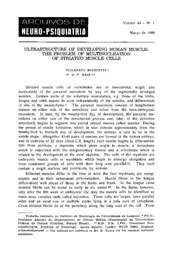
ULTRASTRUCTURE OF DEVELOPING HUMAN MUSCLE: THE PROBLEM OF MULTINUCLEATION
OF STRIATED MUSCLE CELLS
GUILBERTO MINGUETTI * W. G. P. MAIR **
Striated muscle cells of ver tebrates are of mesodermal origin and particularly of the paraxial mesoderm by way of the segmentally a r ranged somites. Centain par t s of the voluntary musculature, e.g. those of the limbs, tongue and orbit appear to arise independently of the somites, and differentiate in situ in the mesenchyme l. The paraxial mesoderm consists of longitudinal masses on either side of the notochord and arises from the intra-embryonic mesoderm. In man, by the twenty-first day of development, the paraxial mesoderm on either side of the notochordal process and, later, of the definitive notochord, begins to segment into paired cubical masses called somites. During the period of somite formation, which in man extends approximately from the twenty-first to thirtieth day of development, the embryo is said to be in the somite s tage. Altogether 42-44 pairs of somites are formed in the human embryo, and in embryos of 32 days (5mm C.R. length) each somite begins to differentiate into three por t ions: a myotome which gives origin to muscle, a dermotome which is concerned with the integumentary tissues and a sclerotome which is related to the development of the axial skeleton. The cells of the myotome are embryonic muscle cells or myoblasts which begin to undergo elongation and from condensed groups of cells with their long axis parallel 1 1 . They each contain a single nucleus and proliferate by mitosis.
Different muscles differ in the time at wich the first myoblas ts are recognizable and in their subsequent differentiation. Muscle fibres in the tongue differentiate well ahead of those in the limbs and trunk. In the tongue cross str iated fibrils can be found as early as six weeks 10. In the limbs, however, only after the 8th week of embryonic life may the muscle cells be identified as much more complex units called myotubes. These cells are larger, have parallel sides and an axial row of multiple nuclei lying in a pale core of cytoplasm. Cross str iated fibrils lie at the periphery along the long axis of the cell. From
Trabalho realizado no Instituto de Neurologia da Universidade de Londres: * PH.D. , Professor Adjunto do Departamento de Clínica Médica (Neurologia) da Universidade Federal do Paraná (Curitiba, Paraná Brasi l ) ; ** M.D., F.R. C.Pth., Consultant Neuropathologist, Institute of Neurology. Queen Square, London WC1N 3B6G, England. Acknowledgement — The authors are indebted to Mr. B. Young for his excelent technical assistance.
the 8th week onwards new myoblasts continue to differentiate from the mesenchyme. Later, during the 5th month of development the nuclei of mature myotubes begin to move to their permanent surface position underneath the plasma membrane, the cells reaching the s tage of myofibres. Now, very numerous and well organised myofibrils occupy the central par t of the fibre. It is the general belief that from this s tage onwards voluntary muscle continues to grow in two ways : by recruitment of new myoblasts from adjacent mesenchyme, and by progressive enlargement of the muscle fibres, the last becoming relatively more and more important the older the foetus 6 , 7 , 9 , 1 0 , 2 7 .
The mechanism of how muscle fibres become multinucleated during embryonic myogenesis and regeneration in vivo, or during the process of differentiation in tissue cultures in vitro has been the subject of continuous discussion. No definite solution has yet been found. Three explanations exist as to how the large, multinucleated skeletal muscle fibres arise from myoblasts — a) Each muscle fibre is a syncytium resulting from the fusion of many separa te cells. b) Each myoblast grows markedly in length but the rapid multiplication of the nuclei by amitosis is not accompanied by division of the cytoplasm. Hence a multinucleated cell is produced. c) Both processes of development may occur. The aim of the present investigation is to bring some contribution for better knowledge of this matter.
MATERIAL, AND METHODS
The material used in this investigation is derived from twenty seven human foetuses ranging from nine weeks to nine months development. They have been studied in detail in longitudinal and transverse sections by electron microscopy; some 300 grids were examined. Foetuses of 9, 10, 11, 12, 13, 14, 15, 16, 17, 18, 20, 24, 28, 32, 36 and 40 weeks were studied. The age of minute foetuses was established by the obstetricians who supplied the specimens and was calculated according to the crown-rump length (C.R. length). The ages of foetuses of 5, 7, 8 and 9 months development were based on the menstrual age. They were newborn prematures who died within 24 hours of birth. Specimens were removed soon after death in these cases to avoid the occurrence of post mortem changes in the muscle. The specimens were obtained from various gynaecological centres in England and Brazil. The age of the earlier specimens, 9-18 weeks, was based on the crown-rump measurement while that of the older specimens, 20-40 weeks, was determined by the menstrual age. In both instances this information was provided by the gynaecologist who attended the patients.
The muscle was usually taken from the thigh, even muscle from the minute foetuses, and laid immediately on a piece of card and kept sl ightly stretched by means of pins applied to either end of the specimen. The specimens were then immersed in cold 3% glutaraldehyde in S^rensen's phosphate buffer at pH 7.4. After 2 hours of fixation the muscle held by the oins was released and cut in small pieces of about 1-2 mm thick. These were washed in two changes of SIrensen's buffer at pH 7.4. for 15 minutes each. Post fixation was carried out for 2 hours at room temperature in cold 1% osmium tetroxide in Michaelis' veronal-acetate buffer at pH 7.4. After being washed in distilled water and dehydrated in ascending grades of alcohols, they were placed successively in
propylene oxide and in a mixture of equal parts of propylene oxide and Epon 812. Finally they were embedded in fresh Epon mixture. Sections, 1-2^ thick were cut on an LKB Ultratome and stained with toluidine blue by the method of Trump et al. ( 2 6 ) and examined by l ight microscopy. Thin sections of appropriate regions were collected on copper grids, stained by uranyl acetate and lead citrate acc. to Reynolds (22) and examined in Siemens Elmiskop I.
RESULTS
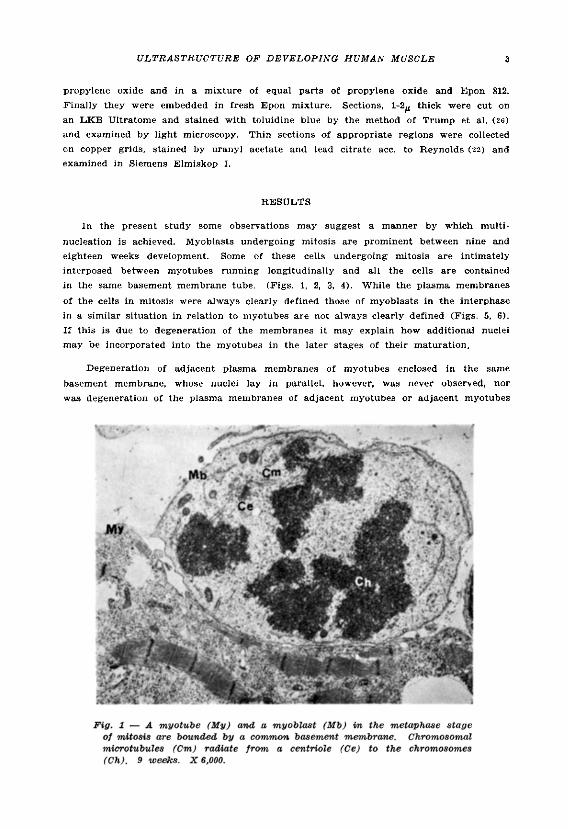
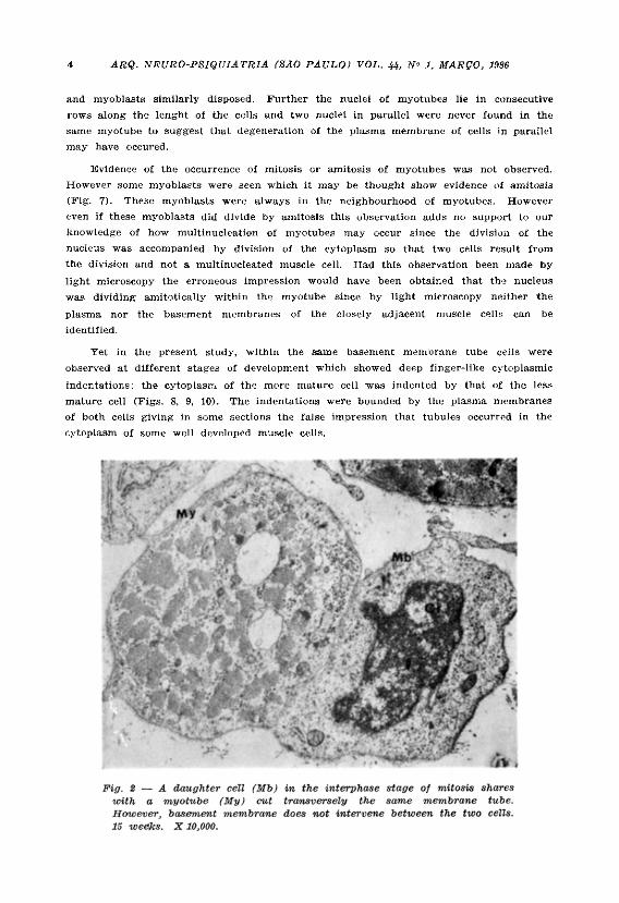
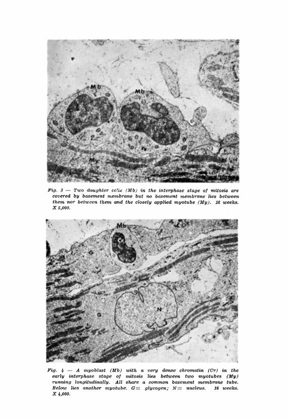
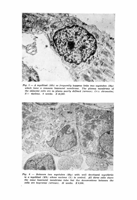
In the present study some observations may suggest a manner by which multi-nucleation is achieved. Myoblasts undergoing mitosis are prominent between nine and eighteen weeks development. Some of these cells undergoing mitosis are intimately interposed between myotubes running longitudinally and all the cells are contained in the same, basement membrane tube. (Figs. 1, 2, 3, 4). While the plasma membranes of the cells in mitosis were always clearly defined those of myoblasts in the interphase in a similar situation in relation to myotubes are not always clearly defined (Figs . 5, 6). If this is due to degeneration of the membranes it may explain how additional nuclei may be incorporated into the myotubes in the later stages of their maturation.
Degeneration of adjacent plasma membranes of myotubes enclosed in the same basement membrane, whose nuclei lay in parallel, however, was never observed, nor was degeneration of the plasma membranes of adjacent myotubes or adjacent myotubes
and myoblasts similarly disposed. Further the nuclei of myotubes lie in consecutive rows along the lenght of the cells and two nuclei in parallel were never found in the same myotube to suggest that degeneration of the plasma membrane of cells in parallel may have occured.
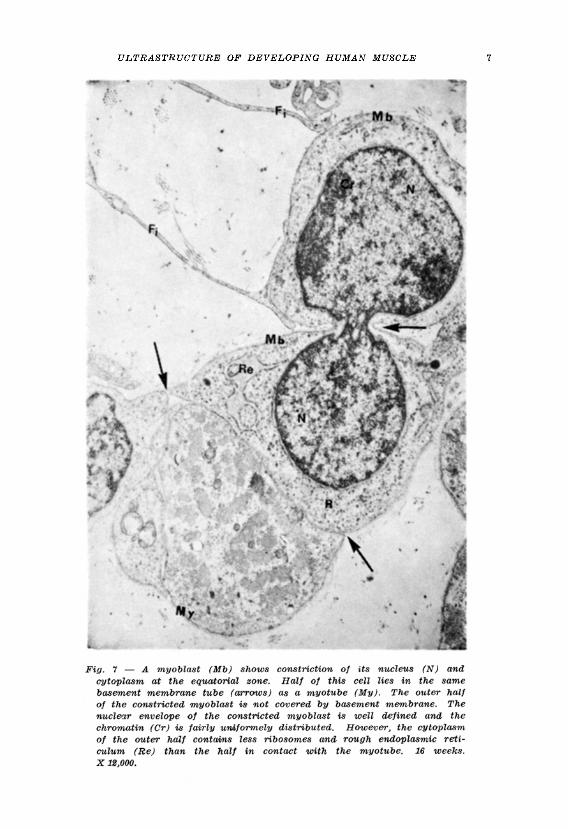
Evidence of the occurrence of mitosis or amitosis of myotubes was not observed. However some myoblasts were seen which it may be thought show evidence of amitosis (Fig. 7). These myoblasts were always in the neighbourhood of myotubes. However even if these myoblasts did divide by amitosis this observation adds no support to our knowledge of how multinucleation of myotubes may occur since the division of the nucleus was accompanied by division of the cytoplasm so that two cells result from the division and not a multinucleated muscle cell. Had this observation been made by light microscopy the erroneous impression would have been obtained that tha nucleus was dividing amitotically within the myotube since by light microscopy neither the plasma nor the basement membranes of the closely adjacent muscle cells can be identified.
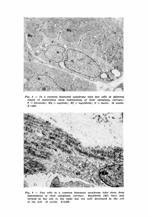
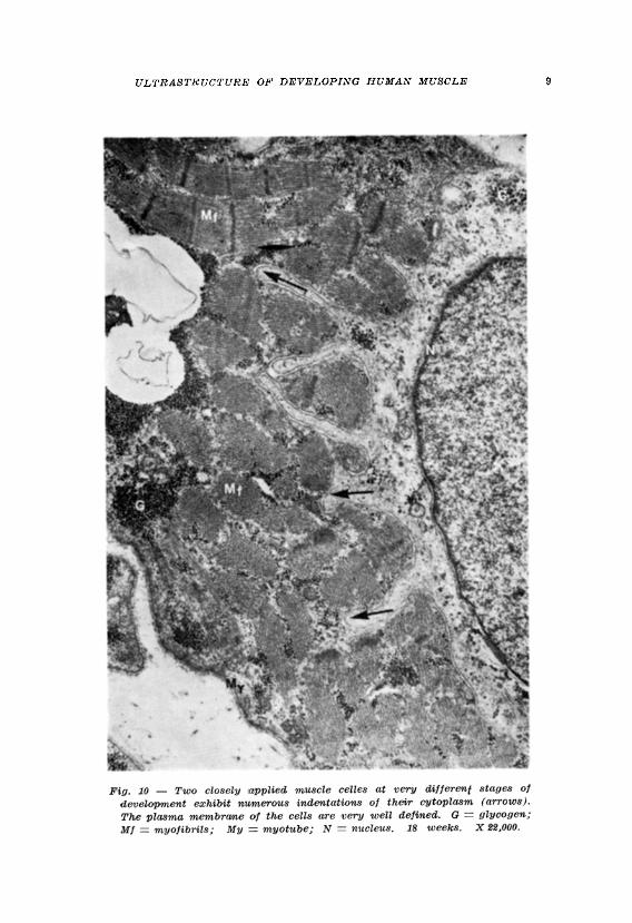
Yet in the present study, within the same basement membrane tube cells were observed at different stages of development which showed deep finger-like cytoplasmic indentations: the cytoplasm of the more mature cell was indented by that of the less mature cell (Figs. 8, 9, 10). The indentations were bounded by the plasma membranes of both cells giving in some sections the false impression that tubules occurred in the cytoplasm of some well developed muscle cells.
COMMENTS
Schwann expressed the opinion that , in the pig, the muscle fibres arose by the alignment of the primitive cells into parallel rows in which these cells were a r ranged in an end-to-end f a s h i o n 2 4 . He considered that linear coalescent of these cells produced the elongated multinucleated muscle fibres. This is called the polygenistic or polycellular theory. This polygenistic view was soon contested by Remak who believed that each multinucleated muscle fibre arose by growth and nuclear division of a single embryonic cell, avoiding the process of c y t o d i e r e s i s 2 1 . Remak's view was termed the monogenistic or unicellular theory. The emphasis placed on amitotic events a t the end of the nineteenth century reinforced the acceptance of the unicellular mechanism of multinuclea-tion of muscle fibres, but brought more confusion to the matter. Many recent reports re-emphasize the thesis that amitotic division of myoblast and myotube nuclei without cytoplasmic division leads to multinucleation 3,18,20.
Numerous other works have been carried out to reinforce both the polygenistic and the monogenistic theories. This l i terature is confusing and there has been much conflict of opinion as to how the myoblast becomes converted into the multinucleated and highly specialized muscle fibre. A modification of the polygenistic theory st ipulates that multinucleation is achieved by the fusion of mitotically dividing mononucleated myoblasts and or with a myotube containing non-dividing nuclei.
Direct observat ions in tissue cultures on the fusion of myoblasts into multinucleated myotubes were reported by Rinaldini ( 1 9 5 7 ) 2 3 , Holtzer ( 1 9 5 8 ) 1 2 , Cooper and Konigsberg (1959)5, Konigsberg (1964 and 1973)13,14 and Okazaki and Holtzer ( 1 9 6 6 ) 1 9 . Muscle syncytium arising as a result of cell fusion has also been demonstrated by several other techniques which will now be discussed. There is no evidence of DNA synthesis either by the microspectrophotometric determination of Feulgen-DNA per nucleus 2,8,15 ( 0 r by radioautographic techniques to detect the incorporation of isotopically labelled DNA precursors . The syncytial nuclei do not incorporate any of the labelled precursors although mononucleated cells do. Later, some of these mononucleated cells appear within the syncytium 4.25.28. The occurrence of fusion of myoblasts in vivo was demonstrated in experiments by Mintz and Baker (1967) 1 ?.
The work of Konigsberg 13,14 and his col laborators has provided not only observations of the fusion of myoblasts but also a partial answer to one of the most serious problems in the investigation of the early s tages of myogenesis (prior to fusion), i.e. the distinction of myogenic cells from fibroblasts. T o obtain this evidence they use the method of Clonal Analysis. By this method in the earliest s tage of myogenesis colonies of myoblasts can be distinguished from colonies of fibroblasts. Cell suspensions are prepared from developing muscle dissected from the embryonic s tage of experimental animals. These cells are pipetted into a series of Petri dishes containing liquid nutrient medium and incubated at body temperature . Fibroblasts form colonies of cells which are rather spread out and stay separate from one another : they exhibit extensive irregular membranes and shapes. Myoblasts on the other hand form colonies
of cells that are usually fusiform with ra ther regular membranes and which, after ceasing to divide, begin to fuse to form a syncytium. T h e work of Konigsberg and his collaborators, however, are based on experiments with embryos of chickens, mice and rats . Human myoblasts are flatter cells than those of birds, mice and ra t s and the only convincing time for these authors to distinguish clonal colonies of human myoblasts is when these cells have already shown some differentiation into myotubes.
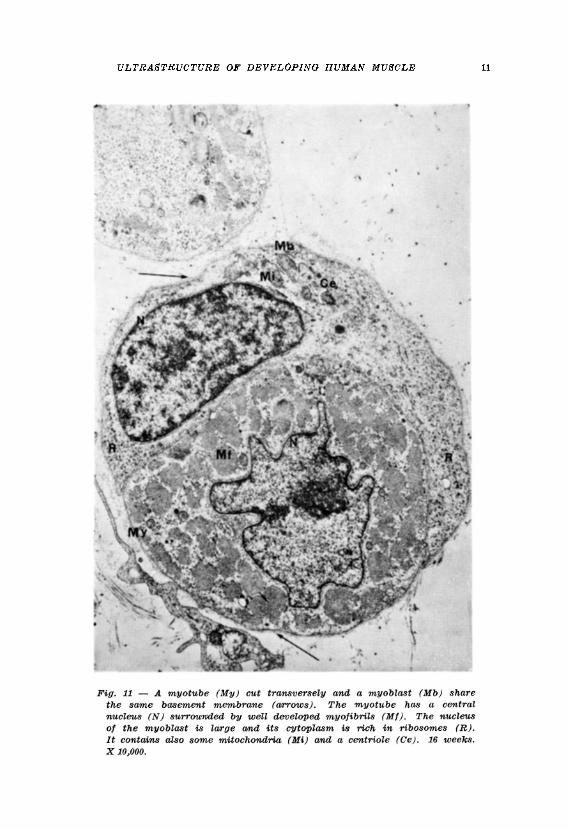
In a previous report the authors observed that myoblasts , the cells from which muscle is derived, are mononucleated cells which during the period from nine to eighteen weeks development show the greatest degree of mitotic a c t i v i t y 1 6 . Dur ing this period the myoblas ts may occur in isolation or in groups and also in association with more mature muscle cells, the myotubes. The myoblasts have a large single nucleus and a relatively small amount of cytoplasm. T h e plasma membrane of the myoblast is a well defined electron dense structure. Basement membrane does not occur around isolated myoblasts but is seen around myoblasts lying in apposition to myotubes (Fig. 11), particularly during mitotic division. Myoblasts which appear to have undergone amitotic division also occur but two daughter cells arise from this division and not a multinucleated cell. Myotubes are also present at nine weeks development and are prevalent in the muscle up to eighteen weeks development. They are multinucleated cells whose nuclei lie in a row in the centre of the cell and whose myofibrils lie peripherally. T h e striated pat tern of the myofibril is evident even a t nine weeks development. Some myotubes exhibit a well defined plasma membrane surrounded by a distinct basement membrane but groups of myotubes also occur which share a common basement membrane tube.
In the present investigation it w a s possible to observe that disintegration of the plasma membranes of adjacent myoblasts and myotubes appears to occur in longitudinally disposed cells of those categories which share a common basement membrane tube and this may help to explain how further nuclei may be incorporated into well developed myotubes. Deep indentations of the cytoplasm and the plasma membranes of adjacent myotubes at different s tages of development are occasionally seen. Th i s gives rise to finger-like processes along the surfaces of contact of both cells. T h e significance of such processes is a subject for speculation. After eighteen weeks development the muscle is formed almost exclusively of muscle fibres, the nuclei of which lie at the periphery of the cell and the greater par t of the cell contains well organised myofibrils. From about twenty weeks development the muscle does not contain myoblasts which are isolated from the other muscle cells but there occur beneath the basement membrane of the muscle fibres mononucleated cells similar in appearance to cells found in the same situation as in adult muscle. These latter cells are satellite cells or myoblasts .
SUMMARY
T h e authors studied by electron microscopy the muscle of 27 human foetuses ranging from 9 weeks to 9 months development. It was possible to
observe tha t disintegration of the plasma membranes of adjacent myoblas ts and myotubes which share a common basement membrane tube appea r s to occur in longitudinally disposed cells of those categories. Th i s may help to explain how further nuclei may be incorporated into well developed myotubes and how the str iated muscle cells become multinucleated during embryonic myogenesis and regeneration in vivo.
RESUMO
Ultraestrutura do músculo humano em desenvolvimento: o problema da multinucleação das células musculares estriadas.
As células musculares es t r iadas dos ver tebrados são de origem mesodérmica, particularmente do mesoderma para-axial localizado nos somitos. No embrião humano de 32 dias cada somito inicia um processo de diferenciação em t rês porções: o miótomo que dá origem ao músculo, o dermátomo e o esclerótomo. No miótomo proliferam as células musculares embrionár ias mononucleadas, os mioblastos. A part i r da oitava semana de vida embrionária j á são observadas células musculares mais diferenciadas, os miotubos: são células com mais de um núcleo de localização central. Ao redor dos núcleos, são observadas as miofibrilas. A part ir do quinto mês de vida intra-interina, os núcleos migram para a periferia do citoplasma, para as porções sub-sarcolêmicas, dando origem às fibras musculares propriamente ditas. Há três teorias sobre o mecanismo de multinucleação das células musculares: a) cada célula muscular multinu¬ cleada é um sincício resultante da fusão de vár ias células musculares mononucleadas; b) divisão do núcleo, sem divisão do citoplasma (ami tose ) ; c) ocorrência de ambos os processos. No presente t rabalho, foram es tudadas por microscopia eletrônica células musculares provenientes de 27 fetos humanos variando de 9 semanas de vida embrionária até 9 meses. Foi possível se observar que mioblastos em mitose são comuns entre os miotubos e que os mesmos localizam-se entre a membrana plasmática e a membrana basal dos miotubos. Foi possível ainda se observar em vár ios campos examinados a presença de mioblastos s i tuados nas extremidades de miotubos ou entre dois miotubos longitudinalmente dispostos cujas membranas plasmáticas jus tapos tas encontravam-se em degeneração sugerindo uma possível anexação das células mais jovens (mioblastos) pelas células mais diferenciadas (miotubos) . Se isso realmente ocorre, o fato poderia em par te explicar o fenômeno da multinucleação das células musculares estr iadas.
REFERENCES
1. ADAMS, R .D. ; DENY-BROWN, D. & PEARSON, CM. — Diseases of Muscle. Harper & Row, New York, 1962.
2. BASSLEER, R. — Etude 1'augmentation du nombre de noyaux dans des bourgeous musculares cultivés in vitro: observations sur le vivant, dosages, cytophotometriques et histoautoradiographies. Z. Anat. Entw. 123: 184, 1962.
3. BEGMANN, R.A. — Observations on the morphogenesis of rat skeletal muscle. Bull. Johns Hopkins Hosp. 110: 187, 1962.
REFERENCES
1. ADAMS, R.D. ; DENY-BROWN, D. & PEARSON, CM. — Diseases of Muscle. Harper & Row, New York, 1962.
2. BASSLiEER, R. — Etude l'augmentation du nombre de noyaux dans des bourgeous musculares cultivés in vitro: observations sur le vivant, dosages, cytophotometriques et histoautoradiographies. Z. Anat. Entw. 123: 184, 1962.
3. BEGMANN, R.A. — Observations on the morphogenesis of rat skeletal muscle. Bull. Johns Hopkins Hosp. 110: 187, 1962.
4. BINTLICFF, S. & WALKER, B.E. — Radioautographic study of skeletal muscle regeneration. Amer. J, Anat. 106 : 233, 1960.
5. COOPER, W.G. & KONIGSBERG, I.R. — Dynamics of myogenesis . Anat. Rec. 140:195, 1959.
6. CUAJUNCO, F. — Development of the neuromuscular spindle in human fetuses. Contrib. Embryol. Carneg. Inst. 28:97, 1940.
7. CUAJUNCO, F. — Development of the human motor end plate. Contrib. Embryol. Carneg. Inst. 30:127, 1942.
8. FIRKET, H. — Recherches sur la synthèse des an des désoxyribonucléiques et la préparation à la mitose dans des cellules cultivées in vitro (étude cytophotometrique et autoradiographique). Arch. Biol. Paris 69: 1, 1958.
9. GODLEWSKI, E. — Ueber der Entwickelung des quergestreiften muskulosen Geweber. Krakauer Ans. 10: 147, 1901.
10. HAMILTON, W.J. & MOSSAMAN, H.W. — Human Embryology. Ed. 4. W. Heffer & Sons, Cambridge, Will iams & Wilkins, Baltimore, 1972.
11. HEWER, E. — The development of muscle in the human foetus. J. Anat. 62 : 72, 1928. 12. HOLTZER, H.; ABBOTT, J. & LASH, J. — On the formation of multinucleated
myotubes. Anat. Rec. 131:567, 1958. 13. KONIGSBERG, I.R. — The embryological origin of muscle. Sci. Amer. 211: 61, 1964. 14. KONIGSBERG, I.R. — The fine structure and control of myogenic fusion in culture.
In B.A. Kakulas (ed.) . Basic Research in Myology (Part I) . Excerpta Medica, Amsterdam, 1973, pg. 327.
15. LASH, J.W. ; HOLTZER, H. & SWIFF, H. — Regeneration of mature skeletal muscle. Anat. Rec. 128 : 679, 1957.
16. MINGUETTI, G. & MAIR, W.G.P. — Ultrastructure of developing human muscle. Biol. Neonate 40: 276, 1981.
17. MINTZ, B. & BAKER; W.W. — Normal mammalian muscle differentiation and gene control of isocitrate dehydrogenase synthesis . Proc. nat. Acad. Sci. 58 : 592, 1967.
18. MURRAY, M.R. — Skeletal muscle t issue in culture. In G.H. Bourne (ed . ) : The Structure and Function of Muscle. Vol. 2. Academic Press, New York, 1960, pg. 111.
19. OKAZAKI, K. & HOLTZER, H. — Myogenesis: myosin synthesis and the mitotic cycle. Proc. nat. Acad. Sci. 56:1484, 1966.
20. PUZA, V.L. ; GAYER, J. & FOREJT., L. — The mechanism of multinuclear muscle cell formations. Folia morph. (Praha) 13 : 294, 1965.
21. REMAK, R. — über die Entwicklung der Muskelprimitivbündel. Froriep's N. Notizer 35 : 305, 1845.
32. REYNOLDS, E.S. — The use of lead citrate at high pH as an electron-opaque stain in electron microscopy. J. cell. Biol. 17: 208, 1963.
23. RINALDINI, L.M. — The preparation of cultures of freshly isolated myoblasts for nutritional and metabolic studies. Abstracts of the International Tissue Culture Meeting, Glasgow, pg. 32, 1957.
24. SCHWANN, T. — Muscle. In : Schwann, T. : Microscopical Researches into the Accordance in the Structure and Growth of Animals and Plants. Translated by H. Smith. Sydenham Society, London, pg. 130, 1847.
25. STOCKDALE, F.E. & HOLTZER, H. — DNA synthesis and myogenesis . Exp. cell. Res. 24 : 508, 1961.
26. TRUMP, B.F., SMUCKLER, E.A. & BENNDITT, E.P. — A method for staining epoxy sections for light microscopy. J. ultrastruc. Res. 5 : 343, 1961.
27. WILLIS, R.A. — The Borderland of Embryology and Pathology. Ed. 2. Butterworths & Co., London, 1962.
28. ZHINKIN, L.N. & ANDREEVA, L.F. — DNA synthesis and nuclear reproduction during embryonic development and regeneration of muscle tissue. J. Embryol. exper. Morphol. 11: 353, 1963.
Neurologia, Departamento de Clínica Médica, Hospital de Clínicas — Rua General Carneiro, 181, 13o andar - 80.000 - Curitiba, PR - Brasil.