Embed Size (px)
DESCRIPTION
UNC Neuroradiology-Neuropathology Conference. November 20 12 Stephen Bagg, MD Fellow in Neuroradiology. Case 1- 57 y/o female with no sig PMHx 1 month hx of progressive Dizziness Diplopia Falls Physical Exam CN 6 palsy R facial droop and R tongue deviation L facial numbness - PowerPoint PPT Presentation
Citation preview
UNC Neuroradiology-Neuropathology Conference
November 2012
Stephen Bagg, MD
Fellow in Neuroradiology
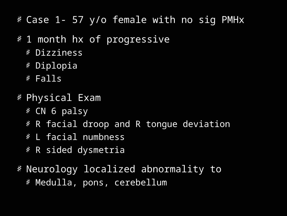
Case 1- 57 y/o female with no sig PMHx
1 month hx of progressiveDizziness
Diplopia
Falls
Physical ExamCN 6 palsy
R facial droop and R tongue deviation
L facial numbness
R sided dysmetria
Neurology localized abnormality toMedulla, pons, cerebellum
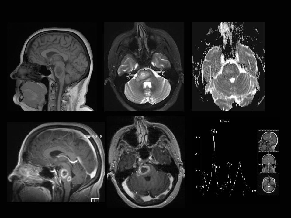
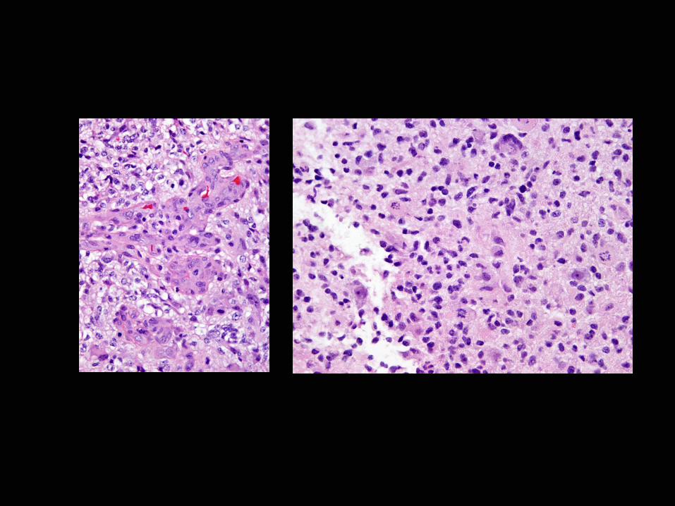
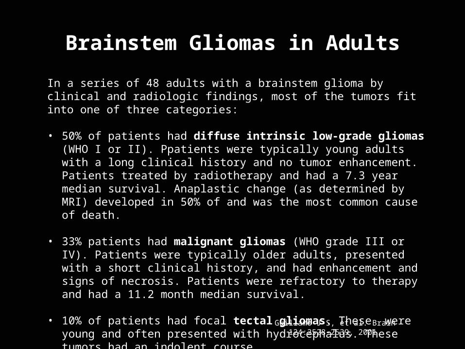
In a series of 48 adults with a brainstem glioma by clinical and radiologic findings, most of the tumors fit into one of three categories:
• 50% of patients had diffuse intrinsic low-grade gliomas (WHO I or II). Ppatients were typically young adults with a long clinical history and no tumor enhancement. Patients treated by radiotherapy and had a 7.3 year median survival. Anaplastic change (as determined by MRI) developed in 50% of and was the most common cause of death.
• 33% patients had malignant gliomas (WHO grade III or IV). Patients were typically older adults, presented with a short clinical history, and had enhancement and signs of necrosis. Patients were refractory to therapy and had a 11.2 month median survival.
• 10% of patients had focal tectal gliomas. These were young and often presented with hydrocephalus. These tumors had an indolent course.
Guillamo J-S, et al. Brain 124:2528-2539, 2001.
Brainstem Gliomas in Adults
Case 2- 21 month old malePreviously healthy
Recently noted by PCP to have macrocephaly
Developed Ataxia and N/V
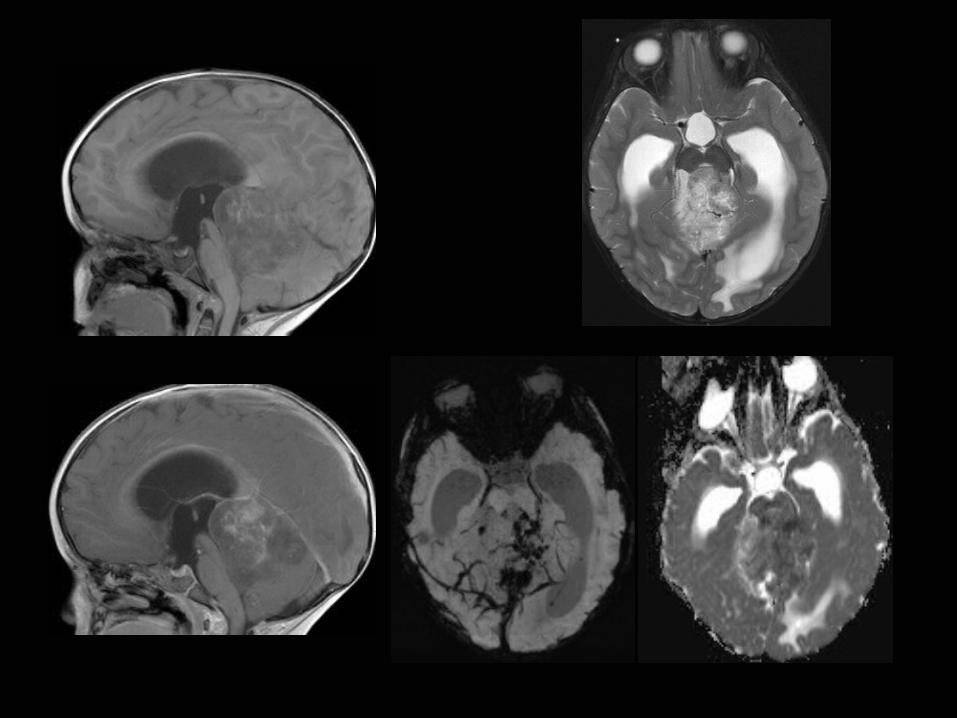
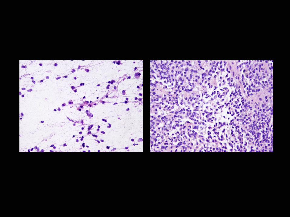
Atypical teratoid rhabdoid tumor (ATRT)
• ATRTs are rare tumors that comprise approximately 1-2% of all pediatric brain tumors; however, in patients less than 3 years of age this tumor accounts for up to 20% of cases.
• ATRT usually occurs in posterior fossa or supratentorial location and are only rarely in spinal region
• ATRT is characterized by loss of the long arm of chromosome 22, which results in loss of the hSNF5/INI-1 gene and loss of INI-1 expression.
• INI1, a member of the SWI/SNF chromatin remodeling complex, is important in maintenance of the mitotic spindle and cell-cycle control.
• WHO grade IV. Overall survival in ATRT is poor with median survival of around 17 months.
Ginn KF, Gajjar A. Front Oncol. 2012;2:114. Epub 2012 Sep 12.
Imaging Features, ATRT
Heterogeneous mass/enhancement
Commonly contains cysts & hemorrhage
Leptomeningeal enhancement is common
Restricted Diffusion is common
Imaging Diff Dx = medulloblastomaATRT are generally more heterogeneous than medulloblastoma
Medulloblastoma is more commmon
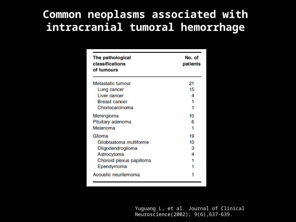
Yuguang L, et al. Journal of Clinical Neuroscience(2002); 9(6),637-639.
Common neoplasms associated with intracranial tumoral hemorrhage
Case 3- 36 y/o male
1 year history of R hand and foot numbness
Otherwise healthy- No significant past medial history
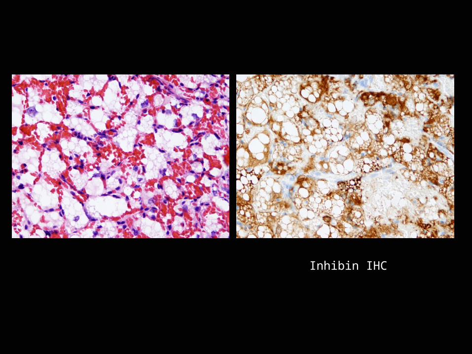
Inhibin IHC
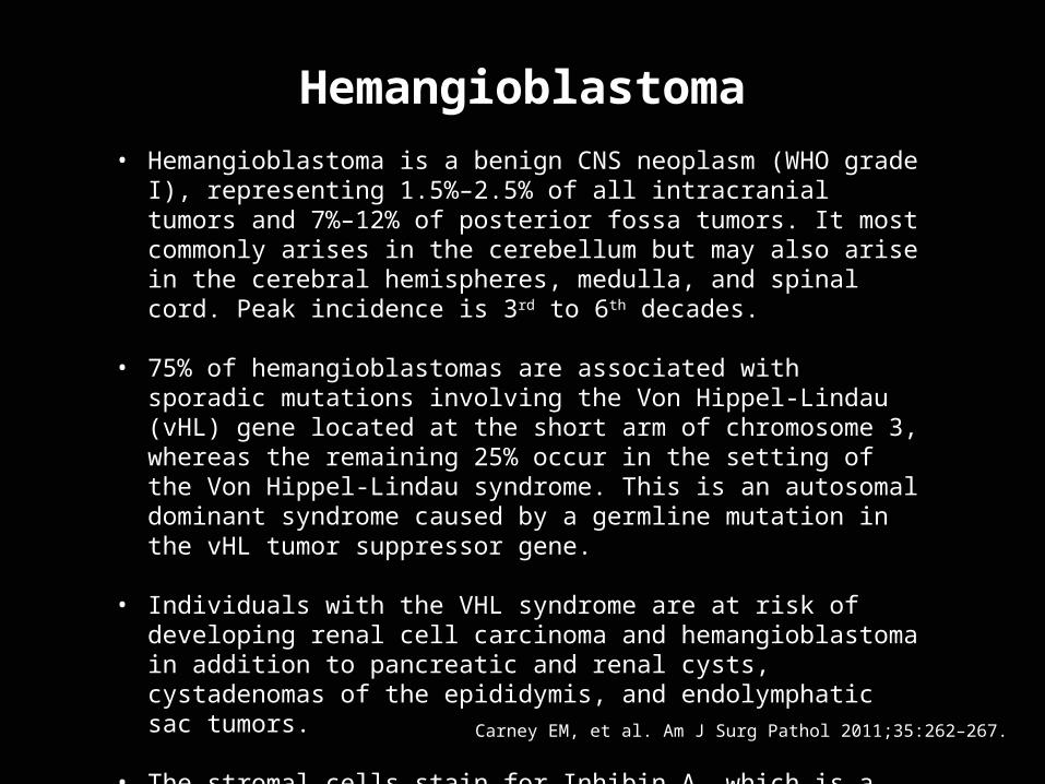
Hemangioblastoma• Hemangioblastoma is a benign CNS neoplasm (WHO grade I),
representing 1.5%–2.5% of all intracranial tumors and 7%–12% of posterior fossa tumors. It most commonly arises in the cerebellum but may also arise in the cerebral hemispheres, medulla, and spinal cord. Peak incidence is 3rd to 6th decades.
• 75% of hemangioblastomas are associated with sporadic mutations involving the Von Hippel-Lindau (vHL) gene located at the short arm of chromosome 3, whereas the remaining 25% occur in the setting of the Von Hippel-Lindau syndrome. This is an autosomal dominant syndrome caused by a germline mutation in the vHL tumor suppressor gene.
• Individuals with the VHL syndrome are at risk of developing renal cell carcinoma and hemangioblastoma in addition to pancreatic and renal cysts, cystadenomas of the epididymis, and endolymphatic sac tumors.
• The stromal cells stain for Inhibin A, which is a glycoprotein normally secreted by ovarian granulosa cells and testicular Sertoli and Leydig cells to inhibit pituitary follicle-stimulating hormone.
Carney EM, et al. Am J Surg Pathol 2011;35:262–267.
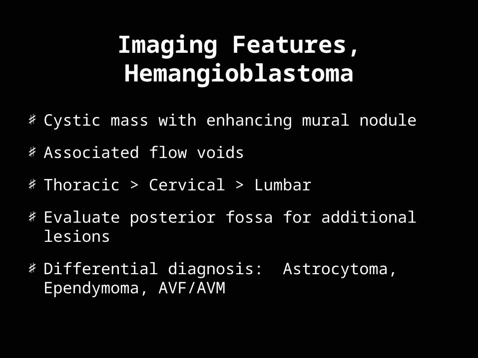
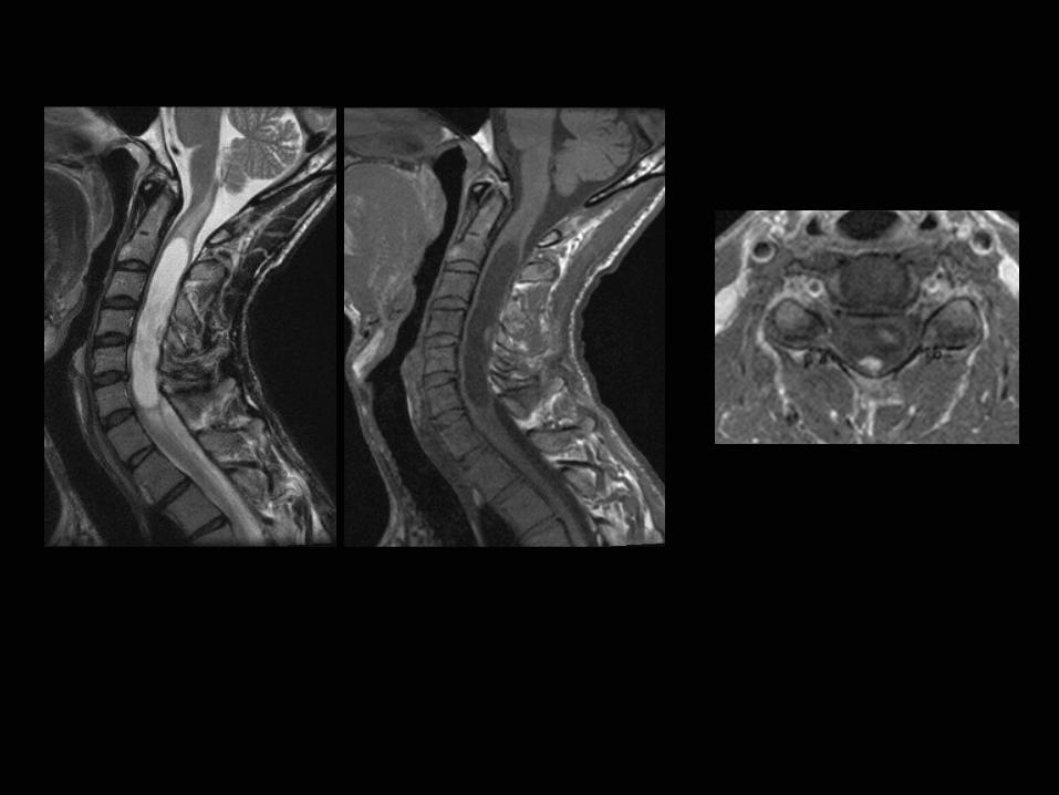
Imaging Features, Hemangioblastoma
Cystic mass with enhancing mural nodule
Associated flow voids
Thoracic > Cervical > Lumbar
Evaluate posterior fossa for additional lesions
Differential diagnosis: Astrocytoma, Ependymoma, AVF/AVM
Case 4- 58 y/o F
Presented to outside institution with altered mental status
Possible complaint of blurry vision
PMHxDiabetes, Hypertension
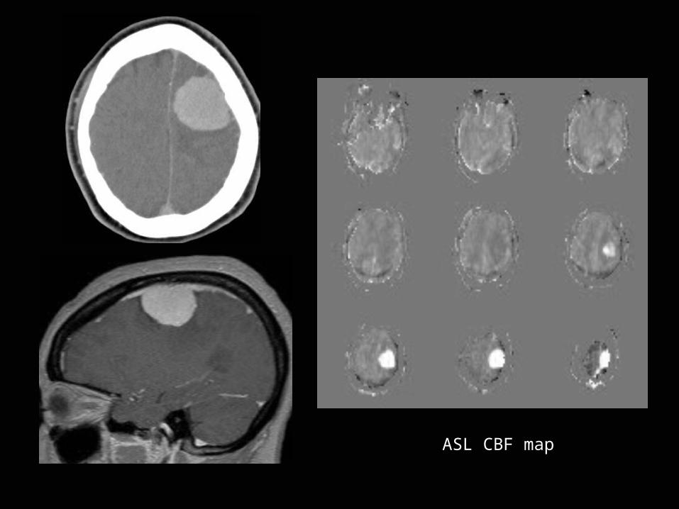
ASL CBF map
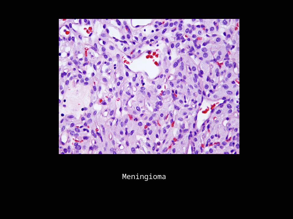
Meningioma

Thank you