Embed Size (px)
Citation preview
© 2017 The Korean Academy of Medical Sciences.This is an Open Access article distributed under the terms of the Creative Commons Attribution Non-Commercial License (http://creativecommons.org/licenses/by-nc/4.0) which permits unrestricted non-commercial use, distribution, and reproduction in any medium, provided the original work is properly cited.
pISSN 1011-8934eISSN 1598-6357
Unsuspected Duplicated Gallbladder in a Patient Presenting with Acute Cholecystitis
Duplicated gallbladder (GB) is a rare congenital disease. Surgical management of a duplicated GB needs special care because of concurrent bile duct anomalies and the risk of injuring adjacent arteries during surgery. An 80-year-old man visited an emergency room with right upper quadrant abdominal pain. Computed tomography (CT) revealed cholecystitis with a 2-bodied GB. Because of this unusual finding, magnetic resonance choledochopancreatography was performed to detect possible biliary anomalies. The 2 GB bodies were unified at the neck with a common cystic duct, a so-called V-shaped duplicated GB. The patient’s right posterior hepatic duct joined the common bile duct (CBD) near the cystic duct. The patient underwent laparoscopic cholecystectomy without adjacent organ injury, and was discharged uneventfully. Surgeons should carefully evaluate the patient preoperatively and select adequate surgical procedures in patients with suspected duplicated GB because of the risk of concurrent biliary anomalies.
Keywords: Duplicated Gallbladder; Cholecystitis; Cholecystectomy; Laparoscopy
Woohyung Lee,1 Dae Hyun Song,2 Jin-Kwon Lee,1 Ji-Ho Park,1 Ju-Yeon Kim,1 Seung-Jin Kwag,1 Taejin Park,1 Sang-Ho Jeong,1 Young-Tae Ju,1 Eun-Jung Jung,1 Young-Joon Lee,1 Soon-Chan Hong,1 Sang-Kyung Choi,1 and Chi-Young Jeong1
1Department of Surgery, Gyeongsang National University Hospital, Gyeongsang National University Postgraduate School of Medicine, Jinju, Korea;2Department of Pathology, Gyeongsang National University Hospital, Gyeongsang National University Postgraduate School of Medicine, Jinju, Korea
Received: 24 November 2015Accepted: 7 February 2016
Address for Correspondence:Chi-Young Jeong, MDDepartment of Surgery, Gyeongsang National University Hospital, Gyeongsang National University Postgraduate School of Medicine, 79 Gangnam-ro, Jinju 52727, Republic of KoreaE-mail: [email protected]
https://doi.org/10.3346/jkms.2017.32.3.552 • J Korean Med Sci 2017; 32: 552-555
INTRODUCTION
Duplicated gallbladder (GB) is a rare congenital anomaly that is found in 0.026% of autopsies (1). Although the disease is rare, preoperative diagnosis has some clinical implications. Undiag-nosed duplicated GB may lead to cholecystectomy, leaving an-other GB or injury to adjacent structures (2). Preoperative diag-nosis of duplicated GB enables the surgeon to evaluate its type before selecting the most appropriate surgical technique (3). We describe a patient with acute cholecystitis caused by dupli-cated GB, which was evaluated preoperatively and treated lapa-roscopically.
CASE DESCRIPTION
An 80-year-old man presented at an emergency department with right upper quadrant abdominal pain lasting for 3 days. The patient was healthy without any underlying disease. Physi-cal examination revealed right upper quadrant tenderness. Lab-oratory tests revealed elevated C-reactive protein (48.3 mg/dL), bilirubin (1.71 mg/dL), aspartate aminotransferase (310 IU/L), and alanine transaminase (204 IU/L) concentrations. Abdomi-nal computed tomography (CT) showed thickened GB wall with pericholecystic fluid collection, consistent with acute cholecys-
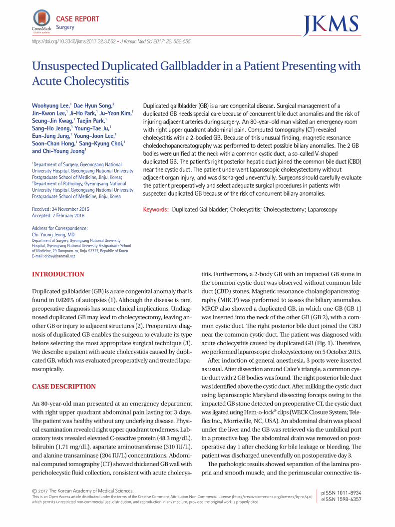
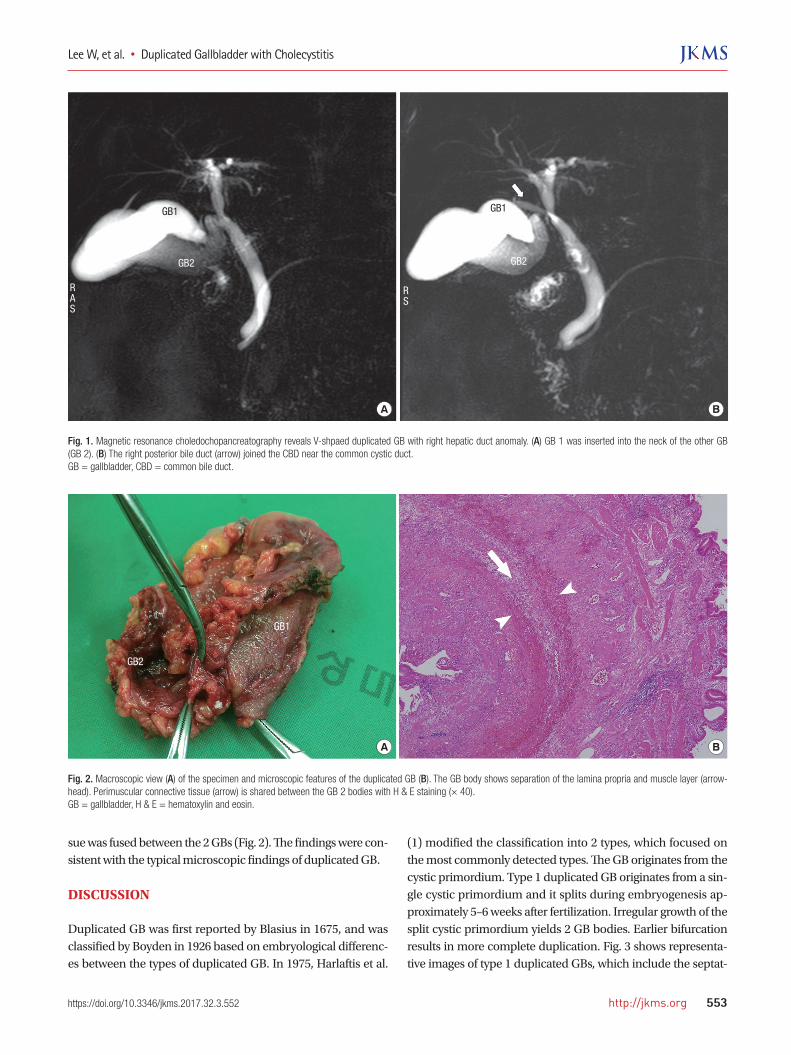
titis. Furthermore, a 2-body GB with an impacted GB stone in the common cystic duct was observed without common bile duct (CBD) stones. Magnetic resonance cholangiopancreatog-raphy (MRCP) was performed to assess the biliary anomalies. MRCP also showed a duplicated GB, in which one GB (GB 1) was inserted into the neck of the other GB (GB 2), with a com-mon cystic duct. The right posterior bile duct joined the CBD near the common cystic duct. The patient was diagnosed with acute cholecystitis caused by duplicated GB (Fig. 1). Therefore, we performed laparoscopic cholecystectomy on 5 October 2015. After induction of general anesthesia, 3 ports were inserted as usual. After dissection around Calot’s triangle, a common cys-tic duct with 2 GB bodies was found. The right posterior bile duct was identified above the cystic duct. After milking the cystic duct using laparoscopic Maryland dissecting forceps owing to the impacted GB stone detected on preoperative CT, the cystic duct was ligated using Hem-o-lock® clips (WECK Closure System; Tele-flex Inc., Morrisville, NC, USA). An abdominal drain was placed under the liver and the GB was retrieved via the umbilical port in a protective bag. The abdominal drain was removed on post-operative day 1 after checking for bile leakage or bleeding. The patient was discharged uneventfully on postoperative day 3. The pathologic results showed separation of the lamina pro-pria and smooth muscle, and the perimuscular connective tis-
CASE REPORTSurgery
Lee W, et al. • Duplicated Gallbladder with Cholecystitis
http://jkms.org 553https://doi.org/10.3346/jkms.2017.32.3.552
sue was fused between the 2 GBs (Fig. 2). The findings were con-sistent with the typical microscopic findings of duplicated GB.
DISCUSSION
Duplicated GB was first reported by Blasius in 1675, and was classified by Boyden in 1926 based on embryological differenc-es between the types of duplicated GB. In 1975, Harlaftis et al.
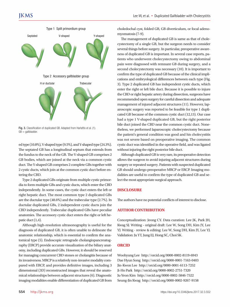
(1) modified the classification into 2 types, which focused on the most commonly detected types. The GB originates from the cystic primordium. Type 1 duplicated GB originates from a sin-gle cystic primordium and it splits during embryogenesis ap-proximately 5–6 weeks after fertilization. Irregular growth of the split cystic primordium yields 2 GB bodies. Earlier bifurcation results in more complete duplication. Fig. 3 shows representa-tive images of type 1 duplicated GBs, which include the septat-
Fig. 1. Magnetic resonance choledochopancreatography reveals V-shpaed duplicated GB with right hepatic duct anomaly. (A) GB 1 was inserted into the neck of the other GB (GB 2). (B) The right posterior bile duct (arrow) joined the CBD near the common cystic duct.GB = gallbladder, CBD = common bile duct.
A B
RAS
GB2 GB2
GB1 GB1
RS
Fig. 2. Macroscopic view (A) of the specimen and microscopic features of the duplicated GB (B). The GB body shows separation of the lamina propria and muscle layer (arrow-head). Perimuscular connective tissue (arrow) is shared between the GB 2 bodies with H & E staining (× 40).GB = gallbladder, H & E = hematoxylin and eosin.
A B
GB2
GB1
Lee W, et al. • Duplicated Gallbladder with Cholecystitis
554 http://jkms.org https://doi.org/10.3346/jkms.2017.32.3.552
ed type (10.8%), V-shaped type (9.5%), and Y-shaped type (24.3%). The septated GB has a longitudinal septum that extends from the fundus to the neck of the GB. The V-shaped GB comprises 2 GB bodies, which are joined at the neck via a common cystic duct. The Y-shaped GB comprises 2 complete GBs together with 2 cystic ducts, which join at the common cystic duct before en-tering the CBD. Type 2 duplicated GBs originate from multiple cystic primor-dia to form multiple GBs and cystic ducts, which enter the CBD independently. In some cases, the cystic duct enters the left or right hepatic duct. The most common type 2 duplicated GBs are the ductular type (48.6%) and the trabecular type (2.7%). In ductular duplicated GBs, 2 independent cystic ducts join the CBD independently. Trabecular duplicated GBs have peculiar anatomies. The accessory cystic duct enters the right or left he-patic duct (1,4). Although high-resolution ultrasonography is useful for the diagnosis of duplicated GB, it is often unable to delineate the anatomic relationship, which is essential to confirm the ana-tomical type (5). Endoscopic retrograde cholangiopancreatog-raphy (ERCP) provide accurate visualization of the biliary anat-omy, including duplicated GBs. However, it should be reserved for managing concurrent CBD stones or cholangitis because of its invasiveness. MRCP is a relatively non-invasive modality com-pared with ERCP, and provides definitive images, including 3 dimensional (3D) reconstructed images that reveal the anato-mical relationships between adjacent structures (6). Diagnostic imaging modalities enable differentiation of duplicated GB from
choledochal cyst, folded GB, GB diverticulum, or focal adeno-myomatosis (7-9). The management of duplicated GB is same as that of chole-cystectomy of a single GB, but the surgeon needs to consider several things before surgery. In particular, preoperative aware-ness of duplicated GB is important. In several case reports, pa-tients who underwent cholecystectomy owing to abdominal pain were diagnosed with remnant GB during surgery, and a second cholecystectomy was necessary (10). It is important to confirm the type of duplicated GB because of the clinical impli-cations and embryological differences between each type (Fig. 3). Type 2 duplicated GB has independent cystic ducts, which enter the right or left bile duct. Because it is possible to injure the CBD or right hepatic artery during dissection, surgeons have recommended open surgery for careful dissection and adequate management of injured adjacent structures (11). However, lap-aroscopic surgery was reported to be feasible for type 1 dupli-cated GB because of the common cystic duct (12,13). Our case had a type 1 V-shaped duplicated GB, but the right posterior bile duct joined the CBD near the common cystic duct. None-theless, we performed laparoscopic cholecystectomy because the patient’s general condition was good and his cholecystitis was not severe based on preoperative imaging. The common cystic duct was identified in the operative field, and was ligated without injuring the right posterior bile duct. Although duplicated GB is very rare, its preoperative detection allows the surgeon to avoid injuring adjacent structures during surgery or repeated surgery. Patients with suspected duplicated GB should undergo preoperative MRCP or ERCP. Imaging mo-dalities are useful to confirm the type of duplicated GB and se-lect the most appropriate surgical approach.
DISCLOSURE
The authors have no potential conflicts of interest to disclose.
AUTHOR CONTRIBUTION
Conceptualization: Jeong CY. Data curation: Lee JK, Park JH, Kwag SJ. Writing - original draft: Lee W, Song DH, Kim JY, Lee YJ. Writing - review & editing: Lee W, Song DH, Kim JY, Lee YJ. Validation: Ju YT, Jung EJ, Hong SC, Choi SK.
ORCID
Woohyung Lee http://orcid.org/0000-0002-8119-6943Dae Hyun Song http://orcid.org/0000-0001-7163-0403Jin-Kwon Lee http://orcid.org/0000-0001-6113-7252Ji-Ho Park http://orcid.org/0000-0002-2751-7320Ju-Yeon Kim http://orcid.org/0000-0002-5846-7522Seung-Jin Kwag http://orcid.org/0000-0002-9267-9158
Fig. 3. Classification of duplicated GB. Adapted from Harlaftis et al. (1).GB = gallbladder.
Type 2 Accessory gallbladder group
H or ductular Trabecular
Type 1 Split primordium group
Septated V-shaped Y-shaped
Lee W, et al. • Duplicated Gallbladder with Cholecystitis
http://jkms.org 555https://doi.org/10.3346/jkms.2017.32.3.552
Taejin Park http://orcid.org/0000-0002-8508-2353Sang-Ho Jeong http://orcid.org/0000-0001-9786-5710Young-Tae Ju http://orcid.org/0000-0002-1785-8500Eun-Jung Jung http://orcid.org/0000-0001-8413-613XYoung-Joon Lee http://orcid.org/0000-0002-1735-3385Soon-Chan Hong http://orcid.org/0000-0003-4499-8741Sang-Kyung Choi http://orcid.org/0000-0003-3691-0359Chi-Young Jeong http://orcid.org/0000-0003-4121-6695
REFERENCES
1. Harlaftis N, Gray SW, Skandalakis JE. Multiple gallbladders. Surg Gynecol
Obstet 1977; 145: 928-34.
2. Shiba H, Misawa T, Ito R, Ohki K, Igarashi T, Yanaga K. Duplicated gall-
bladder. Int Surg 2014; 99: 77-8.
3. Paraskevas GK, Raikos A, Ioannidis O, Papaziogas B. Duplicated gallblad-
der: surgical application and review of the literature. Ital J Anat Embryol
2011; 116: 61-6.
4. Singh B, Ramsaroop L, Allopi L, Moodley J, Satyapal KS. Duplicate gall-
bladder: an unusual case report. Surg Radiol Anat 2006; 28: 654-7.
5. Gocmen R, Yesilkaya Y. Imaging findings of gallbladder duplication due
to two cases: case report and review of literature. Med Ultrason 2012; 14:
358-60.
6. Botsford A, McKay K, Hartery A, Hapgood C. MRCP imaging of duplicate
gallbladder: a case report and review of the literature. Surg Radiol Anat
2015; 37: 425-9.
7. Huang BK, Chess MA. Cholecystitis of a duplicated gallbladder compli-
cated by a cholecystoenteric fistula. Pediatr Radiol 2009; 39: 385-8.
8. Ozaki N, Hashimoto D, Ikuta Y, Chikamoto A, Takamori H, Baba H. De-
finitive diagnosis of a duplicate gallbladder can only be made intraopera-
tively: report of a case. Clin J Gastroenterol 2014; 7: 338-41.
9. Han JY, Jeong S, Lee DH. Percutaneous papillary large balloon dilation
during percutaneous cholangioscopic lithotripsy for the treatment of large
bile-duct stones: a feasibility study. J Korean Med Sci 2015; 30: 278-82.
10. Reinisch A, Brandt L, Fuchs KH. Gallbladder duplication--laparoscopic
cholecystectomy 17 years after open cholecystectomy. Zentralbl Chir
2009; 134: 576-9.
11. Causey MW, Miller S, Fernelius CA, Burgess JR, Brown TA, Newton C. Gall-
bladder duplication: evaluation, treatment, and classification. J Pediatr
Surg 2010; 45: 443-6.
12. Walbolt TD, Lalezarzadeh F. Laparoscopic management of a duplicated
gallbladder: a case study and anatomic history. Surg Laparosc Endosc
Percutan Tech 2011; 21: e156-8.
13. Shirahane K, Yamaguchi K, Ogawa T, Shimizu S, Yokohata K, Mizumoto
K, Tanaka M. Gallbladder duplication successfully removed laparoscopi-
cally using endoscopic nasobiliary tube. Surg Endosc 2003; 17: 1156.