Embed Size (px)
Citation preview
International Journal of Collaborative Research on Internal Medicine & Public
Health (IJCRIMPH)
ISSN 1840-4529 | Journal Type: Open Access | Volume 4 Number 4
Journal details including published articles and guidelines for authors can be found at:
http://www.iomcworld.com/ijcrimph/
Correspondence concerning this article should be addressed to Dr. Smitha Rani Thada; Flat no 204,
Anand apartments, Karangalpady, Mangalore – 575003, Karnataka, India | Mobile no – 09880813732 |
Email – [email protected]
Unusual Clinical Presentation of Generalised Gingival
Enlargement – A Report of 3 Cases
Smitha Rani Thada, Vineetha R, Keerthilatha M Pai
International Journal of Collaborative Research on Internal Medicine & Public Health
Vol. 4 No. 4 (April 2012)

International Journal of Collaborative Research on Internal Medicine & Public Health
Vol. 4 No. 4 (2012)
240
Unusual Clinical Presentation of Generalized Gingival
Enlargement – A Report of 3 Cases
Smitha Rani Thada (1) *, Vineetha R (2), Keerthilatha M Pai (3)
1) MDS; Assistant Professor; Manipal College of Dental Sciences, Manipal, India
2) MDS; Reader; Manipal College of Dental Sciences, Manipal, India
3) MDS; Professor & Head, Department of Oral Medicine & Radiology, Manipal College of Dental Sciences,
Manipal, India
* Corresponding Author
ABSTRACT
Gingival hyperplasia is an aesthetically disfiguring condition causing psychological &
masticatory disturbance of the oral cavity. There are wide varieties of causes of gingival
enlargement ranging from most common causes like plaque accumulation, poor oral hygiene to
serious systemic illnesses including blood dyscrasias, syndromes & side effects of several drugs.
Here we report a case series of a neoplastic, a syndrome associated & a drug induced gingival
enlargement along with a concise review on various etiologies, pathogeneses of gingival
enlargement & an emphasis on the multidisciplinary approach required for the management of
such distressing & functionally compromising gingival pathologies.
Keywords: Gingival enlargement, Chronic Myeloid Leukemia, Zimmermann–Laband syndrome,
Nifedipine
Introduction
Gingival enlargement (GE) is defined as an
abnormal overgrowth of gingival tissues. As the
GE is not merely due to increase in number or size
of cells but due to inflammatory component as
well, the term “gingival overgrowth” or “gingival
enlargement” is preferred over hyperplasia &
hypertrophy.1,2
GE is an unusual condition causing
aesthetic, functional, & psychological disturbance
in an individual. It may be easy for a dentist to
arrive at a clinical diagnosis of GE if the cause is
clearly evident, but at times it becomes necessary
to seek medical advice to explore the cause and
identify the underlying diseases, drug interactions
or the natural body changes & to develop an
effective treatment plan. When the exact cause
cannot be elucidated, it becomes challenging to the
dentist to establish an accurate diagnosis. We
report 3 cases of aesthetically disfiguring GE,
where all the three seem to have a varying
etiology.
CASE 1:
A 48 years old female presented with a complaint
of gradual enlargement of the entire upper & lower
gums since 3 years. The enlargement was so
extensive that it interfered with her speech,
mastication & mouth closure. She also reported of
bad breath & occasional bleeding of gums. She
was a known hypertensive, receiving 20 mg of
Nifedipine twice daily since 2 years. Patient had a
convex profile with open bite and incompetent lips
International Journal of Collaborative Research on Internal Medicine & Public Health
Vol. 4 No. 4 (2012)
241
with nodular masses of gingiva protruding
between the teeth (Figure 1). Intra-oral
examination revealed of bulbous, fibrotic
enlargement of gingiva showing cobble stone
appearance & areas of gingival inflammation. Two
third portions of almost all the teeth crowns were
covered with growing gums with resultant
displacement of teeth & midline shift (Figure 2).
On the panoramic view all complement of teeth
was present with moderate amount of interdental
bone loss & increased spacing between the teeth
was seen. A clinical diagnosis of combined effect
of drug induced (Nifedipine) & inflammatory GE
was given.
CASE 2:
A 17 year old girl reported with a complaint of
gum enlargement since 4 years of age. She gave a
history of few embedded milk teeth in the gums
which were surgically extracted at the age of 6
years. Even the permanent teeth were covered by
the overgrowing gums soon after their eruption
making it difficult for her to maintain her oral
hygiene. Surgical exposure of all anterior teeth
was done 11 years back but it recurred. She also
presented with delayed milestones, challenged
speech & hearing since childhood. Her medical
records revealed of a single episode of epileptic
attack 1 ½ years back for which she was on
sodium valproate since then. Her parents had
consanguineous marriage.
On examination, she was well oriented &
cooperative. She had a short stature with short &
stout fingers & toes (Figure 3), mild facial
hypertrichosis, depressed nasal bridge, thick lips &
a nodular iatrogenic scar on the right lower lip
(Figure 4). On intra oral examination, there was
generalized irregular fibrotic enlargement of
gingiva covering two third of most of the teeth
with areas of inflammation, resultant displacement
of teeth & midline shift (Figure 5). Second & third
molars in all the quadrants were not clinically
visible. However on the panoramic radiograph,
full complement of teeth was present with mild
interdental bone loss & increased spacing between
the teeth. Provisional diagnosis of generalized GE
associated with an unidentified syndrome was
given.
CASE 3:
A 46 years old male, a known case of chronic
myeloid leukemia reported with gum enlargement
since 2 months. He noticed an increase in the size
of his gums after removal of decayed lower right
& left back teeth. There was associated pain of
gums while chewing & severe bleeding while
brushing. He was not able to maintain a good oral
hygiene. He was diagnosed with Chronic Myeloid
Leukemia (CML) (blast crisis) 2½ years back &
was on Tab Imatinib since then. He had mild
Bell’s palsy on left side of face (Figure 6). Intra
orally, there was presence of generalized
erythematous bulbous GE with spontaneous
bleeding & exudate from gums (Figure 7). His oral
hygiene was poor. Panoramic view showed
moderate interdental bone loss & increased
spacing between the teeth with no bone changes.
Clinical diagnosis of leukemia induced GE was
made.
Discussion
Various causes of GE can be grouped as follows:
1) Inflammatory, 2) Medication-induced, 3)
Idiopathic gingival fibromatosis
(hereditary/syndrome associated), 4) Systemic
causes of GE, 5) False GE (underlying osseous
lesions, dental tissues) & 6) Others (mouth
breathers). GE can be inflammatory or fibrotic in
nature. Inflammatory GE is the most common & is
completely reversible in otherwise healthy
International Journal of Collaborative Research on Internal Medicine & Public Health
Vol. 4 No. 4 (2012)
242
individuals if the local causative agent, microbial
plaque; is regularly & effectively removed by
mechanical teeth - cleaning procedures.
Hereditary, drug related, & syndrome associated
GE are usually fibrotic in nature.2,3
Oral
prophylaxis alone will not be sufficient to control
the fibrotic gingival overgrowth, but even surgical
excision of hyperplastic tissues is essential.
GE can be localized or generalized. Initially it may
involve just the papillary & marginal portion of
gingiva but may slowly progress to involve the
attached gingiva; if the causative factor is still
persisting. GE can also be present as discrete
forms either as pedunculated or sessile masses.
GE can be graded by three methods – Cast
method4, Photographic method
5,6 & Clinical
measurement method2,7
. The scoring for GE is
given by many authors, but the most accepted one
is given by Bokenkamp8 in 1994 as – Grade 0 – no
signs of enlargements; Grade 1 – enlargement
confined to interdental papilla; Grade 2 –
enlargement involves papilla & marginal gingiva;
Grade 3 – enlargement covering three quarters or
more of the crown.
1) Inflammatory GE
Inflammatory GE may result from chronic or acute
changes. Chronic inflammatory GE is caused by
prolonged exposure to dental plaque, chronic
irritation due to improper restorative &
orthodontic appliances, or mouth breathing habit.
Initially, life-preserver shaped enlargement is seen
in marginal gingiva. It slowly increases in size &
involves the papilla. Gingiva is soft, friable &
deep red in colour with increased tendency to
bleed. This can be treated by removal of local
factors with scaling & root planing after which the
gingiva shrinks & becomes firm. The persisting
soft GE even after the conventional therapy is best
treated by gingivectomy while the persisting firm
GE is best treated by flap surgery. Acute
inflammatory enlargement is usually in the form
of gingival & periodontal abscess. Gingival
abscess is a purulent infection involving marginal
or interdental gingiva which is mainly caused by
bacteria that are carried deep into the tissues by
tooth brush bristles or orthodontic appliances.
Initially it begins as a small red painful swelling
with smooth/shiny surface. In 24 - 48 hours,
swelling becomes fluctuant & pointed. If allowed
to progress, it will rupture spontaneously with
release of purulent discharge. Periodontal abscess
is caused due to the extension of infection from
pocket into supporting periodontal tissues which
results in gingival swelling with presence of deep
pocket & affected tooth can be depressed into the
socket. Pus may drain through sulcus (or) orifice.
Diffuse gingival/periodontal abscess are preferably
managed through drainage along the sulcus along
with removal of the etiological agent but when
abscess is pointed then vertical stab incision &
drainage is preferred followed by systemic
antibiotics & NSAID’s depending on patient’s
condition.
2) Medication induced GE
The three main groups of drugs that cause GE are
anticonvulsants, immunosuppressants, & calcium
channel blockers.1,2,9
Theoretically, all the drugs of
these groups can cause gingival overgrowth, but
few drugs like phenytoin sodium (50%),
cyclosporine (30%), nifedipine (10%),2 are
associated with high prevalence of overgrowths.
Apart from these drugs, some authors2 have
reported overgrowths after long term use of
erythromycin for tuberculosis. Among the anti-
convulsants, phenytoin is the most common drug
associated with GE & its incidence rate ranges
from 0 - 89%.10,11,12
It is commercially available as
Dilantin Sodium. Kinball13
was the first to report a
case of Phenytoin induced GE. Other
anticonvulsants associated with overgrowth are
carbamazepine, primidone, phenobarbital,
International Journal of Collaborative Research on Internal Medicine & Public Health
Vol. 4 No. 4 (2012)
243
ethosuximide, methosuximide, valproic acid.1
Minimal plasma level needed for seizure control is
10 - 20µg/ml & minimal concentration of drug
needed to produce overgrowth is higher than this
concentration.14
It usually starts after 3 - 6 months
of therapy depending upon the periodontal status
& may reach maximum in 9 - 18 months.15
Among
the immunosuppressants, cyclosporine is more
often associated with gingival overgrowth.1,2
To
maintain immunosuppression, oral therapeutic
dose of 10 – 20mg/kg body weight/day is required.
It will result in a serum concentration of 100 –
400ng/ml. Investigations by Daley et al (1986),16
found that all patients taking more than 700 mg of
cyclosporine per day displayed at least mild GE &
suggested a "threshold" dose exists above which
GE occurs. Overall incidence of cyclosporine
induced GE is 25% to 81%.17
The major side
effect of cyclosporine is nephrotoxicity &
hypertension.2 To counteract these side effects,
usually nifedipine is given. This combination in
turn increases the severity of gingival overgrowth.8
Calcium channel - blocking agents are used
extensively for the management of cardiovascular
conditions & hypertension. Nifedipine is the most
prescribed pharmacologic agent in this group &
was the first documented to be associated with GE
in 1984.18
The onset of GE usually becomes
clinically apparent within two months following
initiation of therapy with nifedipine. Incidence rate
of nifedipine induced GE is around 15% – 21%.2
Other calcium channel blockers that cause GEs are
Verapamil, Diltiazem, & Amlodipine.1,2
Drug induced gingival overgrowth starts as a bead
like fibrotic generalized papillary enlargement &
involves the attached gingiva in later stages.
Enlargement create pesudopocket resulting in
plaque accumulation giving a clinical picture of
combined enlargement (fibrous & inflammatory).
Withdrawal or substitution of the offending
medication is the prime treatment choice but it
should be considered after discussing with the
patient’s general physician. It has been
documented that nifedipine induced GE may be
reduced within one week upon discontinuation of
the drug.2 Furthermore, once drug induced GE sets
in; it usually does not respond well to plaque
control thus requiring surgical excision of
hyperplastic tissues. The patient should be
motivated to maintain strict plaque control
regimen following the periodontal surgery,
including regular professional cleaning then the
hyperplastic gingival lesions may not recur inspite
of continuation of drug.2
In the first case reported here, the patient had
inflammatory GE to begin with, due to poor oral
hygiene. The gingival overgrowth became severe
when she was started on calcium channel blocker
(Nifedipine), 2 years back for the treatment of
hypertension, due to the unwanted effects of the
drug. Hence the clinical diagnosis of combined
effect of drug induced (Nifedipine) &
inflammatory GE was made for this case.
The mechanism of pathogenesis of GE is an
enigma that has intrigued researchers for decades.
Several hypotheses have been proposed by
different investigators on the mechanisms of drug
induced GE. Seymour et al19,20
in their review on
the pathogenesis of drug induced gingival
overgrowth; consider it as a multifactorial model;
involving an interaction of several factors like the
age, gender, genetic predisposition,
pharmacokinetic variables, drug interactions, &
periodontal status.
1. Age & gender – GEs are seen in any age groups
depending on the drug intake. Children, teenagers
are at increased risk of developing phenytoin
induced GE as epilepsy is more prevalent in this
age group, middle & older age group individuals
are prone for GE secondary to calcium channel
blockers & cyclosporine induced GE is seen in all
age groups. Gender & age are not important risk
factors, however males are three times as likely to
International Journal of Collaborative Research on Internal Medicine & Public Health
Vol. 4 No. 4 (2012)
244
develop drug induced gingival overgrowth, & age
is inversely correlated.2,20,21,22,23
2. Genetic Predisposition - Not all patients taking
phenytoin, cyclosporine or a calcium channel
blocker develop GE. Following mechanisms were
put forward to explain the genetic basis:-
1) P-450-gene polymorphism:- All the
above drugs are metabolized in liver by
cytochrome p-450 group of enzyme. Genetic
polymorphism in p-450 gene will result in altered
metabolic activity of these drugs resulting in gum
enlargement.20
2) HLA-polymorphism:- patients who
express HLA-DR-1 show protection against
cyclosporine overgrowth & patients who express
HLA-DR–2 are susceptible to develop
cyclosporine overgrowth,24
however a different
school of thought was proposed saying -
examination of tissue typing data in transplant
recipients has shown that HLA-B37 positive
patients are significantly more likely to show
severe GE, whereas the opposite is true about
HLA-DR-1 positive patients.2
3) Fibroblasts heterogenicity:- phenytoin &
its major metabolite 5-(4-hydroxyphenyl)-5-
phenylhydantoin (4-HPPH) react with
phenotypically distinct subpopulation of gingival
fibroblasts & cause an increase in protein
synthesis & cell proliferation rate.19
4) Macrophage phenotypes:- Responders
usually contain different phenotypic macrophages
which produce proliferative cytokines, resulting in
alteration of connective tissue metabolism.25
3. Pharmacokinetics of drugs - It would seem that
certain threshold concentrations of the drug or its
metabolite is necessary to activate gingival
fibroblasts or to alter connective tissue
homeostasis, but this concentration may vary
markedly between patients. Some studies suggest
that whole blood & salivary concentrations of the
drug are important determinants in the expression
of gingival overgrowth2,16,26,27
others have failed to
substantiate these findings.2,22,28
4. Drug interactions - Interactions between
simultaneously administered medications affecting
GE have also been reported. Chronic co-
medication with phenytoin & other anticonvulsant
agents does not affect the degree of GE in adult
epileptic patients.2 However, cyclosporine treated
patients are often on prednisolone or azathioprine
as well, which can modify the severity of GE.2 On
the other hand, patients on cyclosporine A, who
are also receiving a calcium channel blocker
present with a greater severity of the gingival
lesions than patients medicated with cyclosporine
alone.2
5. Periodontal variable - Even though gingival
overgrowth can occur in absence of plaque,
presence of plaque & gingival inflammation
appears to exacerbate the severity of enlargement.
Although drug induced GE has been extensively
studied, the pathogenesis of this disorder has not
been clarified to date. To explain this, several
mechanisms were put forward –
a) Role of growth factor - It has been
suggested that healthy gingiva is in a continuous
state of wound repair due to constant insult from
bacterial plaque & that growth factor may play an
important role in this reparative or maintenance
process. Thus one might expect to find cells in
normal gingiva producing growth factors
associated with wound healing such as platelet-
derived growth factor B chain (PDGF-B). One
might also expect to find increased numbers of
these cells or increased amounts of these growth
factors in conditions which involve increased
tissue volume such as drug induced gingival
overgrowth. Such growth factors are obvious
targets for drugs & their activation may be
important in the pathogenesis of drug induced
gingival overgrowth.2,19
International Journal of Collaborative Research on Internal Medicine & Public Health
Vol. 4 No. 4 (2012)
245
b) Inflammation from plaque - Oral prophylaxis &
good oral hygiene reduces the severity &
recurrence rate after excision of gingival
overgrowth. This highlights the role of
inflammatory mediators as follows-
o Patient with phenytoin therapy show increase
number of langerhans cells which in turn is related
to increase in Interleukin – 1 (IL-1) & Tumour
Necrosis Factor (TNF) – α. This induces a dose
dependent stimulation of Prostaglandin E2 in
fibroblasts resulting in increase in fibroblastic
proliferation in presence of primary growth
factors.29
o Cyclosporine upregulates IL - 6 expressions. IL –
6 appear to target gingival connective tissue cells
both by enhanced proliferation & by positive
regulation of collagen & glycosaminoglycan
synthesis.1
o Also cyclosporine, nifedipine, phenytoin were
found to synergize with IL – β to further enhance
secretion of this cytokine by gingival fibroblasts in
vitro there by resulting in increased collagenous
protein synthesis.30
c) Drug - induced Alterations in Gingival
Connective Tissue Homeostasis - the connective
tissue in phenytoin overgrowth has a significantly
higher volume density of non-collagenous matrix
than of collagenous matrix.28
A further
investigation into the tissue contents of
proteoglycans & glycosaminoglycans has
confirmed this finding.2 The effect of cyclosporine
& nifedipine on non-collagenous matrix has been
investigated with respect to ‘H-glucosamine
utilisation.2 Fibroblasts obtained from a patient
with gingival overgrowth secondary to both
nifedipine & cyclosporine therapy metabolised ‘H-
glycosamine differentially from those exposed to
cyclosporine in vitro & normal gingival
fibroblasts. Extrapolation of these results to the in
vivo situation would suggest that both
cyclosporine & nifedipine can cause increased
tissue level of non-sulphated glycosaminoglycans.
d) Immunoglobulins - immunological reaction
mediated by T-cells may play a role in
pathogenesis.28
Aarli (1976)31
reported that
phenytoin induces significant increase in both
salivary IgA levels & the IgA rate by parotid
gland.
e) Collagenase activation - In vitro studies,
showed that phenytoin induced GE may be more
related to a lack of collagen breakdown as opposed
to an increase in collagen production.2
f) Gingival fibroblast phenotypes -
Phenytoin reacts with some of phenotypically
distinct sub-population of gingival fibroblasts &
resultant clinical picture is a reference of such
population32
g) Description of fibroblast cellular Na+
Ca+2 function - All three groups of drugs influence
the Ca+2
/Na+ flux. Inhibition of Ca
+2 intake by
fibroblasts may be correlated with the rate of
fibroblastic proliferation & overgrowth.33
h) Folic acid - is involved in the synthesis of
purines & pyrimidines which are necessary for
DNA synthesis. Folic acid is also required for the
activation of collagenase. This folic acid taken up
by cells through Na+ - coupled, Na
+ - dependent
active transport. Phenytoin interferes with Ca+/Na
+
transport at cellular level, resulting in decreased
uptake of folic acid. High doses of folic acid (IV)
gives protection against phenytoin overgrowth.34
It
was explained by:
1. Close structural resemblance between
folic acid & phenytoin. Folic acid could act as
competitive antagonist.
2. Folic acid – decrease the metabolite of
phenytoin resulting in decrease in severity &
incidence of enlargement.
3. Folic acid binds to plaque – derived
endotoxin & prevents stimulation of endotoxin
complement immune system. This will decrease
local hyperplastic changes
i) Combination hypothesis - Combination of
several factors like the drug intake, periodontal
status & tooth - sulcular epithelial integrity is also
responsible for gingival overgrowth.
International Journal of Collaborative Research on Internal Medicine & Public Health
Vol. 4 No. 4 (2012)
246
In the second case reported here, the 17 year old
girl presented with gingival overgrowth since
childhood which affected both her primary &
permanent teeth, the cause for which was not
known. Even though the patient was on
anticonvulsant for 1 ½ years at the time of
presentation, drug induced GE was not considered,
as the primary cause for childhood presentation
was not known. Several complications of sodium
valproate are known35
but GE as a potential side
effect was discussed only in very few case reports.
Several authors implicated the role of mast cells in
the pathogenesis of GE due to sodium valproate.4
In contrary, the effect of sodium valproate on the
periodontal & oral health of epileptic patients has
been carried out in few prospective studies which
showed no unwanted effects on oral & dental
health.4,36
3) Idiopathic gingival fibromatosis
Idiopathic gingival fibromatosis is a rare
hereditary condition that has no definite cause.37,38
This condition may manifest as an autosomal
dominant or, less commonly, an autosomal
recessive mode of inheritance, either as an isolated
disorder or as part of a syndrome. Autosomal
dominant forms of gingival fibromatosis are
usually nonsyndromic. Idiopathic gingival
fibromatosis is a gradually progressive benign
enlargement that affects the marginal, interdental,
& attached gingiva. The fibromatosis may
potentially cover the exposed tooth surfaces,
thereby hampering the functioning of the
stomatognathic system. The gingival tissues are
usually pink & non-hemorrhagic & have a firm,
fibrotic consistency. The autosomal dominant
form is often associated with hypertrichosis,
corneal dystrophy, nail defects, deafness, &
craniofacial deformities. Children suffering from
autosomal dominant form may suffer from mental
retardation & epilepsy. In autosomal recessive
form facial anomalies with hypertelorism have
been observed but most forms are without defects,
other than GE. Consanguinity has been observed
in recessive form.39
Genetic conditions often present at birth (all are
rare conditions) & which are associated with
hereditary fibromatosis include I-cell disease,
mucopolysaccharidoses, fucosidosis, aspartyl
glucosaminuria, Pfeiffer’s syndrome, infantile
systemic hyalinosis, & primary amyloidosis.
Those which cause localised GE include Fabry’s
syndrome, Cowden’s syndrome, tuberous
sclerosis, Sturge - Weber angiomatosis & gingival
granular cell tumor.
Hereditary Gingival Fibromatosis (HGF) is rare,
affecting only one in 7,50,000 people.40
Gingival
Fibromatosis is most commonly associated with
hypertrichosis with or without mental
retardation.41
Syndromes such as Murray–Puretic–
Drescher (juvenile hyaline fibromatosis) present
with multiple hyaline fibromas, osteolysis of
terminal phalanges, recurrent infection, stunted
growth & premature death. Cross syndrome
presents with microphalmia, mental retardation,
athetosis, & hypopigmentation. Ruthufard
syndrome is associated with corneal dystrophy.
Jones syndrome presents with progressive
deafness.42
Zimmermann–Laband syndrome is
characterized by abnormalities of the nose &/or
ears, absence &/or hyperplasia of the nails or
terminal phalanges of the hands & feet,
hyperextensibility of joints, hepatosplenomegaly,
mild hirsutism, & mental retardation.43
The
condition results from autosomal dominant
inheritance & involves a highly variable
phenotype.43
The interesting feature in the third case presented
here was that the patient was short statured girl
with short, stout fingers & toes, with mild facial
hypertrichosis. She also had delayed milestones.
Her parents had a consanguineous marriage.
Considering all these features, we found it
appropriate to give a clinical diagnosis as
International Journal of Collaborative Research on Internal Medicine & Public Health
Vol. 4 No. 4 (2012)
247
idiopathic or syndrome associated GE & all these
clinical features presented by patient fits into the
description of Zimmermann–Laband syndrome.
However our patient did not present with hepato or
splenomegaly but she was suffering from epilepsy.
Zimmermann–Laband syndrome associated with
epilepsy has not been reported so far. Hence
identification of the genetic pathways &
mechanisms of Zimmermann–Laband syndrome
will be useful in clarifying this disorder. In the
present case, GE began when the patient was less
than 1 year old, & she had gingivectomy &
gingivoplasty at 6 years. But the GE recurred
again to greater extent by 17 years of age. The
timing of gingivectomy & gingivoplasty for
gingival fibromatosis patients is controversial.
According to several authors, the ideal time is
when all permanent dentition has erupted, because
the risk of recurrence is higher before this. Growth
may worsen through adolescence, suggesting the
influence of sex hormones.43
In some cases, delay
in surgical treatment may lead to deciduous
dentition retention, alveolar bone resorption,
mastication difficulties, disadvantageous esthetic
& phonation effects, & psychological problems.43
For the patient in this study, the local &
psychological benefits & risk of recurrence were
considered & early treatment was suggested.
4) Systemic causes of GE
Systemic causes of GE may be further classified as
conditioned enlargement (hormonal, nutritional,
allergic, nonspecific enlargement – pyogenic
granuloma) & enlargement secondary to systemic
diseases (leukemia, granulomatous diseases)
A. Conditioned enlargement
Hormonal - Hormonal changes occurring during
pregnancy & puberty, however, have long been
known to be associated with varying types of GE.
Hormonal changes can significantly potentiate the
effects of local irritants on gingival connective
tissue.44
Enlargement in puberty occurs in both
male & female adolescents & appear in areas of
plaque accumulation. After puberty the
enlargement undergoes spontaneous reduction but
does not disappear until plaque & calculus are
removed. Incidence of gingivitis in pregnancy
varies from around 50% to 100%.45
Pregnancy
does not alter healthy gingiva; it affects the
severity of previously inflamed area. Kornman &
Loesch (1980)46
have reported that the subgingival
flora changes to a more anaerobic flora as
pregnancy progresses. “Prevotella intermedia” is
the only microorganism that increases
significantly during pregnancy. They also stated
that the increase is due to elevations of levels of
systemic estradiol & progesterone, which, by the
end of the third trimester, reach levels ten & thirty
times the levels during the menstrual cycle,
respectively. It is generally accepted that increase
in gingival inflammation typically begins in the
second month & reach the maximal level during
the eighth month of pregnancy.44
This altered
gingival tissue response to plaque is due to
depression of the maternal T-lymphocyte.47
These
inflammatory changes may lead to gingiva that
appears oedematous, hyperplastic & erythematous.
The changes may be localized or generalized, &
are usually noted on the marginal gingiva &
interdental papilla, prevalence rate being 10%
according to Butter (1987),48
& 70% according to
Ziskin (1933)49
respectively. In some cases the
inflamed gingiva forms a discrete mass referred to
as pregnancy tumor. It is non - neoplastic
enlargement that usually appears during first or
second trimester. Its incidence is 1.8% - 5%.
Pregnancy does not cause the condition, but the
altered tissue metabolism in pregnancy
accentuates the response to local irritants.44
Therefore, the maintenance of oral hygiene before
& during pregnancy is very important in order to
reduce the incidence & the severity of gingival
inflammation. Lesions that do not cause
significant functional or esthetic problems should
International Journal of Collaborative Research on Internal Medicine & Public Health
Vol. 4 No. 4 (2012)
248
not be excised during pregnancy because, first,
they may reoccur &, secondly they may resolve
spontaneously post-partum.44
Nutritional - Enlargement of the gingiva is
generally included in classic description of scurvy
but incidence of occurrence of scurvy is rare in the
present generation population. Acute vitamin C
deficiency itself does not cause gingival
inflammation, but it causes hemorrhage, collagen
degeneration & oedema of the gingival connective
tissue. These changes modify the response of the
gingiva to plaque to the extent that the normal
delimiting reaction is inhibited & the extent of
inflammation is exaggerated resulting in the
massive GE. Enlargement of marginal gingiva is
usually seen with gingiva appearing bluish red,
soft & friable & has a smooth shiny surface.
Spontaneous bleeding on slight provocation,
hemorrhagic areas & surface necrosis with
pseudomembrane formation are common features.
Allergic - Plasma cell gingivitis (Synonyms:
Atypical gingivitis, Plasma cell gingivostomatitis)
is considered to be an allergic response or
hypersensitivity reaction to some component of
chewing gum, dentifrices or diet. It is commonly
seen in young females. It is associated with
burning sensation, intense hyperaemia & oedema
of free & interdental gingival. Patient usually
gives a history of shifting to new toothpaste / oral
rinse or chewing gum. Identification of allergic
agent & removal from diet / ideally use is the first
treatment strategy along with scaling & root
planning.
Nonspecific enlargement – Pyogenic granuloma
is a non - neoplastic inflammatory hyperplasia of
skin & oral cavity. Various etiologic factors such
as chronic low - grade local irritation, trauma,
hormonal changes, certain drugs, bone marrow
transplant, & reactions to grafts have shown to
induce its initiation. The most common intraoral
site is the gingiva (nearly 75%), but it also affects
the lips, mucosa, & tongue. The size of the lesion
usually ranges between 0.5 cm – 2 cm, & they
may grow at an alarming rate reaching that size in
just 4 - 7 days. There are 2 types of Pyogenic
granuloma - Lobular capillary hemangioma (LCH)
sessile form (66%) & Non – LCH pedunculated
form (77%).50
Bright red / purple in colour with a
friable (or) firm consistency & bleeds on slight
provocation. Sometimes growth may involute
spontaneously on its own. The lesion is treated by
removal of irritating factor like calculus, root
stumps, overhanging restorations, followed by
surgical excision of lesion. Recurrence rate is
around 15%.50
B. Enlargement secondary to systemic
diseases
a. Granulomatous diseases - Wegener’s
Granulomatosis is a pathologic triad of
necrotizing granulomas of nose, paranasal sinuses
& lungs, vasculitis & glomerulonephritis. Growth
is either localized / generalized. It is referred as
“Over-ripened strawberry” appearance due to
reddish purple colour & tendency to bleed. As it is
an immunologically mediated tissue injury,
corticosteroids or immunosuppressants are the
drug of choice for the treatment of the disease.
Other granulomatous diseases producing
enlargement are: Sarcoidosis, Chrohn’s disease,
Merkellson – Rosenthal syndrome etc.
b. Neoplastic GE: Neoplastic enlargement
consists of 8% of all oral neoplasms. The most
common benign tumors that cause GE include -
Fibroma, Papilloma, Peripheral giant cell
granuloma etc. They are usually treated by
surgical excision. Among the malignant lesions,
leukemia is the most common neoplasm that
produces gingival overgrowths.
Leukemia - Leukemia is a malignant neoplasm of
WBC characterized by infiltration of leukemic
cells in the bone marrow & other tissues.
Leukemia is classified based on cell involvement
as Lymphocytic, Monocytic, & Myelocytic &
International Journal of Collaborative Research on Internal Medicine & Public Health
Vol. 4 No. 4 (2012)
249
based on the course of the disease as acute &
chronic. Leukemic infiltration of gingiva may
produce GE in these patients with a prevalence
rate of 3.6%.51
Highest incidence of GE is seen in
acute monocytic leukemia (66.7%). Chronic
Myeloid Leukemia (CML) is a malignancy of the
myeloid line of cells in the bone marrow. The
three clinical stages of CML include chronic-
phase, accelerated-phase & blastic-phase. Extra-
medullary involvement with myeloid cells in CML
is a rare but may be seen in blastic stage. The most
common sites involved with extra-medullary
disease are lymph nodes (10 - 61%), bone (33 -
37%) & soft tissues (30%).51
Dreizen et al52
studied the clinicopathologic &
histopathologic features of leukemic gingival &
cutaneous infiltrates in 1,076 adults hospitalized
for cancer chemotherapy but found no cases of
gingival involvement with CML, thus making the
case reported in this article particularly interesting.
To the best of our knowledge, this case reported
by us is the first report of bimaxillary aggressive
GE as the presenting feature of CML. Imatinib is
the drug of choice in treatment of CML. Imatinib
is the first member of a new class of agents that
act by specifically inhibiting a certain enzyme that
is characteristic of a particular cancer cell. Usage
of Imatinib has no side effects on gingiva or oral
tissues.
5) False GE:
They relate to apparent increase in the size of
gingiva due to increase in size of underlying
osseous & dental tissues. Gum enlargement due to
underlying osseous lesions like Tori, exostosis,
Fibrous dysplasia, Central cysts, Central
neoplasms (Neurofibroma, Hemangioma,
Neurilemmoma, squamous cell carcinoma etc.) &
gum enlargement due to underlying dental tissues
like at time of eruption of teeth, or
“Developmental enlargement” due to
superimposition of bulk of gingiva on the normal
prominence of enamel.
Conclusion
Eventhough generalized GE can result from
multiple causes, the clinical manifestation appear
similar in many cases. Identification of the cause
usually poses no great challenge to the clinicians,
provided they have thorough knowledge regarding
those causative conditions. Rarely, diagnosis
becomes difficult when associated with syndromes
or has unusual pattern of presentation. Moreover,
the esthetic disfigurement & the functional
impairment resulting from severe gingival
overgrowth can have a negative impact in the
physical social & emotional well - being of the
patient. This article puts an effort in highlighting
the various causes & pathogeneses of GE & an
emphasis on the multidisciplinary approach
required for the management of such distressing &
aesthetically & functionally compromising
gingival pathologies.
Conflict of interest: None to declare.
References
1. Dongari-Bagtzoglou A. Informational Paper
– “Drug-Associated Gingival Enlargement. J
Periodontol 2004; 75:124-1431.
2. Pradhan S, Mishra P, Joshi S. Drug induced
gingival enlargement – A review. PGNM,
2009; Volume 8, Number 2.
3. Ricardo D. Coletta, Edgard Graner.
Hereditary Gingival Fibromatosis: A
Systematic Review. J Periodontol, May
2006; Volume 77, Number 5, page 753-764.
4. Seymour RA, Smith DG, Turnbull DN. The
effects of phenytoin & sodium valproate on
the periodontal health of adult epileptic
patients. J Clin Peridontol 1985; 12:413-419.
International Journal of Collaborative Research on Internal Medicine & Public Health
Vol. 4 No. 4 (2012)
250
5. J. S. Ellis, R. A. Seymour, P. Robertson, T.
J. Butler & J. M. Thomason. Photographic
scoring of gingival overgrowth. J Clin
Periodontol 2001; 28: 81–85.
6. Abdul Aziz Hasan, Sebastian Ciancio.
Relationship between Amphetamine
Ingestion & Gingival Enlargement. Pediatric
Dentistry, 2004; 26:5, 396-400.
7. Cliciane Portela Sousa, ClaudiaMaria
Navarro, & Maria Regina Sposto, Research
Article - Clinical Assessment of Nifedipine-
Induced Gingival Overgrowth in a Group of
Brazilian Patients. ISRN Dentistry Volume
2011, Article ID 102047, 5 pages.
8. Bo¨kenkamp A, Bahuharst B, Beier C,
Albers N, Offner G, Brondehl J. Nifedipine
aggravates cyclosporine A-induced gingival
hyperplasia. Pediatr Nephrol 1994; 8: 181–
185.
9. Masatoshi Kataoka, Jun-ichi Kido, Yasuo
Shinohara, & Toshihiko Nagata. Drug-
Induced Gingival Overgrowth—a Review.
Biol. Pharm. Bull. 28(10) 1817—1821
(2005).
10. Prasad VN, Chawla HS, Goyal A, Gauba K,
Singhi P. Incidence of phenytoin induced
gingival overgrowth in epileptic children: a
six month evaluation. J Indian Soc Pedod
Prev Dent 2002; 20:73–80.
11. Angelopoulos AP, Goaz PW. Incidence of
diphenylhydantoin gingival hyperplasia.
Oral Surg Oral Med Oral Pathol 1972;
34:898–906.
12. Arya R, Gulati S, Kabra M, Sahu JK, Kalra
V. Folic acid supplementation prevents
phenytoin induced gingival overgrowth in
children. Neurology 2011; 76:1338–43.
13. Kimball OP, Horan TN. The use of Dilantin
in the treatment of epilepsy. Ann Intern Med
1939; 13:787-93.
14. Joyce M. Brewer, Patricia A. Waltman.
Epilepsy & Pregnancy: Maternal & Fetal
Effects of Phenytoin. Critical Care Nurse,
April 2003; 23:2, 93-98.
15. R. Arya, S. Gulati. Review Article -
Phenytoin-induced gingival overgrowth.
Acta Neurol Scand DOI: 10.1111/j.1600-
0404.2011.01535.x page 1- 7
16. Daley. T, Wysocki. G & Day. C. Clinical &
pharmacologic correlations in cyclosporine -
induced gingival hyperplasia. Oral Surgery.
Oral Medicine. Oral Pathology 1986; 62,
417-421.
17. Hessam Nowzari; & Sandra K. Rich. The
Impact of Systemic Disease-Associated
Gingival Enlargement on Pediatric Patients.
Compendium January | February 2008—
Volume 29, Number 1
18. Lee H. Silverstein, J. Paul Koch, Michael D.
Lefkove, Jerry J. Garnick, Baldev Singh,
David E. Steflik. Nifedipine – induced
Gingival Enlargement around Dental
Implants: A Clinical Report. Journal of Oral
Implantology, Vol XXI, No : 2, 1995, page
116 - 120.
19. Seymour RA, Thomason JM, Ellis JS. The
pathogenesis of drug-induced gingival
overgrowth”. J Clin Periodontol 1996;
23:165-75.
20. Robin A. Seymour. Effects of medications
on the periodontal tissues in health &
disease. Periodontology 2000, Vol. 40,
2006, 120–129.
21. Seymour RA, Heasman PA. Drugs & the
periodontium. J Clin Periodontol 1988: 15:
1–16.
22. King G. N, Fullinfaw R, Higgins T. J,
Walker R. G, Francis D. M & Wiesenfeld D.
Gingival hyperplasia in renal allograft
recipients receiving cyclosporin- A &
calcium antagonists. Journal of Clinical
Periodontology 1993; 20, 286–293.
International Journal of Collaborative Research on Internal Medicine & Public Health
Vol. 4 No. 4 (2012)
251
23. Ellis J, Seymour R, Steele J, Robertson P,
Butler T & JM T. Prevalence of gingival
overgrowth induced by calcium channel
blockers: a community based study. Journal
of Periodontology 1999; 70, 63–67.
24. Pernu E. H, Knuuttila M. L. E, Huttenen K.
R. H & Tiilikainen A. S. K. Drug-induced
gingival overgrowth & class II major
histocompatibility antigens. Transplantation
1994; 57, 1811–1813.
25. Petri K. Nurmenniemi, Hilkka E. Pernu,
Paivi Laukkanen, & Matti L.E. Knuuttila.
Macrophage subpopulations in gingival
overgrowth induced nifedipine &
immunosuppressive medication. J
Periodontol November 2002, 73 (11): 1323-
1330.
26. Hassell T, O’Donnell J, Pearlman J, Tesini
D, Best H & Murphey T. Salivary phenytoin
levels in institutionalised epileptics. Journal
of Chronic Diseases 1983; 36, 899–906.
27. Hefti A. F, Eshenaur A. E, Hassell T. M. &
Stone C. Gingival overgrowth in
cyclosporine A treated multiple sclerosis
patients. Journal of Periodontology 1994;
65, 744–749.
28. Dahllof G. & Mode´er T. The effect of a
plaque control program on the development
of phenytoin-induced gingil overgrowth. A 2
year longitudinal study. Journal of Clinical
Periodontology 1986; 13, 845– 984.
29. Thomas Modeer, Gustaf Brunius, Mitsuo
Linuma & Ulf H. Lerner, “Phenytoin
potentiates interleukin-1 –induced
prostaglandin biosynthesis in human
gingival fibroblasts”, Br. J. Pharmacol,
1992; 106, 574-578.
30. Sinha-Morton R, Dongari-Bagtzoglou AI.
Regulation of gingival fibroblast interleukin-
6 secretion by cyclosporine A. J Periodontol
1999; 70:1464-1471.
31. Aarli J.A, Phenytoin-induced depression of
salivary IgA & gingival enlargement.
Epilepsia 1976; 17:283-291.
32. Hassell T. M. & Hefti A. F. Drug induced
gingival overgrowth: old problem, new
problem. Critical Reviews in Oral Biology
& Medicine, 1991; 2, 103–137.
33. Fujii A, Kobayashi S. Nifedipine inhibits
calcium uptake of nifedipine sensitive
gingival fibroblasts. J Dent Res 1990; 69:
332.
34. Timothy D. Poppell, Stephen D. Keeling, J.
Frank Collins, Thomas M. Hassell. Effect of
folic acid on recurrence of phenytoin-
induced gingival overgrowth following
gingivectomy. Journal of Clinical
Periodontology, February 1991; Volume 18,
Issue 2, pages 134–139.
35. M Behari, Letter to Editor - “Gingival
hyperplasia due to sodium valproate”,
Journal of Neurology, Neurosurgery, &
Psychiatry, 1991; 54: page 279 – 283.
36. Eeg-Ollofsson O, Lundstrom A, Hamp SE,
“Oral state of children with epilepsy on
treatment with sodium valproate”, Scand J
Dent Res 1983; 91: page 219-23.
37. Ferhat Cekmez, Ozgur Pirgon, Ilhan Asya
Tanju. Idiopathic Gingival Hyperplasia. Int J
Biomed Sci 2009; Vol: 5 Issue: 2, page 198-
200.
38. F. A. Carranza & E. L. Hogan. “Gingival
enlargement,” in Clinical Periodontology,
M.G.Newman, H.H.Takei, & F.A. Carranza,
International Journal of Collaborative Research on Internal Medicine & Public Health
Vol. 4 No. 4 (2012)
252
Eds., pp. 279–296, Saunders, Philadelphia,
Pa, USA, 9th edition, 2002.
39. Prashant P. Jaju, Ankit Desai, Rajiv S.
Desai, & Sushma P. Jaju. Idiopathic
Gingival Fibromatosis: Case Report & Its
Management. International Journal of
Dentistry, Volume 2009, Article ID 153603,
page 1-6.
40. Pappachan B, Narayan JV, Nayak A.
Idiopathic gingival fibromatosis—a
neglected case. Indian Journal of
Radiological Imaging 2002; 12:335-8.
41. Wynne SE, Aldred MJ, Bartold PM.
Hereditary gingival fibromatosis associated
with hearing loss & supernumerary teeth—a
new syndrome. J Periodontol 1995; 66: 75–
79.
42. Coletta RD, Graner E. Hereditary gingival
fibromatosis: a systematic review. J
Periodontol 2006; 77: 753–764.
43. Z. Lin, T. Wang, G. Sun, X. Huang. Report
of a case of Zimmermann–Laband syndrome
with new Manifestations. Int J Oral
Maxillofac Surg. 2010 Sep; 39(9):937-41.
Epub 2010 May 10.
44. Dr. Anoop Kapoor, Ranjan Malhotra,
Vishakha Grover, Divya Saxena. Pregnancy
Associated Gingival Enlargement – case
report. J Oral Health Comm Dent 2010;
4(2):48-51.
45. Maier AW, Orban B. Gingivitis in
Pregnancy. Oral Surg 1949; 2:234.
46. Kornman KS, Loesch WJ. The subgingival
microbial flora during pregnancy. J Perio
dont Res 1980; 15:111.
47. O’Neil TCA. Maternal T-lymphocyte
response & gingivitis in pregnancy. J
Periodontal 1979; 50: 178.
48. Butler RT, Kalkwarf KL, Kaldahl WB. Drug
induced hyperplasia: Phenytoin,
cyclosporine & nifedipine. J Am Dent Assoc
1998, 114: 56- 60.
49. Ziskin, D.E.; Blackberg, S.N.; & Stout, A.P.
(1933): The Gingivae during Pregnancy,
Surg Gynec Obst 57:719-726.
50. Jafarzadeh H, Sanatkhani M, Mohtasham N,
“Oral pyogenic granuloma: a review”,
JOralSci2006; 48:167–75.
51. Aleem A, Siddiqui N, “Chronic myeloid
leukemia presenting with extramedullary
disease as massive ascites responding to
imatinib mesylate”, Leuk Lymphoma. 2005
Jul; Vol : 46, Issue : 7, page 1097-9.
52. Samuel Dreizen, Kenneth B. McCredie,
Michael J. Keating, & Mario A. Luna,
“Malignant gingival & skin “infiltrates” in
adult leukemia”, Oral Surgery, Oral
Medicine, Oral Pathology, Volume 55, Issue
6, June 1983, pages 572-579.
International Journal of Collaborative Research on Internal Medicine & Public Health
Vol. 4 No. 4 (2012)
253
Legends of Pictures
Case 1
Figure 1
Figure 2
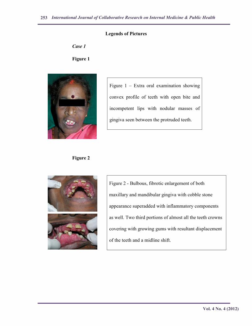
Figure 1 – Extra oral examination showing
convex profile of teeth with open bite and
incompetent lips with nodular masses of
gingiva seen between the protruded teeth.
Figure 2 - Bulbous, fibrotic enlargement of both
maxillary and mandibular gingiva with cobble stone
appearance superadded with inflammatory components
as well. Two third portions of almost all the teeth crowns
covering with growing gums with resultant displacement
of the teeth and a midline shift.
International Journal of Collaborative Research on Internal Medicine & Public Health
Vol. 4 No. 4 (2012)
254
Case 2
Figure 3
Figure 4
Figure 5
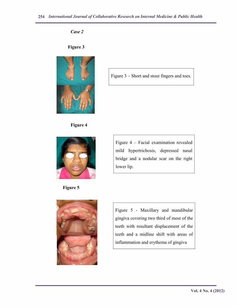
Figure 3 – Short and stout fingers and toes.
Figure 4 – Facial examination revealed
mild hypertrichosis, depressed nasal
bridge and a nodular scar on the right
lower lip.
Figure 5 - Maxillary and mandibular
gingiva covering two third of most of the
teeth with resultant displacement of the
teeth and a midline shift with areas of
inflammation and erythema of gingiva
International Journal of Collaborative Research on Internal Medicine & Public Health
Vol. 4 No. 4 (2012)
255
Case 3
Figure 6
Figure 7
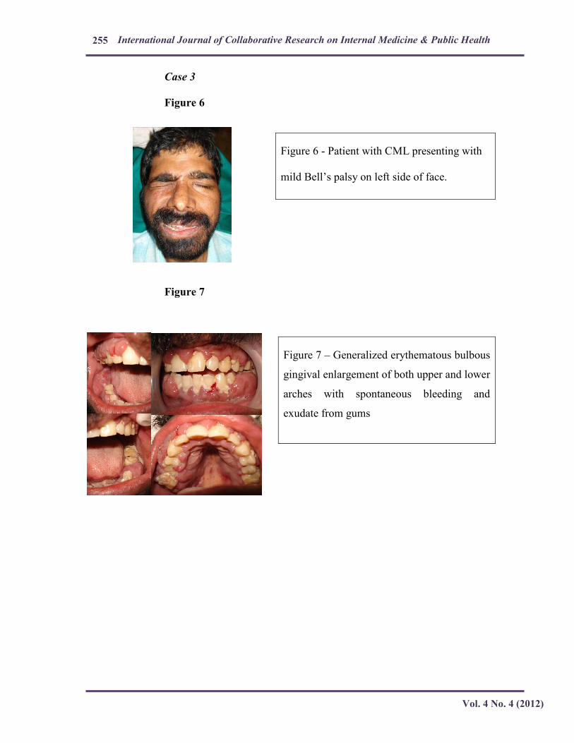
Figure 6 - Patient with CML presenting with
mild Bell’s palsy on left side of face.
Figure 7 – Generalized erythematous bulbous
gingival enlargement of both upper and lower
arches with spontaneous bleeding and
exudate from gums