Embed Size (px)
Citation preview
Update on Thyroid FNA Update on Thyroid FNA –– The Bethesda System The Bethesda System
Shikha Bose M.D.Associate Professor
Cedars Sinai Medical Center
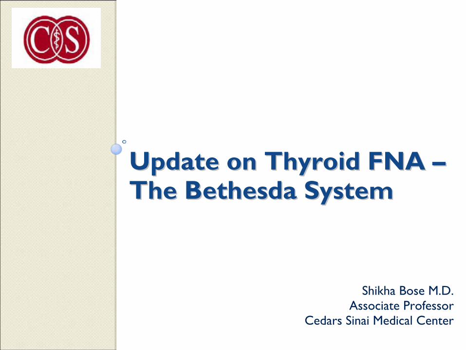
Thyroid NodulesThyroid Nodules
Frequent occurrence ◦
Palpable: 4-7% of adults◦
Ultrasound: 10-31%
Majority benign
FNA has proven to be an effective management tool in patients with thyroid nodules
Thyroid FNAThyroid FNA
Safe, inexpensive, easily performed, minimal patient discomfort
High diagnostic accuracy rate ~ 90-100%
False positive diagnoses: <1% ◦
over-interpretation of reparative and reactive nuclear changes as papillary thyroid carcinoma (PTC)
False negative rates: 1-11%◦
unsatisfactory specimens
◦
sampling errors
◦
interpretation errors
◦
cystic neoplasms, especially PTC
Major limitations include:◦
difficulty to distinguish hypercellular non-neoplastic nodules from follicular neoplasm
◦
difficulty to obtain an adequate specimen on some occasions
THYROID FNA THYROID FNA
Its main purpose is ◦
to provide a rational approach to management and ◦
to determine the correct surgical procedure when surgery is required
The Bethesda ConferenceThe Bethesda Conference
Aim: to provide the current “state of the science”
in the practice of thyroid FNA.
Held -
Oct 2007 Bethesda, Maryland
6 committees were formed for review of English medical literature extending back to 1995
Final document containing the review and conclusions
–
posted on Feb 2008
The text was not endorsed as standard of practice guidelines
Fine Needle Aspiration Fine Needle Aspiration Biopsy TechniqueBiopsy Technique
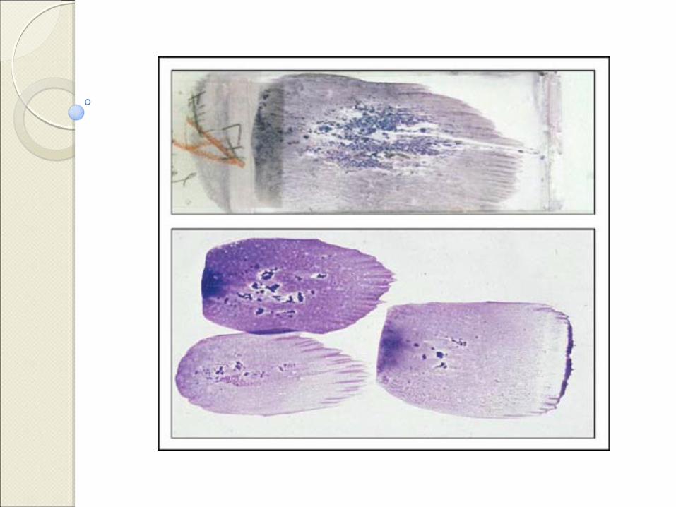
HIGH Quality smears are HIGH Quality smears are essential for diagnosisessential for diagnosis
Thyroid FNAThyroid FNA
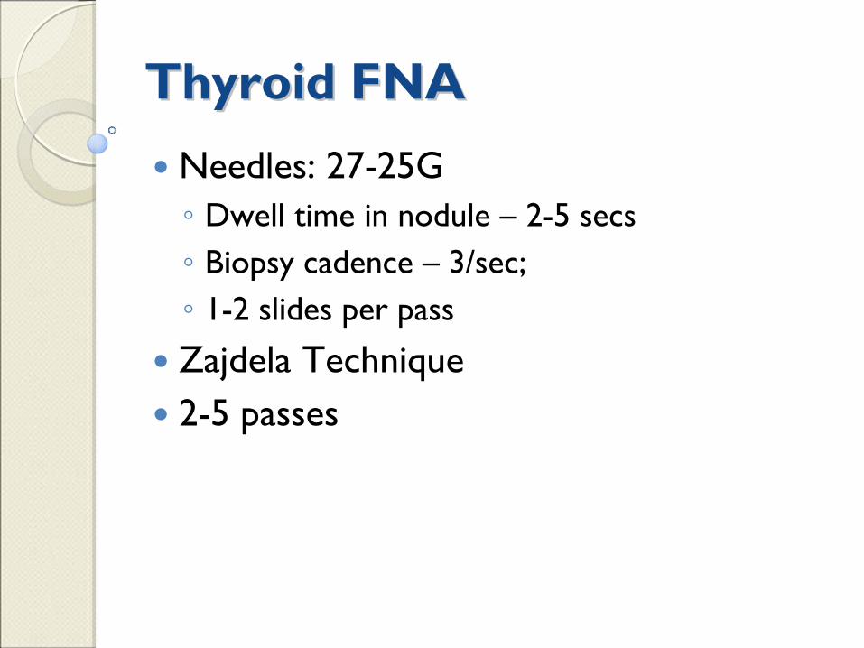
Needles: 27-25G◦
Dwell time in nodule –
2-5 secs
◦
Biopsy cadence –
3/sec; ◦
1-2 slides per pass
Zajdela Technique
2-5 passes
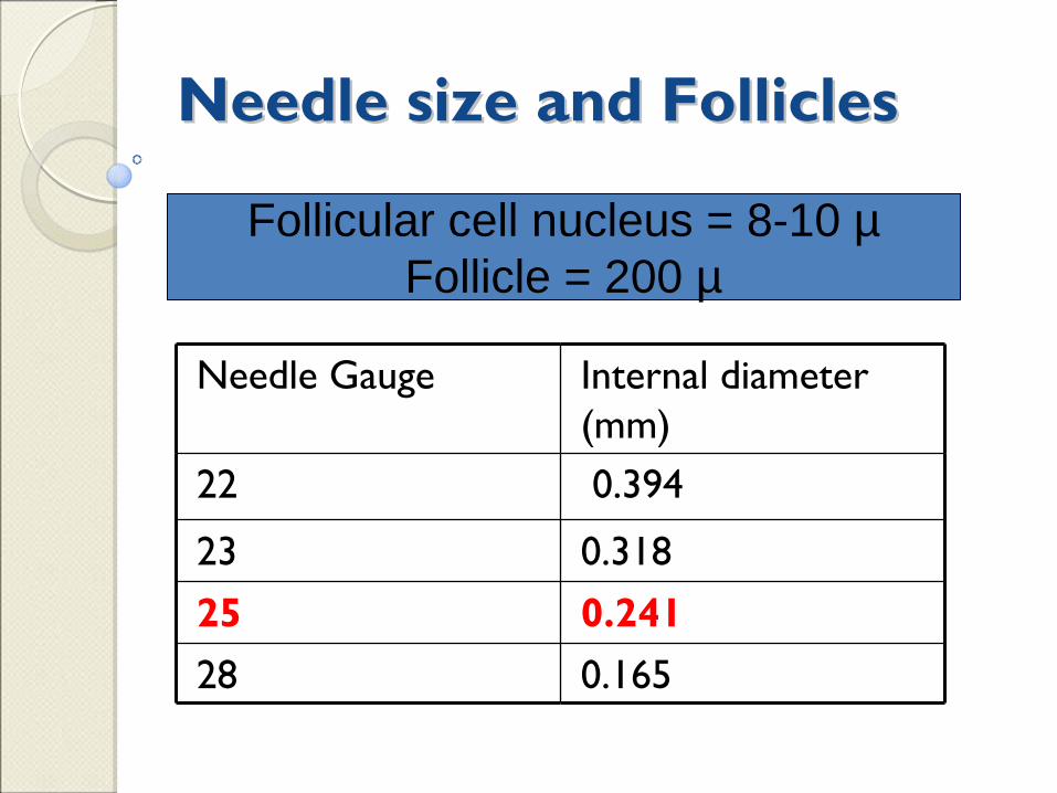
Needle size and FolliclesNeedle size and Follicles
Needle Gauge Internal diameter (mm)
22 0.394
23 0.31825 0.24128 0.165
Follicular cell nucleus = 8-10 µFollicle = 200 µ
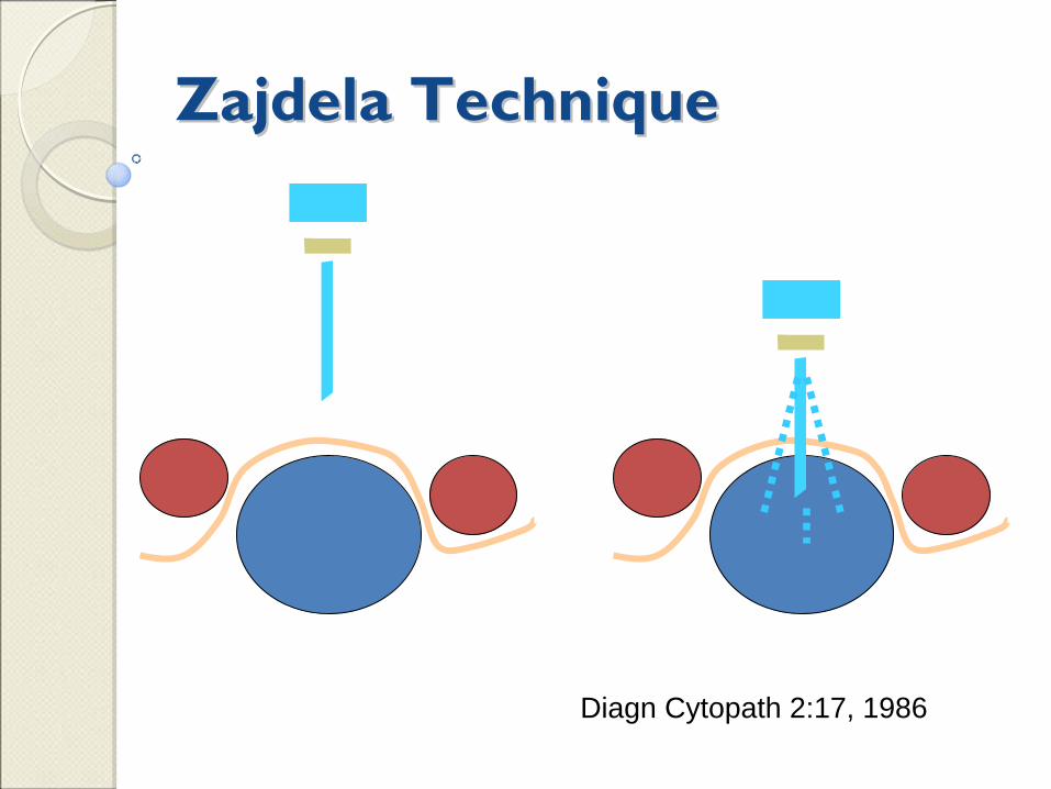
Zajdela TechniqueZajdela Technique
Diagn Cytopath 2:17, 1986
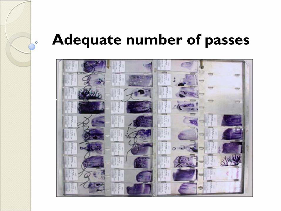
Adequate number of passes
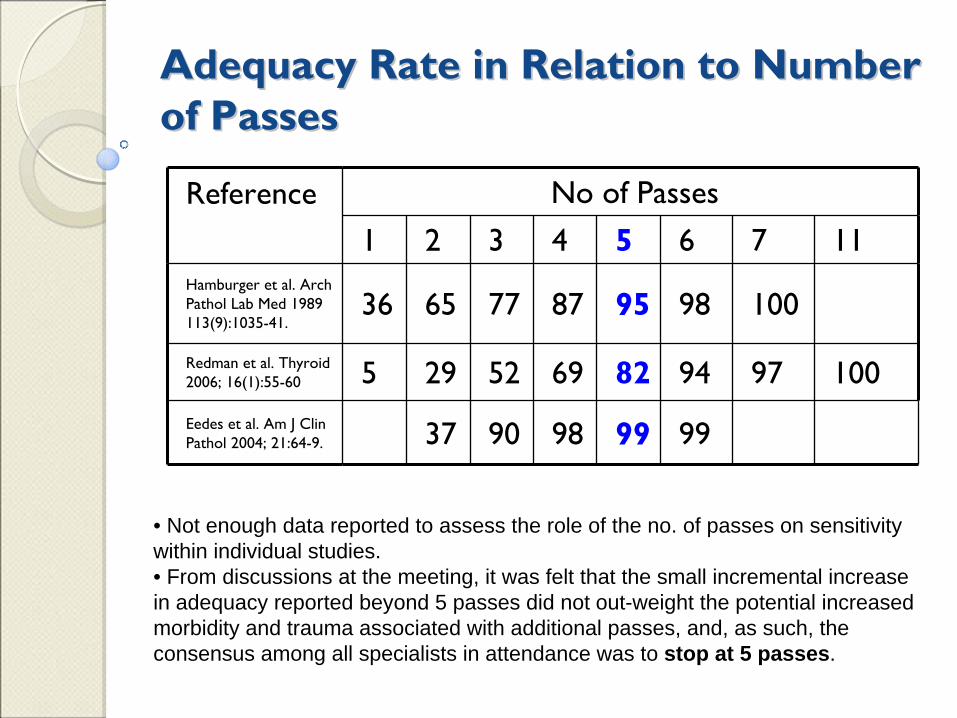
Adequacy Rate in Relation to Number Adequacy Rate in Relation to Number of Passesof Passes
Reference No of Passes1 2 3 4 5 6 7 11
Hamburger et al. Arch Pathol Lab Med 1989 113(9):1035-41.
36 65 77 87 95 98 100
Redman et al. Thyroid 2006; 16(1):55-60 5 29 52 69 82 94 97 100Eedes et al. Am J Clin Pathol 2004; 21:64-9. 37 90 98 99 99
• Not enough data reported to assess the role of the no. of passes on sensitivitywithin individual studies. • From discussions at the meeting, it was felt that the small incremental increase in adequacy reported beyond 5 passes did not out-weight the potential increased morbidity and trauma associated with additional passes, and, as such, the consensus among all specialists in attendance was to stop at 5 passes.
Optimal Number of Passes for Optimal Number of Passes for Solid or Cystic Lesions Solid or Cystic Lesions
2-5 biopsies from different sites
representative tissue from each pass smeared on a
slide (or 2) and the
remaining rinsed into a collection tube with transport fluid medium
Preparing a good smearis as important as
performing the procedure
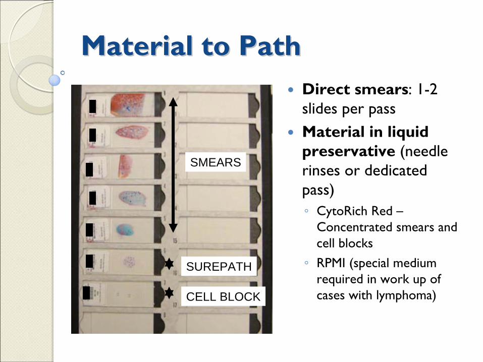
Material to PathMaterial to Path
Direct smears: 1-2 slides per pass
Material in liquid preservative
(needle
rinses or dedicated pass)◦
CytoRich Red –
Concentrated smears and cell blocks
◦
RPMI (special medium required in work up of cases with lymphoma)
SMEARS
SUREPATH
CELL BLOCK
Diagnostic terminology / Diagnostic terminology / classification scheme and classification scheme and morphologic criteria for morphologic criteria for cytologic diagnosis of thyroid cytologic diagnosis of thyroid lesionslesions
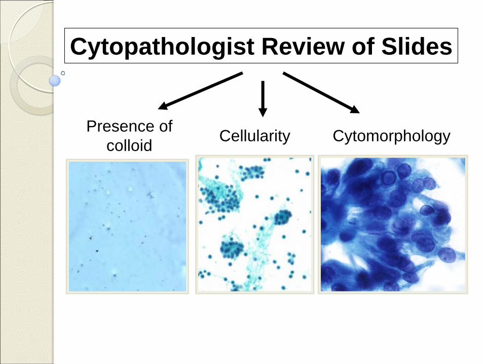
Cytopathologist Review of Slides
CellularityPresence of colloid Cytomorphology
What most of us have been saying What most of us have been saying on our reportson our reports
Benign
Indeterminate (Don’t know) ◦
Follicular lesion/nodule
Suspicious
Malignant
Non Diagnostic
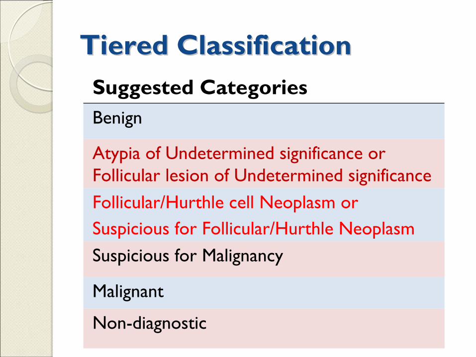
Tiered ClassificationTiered ClassificationSuggested CategoriesBenign
Atypia of Undetermined significance or Follicular lesion of Undetermined significanceFollicular/Hurthle cell Neoplasm or Suspicious for Follicular/Hurthle NeoplasmSuspicious for Malignancy
Malignant
Non-diagnostic
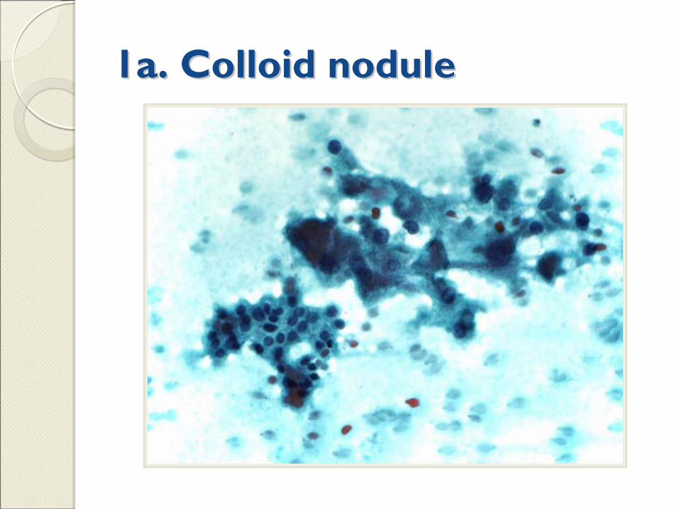
1. Benign 1. Benign
Low risk of malignancy <1%
The diagnostic terms include ◦
nodular goiter◦
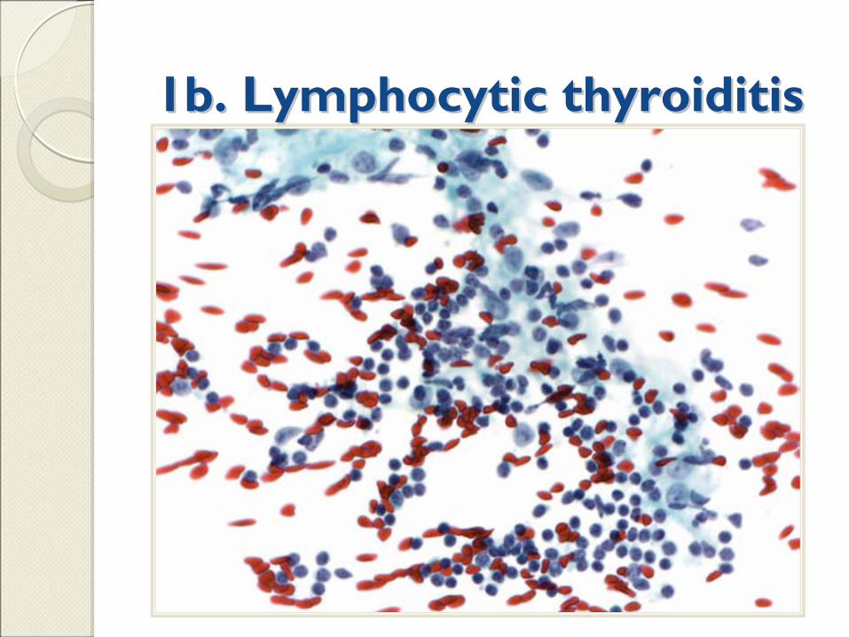
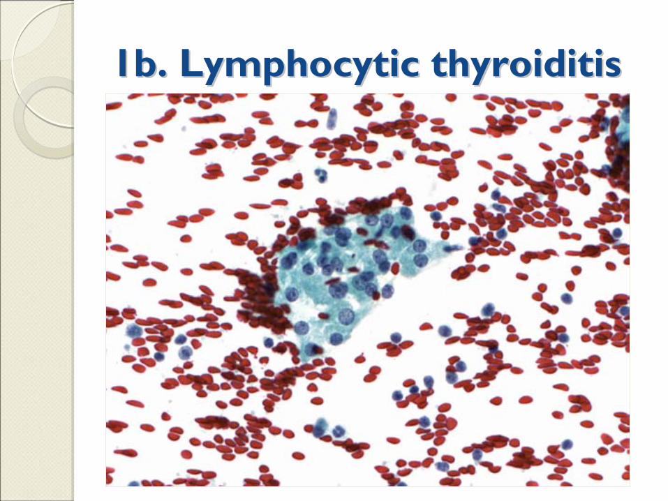
chronic lymphocytic thyroiditis◦
hyperplastic/adenomatoid nodule in goiter◦
colloid nodule.
Follow up◦
by clinical and periodic radiologic exam◦
repeat FNA due to increase in the size of nodule.
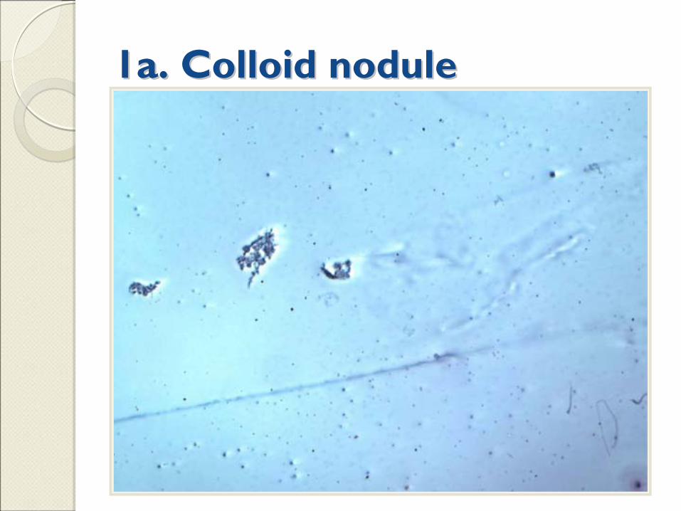
1a. Colloid nodule1a. Colloid nodule
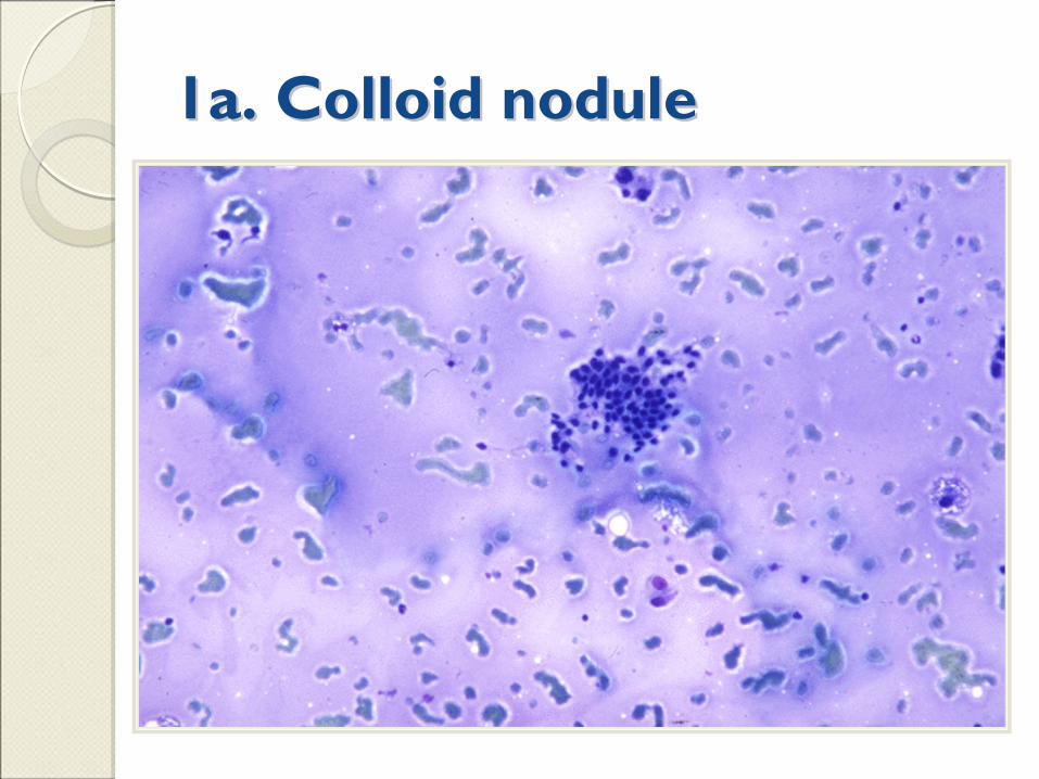
1a. Colloid nodule1a. Colloid nodule
1a. Colloid nodule1a. Colloid nodule
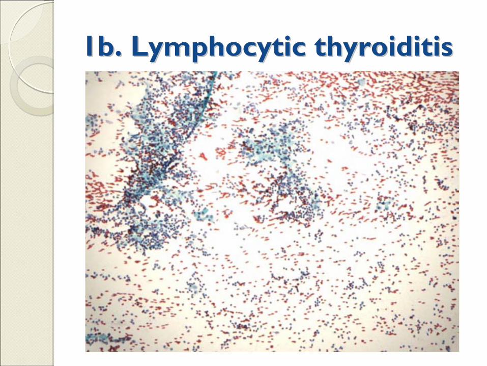
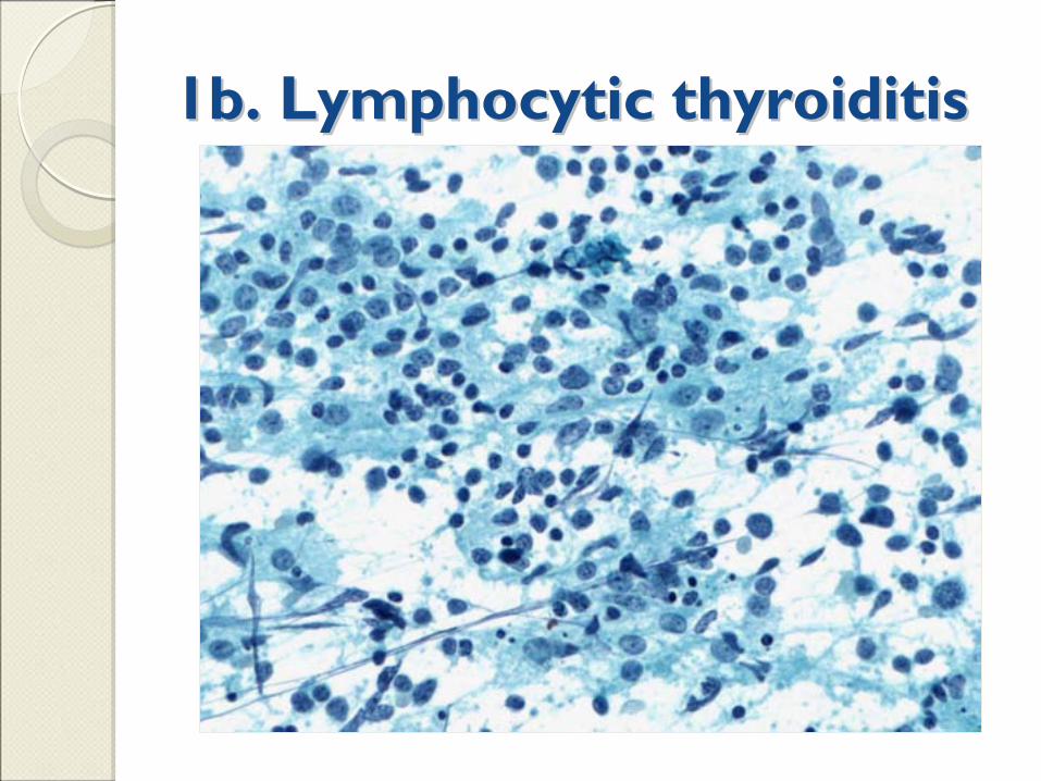
1b. Lymphocytic thyroiditis1b. Lymphocytic thyroiditis
1b. Lymphocytic thyroiditis1b. Lymphocytic thyroiditis
1b. Lymphocytic thyroiditis1b. Lymphocytic thyroiditis
1b. Lymphocytic thyroiditis1b. Lymphocytic thyroiditis
D/D of lymphocytes in thyroidD/D of lymphocytes in thyroid
Chronic inflammation in goiter
Chronic lymphocytic thyroiditis
Graves disease
Radiation/drug induced thyroiditis
Intrathyroidal lymph node
Malignant lymphoma –
NHL, HD
3. Follicular Neoplasm/ Suspicious for 3. Follicular Neoplasm/ Suspicious for Follicular Neoplasm Follicular Neoplasm
Low to intermediate risk of malignancy 20-30%
(higher in Hurthle cell lesions if the nodule is equal to or larger than 3.5 cm)
Includes:◦
Non-papillary follicular patterned lesions/neoplasms ◦
Hurthle cell lesions/neoplasms. ◦
Other suggested diagnostic terms -
micro-follicular
proliferation/lesion, suggestive of neoplasm and follicular lesion.
Lobectomy/hemithyroidectomy
Diagnosis rendered on surg. pathology exam -
adenomatoid nodule or adenoma or carcinoma
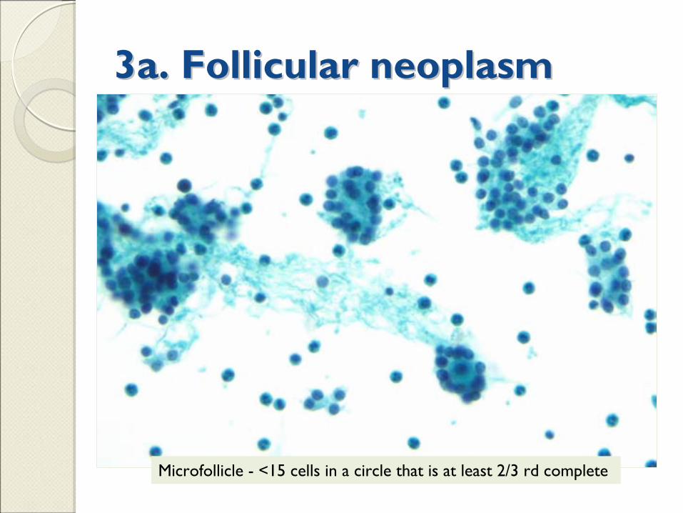
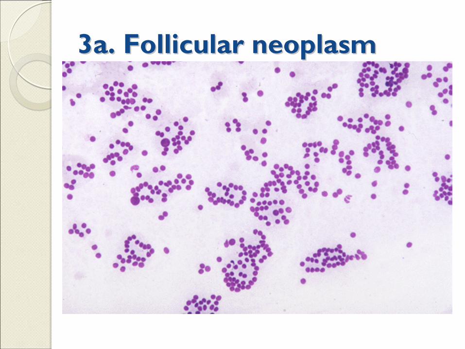
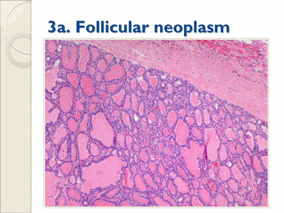
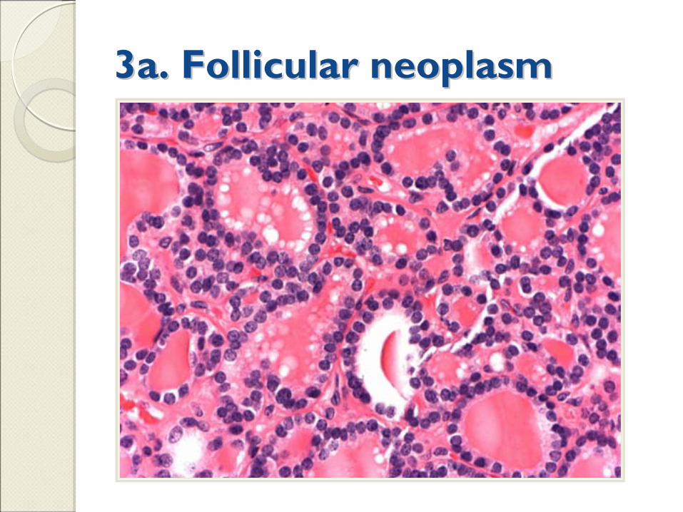
3a. Follicular neoplasm3a. Follicular neoplasm
Microfollicle -
<15 cells in a circle that is at least 2/3 rd complete
3a. Follicular neoplasm3a. Follicular neoplasm
3a. Follicular neoplasm3a. Follicular neoplasm
3a. Follicular neoplasm3a. Follicular neoplasm
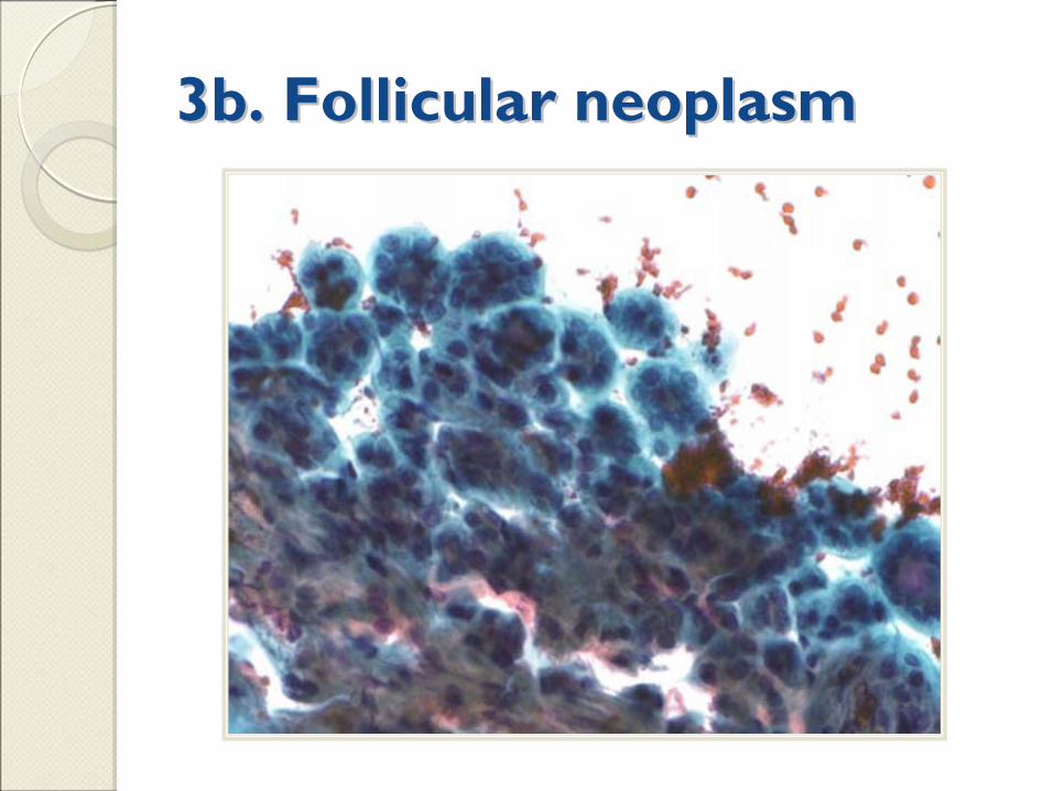
3b. Follicular neoplasm3b. Follicular neoplasm
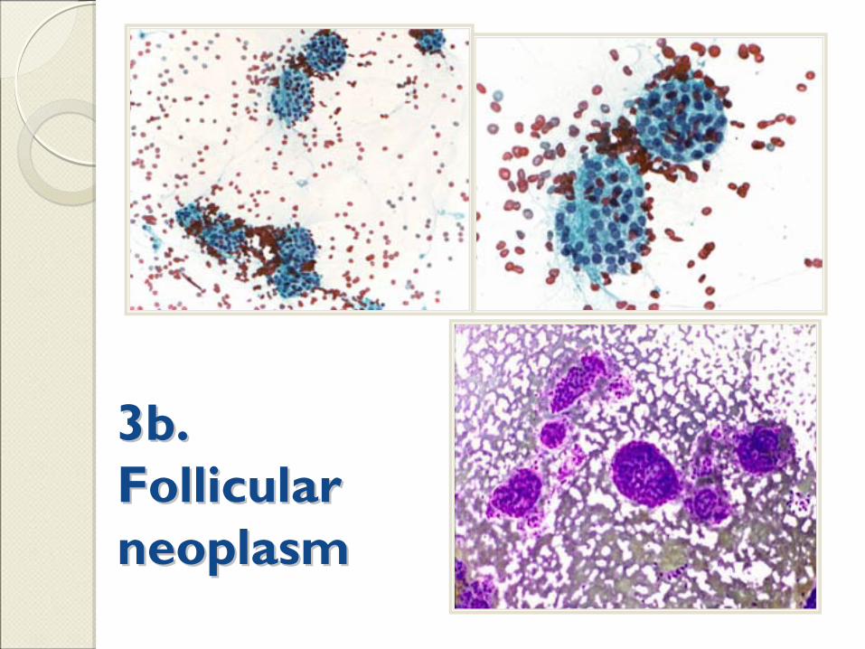
3b. 3b. Follicular Follicular neoplasmneoplasm
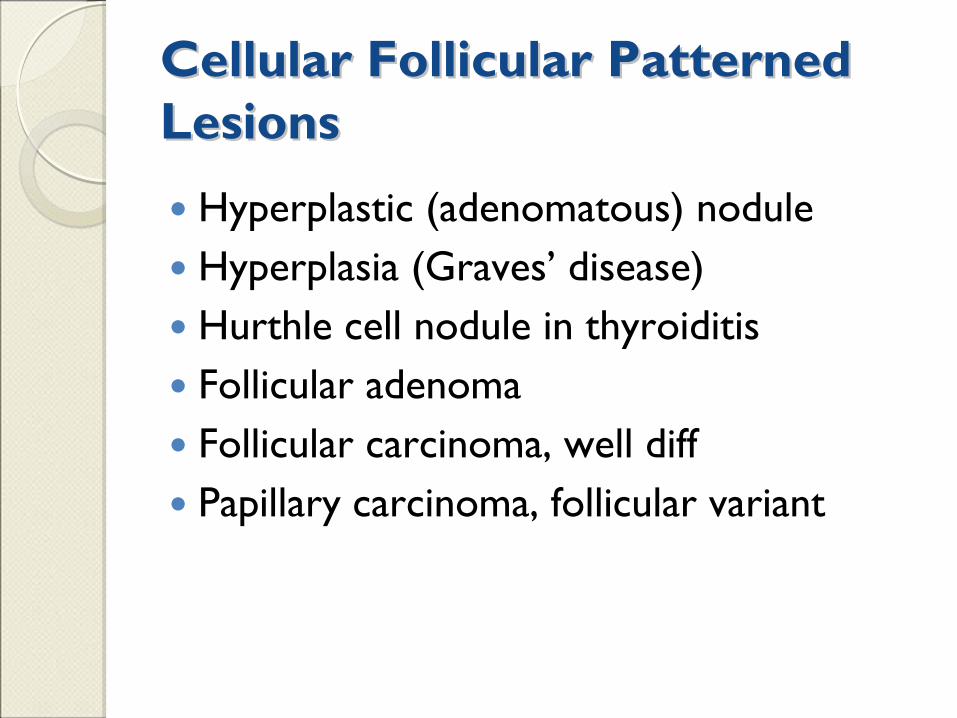
Cellular Follicular Patterned Cellular Follicular Patterned LesionsLesions
Hyperplastic (adenomatous) nodule
Hyperplasia (Graves’
disease)
Hurthle cell nodule in thyroiditis
Follicular adenoma
Follicular carcinoma, well diff
Papillary carcinoma, follicular variant
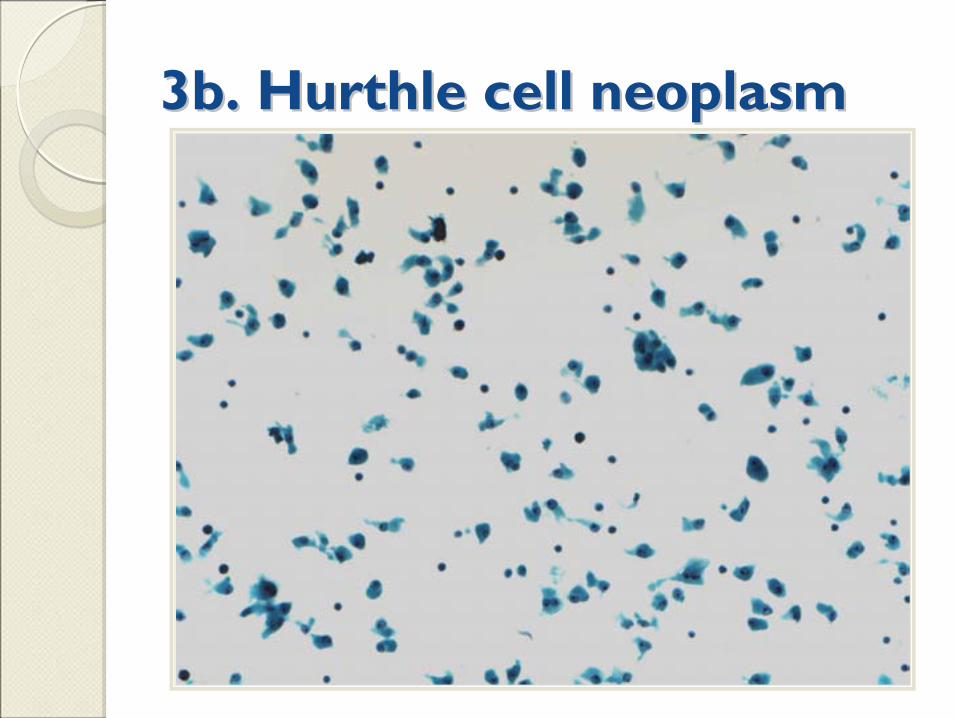
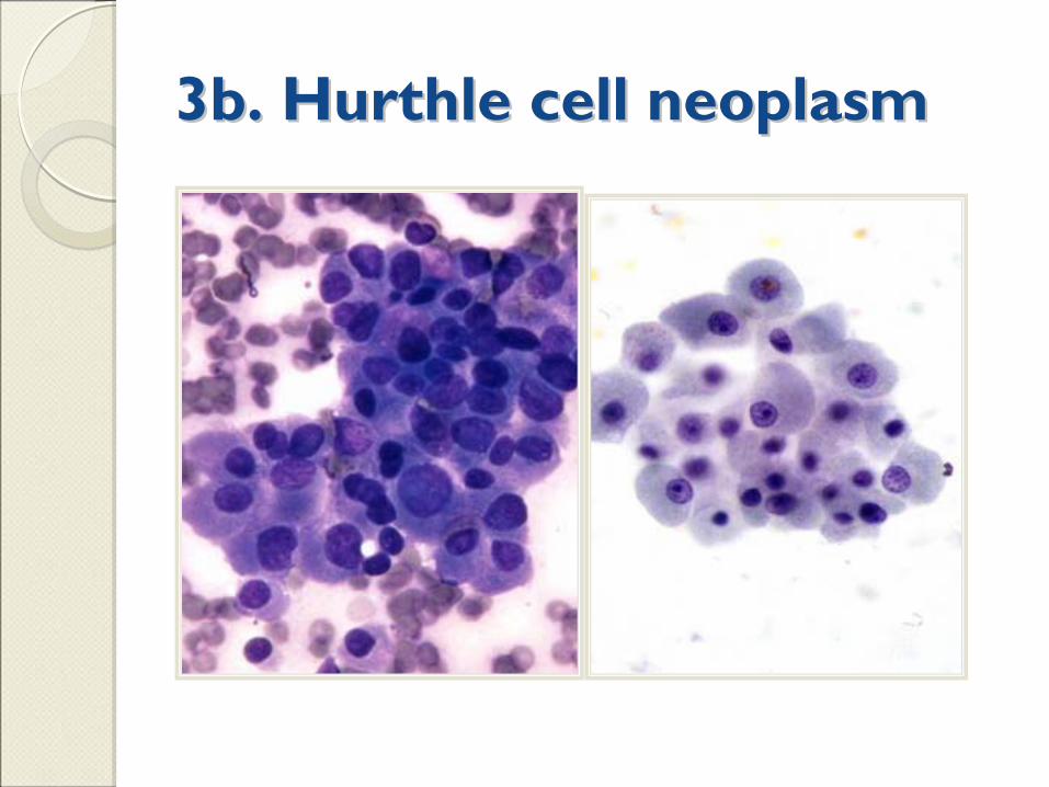
3b. Hurthle cell neoplasm3b. Hurthle cell neoplasm
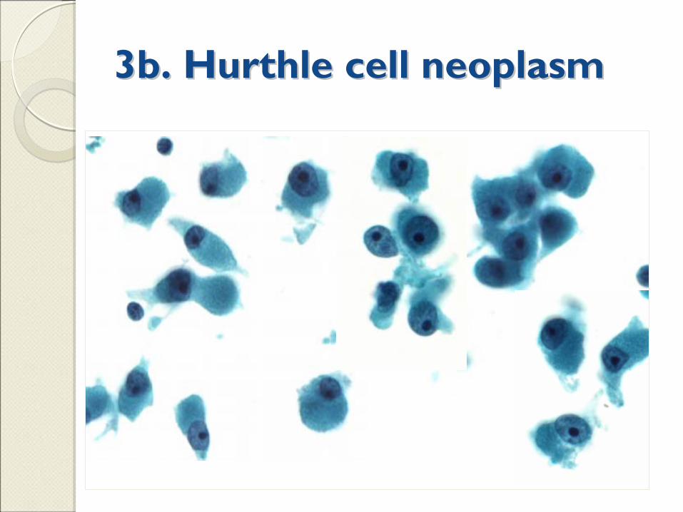
3b. Hurthle cell neoplasm3b. Hurthle cell neoplasm
3b. Hurthle cell neoplasm3b. Hurthle cell neoplasm
Hurthle Cell ProliferationsHurthle Cell Proliferations
Hurthle cell nodule in thyroiditis
Hurthle cell adenoma
Hurthle cell carcinoma
Papillary carcinoma, oncocytic variant
Medullary carcinoma
2. Follicular Lesion/Atypia of 2. Follicular Lesion/Atypia of Undetermined SignificanceUndetermined Significance
Risk of malignancy 5-10%
Heterogeneous category
that includes−cases where cytologic findings are not convincingly
benign, yet the degree of cellular or architectural atypia is not sufficient for an interpretation of "Follicular Neoplasm" or "Suspicious for Malignancy". −compromised specimen (e.g. low cellularity, poor
fixation, obscuring blood).
Benefit from repeat FNA
and correlation with clinical and radiologic findings.
When utilized should ideally represent <7%
of all thyroid FNA interpretations.
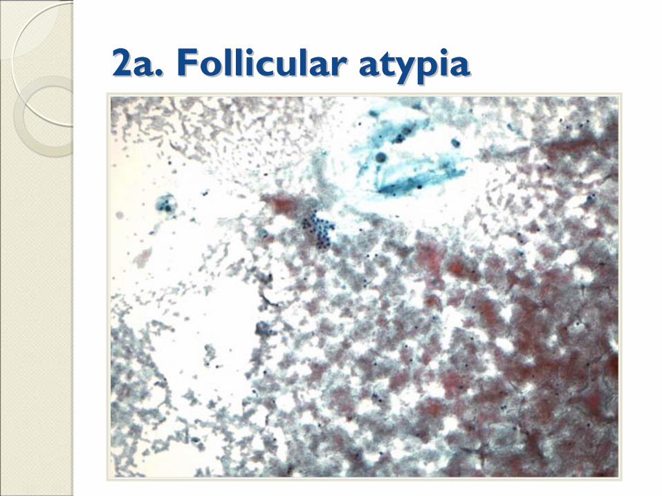
2a. Follicular atypia2a. Follicular atypia
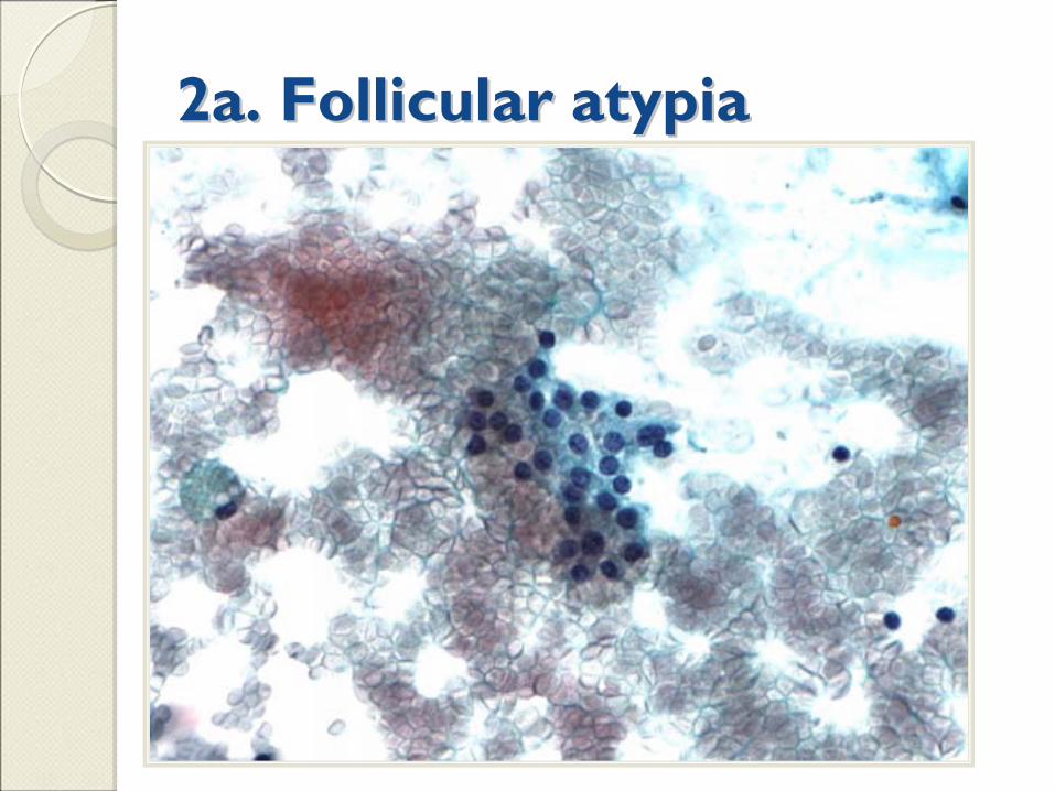
2a. Follicular atypia2a. Follicular atypia
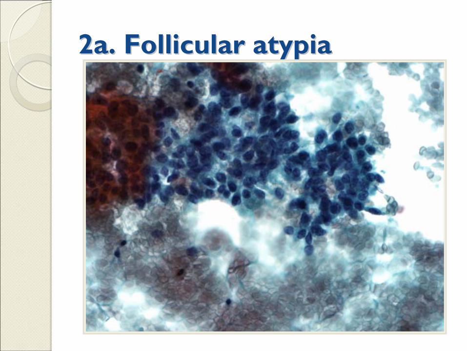
2a. Follicular atypia2a. Follicular atypia
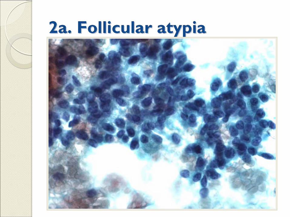
2a. Follicular atypia2a. Follicular atypia
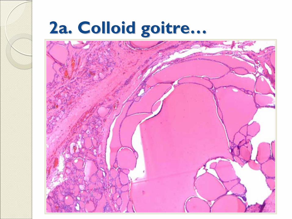
2a. Colloid goitre2a. Colloid goitre……
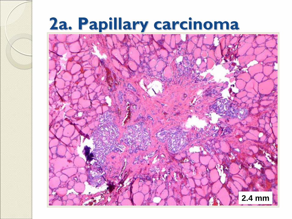
2a. Papillary carcinoma 2a. Papillary carcinoma
2.4 mm
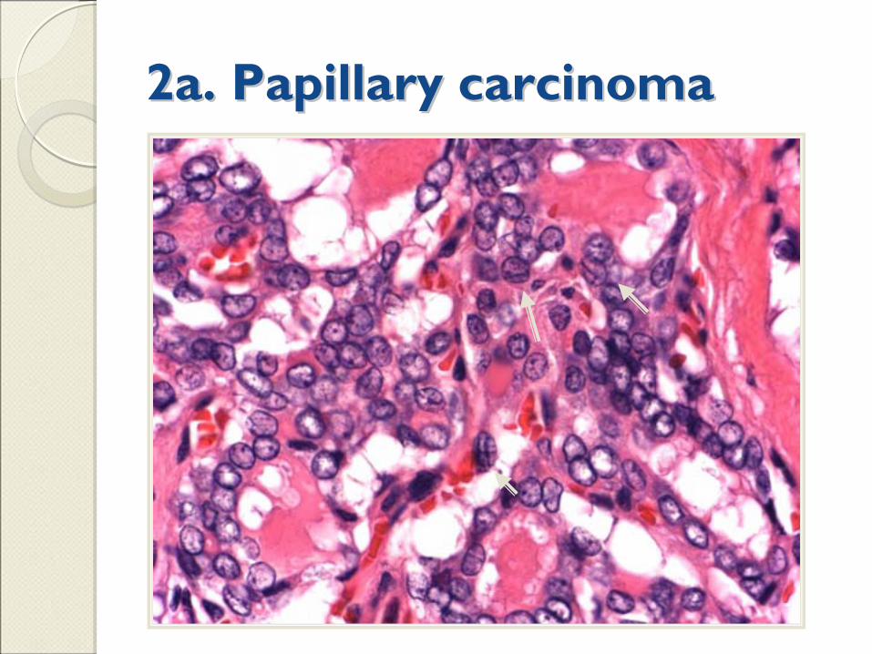
2a. Papillary carcinoma2a. Papillary carcinoma
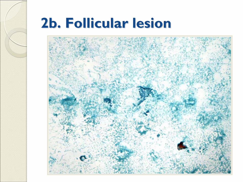
2b. Follicular lesion2b. Follicular lesion
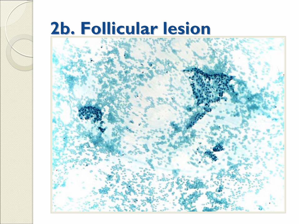
2b. Follicular lesion2b. Follicular lesion
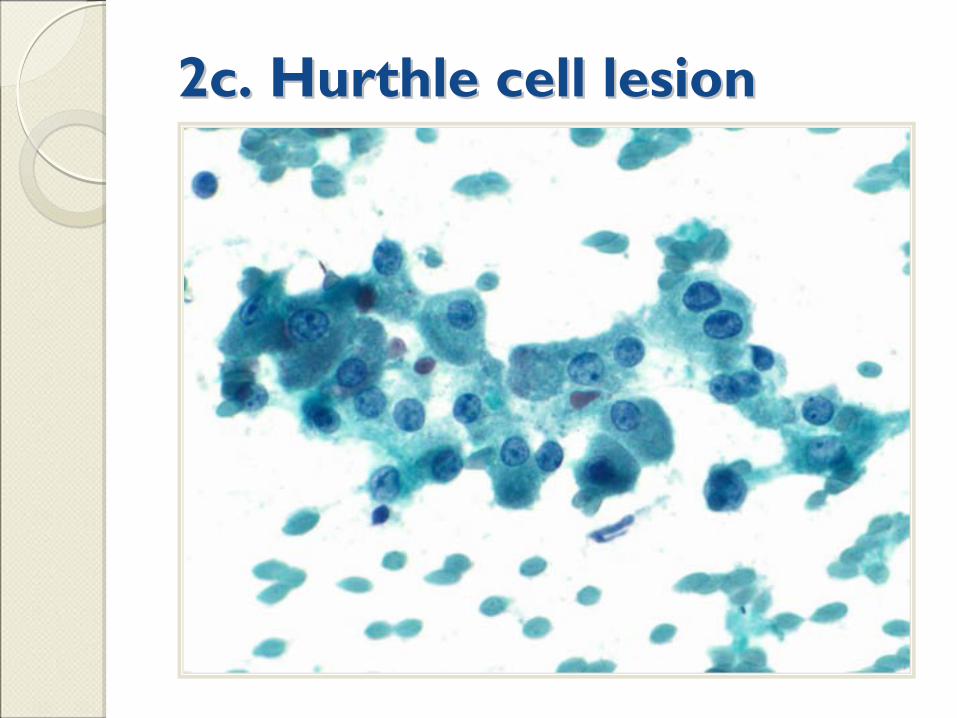
2c. Hurthle cell lesion2c. Hurthle cell lesion
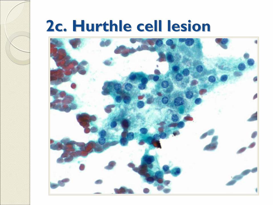
2c. Hurthle cell lesion2c. Hurthle cell lesion
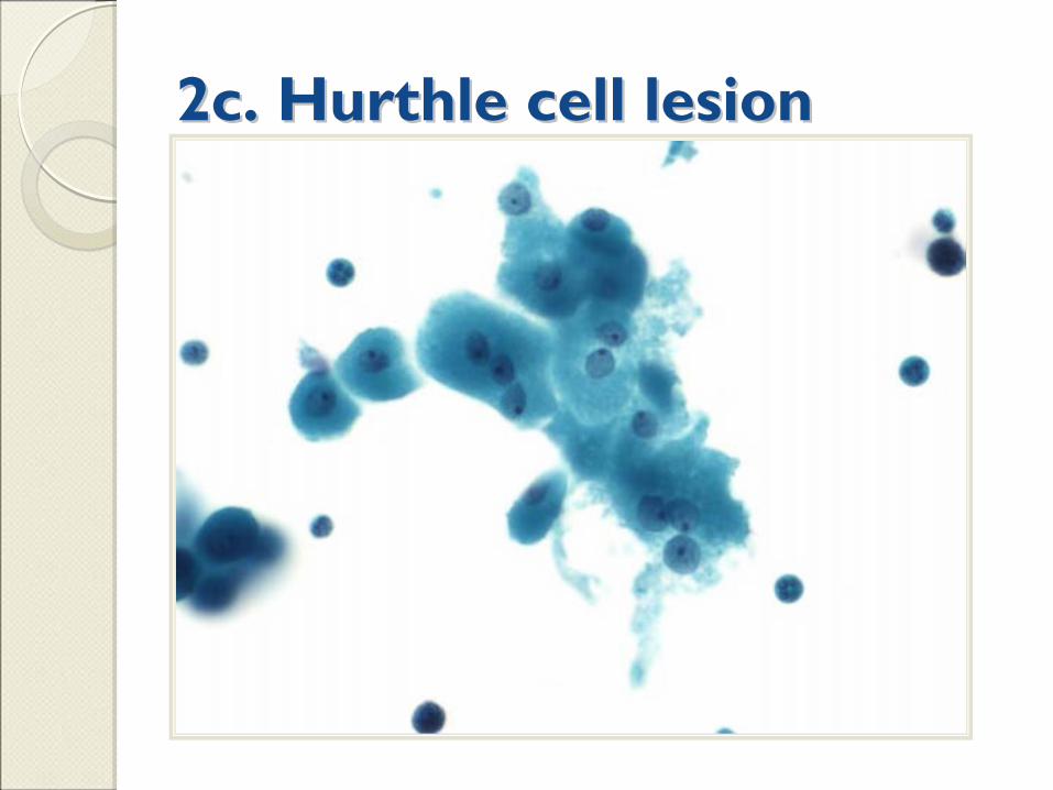
2c. Hurthle cell lesion2c. Hurthle cell lesion
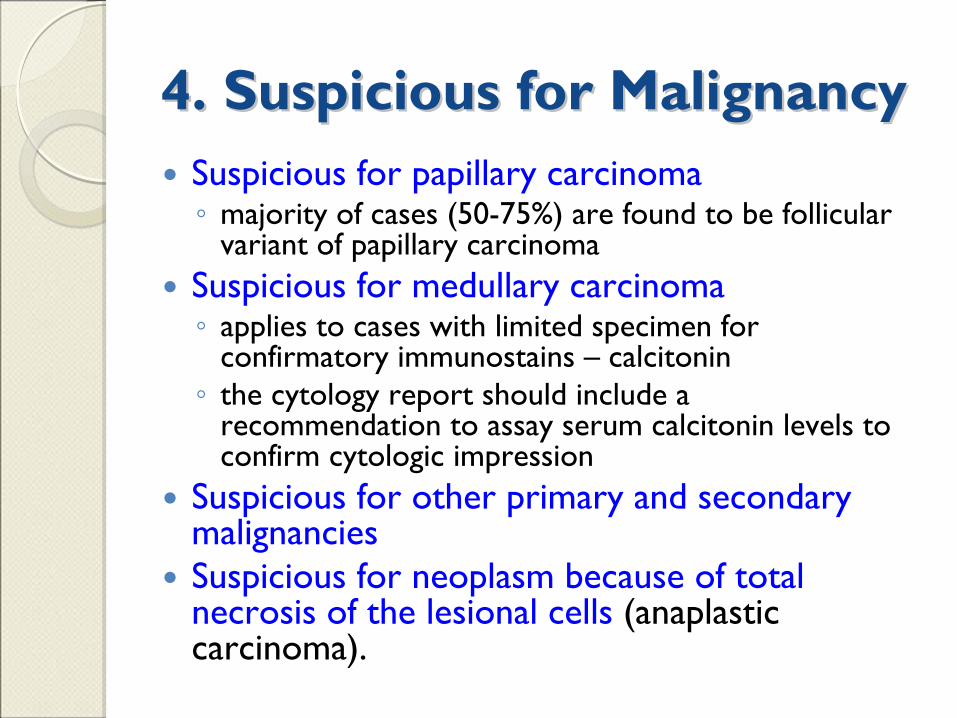
4. Suspicious for Malignancy4. Suspicious for Malignancy
Suspicious for papillary carcinoma◦
majority of cases (50-75%) are found to be follicular variant of papillary carcinoma
Suspicious for medullary carcinoma◦
applies to cases with limited specimen for confirmatory immunostains –
calcitonin
◦
the cytology report should include a recommendation to assay serum calcitonin levels to confirm cytologic impression
Suspicious for other primary and secondary malignancies
Suspicious for neoplasm because of total necrosis of the lesional cells
(anaplastic
carcinoma).
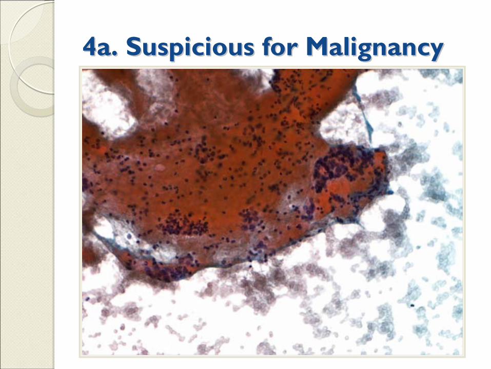
4a. Suspicious for Malignancy 4a. Suspicious for Malignancy
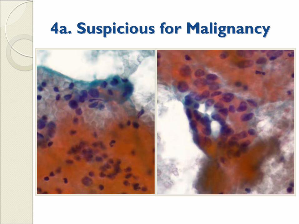
4a. Suspicious for Malignancy4a. Suspicious for Malignancy
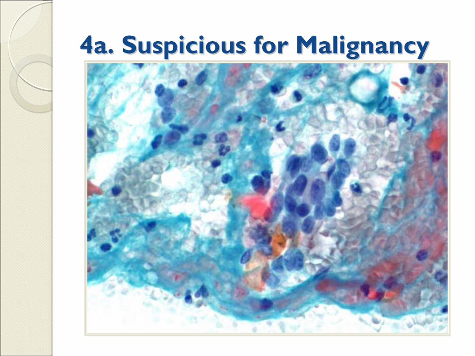
4a. Suspicious for Malignancy4a. Suspicious for Malignancy
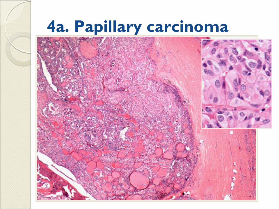
4a. Papillary carcinoma
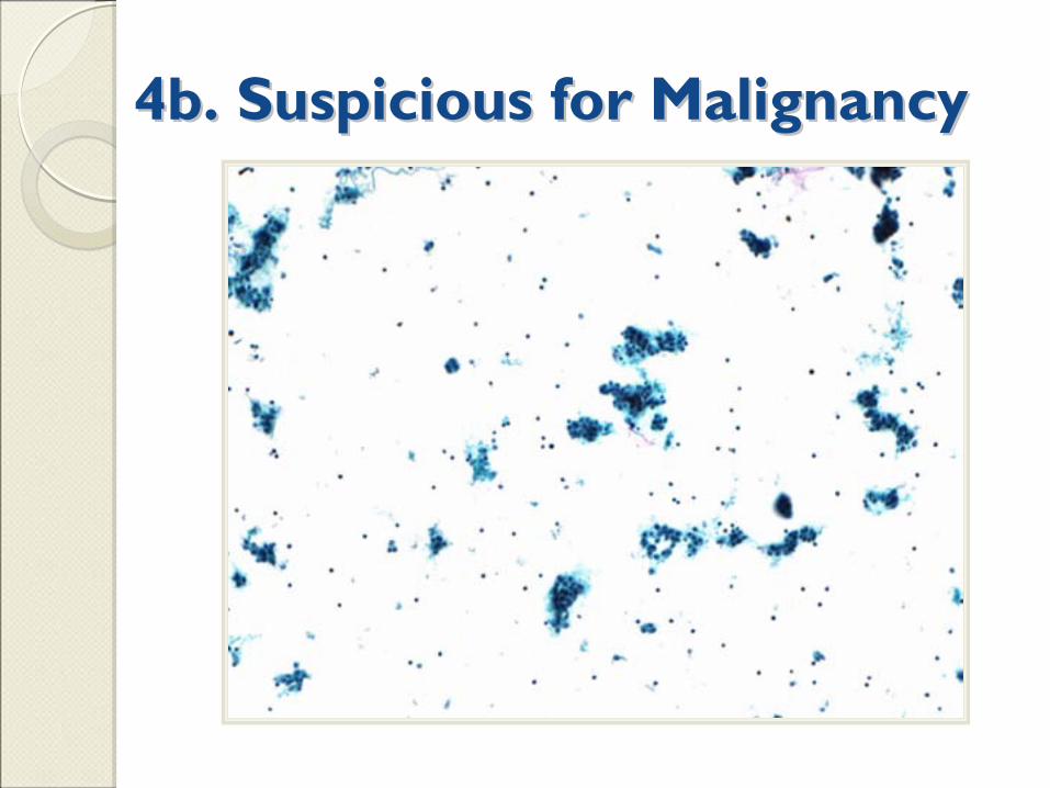
4b. Suspicious for Malignancy4b. Suspicious for Malignancy
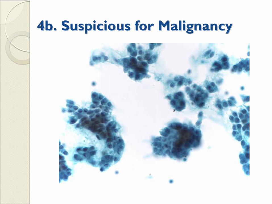
4b. Suspicious for Malignancy4b. Suspicious for Malignancy
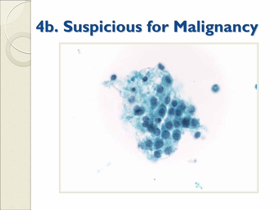
4b. Suspicious for Malignancy4b. Suspicious for Malignancy
5. Malignant5. Malignant
Papillary carcinoma and its variants
Poorly differentiated carcinoma
Anaplastic carcinoma
Medullary carcinoma
Lymphoma
Metastases
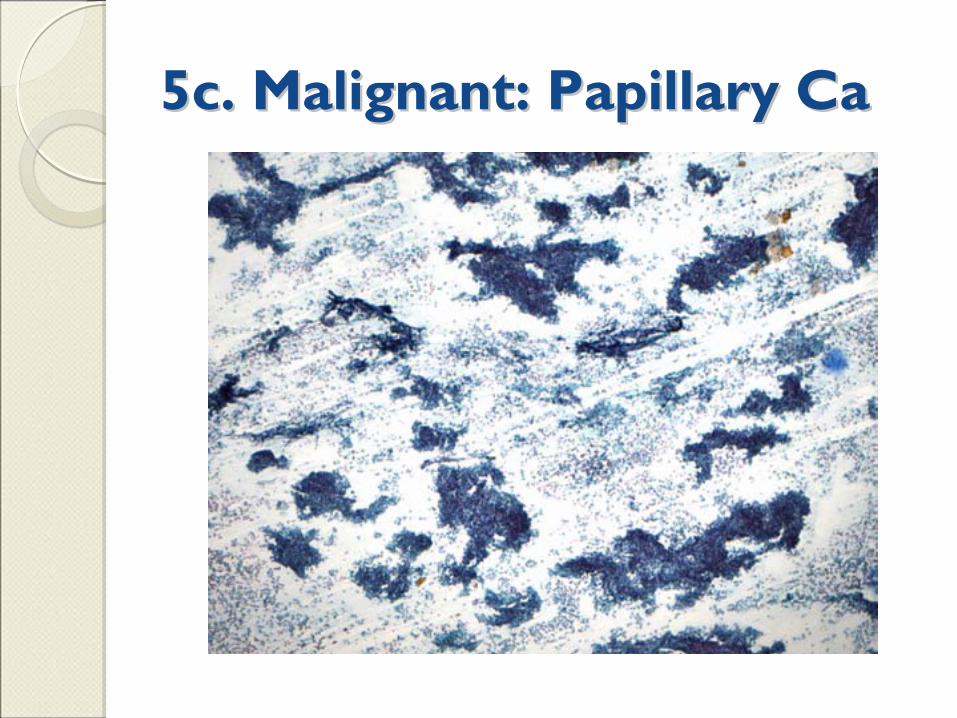
5c. Malignant: Papillary Ca5c. Malignant: Papillary Ca
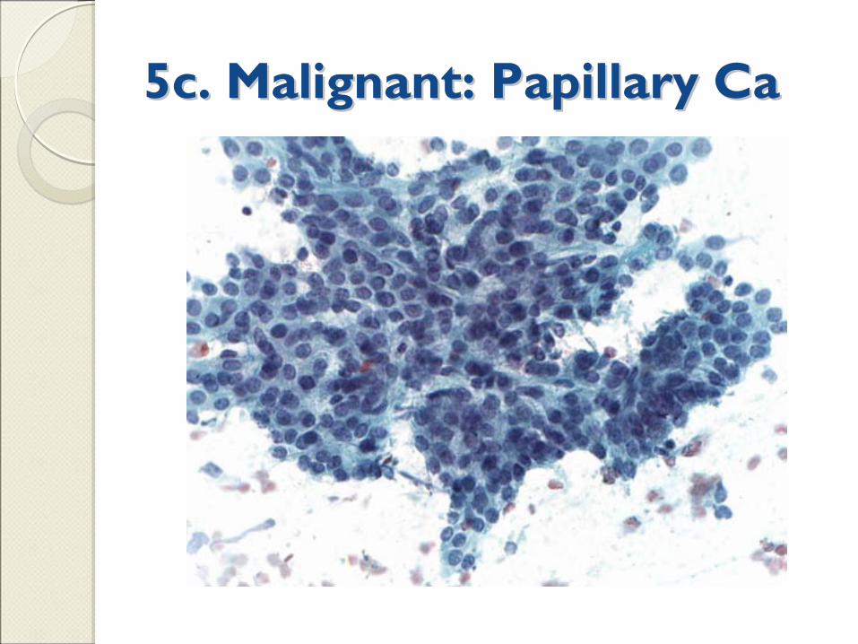
5c. Malignant: Papillary Ca5c. Malignant: Papillary Ca
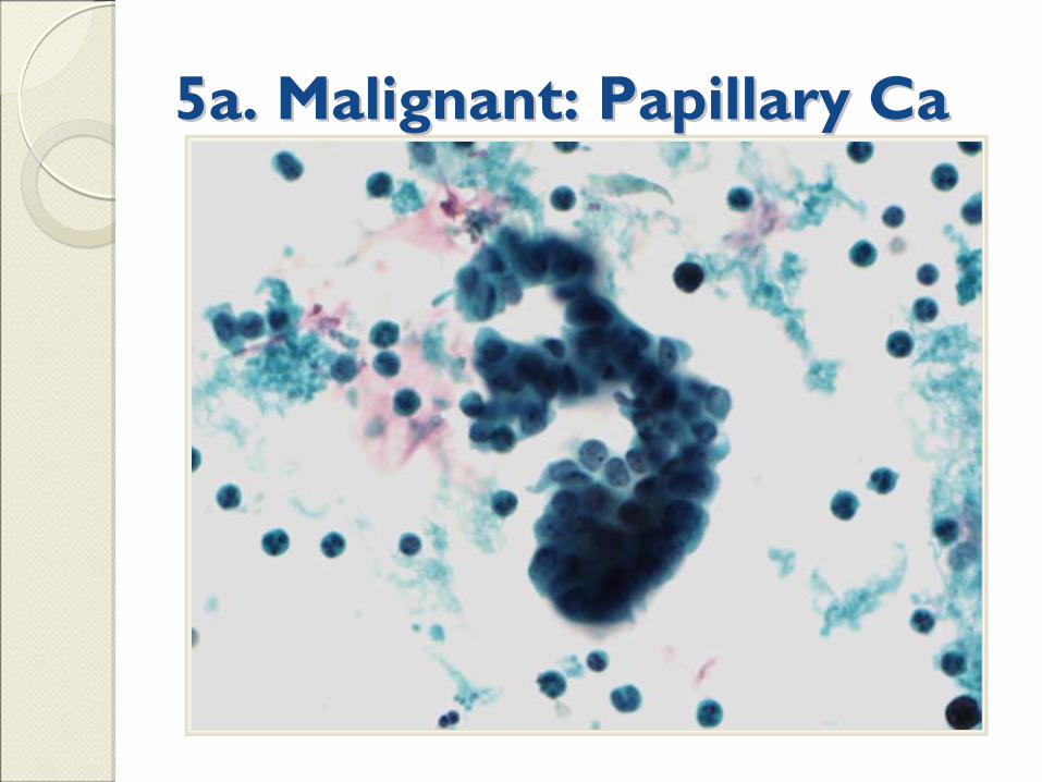
5a. Malignant: Papillary Ca5a. Malignant: Papillary Ca
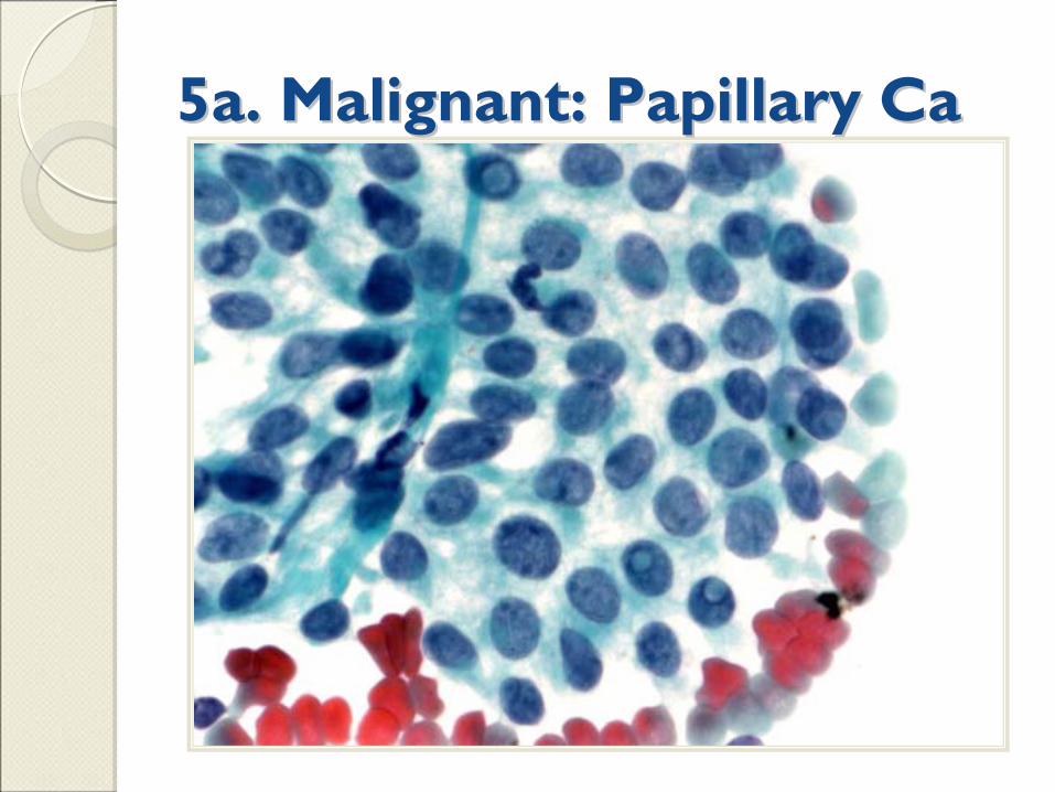
5a. Malignant: Papillary Ca5a. Malignant: Papillary Ca
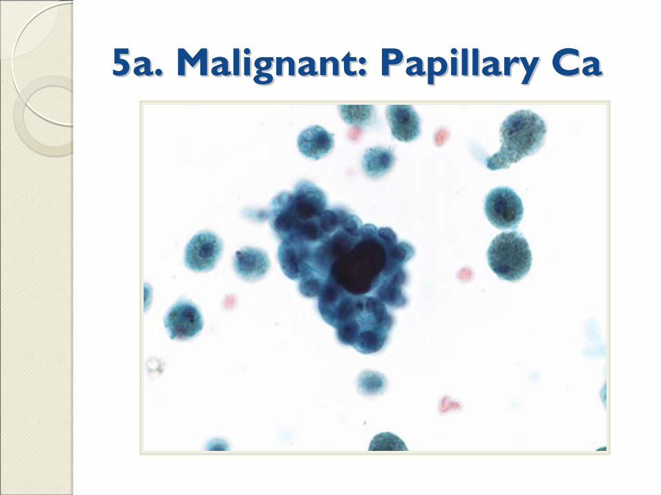
5a. Malignant: Papillary Ca5a. Malignant: Papillary Ca
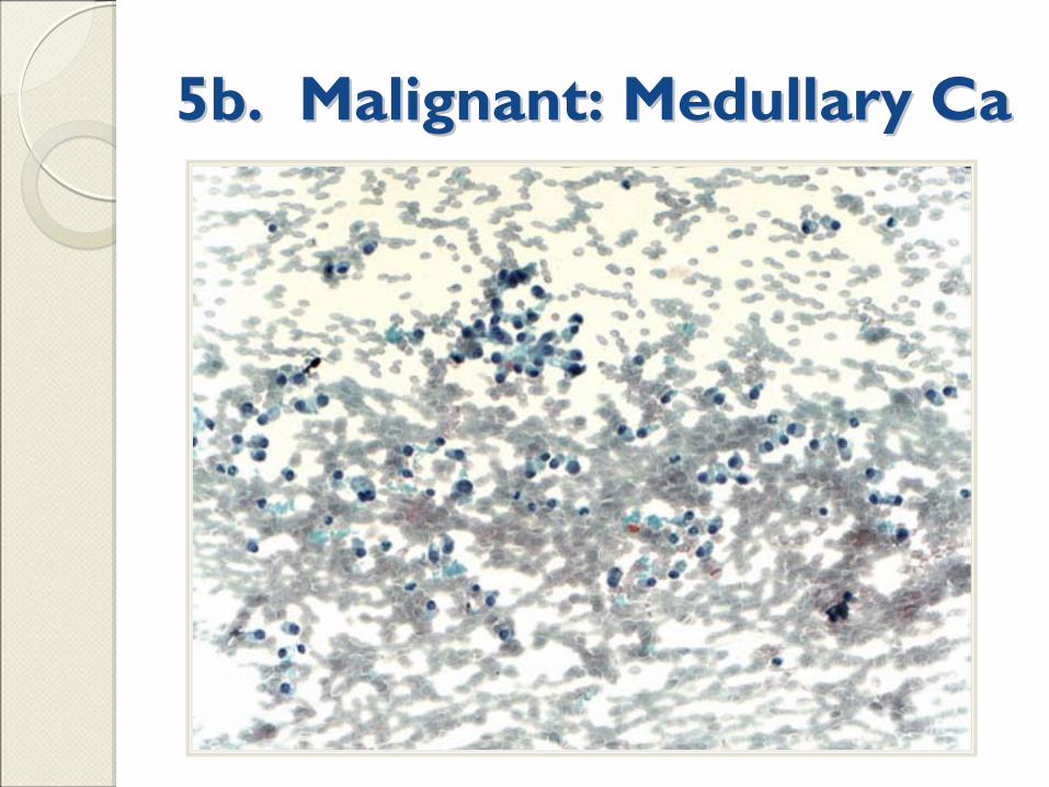
5b. Malignant: Medullary Ca5b. Malignant: Medullary Ca
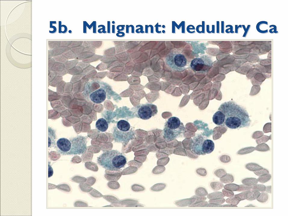
5b. Malignant: Medullary Ca5b. Malignant: Medullary Ca
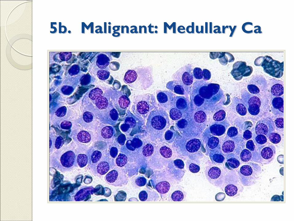
5b. Malignant: Medullary Ca5b. Malignant: Medullary Ca
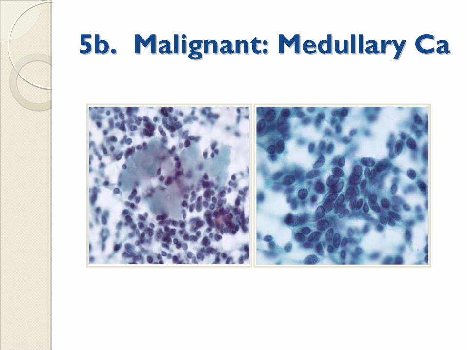
5b. Malignant: Medullary Ca5b. Malignant: Medullary Ca
Metastatic carcinomaMetastatic carcinoma
Rare -
<10% of thyroid neoplasms
sampled by FNA
Multiple/solitary lesion/s
Most common primaries –
Breast, Lung,
Kidney, Colon, Melanoma
History of primary neoplasm is usually present
6. Non6. Non--diagnosticdiagnostic
Processed and examined, but non-
diagnostic due to ◦
limited cellularity◦
no follicular cells or ◦
poor fixation and preservation
Repeat FNA can be recommended in these cases (6-18 mos)
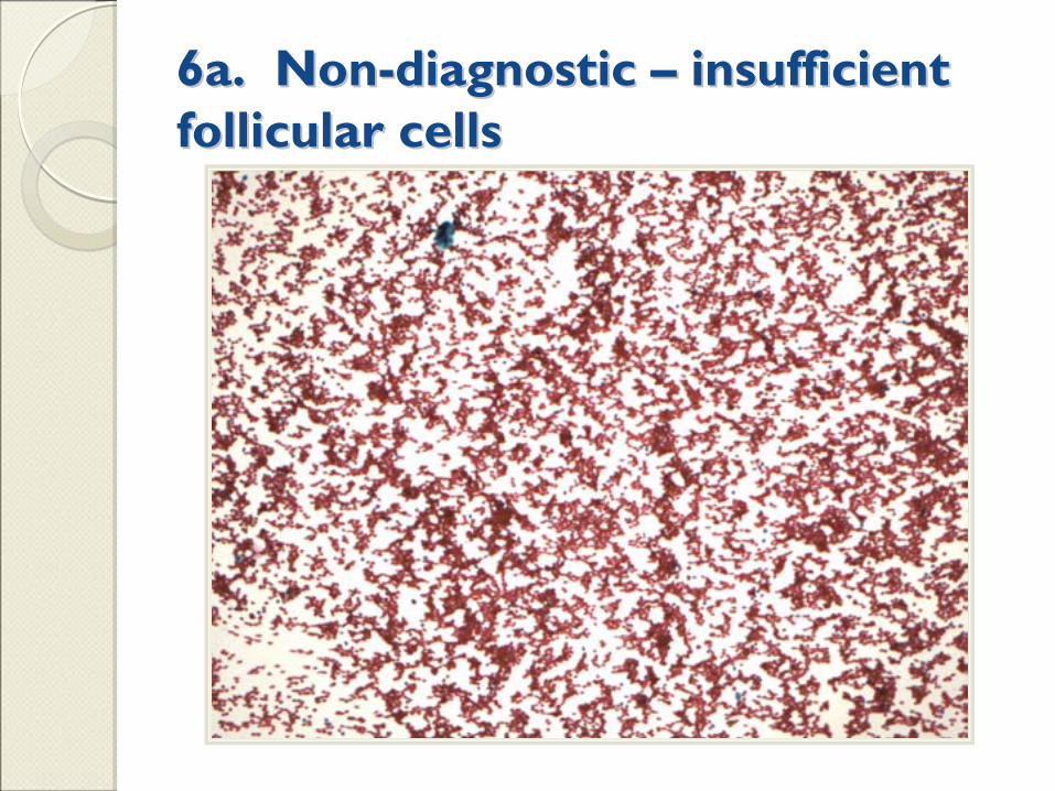
6a. Non6a. Non--diagnostic diagnostic ––
insufficient insufficient follicular cellsfollicular cells
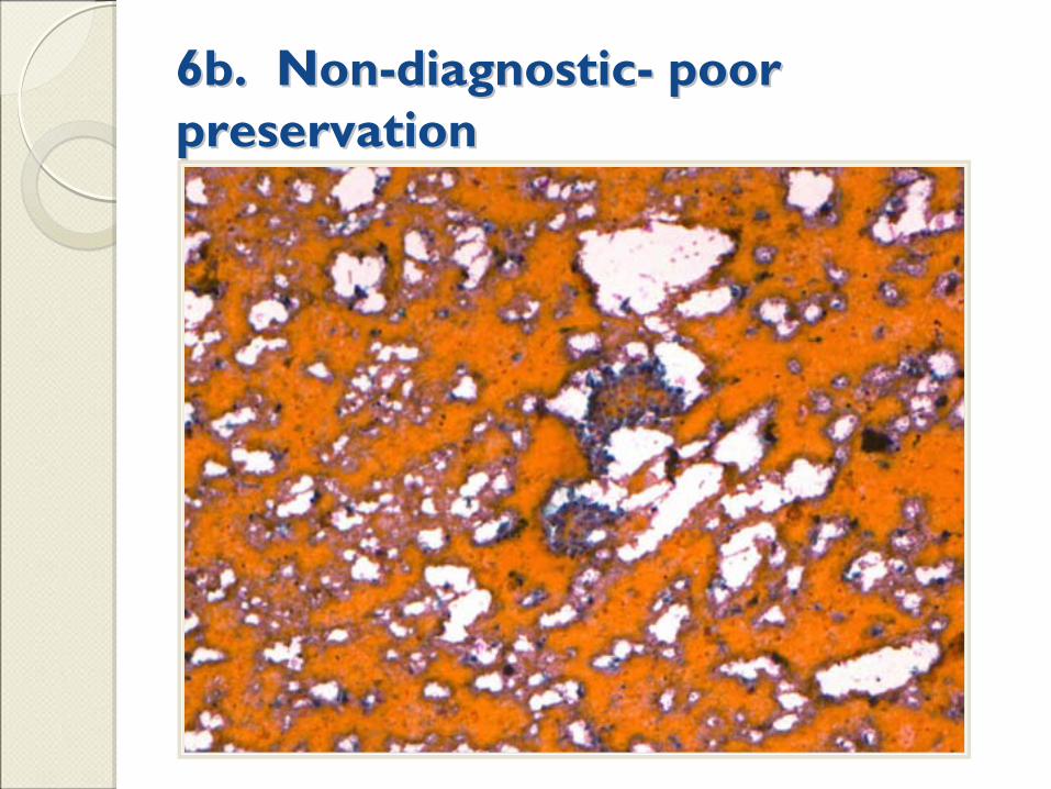
6b. Non6b. Non--diagnosticdiagnostic--
poor poor preservationpreservation
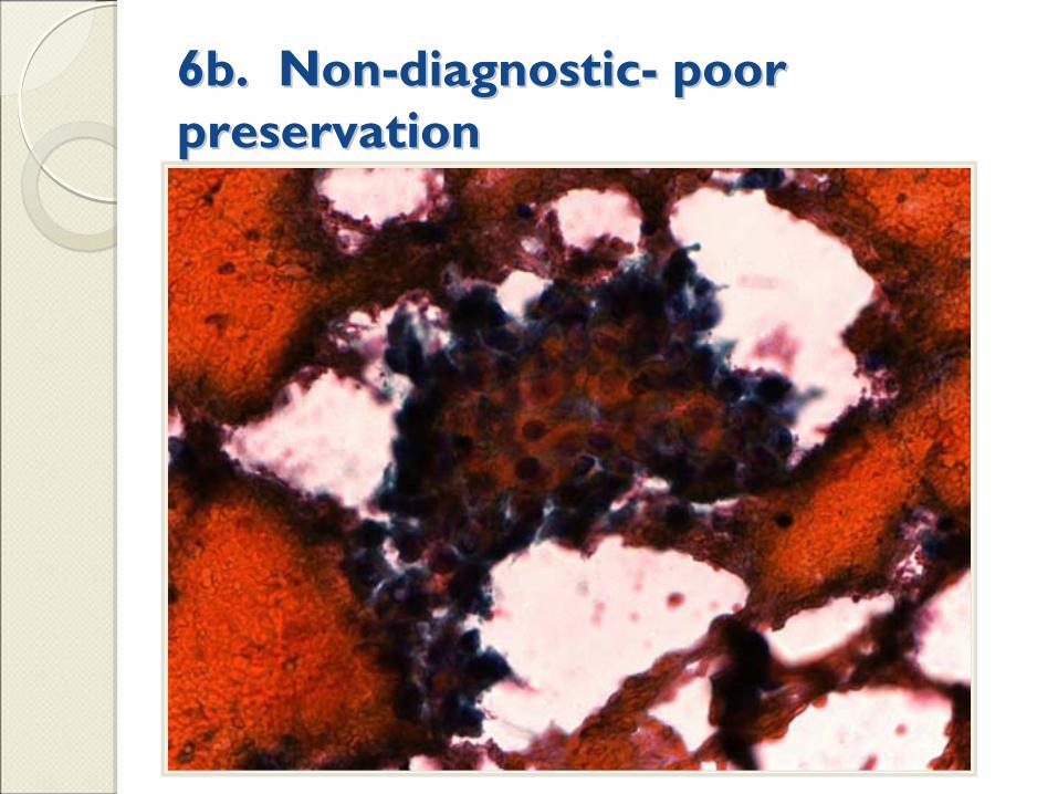
6b. Non6b. Non--diagnosticdiagnostic--
poor poor preservationpreservation
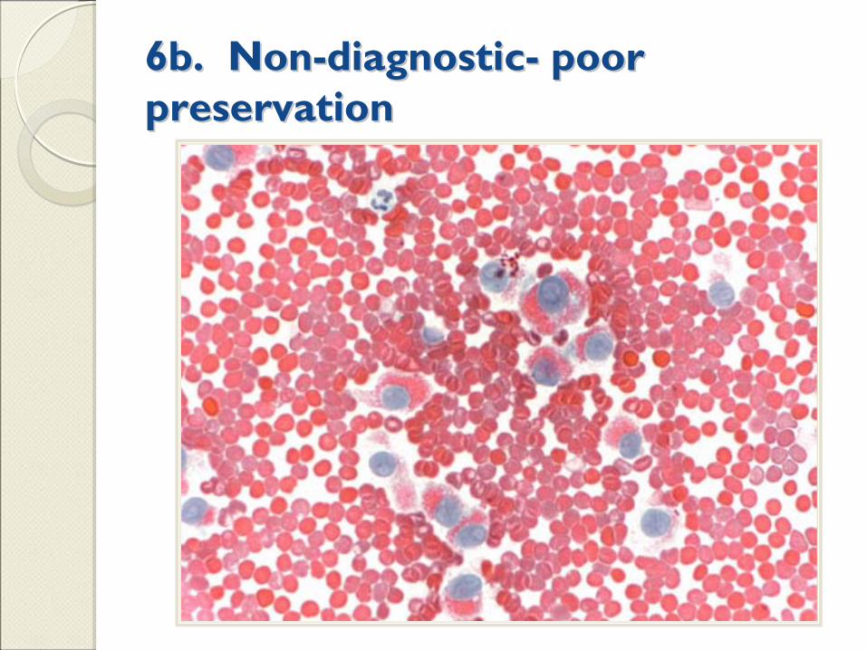
6b. Non6b. Non--diagnosticdiagnostic--
poor poor preservationpreservation
AdequacyAdequacy
All thyroid FNAs must be technically adequate with well preserved and well prepared tissue for examination
Solid nodules with less than abundant colloid:◦
5-6 groups with at least 10 cells is recommended
Minimum no. of follicular cells are not required◦
Smears with abundant thick colloid◦
Inflammatory processes eg thyroiditis◦
Cystic lesions
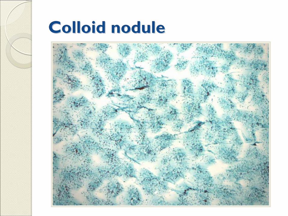
Colloid noduleColloid nodule
Cystic lesionsCystic lesions
Most commonly occur as a result of cystic degeneration of adenomatous nodule
Low risk of malignancy (1-4%) in simple non complex cysts
Higher risk of malignancy (~14%) in ◦
Mixed cystic and solid nodules◦
Cysts > 3cm◦
Recurring cysts
Of all
aspirated cysts 1% are malignant
Cystic lesion contd.Cystic lesion contd.
Because of low potential of false negative it is recommend that cysts be interpreted as
Negative for malignancy
Clinical and radiologic correlation is a must
Cyst size, complexity
Disclaimer of cystic papillary ca
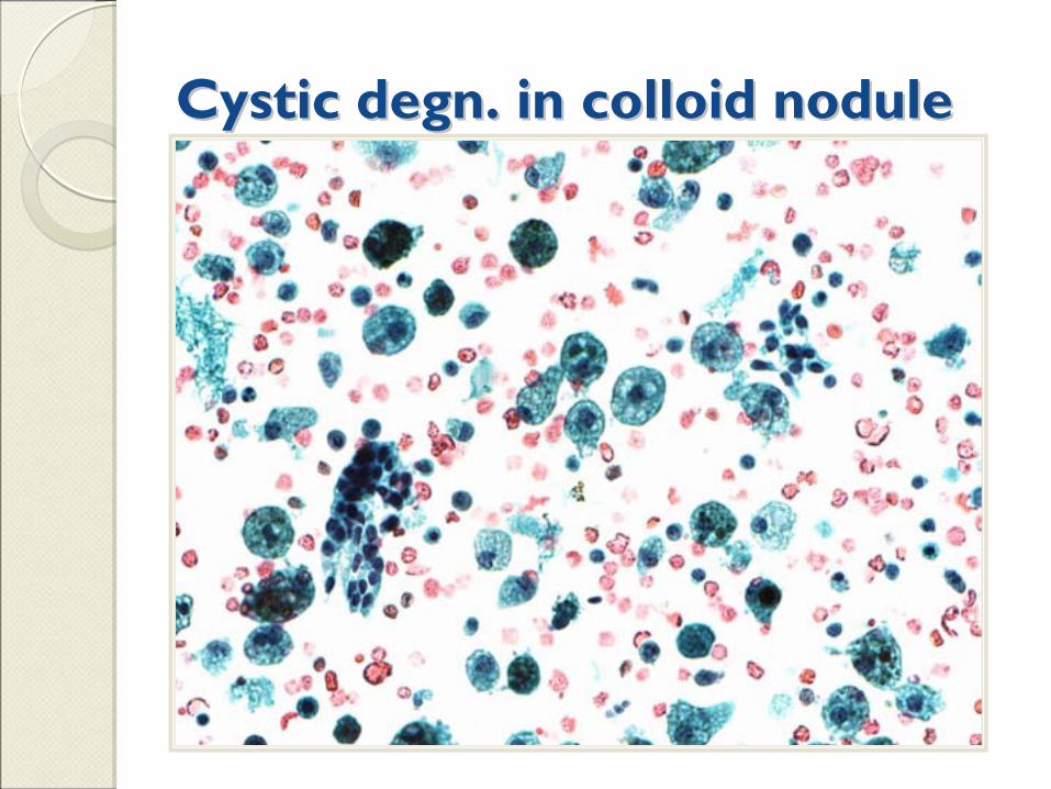
Cystic degn. in colloid noduleCystic degn. in colloid nodule
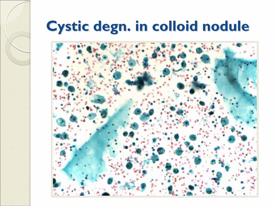
Cystic degn. in colloid noduleCystic degn. in colloid nodule
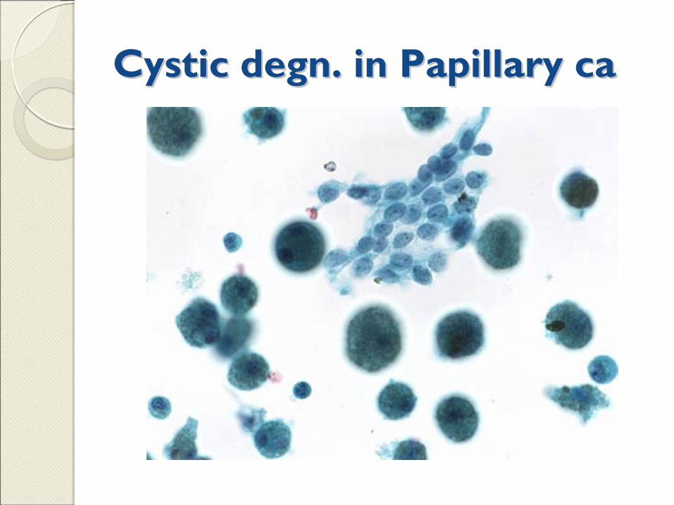
Cystic degn. in Papillary caCystic degn. in Papillary ca
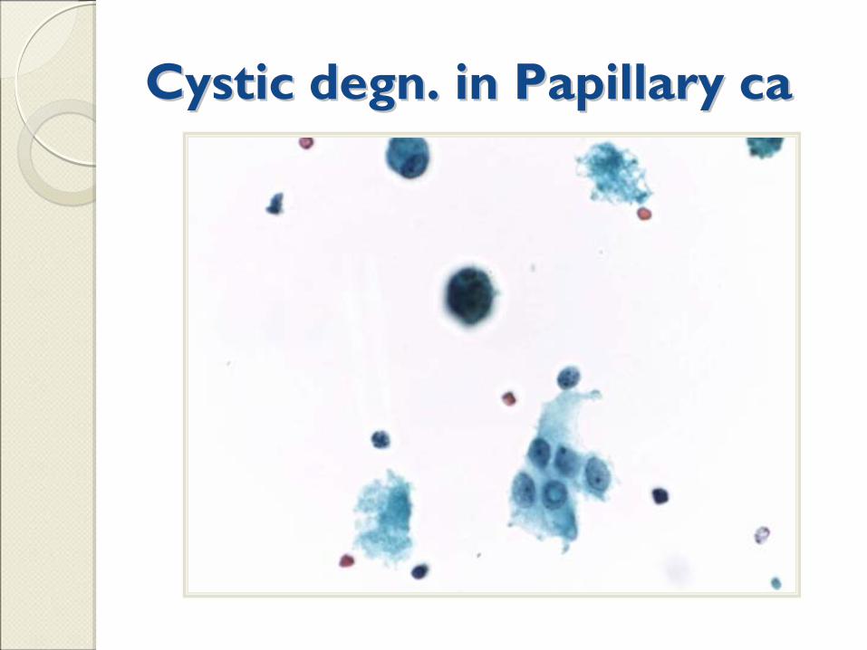
Cystic degn. in Papillary caCystic degn. in Papillary ca
Causes of Diagnostic FailuresCauses of Diagnostic Failures
Unsatisfactory samples ~ 50%
Remaining 50%:◦
short comings in interpretation of adequate samples or◦
pathologists issuing diagnoses on samples with inadequate material
Thus unsatisfactory specimens were the cause or a contributing factor in the majority of failed diagnoses.
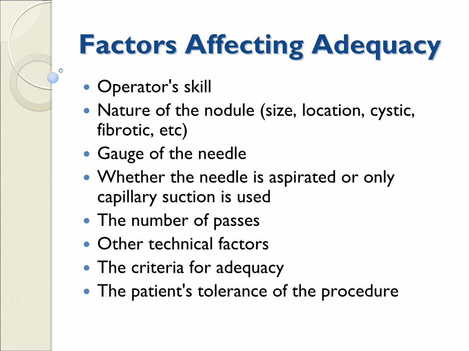
Factors Affecting AdequacyFactors Affecting Adequacy
Operator's skill
Nature of the nodule (size, location, cystic, fibrotic, etc)
Gauge of the needle
Whether the needle is aspirated or only capillary suction is used
The number of passes
Other technical factors
The criteria for adequacy
The patient's tolerance of the procedure
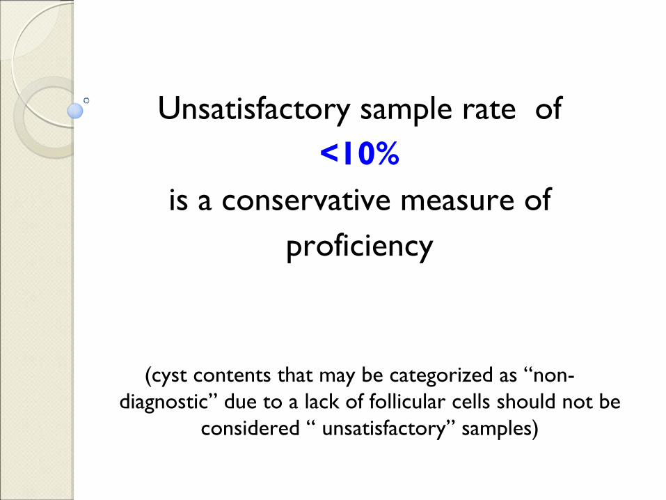
Unsatisfactory sample rate of <10%
is a conservative measure of proficiency
(cyst contents that may be categorized as “non- diagnostic”
due to a lack of follicular cells should not be
considered “
unsatisfactory”
samples)
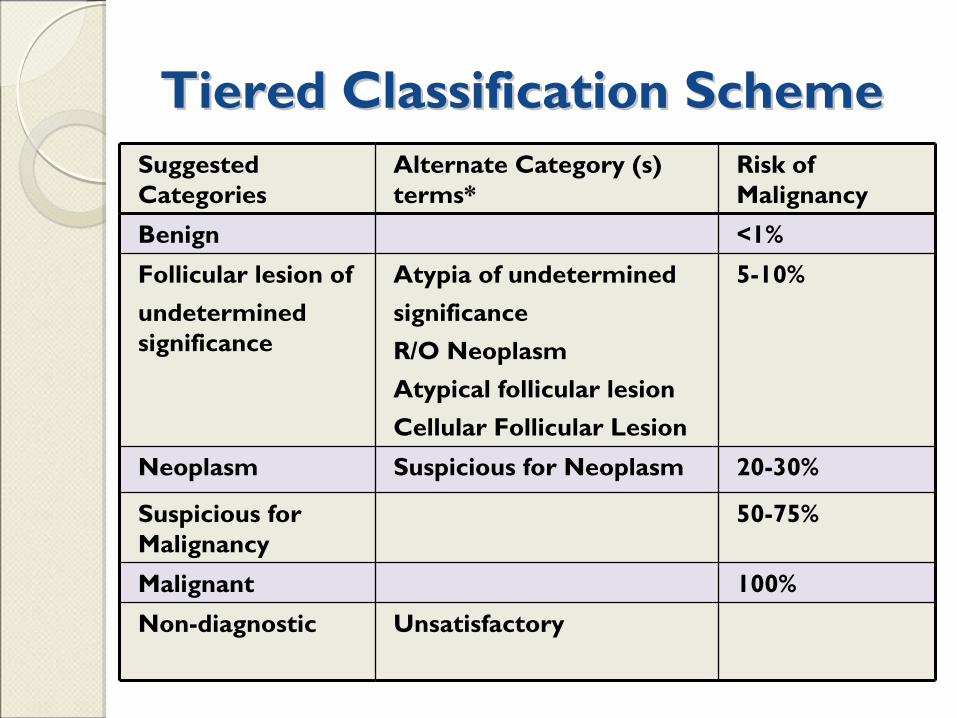
Tiered Classification SchemeTiered Classification SchemeSuggested Categories
Alternate Category (s) terms*
Risk of Malignancy
Benign <1%
Follicular lesion ofundetermined significance
Atypia
of undeterminedsignificanceR/O NeoplasmAtypical follicular lesionCellular Follicular Lesion
5-10%
Neoplasm Suspicious for Neoplasm 20-30%
Suspicious for Malignancy
50-75%
Malignant 100%
Non-diagnostic Unsatisfactory
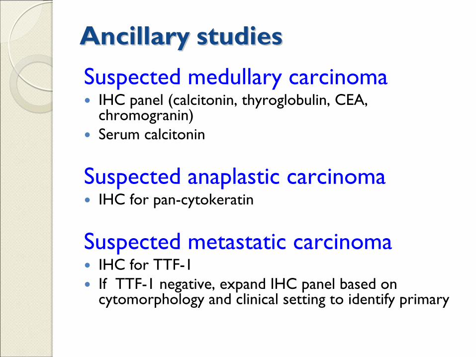
AncillaryAncillary
studiesstudiesSuspected medullary
carcinoma
IHC panel (calcitonin, thyroglobulin, CEA, chromogranin)
Serum calcitonin
Suspected anaplastic
carcinoma
IHC for pan-cytokeratin
Suspected metastatic
carcinoma
IHC for TTF-1
If TTF-1 negative, expand IHC panel based on cytomorphology
and clinical setting to identify primary
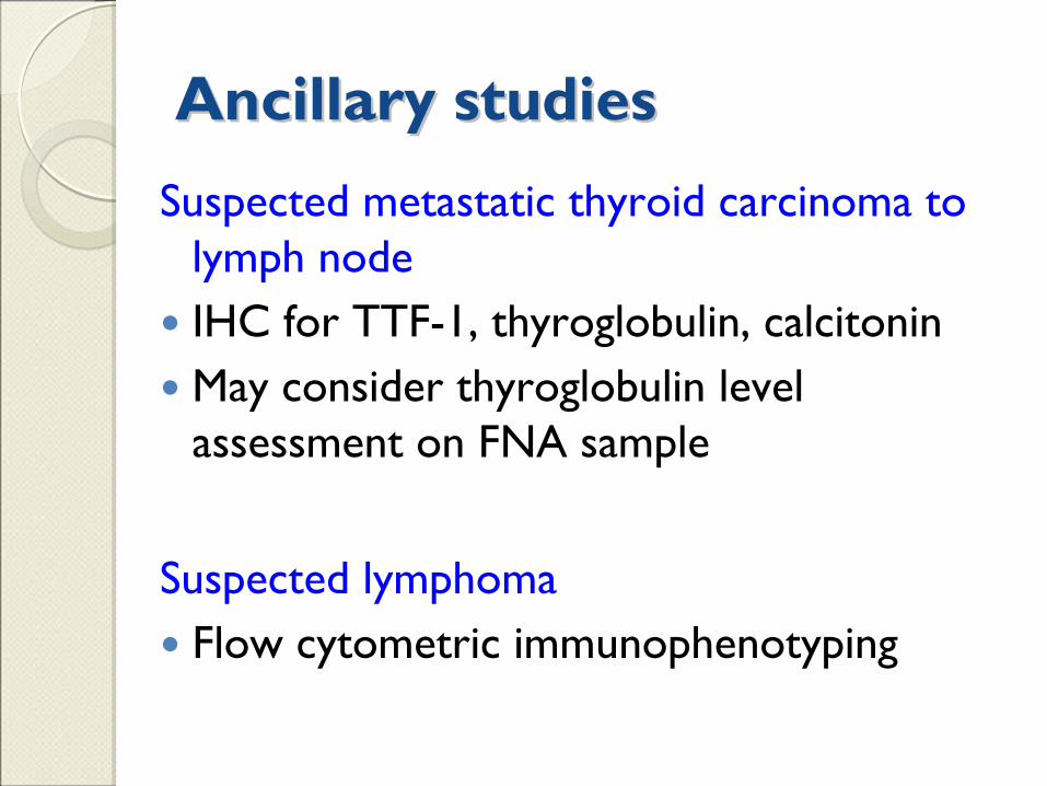
Ancillary studiesAncillary studiesSuspected metastatic
thyroid carcinoma to
lymph node
IHC for TTF-1, thyroglobulin, calcitonin
May consider thyroglobulin
level
assessment on FNA sample
Suspected lymphoma
Flow cytometric
immunophenotyping


![Cytopathologic diagnosis of fine needle aspiration …...Thyroid FNA State of the Science Conference with a group of experts at Bethesda, MD, in October 2007[7]. This conference established](https://img.pdfslide.net/doc/110x75/5f58ba8c2659e94ec243e3b2/cytopathologic-diagnosis-of-fine-needle-aspiration-thyroid-fna-state-of-the.jpg)