Embed Size (px)
Citation preview
Urinary Calcium Excretion in FamilialHypocalciuric HypercalcemiaPERSISTENCE OF RELATIVE HYPOCALCIURIA AFTER
INDUCTION OF HYPOPARATHYROIDISM
M. F. ATTIE, J. R. GILL, JR., J. L. STOCK, A. M. SPIEGEL, R. W. DOWNS,JR.,M. A. LEVINE, and S. J. MARX, Metabolic Diseases Branch, National Instituteof Arthritis, Diabetes, and Digestive and Kidney Diseases, Bethesda,Maryland 20205; Hypertension Endocrine Branch, National Heart Lung andBlood Institute, Bethesda, Maryland 20205
A B S T R A C T Familial hypocalciuric hypercalcemia(FHH) is an autosomal dominant trait comprising hy-percalcemia, hypophosphatemia, parathyroid hyper-plasia, and unusually low renal clearance of calcium.Weevaluated the role of parathyroid hormone in therelative hypocalciuria of FHH and characterized therenal transport of calcium in this disorder using threepreviously hypercalcemic FHHpatients with surgicalhypoparathyroidism and three controls with surgicalhypoparathyroidism. Intravenous infusion of calciumchloride in two patients with FHHand in three con-trols increased serum calcium from a mean basal of5.0 to a mean peak of 6.8 meq/liter in two FHH pa-tients and from 4.2 to 5.7 in three control subjects.Urinary calcium in a third FHH patient was studiedwithout calcium infusion during recovery from hy-percalcemia of vitamin D intoxication. At all serumconcentrations of calcium, calcium clearance waslower in FHH than in controls; at base-line serum cal-cium, the ratio of calcium clearance to inulin clearance(CCa/CIN) in FHHsubjects was 32% of that in controlsand decreased to 19% during hypercalcemia. Calciuminfusion increased the ratio of sodium clearance to in-ulin clearance in controls from a base line of 0.020 to0.053 at peak concentrations of calcium in serum, butdid not affect this parameter in FHH (0.017 at base-line serum calcium vs. 0.019 at peak).
This work was presented in part at the plenary session ofthe Annual Meeting of the American Federation for ClinicalResearch, 1980 and published in abstract form in 1980, Clin.Res., 28:384A.
Dr. Attie's current address is Endocrine Section, Depart-ment of Medicine, University of Pennsylvania, Philadelphia,PA 19104.
Received for publication 18 May 1982 and in revised form7 February 1983.
When calcium infusion studies were performed (intwo patients with FHH and one control) during ad-ministration of acetazolamide, a drug whose principalrenal action causes inhibition of proximal transport ofsolute, CCa/CIN in the patients with FHHwas 29 and7% of that of the control at base-line and peak serumcalcium, respectively. In contrast, ethacrynic acid, adiuretic that acts in the ascending limb of the loop ofHenle, increased CCa/CIN more in the FHH patientsthan in the control subject; CCa/CIN was 65% at base-line and 47% at peak serum calcium, compared withthat of the control subject. The greater calciuric re-sponse to ethacrynic acid than to acetazolamide orcalcium infusion alone in FHH indicates that a majorrenal locus of abnormal calcium transport in this dis-order may be the ascending limb of the loop of Henle.
Decreased clearance of calcium in patients withFHH and hypoparathyroidism when compared withhypoparathyroid controls indicates that relative hy-pocalciuria in FHH is not dependent on hyperpara-thyroidism. Since the parathyroid glands in FHHarenot appropriately suppressed by calcium, this impliesthat FHH represents a disorder of abnormal transportof, and/or response to, extracellular calcium in at leasttwo organs, parathyroid gland and kidney.
INTRODUCTION
Familial hypocalciuric hypercalcemia (FHH)' or fa-milial benign hypercalcemia is an autosomal disorderthat resembles typical primary hyperparathyroidism(1, 2); patients with either disorder have hypercal-
1 Abbreviations used in this paper: FHH, familial hypo-calciuric hypercalcemia; hPTH, human PTH; PTH, para-thyroid hormone.
The Journal of Clinical Investigation Volume 72 August 1983. 667-676 667
cemia and may have hypophosphatemia (3, 4). Al-though patients with FHH and patients with typicalprimary hyperparathyroidism exhibit similar elevationin serum concentration of total ultrafiltrable calciumand similar creatinine clearance (and thus similar fil-trable loads of calcium) (3), patients with FHH havelower urinary excretion rates for calcium, indicatinga lower renal clearance of calcium in FHH (3-5).Renal transport of magnesium also differs; in FHH,there is a normal or high serum concentration of mag-nesium (total and ultrafiltrable), reflecting a renalclearance of magnesium lower than that in typicalprimary hyperparathyroidism (3).
The pathogenesis of the low renal clearance of cal-cium and, in particular, the role of parathyroid hor-mone (PTH) in this process have not been elucidated.Although PTH decreases renal clearance of calciumand magnesium (6-9), indices of circulating PTH(plasma concentration of PTH fragments by immu-noassay, urinary cyclic AMP [cAMP] excretion, andrenal tubular maximum of phosphate transport cor-rected for glomerular filtration rate [TmP/GFR]) areless abnormal in FHH than in typical primary hyper-parathyroidism at any level of serum calcium (4, 10).Thus, factors other than merely the concentration ofcirculating PTH may determine the abnormal renaltransport of divalent cations in FHH.
Attempts at total or subtotal parathyroidectomy inFHHoccasionally have led to profound hypocalcemia(2) and a state resembling hypoparathyroidism (seebelow). In typical hypoparathyroidism, the absence ofPTHeffect on renal tubular calcium reabsorption leadsto a high renal clearance of calcium (6, 8, 11). Wecompared the renal handling of calcium in a groupwith surgical hypoparathyroidism and FHHwith thatin a control group with surgical hypoparathyroidism.The low renal clearance of calcium characteristic ofFHH persists even upon development of hypopara-thyroidism. Moreover, our findings in this group withinfusion of diuretics suggest that the loop of Henle isa possible locus for this abnormal calcium transport.
METHODS
SubjectsWestudied three patients with FHH, who had been referredto the National Institutes of Health (NIH) after having beenrendered hypocalcemic by prior parathyroidectomy, andthree control subjects with postsurgical hypoparathyroidism.Criteria for hypoparathyroidism were as follows: Serum cal-cium was <3.75 meq/liter in the absence of calcium and/or vitamin D supplementation on one or more occasions atleast 6 mo after parathyroidectomy, urinary cAMPwas notelevated during hypocalcemia, and plasma immunoreactivePTH was undetectable at a time of hypocalcemia. The threesubjects with FHHwere previously hypercalcemic membersof typical FHH kindreds.
Subject 1. This 17-yr-old male had been hospitalized at14 d of age with dehydration. The serum calcium was 10.5meq/liter and the serum phosphorus 3.9 mg/100 ml. At neckexploration, three of four hyperplastic parathyroid glandswere removed, but severe hypercalcemia recurred and 1 molater the remaining hyperplastic gland was excised (12).Since that time, he has required calcium and vitamin Dsupplementation to maintain a serum calcium concentration> 4.0 meq/liter. Serum concentrations of calcium have beenbetween 3.5 and 3.8 meq/liter at times of poor compliance.At least 11 other members of this patient's family had hy-percalcemia; urinary calcium, evaluated in five hypercal-cemic members, was <7 meq/d in all (family 0 in reference2, family B in reference 13).
Subject 2. During an evaluation of paresthesiae in 1976,this 28-yr-old male had been found to have a serum con-centration of calcium of 7.0 meq/liter and a serum phos-phorus ranging between 1.7 and 2.1 mg/100 ml. He wasotherwise asymptomatic. At neck exploration, 33/4 hyper-plastic parathyroid glands were excised (total weight 1.6 g)but hypercalcemia persisted. At a subsequent exploration,the gland remnant was removed. Postoperatively, the serumcalcium remained low, despite oral calcium supplements.During periods of poor compliance, the serum calcium hasdropped as low as 3.6 meq/liter. Three other members ofthis subject's family (family N in reference 2) have had hy-percalcemia without hypercalciuria.
Subject 3. This 7-yr-old female was hypercalcemic atbirth (7 meq/liter), with a serum magnesium of 2.34 meq/liter, and a serum phosphorus of 3.4 mg/100 ml. Urine cal-cium was <1 meq/d. Radiographs showed subperiosteal re-sorption and diffuse undermineralization. At 2 wk of age,four hyperplastic parathyroid glands were excised. Since thattime, she has required calcium and vitamin D. Without thesemedications the serum calcium concentration has been aslow as 2.7 meq/liter. This subject and her family have beendescribed in detail (13, 14). Urine calcium excretion in 13of her hypercalcemic relatives was in the range characteristicof FHH (13).
Two of the three control subjects became hypoparathyroidafter total thyroidectomy for nonmetastatic thyroid carci-noma (subjects 4 and 5). A third (subject 6), with primaryhyperplasia, became hypoparathyroid after a second neckexploration 8 mo before study. His brother had primaryhyperparathyroidism and Cushing's disease, which suggeststhat they have multiple endocrine neoplasia, Type I. Noneof the controls had had known episodes of vitamin D intox-ication or urinary tract infections. None of the subjects washypertensive, nor were they taking drugs (other than cal-ciferol) known to alter renal handling of calcium.
Study protocolThe adult subjects were admitted to a metabolic ward andgiven a diet containing "40 meq (800 mg) of calcium, 800-1,000 mg phosphorus, and 4-6 g sodium chloride/d. Dietarycalcium was supplemented with calcium containing medi-cations (Table I). Informed consent was obtained for all ex-perimental procedures.
Serum and urinary electrolyte and phosphorus concentra-tions and serum calcium concentrations were determined ina Technicon-SMAC (Technicon Instruments Corp., Tarry-town, NY) in the Clinical Chemistry Laboratory of the Clin-ical Center, NIH. Serum and urinary magnesium and uri-nary calcium were analyzed by atomic absorption spectro-photometry (model 5000, Perkin-Elmer Corp., InstrumentDiv., Norwalk, CT). Normal ranges (2 SD) for serum com-
668 Attie, Gill, Stock, Spiegel, Downs, Levine, and Marx
ponents were determined from 250 blood donors at the Clin-ical Center Blood Bank. cAMP in urine was measured byradioimmunoassay (10). PTH in plasma was analyzed witha radioimmunoassay specific for the midportion of the mol-ecule, using purified human PTH (hPTH) as standard (15);the normal range was undetectable to 0.24 ng eq hPTH/ml.The detection limit was 0.10 ng eq hPTH/ml and PTH wasdetectable in plasma from 50% of normal subjects. Inulinconcentration was determined in serum and urine by pre-viously described rmethods (16).
Calcium, phosphorus, sodium, and creatinine were ana-lyzed in specimens from 24-h urine collections. Calcium,phosphorus, magnesium, and electrolytes in serum and PTHin plasma were determined in samples obtained after over-night fast. cAMP was analyzed by radioimmunoassay in atleast four sequential 12-h urine collections and adjusted forglomerular filtration rate as follows (17): (UCAMP X SCr)/U,r,where UC,AMP is the urinary concentration of cAMP, and Scrand U,. are serum and urine concentrations of creatinine,respectively.
Clearances of inulin, creatinine, calcium, phosphorus, andsodium were calculated in the usual manner. No adjustmentwas made for ultrafiltrability of calcium in serum. Data wereevaluated using the t test, two-way analysis of variance, andlinear least-squares regression (18).
Clearance studies were performed after an overnight fastwith the subjects recumbent except when voiding. To main-tain urine flow, they drank 200 ml of distilled water at 0700h and then every 30 min for the remainder of the study.Infusion of inulin (35 mg/kg body weight loading dose, fol-lowed by a sustaining infusion consisting of 0.07 mg/kg in250 ml normal saline at 1.1 ml/min) was started at 0800 h.Between 0730 and 0830 h, 5 ml/kg of 0.15 Msodium chlo-ride was given to expand extracellular fluid. Calcium chlo-ride (100 meq elemental calcium/liter of 0.075 M sodiumchloride) was infused at two rates: 75-100 Aeq elementalcalcium/kg -h and 100-160 Ueq/kg -h. Urine from 20-minclearance periods was obtained before infusion of calcium(0830-0910 h) and during each rate of calcium infusion,after allowing 30 min for serum calcium to stabilize. Venousblood was obtained through an indwelling catheter withouthemostasis (in an antecubital vein contralateral to the in-fusion catheter) at the midpoint of each urine collectionperiod for determination of serum concentrations of calcium,sodium, and inulin.
The effects of diuretics were assessed with a modificationof the calcium infusion protocol. Beginning at 0700 h, 200ml of distilled water was ingested every 30 min and 5ml/kg body weight of 0.15 M sodium chloride was givenover a period of 1 h. Inulin was given as described above.
At 0800 h, a loading dose of either ethacrynic acid (0.25mg/kg) or acetazolamide (2.5 mg/kg) was given, followedby maintenance infusion (0.2 mg/kg h ethacrynic acid or2.5 mg/kg h acetazolamide) for the remainder of the in-fusion. Urinary losses of sodium, potassium, and bicarbonatewere estimated from urinary volume and rapid measure-ment of concentrations of urinary sodium and potassium(results obtained within 15 min) and were quantitativelyreplaced with intravenous solutions containing either 0.15or 0.075 Msodium chloride and variable amounts of potas-sium chloride. Clearance periods were 15 min with a mid-point blood collection. Base-line collections were started af-ter urinary volume varied <20% in two consecutive collec-tions: This generally required 45-60 min. Calcium chloridewas then infused at the two rates as described above and two15-min clearance periods were obtained after allowing 30min for serum calcium to stabilize.
RESULTS
Metabolic indices during steady state. With theirusual calcium and vitamin D supplementation (TableI), FHH patients and controls were mildly hypocal-cemic (Table II). Mean serum phosphorous was highin the three FHHpatients and in two of three controlsubjects. Urinary excretion of phosphorus for the twogroups was similar. The ratio of phosphorus clearanceto creatinine clearance was similarly low in bothgroups. Taken together, these data indicate a highthreshold for renal excretion of phosphorus in hypo-parathyroid patients with FHH, in contrast to the lowthreshold in patients with FHH and hypercalcemia(10). At a time when serum calcium was low, immu-noreactive PTH was below the lower limit of detect-ability and urinary cAMPwas not elevated. Thus, bystandard indices of circulating PTH (renal phosphatetransport, plasma immunoreactive PTH, and urinarycAMP), patients with FHHwere similar to the controlsubjects.
Table III shows means of serum and urinary calciumand magnesium, urinary sodium, and creatinine clear-ance obtained throughout the hospitalization. Subjectsin both groups had similarly low to normal serum con-
TABLE IMedications for Treatment of Hypoparathyroidism in Patients and Controls
Subject Group Age Sex Daily calciferol supplement Daily calcium supplement'
yr
1 FHH 18 M 1.25 mg Ergocalciferol 34 g Calcium gluconate (3 g)2 FHH 32 M 1.25 mg Ergocalciferol 10 g Calcium gluconate (0.9 g)3 FHH 7 F 1.25 mg Ergocalciferol 11 g Calcium glubionate (0.7 g)4 Control 38 F 0.25 mg Dihydrotachysterol 4 g Calcium gluconate (0.4 g)5 Control 45 F 1.88 mg Ergocalciferol 9.6 g Calcium lactate (1.2 g)6 Control 22 M 0.5 jig 1,25-Dihydroxycholecalciferol 30 g Calcium gluconate (2.7 g)
Calcium as elemental calcium is given in parentheses.
Hypocalciuria in Hypercalcemia with Hypoparathyroidism 669
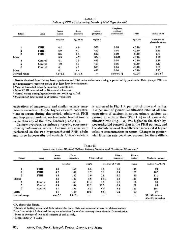
TABLE IIIndices of PTHActivity during Periods of Mild Hypocalcemia'
PhosphorusSerum Serum Urinary creatinine
Subject Group calcium phosphorus phosphorus clearance ratio PTH Urinary cAMP
meq/liter mg/1OO ml mg/24 h ng eq/ml nmol/100 mlglomerular filtrate
1 FHH 4.2 4.6 558 0.05 <0.10 1.822 FHH 3.9 4.7 490 0.04 <0.10 2.453 FHH 3.5 6.5 322 0.09 <0.10 1.61
Mean 3.9 4.71 524t 0.051 <0.10 1.964 Control 4.1 3.5 469 0.03 <0.10 1.865 Control 4.0 5.1 453 0.05 <0.10 ND6 Control 4.1 4.7 388 0.04 <0.10 2.67
Mean 4.1 4.4 436 0.04 <0.10 2.27Normal range 4.5-5.3 2.1-3.8 - 0.08-0.17§ <0.2411 1.2-3.61
a Results obtained from fasting blood specimens and 24-h urine collections during a period of hypocalcemia. Data (except PTH ra-dioimmunoassay) represent means of at least four determinations.I Mean of two adult subjects (numbers 1 and 2) only.§ Mean±2 SD determined in 10 normal volunteers.
Normal values during hypocalcemia are >0.24 ng eq/ml.¶f Mean±2 SD determined in 20 normal volunteers.
centrations of magnesium and similar urinary mag-nesium excretion. Despite higher calcium concentra-tions in serum during this period, adults with FHHand hypoparathyroidism each excreted less calcium inurine than any of the three controls (Table III).
Calcium transport by kidney at varying concentra-tions of calcium in serum. Calcium infusions wereperformed on the two hypoparathyroid FHH adultsand three hypoparathyroid controls. Urinary calcium
is expressed in Fig. 1 A per unit of time and in Fig.1 B per unit of glomerular filtration rate. At all con-centrations of calcium in serum, urinary calcium ex-pressed in units of time (Fig. 1 A) or of glomerularfiltration rate (Fig. 1 B) was higher in the three hy-poparathyroid controls than in the FHHpatients, andthe absolute value of this difference increased at highercalcium concentrations in serum. Changes in glomer-ular filtration rate could not account for these differ-
TABLE IIISerum and Urine Divalent Cations, Urinary Sodium, and Creatinine Clearances'
Serum Serum Urinary UrinarySubject Group calcium magnesium Urinary calcium magnesium sodium Creatinine clearance
meq/ltter meq/d meqlliter GF X 100 men/d ml/(min X 1.73 m')
1 FHH 4.8 1.55 5.5 3.5 6.2 118 992 FHH 4.2 1.39 1.7 1.1 5.4 167 1073 FHH 3.5 1.30 1.6 1.8 5.0 80 111
Meant 4.5 1.47 3.6 2.3§ 5.8 143 1034 Control 4.2 1.53 11.4 7.5 6.7 86 985 Control 3.9 1.34 12.2 11.5 6.4 69 856 Control 4.1 1.57 8.2 6.0 5.4 142 107
Mean 4.1 1.48 10.6 8.3§ 6.2 99 97Normal range 4.4-5.3 1.4-2.1 - - - 97-140 (males)
85-125 (females)
GF, glomerular filtrate.° Results of fasting serum and 24-h urine collections. Data are means of at least six determinations.Data from subject 3 obtained during an admission 5 mo after recovery from vitamin D intoxication.I Mean is average of two adult subjects (1 and 2) only.§ Means differ P < 0.025.
670 Attie, Gill, Stock, Spiegel, Downs, Levine, and Marx

10 r
0
x
C
E
E
0
0
cc
zcc
0o A
8 h
6
4
2
0
A
05Jpo
060 A
0 AA 6 A A A
O ° t A
I Mm 4- Ai4 54 5 6 7
TOTAL SERUMCALCIUM (meq/1)
80 r-
60 _-
40 k
20 _-
0
0
0 00°0
0 a0
0
m0 A~~~A4 A
4 5 6
TOTAL SERUMCALCIUM (meq/
FIGURE 1 Effect of calcium load on urinary e)
calcium in two FHH subjects (1, *; 2, *) and tiparathyroid controls (4, 0; 5, A; 6, 0) during icalcium chloride. Calcium excretion is expressedtion of time in A and glomerular filtration rate infrom 12- and 24-h urine collections from subjecan episode of vitamin D intoxication are plotted(c) as a function of creatinine clearance. For e
subject 3, each point represents one 20-min clearai(Data from patient 3 were omitted from A becauand small size result in data biased toward lowcalcium.)
ences, because there was little change in inance throughout the infusion in either groinulin clearances in the FHHpatients for thperiods at base-line serum calcium conci
were 107 ml/min, and they were 127 ml/mtwo periods at peak calcium concentration; itrol group clearances were 115 and 104 mlspectively.
Subject 3 was not given the calcium infusicof age. Urinary calciums obtained during
from an episode of vitamin D (dihydrotachysterol) in-toxication associated with serum calcium concentra-tions as high as 7.1 meq/liter are shown in Fig. 1 B.The urinary calcium excretion rate at high serum cal-cium in this subject is significantly different from therate in members of the hypoparathyroid control groupand suggests that calcium transport by the kidney ofsubject 3 is similar to that in the two adults with FHHand hypoparathyroidism.
Relation of calcium clearance to sodium clearance.* Base-line sodium clearance before the calcium infusion
was similar in the two groups, but differed strikinglyduring hypercalcemia (Table IV and Fig. 2). In thecontrol group there was a marked increase in sodiumclearance with increased serum calcium, whereas inthe FHH group there was little increase in sodiumclearance in response to the calcium load (Fig. 2).
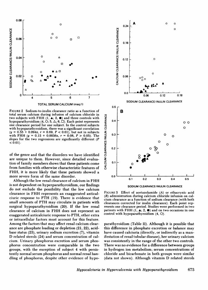
Infusion of diuretics. We further studied renalcalcium transport, using acetazolamide and ethacrynic
B acid, two drugs that alter renal transport at differentsites in the nephron. These studies were performed inthe two adult patients with FHH (subjects 1 and 2)and in one control with hypoparathyroidism (subject4). Base-line sodium excretion rate was affected sim-ilarly by acetazolamide in both groups. However, cal-cium clearance before and during calcium infusionremained lower in the patients with FHH (Table IVand Fig. 3 A). In fact, the absolute value of the dif-
* ference in calcium clearances between groups was in-* creased by the drug-induced inhibition of proximal
sodium reabsorption.Ethacrynic acid produced a greater natriuresis than
7 acetazolamide (Table IV). Natriuresis for the control/l) subject with hypoparathyroidism was, for unknown
reasons, greater than for the two patients with FHH.xcretion of Although the absolute value of the clearance of cal-hree hypo- cium was still higher in the control patient with hy-infusion of 1 .1Ias a fun- poparathyroidism, infusion of ethacrynic acid mark-
B. Results edly increased the ratio of calcium clearance to inulint 3 during clearance in the FHH patients (Table IV); it was in-
only in B creased six- and 23-fold in these two patients at base-ach except line serum calcium and nine- and eightfold at peaknse her age serum calcium vs. fourfold in the control patient at'er urinary both basal and peak serum calcium. In the region of
overlap of sodium clearance (Fig. 3 B), the degree ofcalciuria during ethacrynic acid administration was
ulin clear- similar for patients with FHHand for the control pa-oup. Mean tient.e first twoentrationsin for then the con-
1/min, re-
rn becauserecovery
DISCUSSION
In our three patients with FHH, parathyroidectomyproduced a state resembling chronic hypoparathyroid-ism. As in typical hypoparathyroidism, there was an
increased renal threshold for phosphate excretion anda requirement for pharmacologic doses of calciferols
Hypocalciuria in Hypercalcemia with Hypoparathyroidism
(n01)
0)2_
D .
E> ozr m
z
4
_-
671
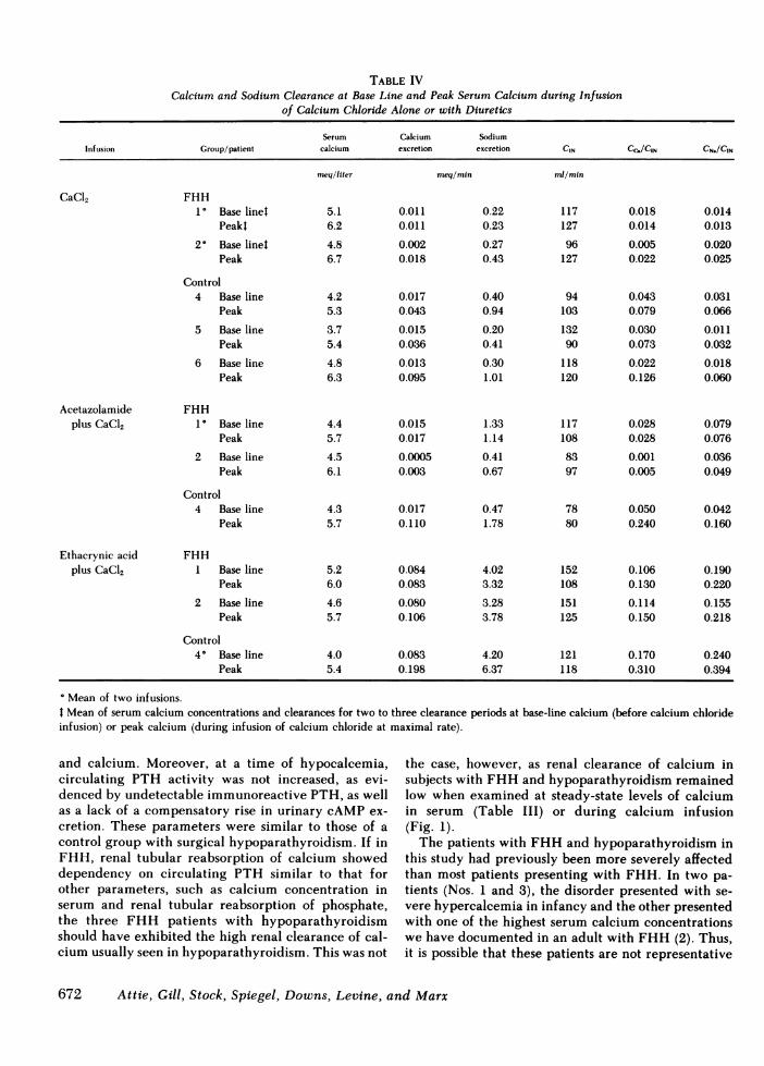
TABLE IVCalcium and Sodium Clearance at Base Line and Peak Serum Calcium during Infusion
of Calcium Chloride Alone or with Diuretics
Serum Calcium SodiumInfusion Group/patient calcium excretion excretion CIN CC,/CIN CN./CIN
FHH1° Base linet
Peakt
2' Base linetPeak
Control4 Base line
Peak5 Base line
Peak6 Base line
Peak
Acetazolamideplus CaCl2
FHH1° Base line
Peak2 Base line
Peak
Control4 Base line
Peak4.3 0.0175.7 0.110
0.471.78
78 0.05080 0.240
FHH1 Base line
Peak2 Base line
Peak
Control4° Base line
Peak
5.26.0
4.65.7
4.05.4
0.0840.083
0.0800.106
0.0830.198
4.023.32
3.283.78
4.206.37
152108
151125
0.1060.130
0.1140.150
121 0.170118 0.310
° Mean of two infusions.I Mean of serum calcium concentrations and clearances for two to three clearance periods at base-line calcium (before calcium chlorideinfusion) or peak calcium (during infusion of calcium chloride at maximal rate).
and calcium. Moreover, at a time of hypocalcemia,circulating PTH activity was not increased, as evi-denced by undetectable immunoreactive PTH, as wellas a lack of a compensatory rise in urinary cAMP ex-
cretion. These parameters were similar to those of a
control group with surgical hypoparathyroidism. If inFHH, renal tubular reabsorption of calcium showeddependency on circulating PTH similar to that forother parameters, such as calcium concentration inserum and renal tubular reabsorption of phosphate,the three FHH patients with hypoparathyroidismshould have exhibited the high renal clearance of cal-cium usually seen in hypoparathyroidism. This was not
the case, however, as renal clearance of calcium insubjects with FHHand hypoparathyroidism remainedlow when examined at steady-state levels of calciumin serum (Table III) or during calcium infusion(Fig. 1).
The patients with FHHand hypoparathyroidism inthis study had previously been more severely affectedthan most patients presenting with FHH. In two pa-
tients (Nos. 1 and 3), the disorder presented with se-
vere hypercalcemia in infancy and the other presentedwith one of the highest serum calcium concentrationswe have documented in an adult with FHH (2). Thus,it is possible that these patients are not representative
672 Attie, Gill, Stock, Spiegel, Downs, Levine, and Marx
CaCl2
meqlliter meq/min ml/min
5.16.2
4.86.7
4.25.3
3.75.4
4.86.3
0.0110.011
0.0020.018
0.0170.043
0.0150.036
0.0130.095
0.220.23
0.270.43
0.400.94
0.200.41
0.301.01
117127
96127
94103
13290
118120
0.0180.014
0.0050.022
0.0430.079
0.0300.073
0.0220.126
0.0140.013
0.0200.025
0.0310.066
0.0110.032
0.0180.060
4.45.7
4.56.1
0.0150.017
0.00050.003
1.331.14
0.410.67
Ethacrynic acidplus CaCl2
117108
8397
0.0280.028
0.0010.005
0.0790.076
0.0360.049
0.0420.160
0.1900.220
0.1550.218
0.2400.394
0.080
0
0 00.06 _
0.041-
0.02
0
00
0 a
0
0 a
80
0t,
0 YO&u
A o aA,* t-. .A A
0
4 5
z
J
4-i
LL
z
zC0z
w-JC.
-J40A A
A
6
0.24A 0 0
0
0.18
0.12
0.06
0
0
000
AAA^ A
U.Uv
0.06 0.127
TOTALSERUMCALCIUM (meq/l)
FIGURE 2 Sodium-to-inulin clearance ratio as a function oftotal serum calcium during infusion of calcium chloride intwo subjects with FHH (1, A; 2, 0) and three controls withhypoparathyroidism (4, 0; 5, A; 6, 0). Each point representsone clearance period for one subject. In the control subjectswith hypoparathyroidism, there was a significant correlation(y = 0.55 + 0.094x, r = 0.69, P < 0.01), but not in subjectswith FHH (y = 0.13 + 0.0036x, r = 0.08, P > 0.05). Theslopes for the two regressions are significantly different (P<0.01).-
of the genre and that the disorders we have identifiedare unique to them. However, since detailed evalua-tion of family members shows that these patients come
from families with otherwise characteristic features ofFHH, it is more likely that these patients showed a
more severe form of the same disorder.Although the low renal clearance of calcium in FHH
is not dependent on hyperparathyroidism, our findingsdo not exclude the possibility that the low calciumclearance in FHH represents an exaggerated antical-ciuric response to PTH (19). There is evidence thatsmall amounts of PTH may circulate in patients withsurgical hypoparathyroidism (20). If the low renalclearance of calcium in FHH does not represent an
exaggerated anticalciuric response to PTH, other extraor intracellular factors must account for this feature.Among the factors that may affect renal calcium clear-ance are phosphate loading or depletion (21, 22), acid-base status (23), urinary sodium excretion (7), vitaminD related sterols (24) and serum concentration of cal-cium. Urinary phosphorus excretion and serum phos-phorus concentration were comparable in the twogroups, with the exception of subject 4 with persis-tently normal serum phosphorus and normal renal han-dling of phosphorus, despite other evidence of hypo-
0.18
SODIUMCLEARANCE/INULIN CLEARANCE
0.5
z
CR
J-j
z
:3z
4
i3
C.)
0.4
0.3
0.2
0.1
0.0
B
0 0
0
0
0
0
0
0
0
* 00
0.1 0.2 0.3 0.4 0.5
SODIUMCLEARANCE/INULIN CLEARANCE
FIGURE 3 Effect of acetazolamide (A) or ethacrynic acid(B) administration during calcium chloride infusion on cal-cium clearance as a function of sodium clearance (with bothclearances corrected for inulin clearance). Each point rep-resents one clearance period. Studies were performed in twopatients with FHH (1, A; 2, 0) and on two occasions in one
control with hypoparathyroidism (4, 0).
parathyroidism (Table II). Although it is possible thatthis difference in phosphate excretion or balance mayhave caused calciuria (directly, or indirectly as a man-
if estation of renal tubular disease), her urinary calciumwas consistently in the range of the other two controls.There was no evidence for a difference between groupsin hydrogen ion metabolism; serum concentrations ofchloride and bicarbonate in both groups were similar(data not shown). Although vitamin D related sterols
Hypocalciuria in Hypercalcemia with Hypoparathyroidism
z
Cr
uJ-J
z
z
zCR
-J
C]
0U,
n n .f X -
'.A
673
in some studies increase renal tubular reabsorption ofcalcium, this effect has not been found consistently andeven opposite effects have been found (24). In thisstudy, all subjects were taking pharmacologic doses ofvitamin D. Circulating levels of calciferols were notmeasured, but it is not likely that the FHH patientshad higher circulating levels of active vitamin D me-tabolites, since oral calcium requirements were similarin both groups.
From the data in Fig. 1, it appears that for anyserum concentration of calcium or filtered load of cal-cium, tubular reabsorption of calcium is greater withFHH than in the controls. Infusion of diuretics whichinhibit calcium transport in specific regions of thenephron provide an opportunity to localize sites of in-creased calcium reabsorption. Infusion of calcium to-gether with acetazolamide increased the calciumclearance in the control patient, but produced virtuallyno change in the patients with FHH (Table IV). Thissuggests that the load of calcium delivered to the distalnephron as a result of calcium infusion and inhibitionof proximal tubular reabsorption by acetazolamideexceeded the capacity of the distal segment to reabsorbcalcium in the control but not in the patients withFHH (25, 26). This also suggests that the principal siteof enhanced renal tubular reabsorption of calcium inFHH is not in the proximal tubule.
Ethacrynic acid, which inhibits sodium and calciumtransport primarily in the thick ascending limb of theloop of Henle (26, 27), increased calcium clearance inthe patients with FHH and in the control patient(Table IV). Although calcium clearance remainedhigher in the control than in the FHH patients afterethacrynic acid, the diuretic had a much greater effecton calcium clearance in the FHHsubjects, so that thediscrepancy between the two groups was proportion-ately less (Table IV). Sodium clearance after ethac-rynic acid infusion was also greater in the control thanin the patients with FHH, but, in those clearance pe-riods where sodium clearance in the FHHpatients wassimilar to that of the control, calcium clearance wasalso equivalent (Fig. 3 B). This is in striking contrastto the results obtained with infusion of calcium aloneor calcium and acetazolamide (Fig. 3 A); at similarsodium clearances, calcium clearance was considera-bly lower in the FHH patients. These results suggestthat the site of supranormal calcium reabsorption inFHHis the thick ascending limb of the loop of Henle.The greater natriuretic response of the control patientto ethacrynic acid raises the possibility that in FHHother ions such as sodium also are reabsorbed moreavidly at this site. An alternative interpretation of thesefindings is that a site of avid calcium reabsorption dis-tal to the loop of Henle in FHH is saturated by thelarge amount of calcium delivered to it as a conse-
quence of the effect of ethacrynic acid. According tothis interpretation, the fact that the calciuria in thepatients with FHHdid not achieve that of the controlsreflects the persistence of some calcium (and perhapssodium) reabsorption at that site.
Analysis of sodium clearance as a function of serumcalcium reveals a much greater natriuretic responseto calcium infusion in the control than the FHHgroup(Fig. 2). There are several possible mechanisms bywhich hypercalcemia could cause natriuresis in hy-poparathyroidism. Calcium infusion could directlystimulate natriuretic factors. One such factor is cal-citonin; in high doses, salmon calcitonin is natriureticin man (28). At present, there is no evidence that in-creased endogenous calcitonin produces this effect.Furthermore, there is no information at present sug-gesting deficient calcitonin secretion in FHH. Calciumcould also act directly on the kidney. Calcium fromthe renal tubular lumen could interfere with sodiumreabsorption, or calcium from the antelumenal (plasma)surface of the tubule could inhibit sodium reabsorp-tion. An abnormality in either of these mechanismsmay explain the differences between FHH and con-trols. According to the first mechanism, in FHHmostof the increment in filtered calcium would be reab-sorbed from the lumenal fluid proximal to sites atwhich it would otherwise interfere with sodium trans-port. According to the second mechanism, renal cellsin FHH would be insensitive to the normal effects ofantelumenal calcium concentration on sodium trans-port. Our data are consistent with either of the lattertwo mechanisms. Moreover, the finding that impair-ment of urinary concentrating ability is less than insimilarly hypercalcemic patients with typical hyper-parathyroidism (29) is also consistent with eithermechanism.
All three patients with FHHand hypoparathyroid-ism (as well as the three controls with hypoparathy-roidism) showed low-to-normal serum magnesiumconcentrations. This contrasts with the findings in pa-tients with FHHand hypercalcemia, where serum con-centrations of magnesium are usually above the normalmean, with decreased renal clearance of magnesium(3). Although formal clearance studies were not per-formed for magnesium, the lower steady-state serumconcentration of magnesium and similar steady-stateurinary excretion of magnesium when compared withcontrols suggest that resolution of the low magnesiumclearance in FHH may have resulted from the low-ering of PTH. Thus, unlike the excessive tubular reab-sorption of calcium, which is partly if not totally in-dependent of PTH, the decreased renal clearance ofmagnesium appears to be dependent on PTH. Thissuggests a tentative mechanism for the difference inrenal magnesium handling between FHHand typical
674 Attie, Gill, Stock, Spiegel, Downs, Levine, and Marx
primary hyperparathyroidism. Magnesium clearancein these disorders is affected by two opposing factors;PTH, which decreases, and hypercalcemia (or hyper-calciuria), which increases magnesium clearance. Intypical primary hyperparathyroidism, these effectsusually balance each other; rarely, the effect of hy-percalcemia predominates, leading to increased mag-nesium clearance and hypomagnesemia (30). In FHH,the low magnesium clearance reflects a predominanteffect of PTH as a result of impaired magnesuric re-sponse to hypercalcemia or less calciuria.
In comparison with that in typical primary hyper-parathyroidism, the kidney in FHH exhibits differ-ences in transport of calcium, magnesium, sodium, andfree water (29), and in the cAMP response to PTH(31). However, a genetic defect confined to the kidneymay not account for all features in FHH. If a renaldefect were the sole cause of chronic hypercalcemiain FHH, the parathyroid glands should be suppressed(unless the renal defect in FHH results in a parathy-roid-directed stimulus of unknown variety). Currentevidence suggests that the parathyroid glands in FHHare not suppressed and may be hyperactive. In somereports, parathyroid glands have been described ashistopathologically normal (1, 4, 32), although a recentdetailed histologic analysis of surgical specimensshowed that at least 13 of 18 patients with FHH ex-hibited parathyroid hyperplasia (33). Furthermore,severely symptomatic primary hyperparathyroidismhas been documented in neonatal members of threeFHHkindreds (13).
Both the parathyroids and the renal tubules manifestan abnormality of calcium metabolism in FHH; para-thyroid gland growth and PTH secretion are not reg-ulated by calcium in a normal fashion (13, 33), andthe renal tubules, via a process that is independent ofhyperparathyroidism, reabsorb an excessive fractionof filtered calcium. A single genetic trait must explainthe abnormalities in both organs. Little is known aboutthe mechanism whereby cells respond to changes inthe concentration of extracellular calcium ion. Thereis indirect evidence that changes in calcium concen-tration outside the parathyroid cell produce parallelchanges in cytosolic calcium which in turn affect hor-mone secretion (34). These effects of calcium may bemediated by intracellular binding proteins such as cal-modulin (35). FHH could represent an abnormalityin the function of an intracellular calcium bindingprotein or in transport of calcium to or from cytosol,or in regulation of these processes by unidentified ex-tracellular factors. Since FHH may be the first rec-ognized example of a multiorgan derangement in cal-cium metabolism, studies of this syndrome may helpelucidate basic mechanisms of cell regulation by cal-cium.
ACKNOWLEDGMENTS
The authors are grateful to Mrs. Joan Folio for her excellentassistance during the infusions, Ms. Judith Simms for helpin preparing the manuscript, and Dr. Gerald D. Aurbach forguidance and helpful suggestions. The authors also wish tothank Dr. Norman W. Thompson for providing one of hispatients.
This work was supported in part by U.S. Public HealthServices Research grant RR 00040, from the Division ofResearch Facilities and Resources, National Institutes ofHealth.
REFERENCES
1. Foley, T. C., H. C. Harrison, C. D. Arnaud, and H. E.Harrison. 1972. Familial benign hypercalcemia. J. Pe-diatr. 81:1060-1067.
2. Marx, S. J., M. F. Attie, M. A. Levine, A. M. Spiegel,R. W. Downs, Jr., and R. Lasker. 1981. The hypocal-ciuric or benign variant of familial hypercalcemia: clin-ical and biochemical features in fifteen kindreds. Med-icine (Baltimore). 60:397-412.
3. Marx, S. J., A. M. Spiegel, E. M. Brown, J. 0. Koehler,D. G. Gardner, M. F. Brennan, and G. D. Aurbach. 1978.Divalent cation metabolism: familial hypocalciuric hy-percalcemia versus typical primary hyperparathyroid-ism. Am. J. Med. 65:235-242.
4. Paterson, C. R., and A. Gunn. 1981. Familial benignhypercalcemia. Lancet. 11:61-63.
5. Marx, S. J., J. L. Stock, M. F. Attie, R. W. Downs, Jr.,D. G. Gardner, E. M. Brown, A. M. Spiegel, J. L. Dopp-man, and M. F. Brennan. 1980. Familial hypocalciurichypercalcemia: recognition among patients referred af-ter unsuccessful parathyroid exploration. Ann. Int. Med.92:351-356.
6. Kleeman, C. R., D. Bernstein, R. Rockney, J. T. Dowling,and M. H. Maxwell. 1961. Studies on the renal clearanceof diffusible calcium and the role of the parathyroidglands in its regulation. Yale J. Biol. Med. 34:1-30.
7. Massry, S. G., J. W. Coburn, L. W. Chapman, andC. R. Kleeman. 1968. Role of serum Ca, parathyroidhormone and NaCl infusion on renal Ca and Na clear-ances. Am. J. Physiol. 214:1403-1409.
8. Bethune, J. E., R. A. Turpin, and H. Ingue. 1968. Effectof parathyroid extract on divalent ion excretion in man.J. Clin. Endocrinol. Metab. 28:673-678.
9. Gill, Jr., J. R., N. H. Bell, and F. C. Bartter. 1967. Effectof parathyroid extract on magnesium in man. J. Appl.Physiol. 22:136-138.
10. Marx, S. J., A. M. Spiegel, E. M. Brown, R. Windeck,D. G. Gardner, R. W. Downs, M. Attie, and G. D. Aur-bach. 1978. Circulating parathyroid hormone activity:familial hypocalciuric hypercalcemia versus typical pri-mary hyperparathyroidism. J. Clin. Endocrinol. Metab.47:1190-1197.
11. Peacock, M., W. G. Robertson, and B. E. C. Nordin.1969. Relation between serum and urinary calcium withparticular reference to parathyroid activity. Lancet.1:384-386.
12. Thompson, N. W., L. C. Carpenter, D. L. Kessler, andR. H. Nishiyama. 1978. Hereditary neonatal hyperpara-thyroidism. Arch. Surg. 113:100-103.
13. Marx, S. J., M. F. Attie, A. M. Spiegel, M. A. Levine,R. D. Lasker, and M. Fox. 1982. An association betweenneonatal severe primary hyperparathyroidism and fa-
Hypocalciuria in Hypercalcemia with Hypoparathyroidism 675
milial hypocalciuric hypercalcemia in three kindreds.N. Engl. J. Med. 306:257-264.
14. Spiegel, A. M., H. E. Harrison, S. J. Marx, E. M. Brown,and G. D. Aurbach. 1977. Neonatal primary hyperpara-thyroidism with autosomal dominant inheritance. J. Pe-diatr. 90:269-272.
15. -Marx, S. J., M. E. Sharp, A. Krudy, M. Rosenblatt, andL. E. Mallette. 1981. Radioimmunoassay for the middleregion of human parathyroid hormone: studies with aradioiodinated synthetic peptide, J. Clin. Endocrinol.Metab. 53:76-84.
16. Walser, M., D. G. Davidson, and J. Orloff. 1955. Therenal clearance of alkali-stable inulin. J. Clin. Invest.34:1520-1523.
17. Broadus, A. E., J. E. Mahaffey, F. C. Bartter, and R. M.Neer, 1977. Nephrogenous cyclic adenosine monophos-phate as a parathyroid function test. J. Clin. Invest.60:771-783.
18. Snedocor, G. W., and W. G. Cochran. 1967. StatisticalMethods. Iowa State University Press, Ames. Sixth ed.104-105,147-152,432-436.
19. Besarab, A., and J. W. Swanson. 1982. Differences ineffect of amino terminal and intact parathyroid hormoneon calcium, phosphate, and cyclic AMPexcretion by theisolated perfused rat kidney. Renal Physiol. 5:261-271.
20. Parfitt, A. M. The spectrum of hypoparathyroidism.1972. J. Clin. Endocrinol. Metab. 34:152-158.
21. Lotz, M., E. Zisman, and F. C. Bartter. 1968. Evidencefor a phosphorus depletion syndrome in man. N. Engl.J. Med. 278:409-415.
22. Coburn, J. W., D. L. Hartenblower, and S. G. Massry.1971. Modification of the calciuretic effect of extracel-lular volume expansion by phosphate infusion. Am. J.Physiol. 220:337-383.
23. Lemann, J., Jr., J. R. Litzow, and E. J. Lennon. 1967.Studies of the mechanism by which chronic metabolicacidosis augments urinary calcium excretion in man. J.Clin. Invest. 46:1318-1328.
24. Levine, B. S., N. Brautbar, and J. W. Coburn. 1978. Doesvitamin D affect the renal handling of calcium and phos-phorus? Miner. Electrolyte Metab. 1:295-300.
25. Beck, L. H., and M. Goldberg. 1973. Effects of acet-
azolamide and parathyroidectomy on renal transport ofsodium, calcium, and phosphate. Am. J. Physiol.224:1136-1142.
26. Eknoyan, G., W. N. Suki, and M. Martinez-Maldonado.1970. Effect of diuretics on urinary excretion of phos-phate, calcium and magnesium in thyroparathyroidec-tomized dogs. J. Lab. Clin. Med. 76:257-266.
27. Demartini, F. E., A. M. Briscoe, and C. Ragan. 1967.Effect of ethacrynic acid on calcium and magnesium.Proc. Soc. Exp. Biol. Med. 124:320-324.
28. Bijvoet, 0. L. M., J. van der Sluys Veer, H. R. de Vries,and A. T. J. van Koppen. 1971. Natriuretic effect ofcalcitonin in man. N. Engl. J. Med. 284:681-688.
29. Marx, S. J., M. F. Attie, J. L. Stock, A. M. Spiegel, andM. A. Levine. 1981. Maximal urine-concentrating abil-ity: familial hypocalciuric hypercalcemia versus primaryhyperparathyroidism. J. Clin. Endocrinol. Metab.52:736-740.
30. King, R. G., and S. W. Stanbury. 1970. Magnesium me-tabolism in primary hyperparathyroidism. Clin. Sci.(Oxf.). 39:281-303.
31. Marx, S. J., A. M. Spiegel, M. E. Sharp, E. M. Brown,D. G. Gardner, R. W. Downs, Jr., M. F. Attie, and J. L.Stock. 1980. Adenosine 3',5'-monophosphate response toparathyroid hormone: familial hypocalciuric hypercal-cemia versus typical primary hyperparathyroidism. J.Clin. Endocrinol. Metab. 50:546-549.
32. Heath, H., III, and D. C. Purnell. 1980. Urinary cyclic3',5'-adenosine monophosphate responses to exogenousand endogenous parathyroid hormone in familial benignhypercalcemia and primary hyperparathyroidism. J.Lab. Clin. Med. 96:974-984.
33. Thorgeirsson, V., J. Costa, and S. J. Marx. 1981. Theparathyroid glands in familial hypocalciuric hypercal-cemia. Human Pathol. 12:229-237.
34. Brown, E. M., and G. D. Aurbach. 1980. Role of cyclicnucleotides in secretory mechanisms and actions of para-thyroid hormone and calcitonin. Vitam. Horm. 38:205-236.
35. Means, A., and J. Dedman. 1980. Calmodulin: an intra-cellular calcium receptor. Nature (Lond.). 285:73-77.
576 Attie, Gill, Stock, Spiegel, Downs, Levine, and Marx


![Excretion [2015]](https://img.pdfslide.net/doc/110x75/55d39c87bb61eb05278b46dd/excretion-2015-55d47f0693bf7.jpg)