Embed Size (px)
Citation preview
Source: Odenweller et al. Adva Physiol Educ 273:24-40, 1997 Adapted for Horton Biology January 2007
Mr. Fuller
1
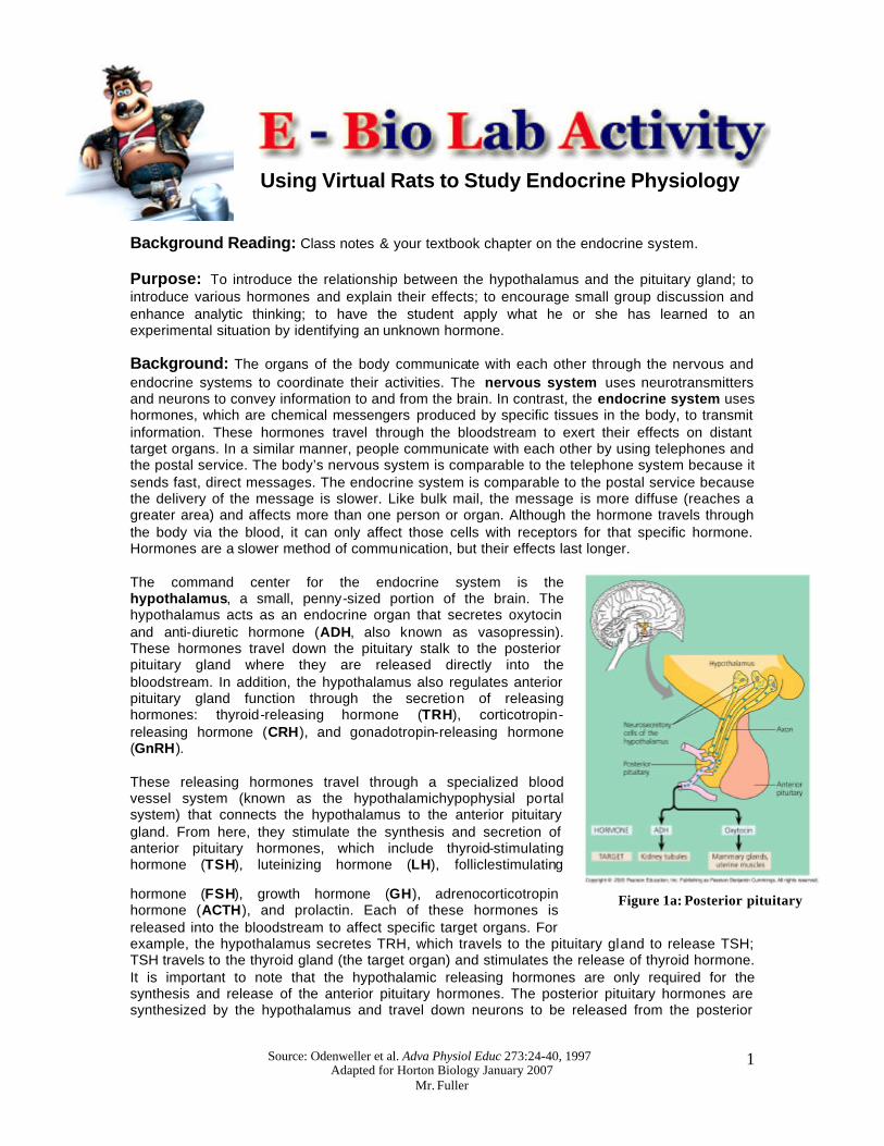
Figure 1a: Posterior pituitary
Using Virtual Rats to Study Endocrine Physiology
Background Reading: Class notes & your textbook chapter on the endocrine system. Purpose: To introduce the relationship between the hypothalamus and the pituitary gland; to introduce various hormones and explain their effects; to encourage small group discussion and enhance analytic thinking; to have the student apply what he or she has learned to an experimental situation by identifying an unknown hormone. Background: The organs of the body communicate with each other through the nervous and endocrine systems to coordinate their activities. The nervous system uses neurotransmitters and neurons to convey information to and from the brain. In contrast, the endocrine system uses hormones, which are chemical messengers produced by specific tissues in the body, to transmit information. These hormones travel through the bloodstream to exert their effects on distant target organs. In a similar manner, people communicate with each other by using telephones and the postal service. The body’s nervous system is comparable to the telephone system because it sends fast, direct messages. The endocrine system is comparable to the postal service because the delivery of the message is slower. Like bulk mail, the message is more diffuse (reaches a greater area) and affects more than one person or organ. Although the hormone travels through the body via the blood, it can only affect those cells with receptors for that specific hormone. Hormones are a slower method of communication, but their effects last longer. The command center for the endocrine system is the hypothalamus, a small, penny-sized portion of the brain. The hypothalamus acts as an endocrine organ that secretes oxytocin and anti-diuretic hormone (ADH, also known as vasopressin). These hormones travel down the pituitary stalk to the posterior pituitary gland where they are released directly into the bloodstream. In addition, the hypothalamus also regulates anterior pituitary gland function through the secretion of releasing hormones: thyroid-releasing hormone (TRH), corticotropin-releasing hormone (CRH), and gonadotropin-releasing hormone (GnRH). These releasing hormones travel through a specialized blood vessel system (known as the hypothalamichypophysial portal system) that connects the hypothalamus to the anterior pituitary gland. From here, they stimulate the synthesis and secretion of anterior pituitary hormones, which include thyroid-stimulating hormone (TSH), luteinizing hormone (LH), folliclestimulating
hormone (FSH), growth hormone (GH), adrenocorticotropin hormone (ACTH), and prolactin. Each of these hormones is released into the bloodstream to affect specific target organs. For example, the hypothalamus secretes TRH, which travels to the pituitary gland to release TSH; TSH travels to the thyroid gland (the target organ) and stimulates the release of thyroid hormone. It is important to note that the hypothalamic releasing hormones are only required for the synthesis and release of the anterior pituitary hormones. The posterior pituitary hormones are synthesized by the hypothalamus and travel down neurons to be released from the posterior
Source: Odenweller et al. Adva Physiol Educ 273:24-40, 1997 Adapted for Horton Biology January 2007
Mr. Fuller
2
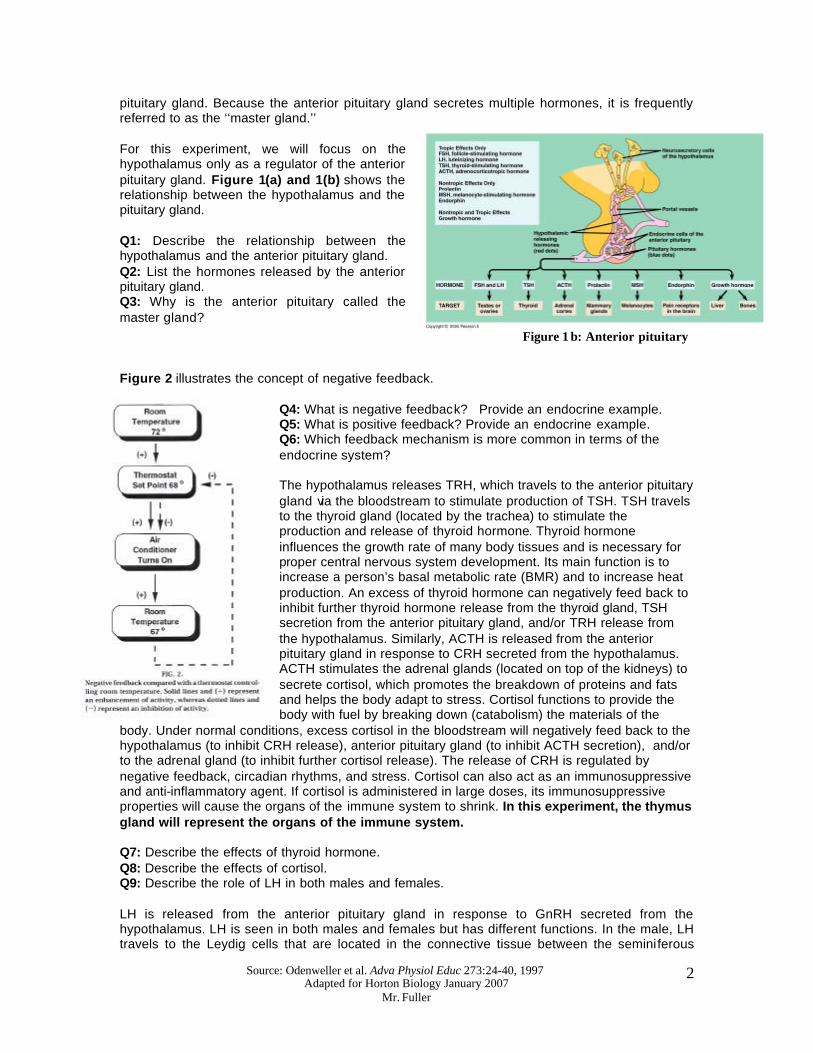
Figure 1 b: Anterior pituitary
pituitary gland. Because the anterior pituitary gland secretes multiple hormones, it is frequently referred to as the ‘‘master gland.’’ For this experiment, we will focus on the hypothalamus only as a regulator of the anterior pituitary gland. Figure 1(a) and 1(b) shows the relationship between the hypothalamus and the pituitary gland. Q1: Describe the relationship between the hypothalamus and the anterior pituitary gland. Q2: List the hormones released by the anterior pituitary gland. Q3: Why is the anterior pituitary called the master gland? Figure 2 illustrates the concept of negative feedback.
Q4: What is negative feedback? Provide an endocrine example. Q5: What is positive feedback? Provide an endocrine example. Q6: Which feedback mechanism is more common in terms of the endocrine system? The hypothalamus releases TRH, which travels to the anterior pituitary gland via the bloodstream to stimulate production of TSH. TSH travels to the thyroid gland (located by the trachea) to stimulate the production and release of thyroid hormone. Thyroid hormone influences the growth rate of many body tissues and is necessary for proper central nervous system development. Its main function is to increase a person’s basal metabolic rate (BMR) and to increase heat production. An excess of thyroid hormone can negatively feed back to inhibit further thyroid hormone release from the thyroid gland, TSH secretion from the anterior pituitary gland, and/or TRH release from the hypothalamus. Similarly, ACTH is released from the anterior pituitary gland in response to CRH secreted from the hypothalamus. ACTH stimulates the adrenal glands (located on top of the kidneys) to secrete cortisol, which promotes the breakdown of proteins and fats and helps the body adapt to stress. Cortisol functions to provide the body with fuel by breaking down (catabolism) the materials of the
body. Under normal conditions, excess cortisol in the bloodstream will negatively feed back to the hypothalamus (to inhibit CRH release), anterior pituitary gland (to inhibit ACTH secretion), and/or to the adrenal gland (to inhibit further cortisol release). The release of CRH is regulated by negative feedback, circadian rhythms, and stress. Cortisol can also act as an immunosuppressive and anti-inflammatory agent. If cortisol is administered in large doses, its immunosuppressive properties will cause the organs of the immune system to shrink. In this experiment, the thymus gland will represent the organs of the immune system. Q7: Describe the effects of thyroid hormone. Q8: Describe the effects of cortisol. Q9: Describe the role of LH in both males and females. LH is released from the anterior pituitary gland in response to GnRH secreted from the hypothalamus. LH is seen in both males and females but has different functions. In the male, LH travels to the Leydig cells that are located in the connective tissue between the seminiferous
Source: Odenweller et al. Adva Physiol Educ 273:24-40, 1997 Adapted for Horton Biology January 2007
Mr. Fuller
3
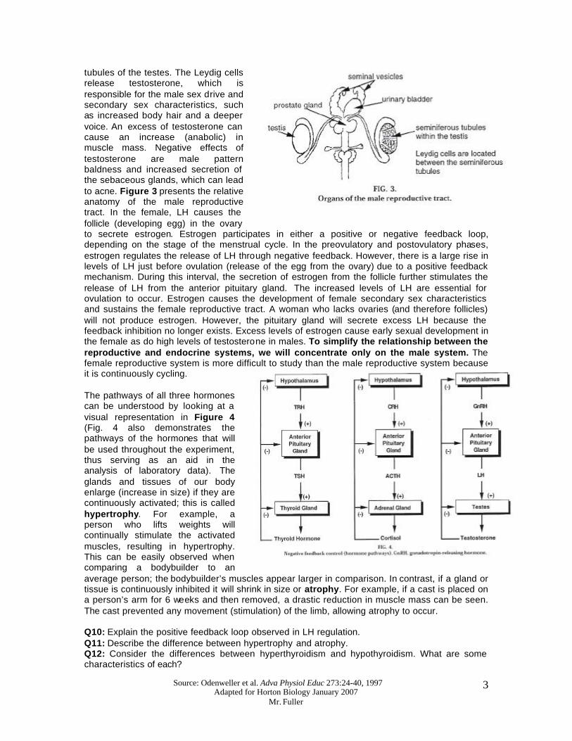
tubules of the testes. The Leydig cells release testosterone, which is responsible for the male sex drive and secondary sex characteristics, such as increased body hair and a deeper voice. An excess of testosterone can cause an increase (anabolic) in muscle mass. Negative effects of testosterone are male pattern baldness and increased secretion of the sebaceous glands, which can lead to acne. Figure 3 presents the relative anatomy of the male reproductive tract. In the female, LH causes the follicle (developing egg) in the ovary to secrete estrogen. Estrogen participates in either a positive or negative feedback loop, depending on the stage of the menstrual cycle. In the preovulatory and postovulatory phases, estrogen regulates the release of LH through negative feedback. However, there is a large rise in levels of LH just before ovulation (release of the egg from the ovary) due to a positive feedback mechanism. During this interval, the secretion of estrogen from the follicle further stimulates the release of LH from the anterior pituitary gland. The increased levels of LH are essential for ovulation to occur. Estrogen causes the development of female secondary sex characteristics and sustains the female reproductive tract. A woman who lacks ovaries (and therefore follicles) will not produce estrogen. However, the pituitary gland will secrete excess LH because the feedback inhibition no longer exists. Excess levels of estrogen cause early sexual development in the female as do high levels of testosterone in males. To simplify the relationship between the reproductive and endocrine systems, we will concentrate only on the male system. The female reproductive system is more difficult to study than the male reproductive system because it is continuously cycling. The pathways of all three hormones can be understood by looking at a visual representation in Figure 4 (Fig. 4 also demonstrates the pathways of the hormones that will be used throughout the experiment, thus serving as an aid in the analysis of laboratory data). The glands and tissues of our body enlarge (increase in size) if they are continuously activated; this is called hypertrophy. For example, a person who lifts weights will continually stimulate the activated muscles, resulting in hypertrophy. This can be easily observed when comparing a bodybuilder to an average person; the bodybuilder’s muscles appear larger in comparison. In contrast, if a gland or tissue is continuously inhibited it will shrink in size or atrophy. For example, if a cast is placed on a person’s arm for 6 weeks and then removed, a drastic reduction in muscle mass can be seen. The cast prevented any movement (stimulation) of the limb, allowing atrophy to occur. Q10: Explain the positive feedback loop observed in LH regulation. Q11: Describe the difference between hypertrophy and atrophy. Q12: Consider the differences between hyperthyroidism and hypothyroidism. What are some characteristics of each?
Source: Odenweller et al. Adva Physiol Educ 273:24-40, 1997 Adapted for Horton Biology January 2007
Mr. Fuller
4
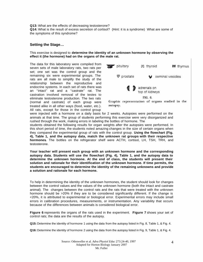
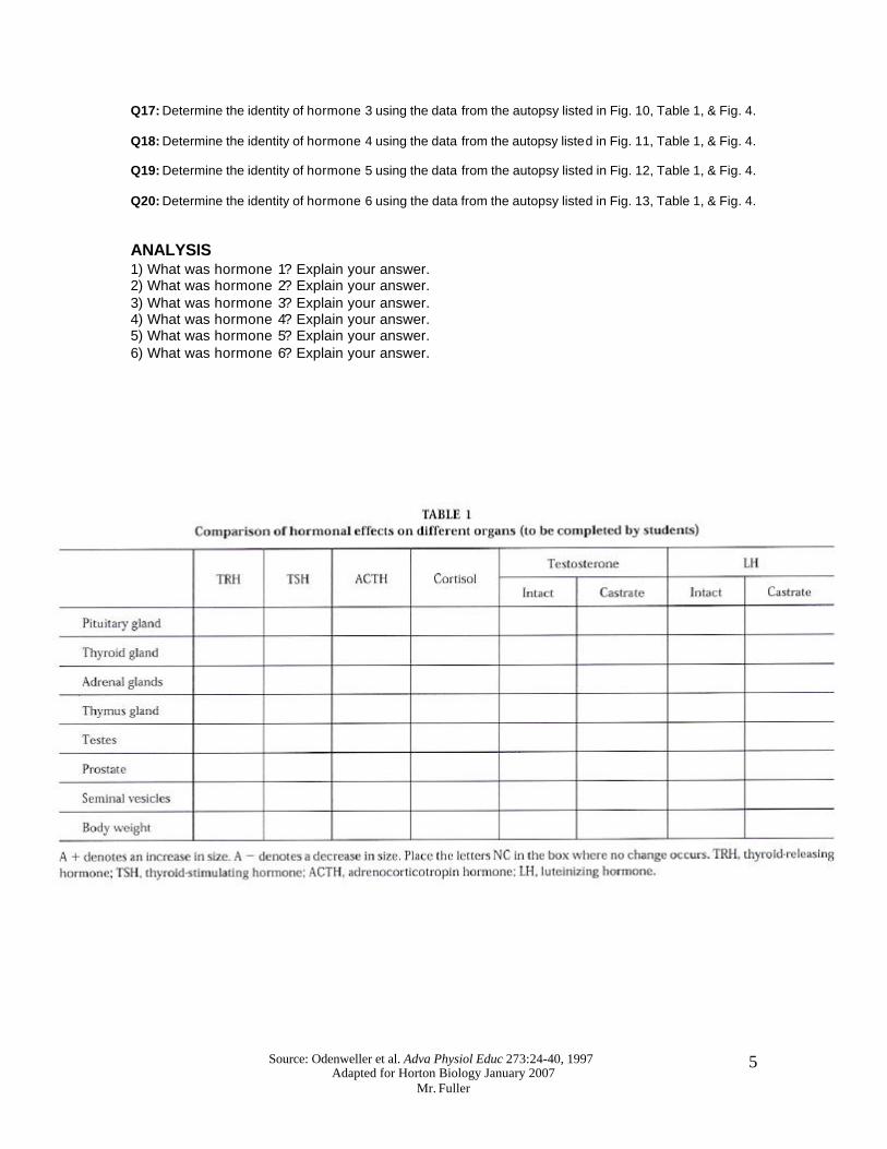
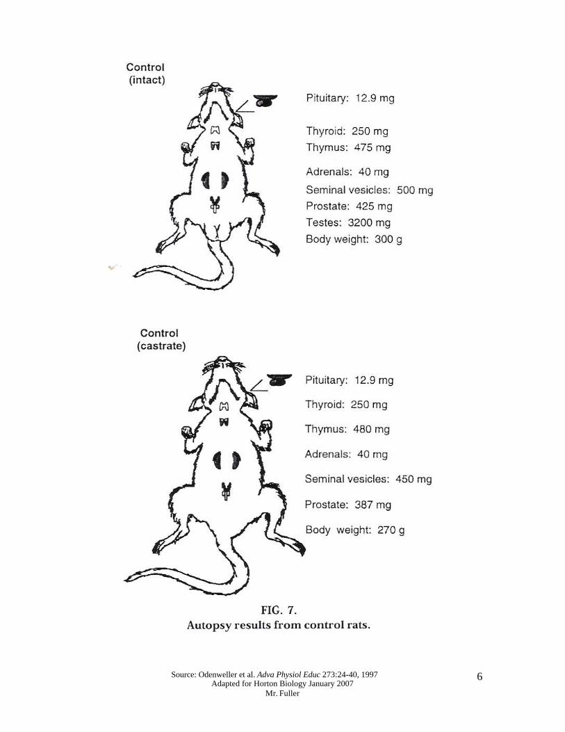
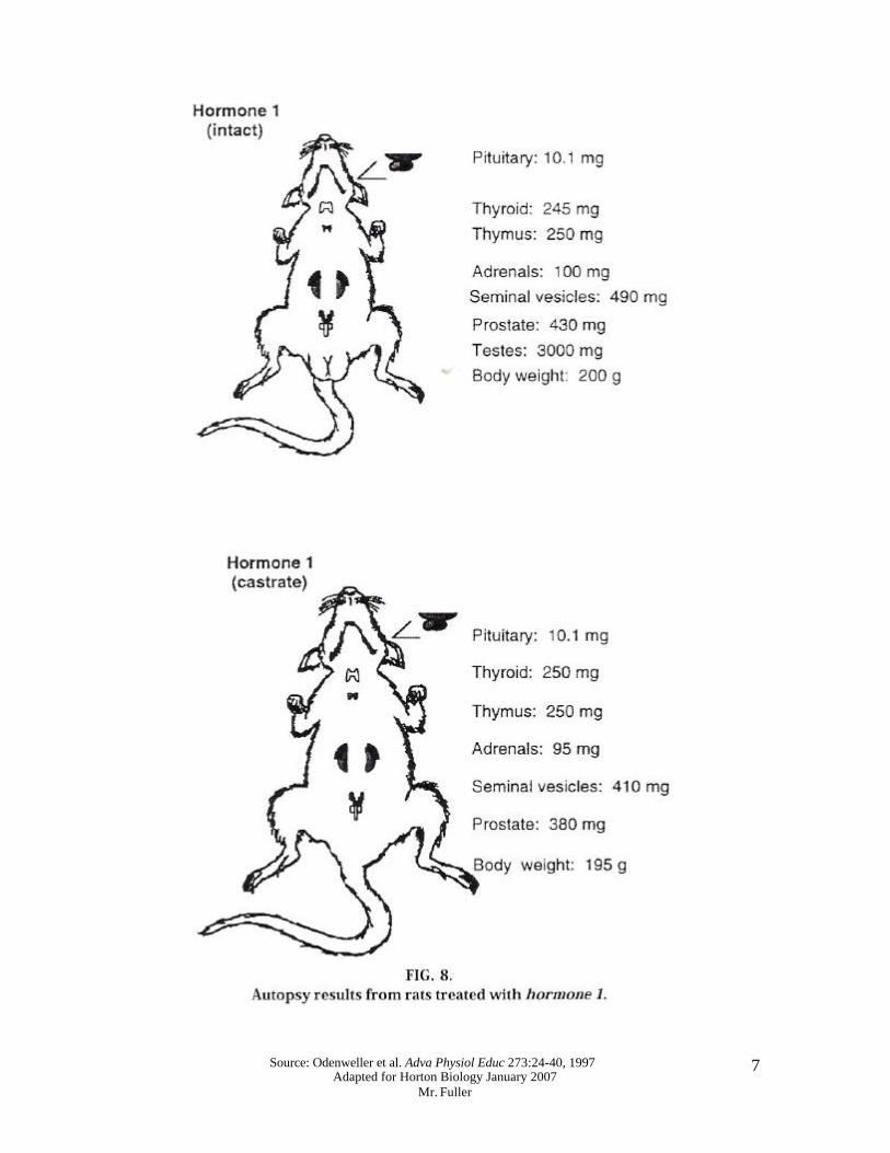
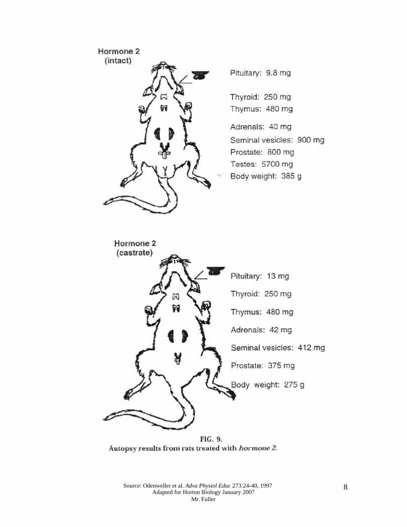
Q13: What are the effects of decreasing testosterone? Q14: What is the result of excess secretion of cortisol? (Hint: it is a syndrome) What are some of the symptoms of this syndrome? Setting the Stage… This exercise is designed to determine the identity of an unknown hormone by observing the effect it (the hormone) had on the organs of the male rat. The data for this laboratory were compiled from seven sets of male laboratory rats, two rats per set; one set was the control group and the remaining six were experimental groups. The rats are all male to simplify the study of the relationship between the reproductive and endocrine systems. In each set of rats there was an ‘‘intact’’ rat and a ‘‘castrate’’ rat. The castration involved removal of the testes to eliminate testosterone production. The two rats (normal and castrate) of each group were treated alike in all other ways (food, water, etc.). All rats, except for those in the control group were injected with a hormone on a daily basis for 2 weeks. Autopsies were performed on the animals at that time. The group of students performing this exercise were very disorganized and rushed through the work, making errors in labeling the bottles of hormone. The students obtained the following results for organ weights after the autopsies were performed. In this short period of time, the students noted amazing changes in the size of certain organs when they compared the experimental group of rats with the control group. Using the flowchart (Fig. 4), Table 1, and the autopsy data, match the unknown rat groups with their respective hormones. The bottles on the refrigerator shelf were ACTH, cortisol, LH, TSH, TRH, and testosterone. Your teacher will present each group with an unknown hormone and the corresponding autopsy data. Students will use the flowchart (Fig. 4), Table 1, and the autopsy data to determine the unknown hormone. At the end of class, the students will present their solution and rationale for their identification of the unknown hormone. If time permits, the students are encouraged to determine the identity of the remaining unknowns and provide a solution and rationale for each hormone. To help in determining the identity of the unknown hormones, the student should look for changes between the control values and the values of the unknown hormone (both the intact and castrate animal). The changes between the control rats and the rats that were treated with the unknown hormone should be >20% if they are to be considered significantly different. If the change is <20%, it is attributed to experimental or biological error. Experimental errors may include small errors in calibration procedures, measurements, or instrumentation. Any variability that occurs because of the differences between animals is considered biological error. Figure 6 represents the organs of the rats used in the experiment. Figure 7 shows your set of control rats; the data are the results of the autopsy. Q15: Determine the identity of hormone 1 using the data from the autopsy listed in Fig. 8, Table 1, & Fig. 4. Q16: Determine the identity of hormone 2 using the data from the autopsy listed in Fig. 9, Table 1, & Fig. 4.
Source: Odenweller et al. Adva Physiol Educ 273:24-40, 1997 Adapted for Horton Biology January 2007
Mr. Fuller
5
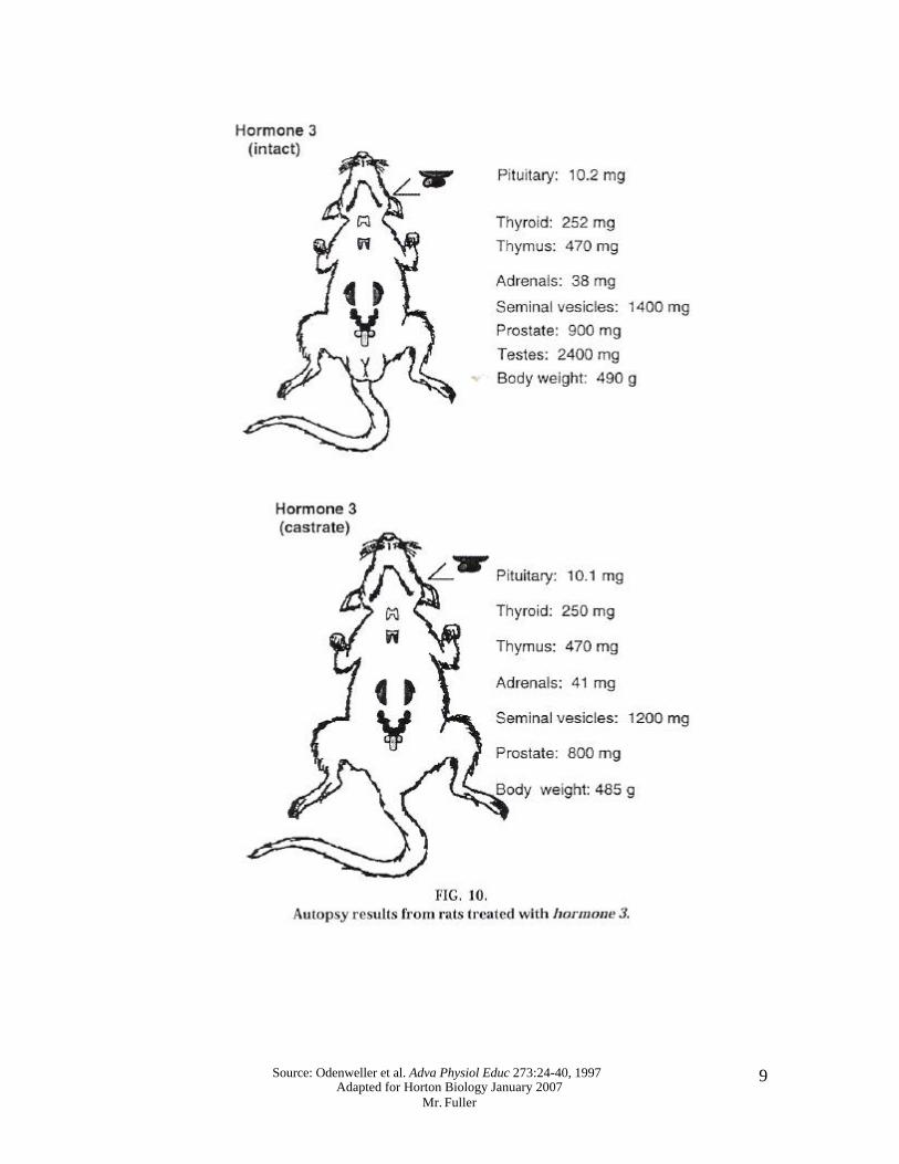
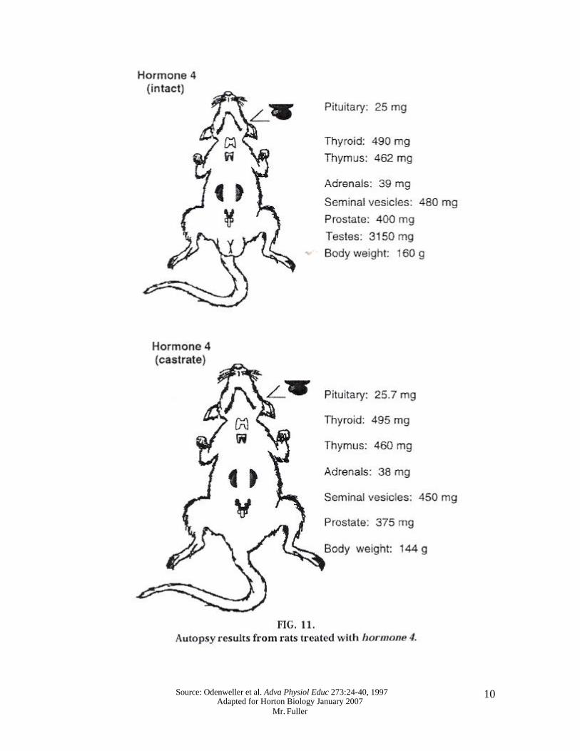
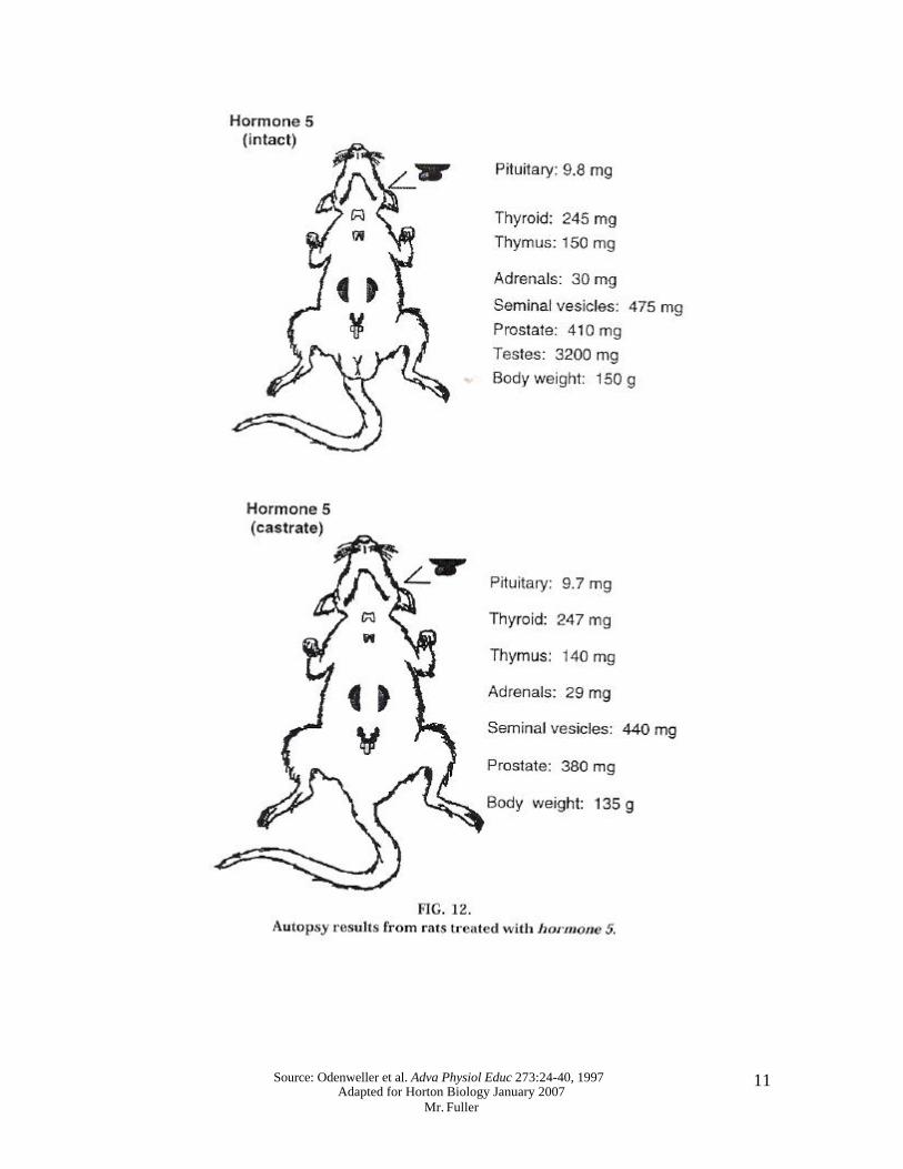
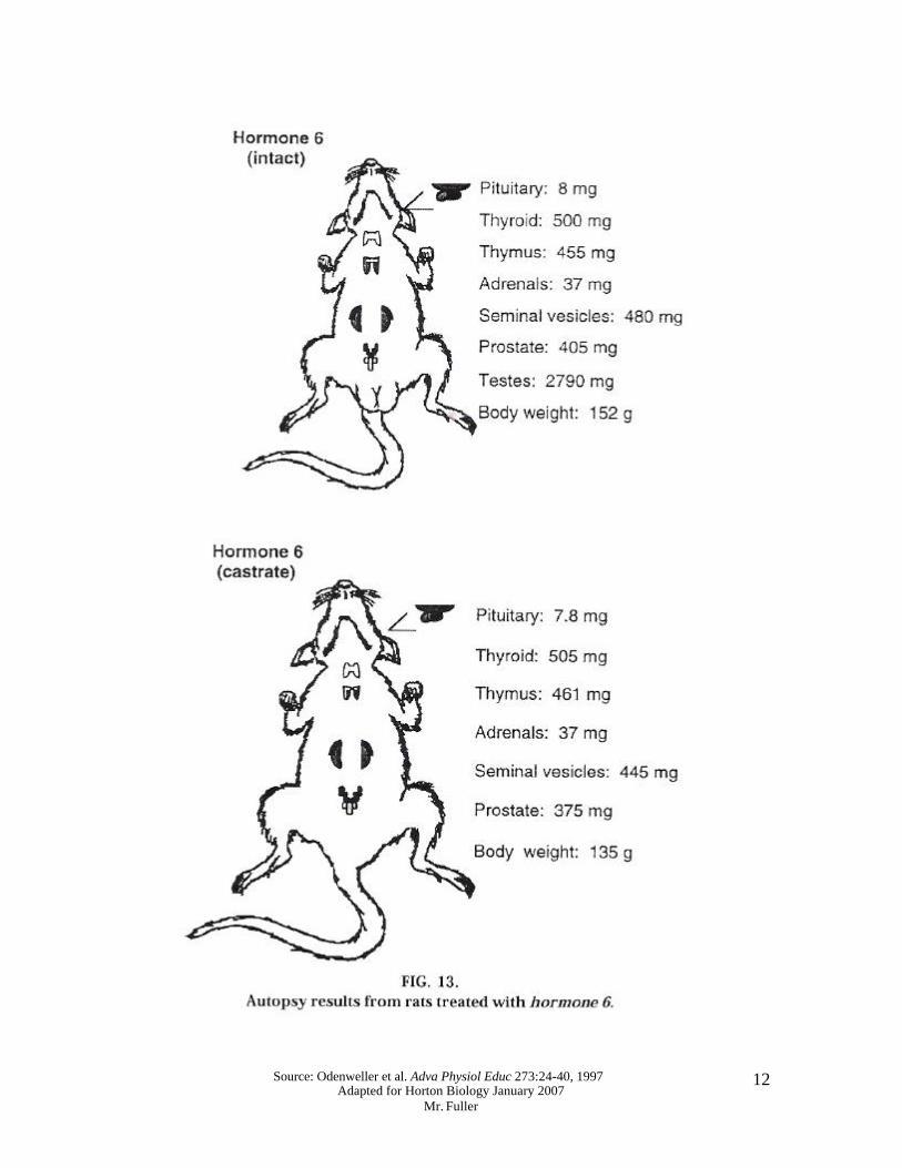
Q17: Determine the identity of hormone 3 using the data from the autopsy listed in Fig. 10, Table 1, & Fig. 4. Q18: Determine the identity of hormone 4 using the data from the autopsy listed in Fig. 11, Table 1, & Fig. 4. Q19: Determine the identity of hormone 5 using the data from the autopsy listed in Fig. 12, Table 1, & Fig. 4. Q20: Determine the identity of hormone 6 using the data from the autopsy listed in Fig. 13, Table 1, & Fig. 4. ANALYSIS 1) What was hormone 1? Explain your answer. 2) What was hormone 2? Explain your answer. 3) What was hormone 3? Explain your answer. 4) What was hormone 4? Explain your answer. 5) What was hormone 5? Explain your answer. 6) What was hormone 6? Explain your answer.
Source: Odenweller et al. Adva Physiol Educ 273:24-40, 1997 Adapted for Horton Biology January 2007
Mr. Fuller
6
Source: Odenweller et al. Adva Physiol Educ 273:24-40, 1997 Adapted for Horton Biology January 2007
Mr. Fuller
7
Source: Odenweller et al. Adva Physiol Educ 273:24-40, 1997 Adapted for Horton Biology January 2007
Mr. Fuller
8
Source: Odenweller et al. Adva Physiol Educ 273:24-40, 1997 Adapted for Horton Biology January 2007
Mr. Fuller
9
Source: Odenweller et al. Adva Physiol Educ 273:24-40, 1997 Adapted for Horton Biology January 2007
Mr. Fuller
10
Source: Odenweller et al. Adva Physiol Educ 273:24-40, 1997 Adapted for Horton Biology January 2007
Mr. Fuller
11
Source: Odenweller et al. Adva Physiol Educ 273:24-40, 1997 Adapted for Horton Biology January 2007
Mr. Fuller
12