Embed Size (px)
Citation preview
Vaginal Bleeding and Abdominal Pain in the Non-Pregnant Patient
December 6, 2005
Eli Denney, DO
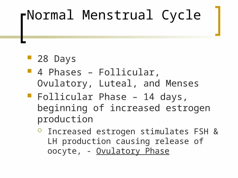
Normal Menstrual Cycle
28 Days 4 Phases – Follicular, Ovulatory,
Luteal, and Menses Follicular Phase – 14 days, beginning
of increased estrogen production Increased estrogen stimulates FSH & LH
production causing release of oocyte, - Ovulatory Phase
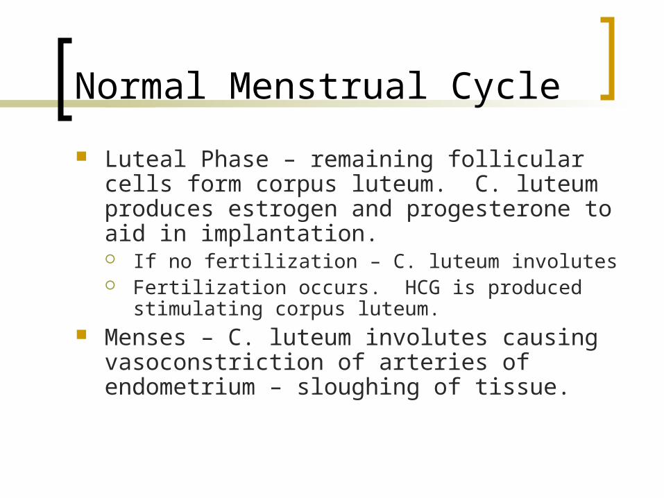
Normal Menstrual Cycle
Luteal Phase – remaining follicular cells form corpus luteum. C. luteum produces estrogen and progesterone to aid in implantation. If no fertilization – C. luteum involutes Fertilization occurs. HCG is produced
stimulating corpus luteum. Menses – C. luteum involutes causing
vasoconstriction of arteries of endometrium – sloughing of tissue.
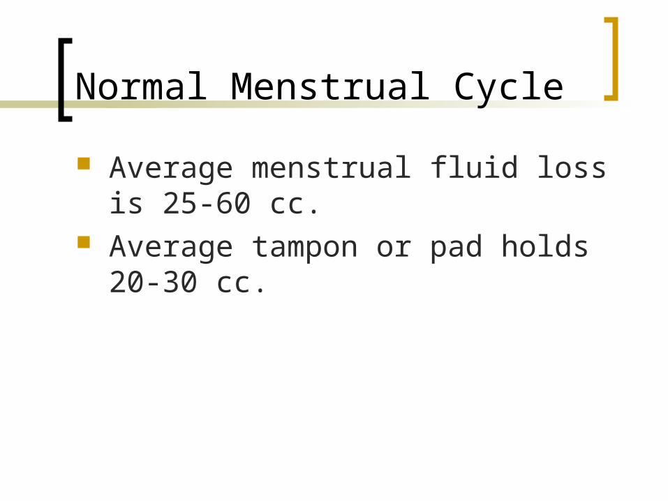
Normal Menstrual Cycle
Average menstrual fluid loss is 25-60 cc.
Average tampon or pad holds 20-30 cc.
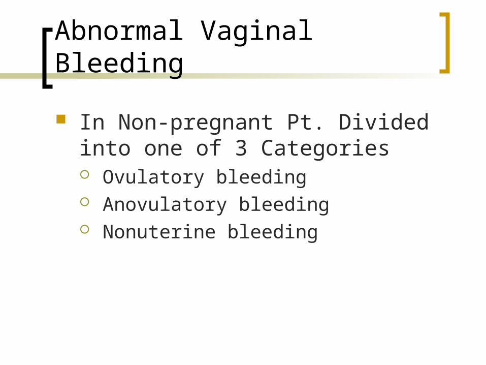
Abnormal Vaginal Bleeding
In Non-pregnant Pt. Divided into one of 3 Categories Ovulatory bleeding Anovulatory bleeding Nonuterine bleeding
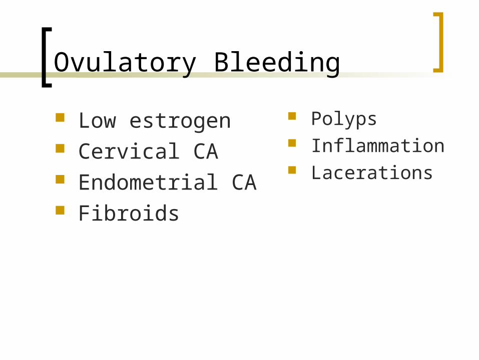
Ovulatory Bleeding
Low estrogen Cervical CA Endometrial CA Fibroids
Polyps Inflammation Lacerations
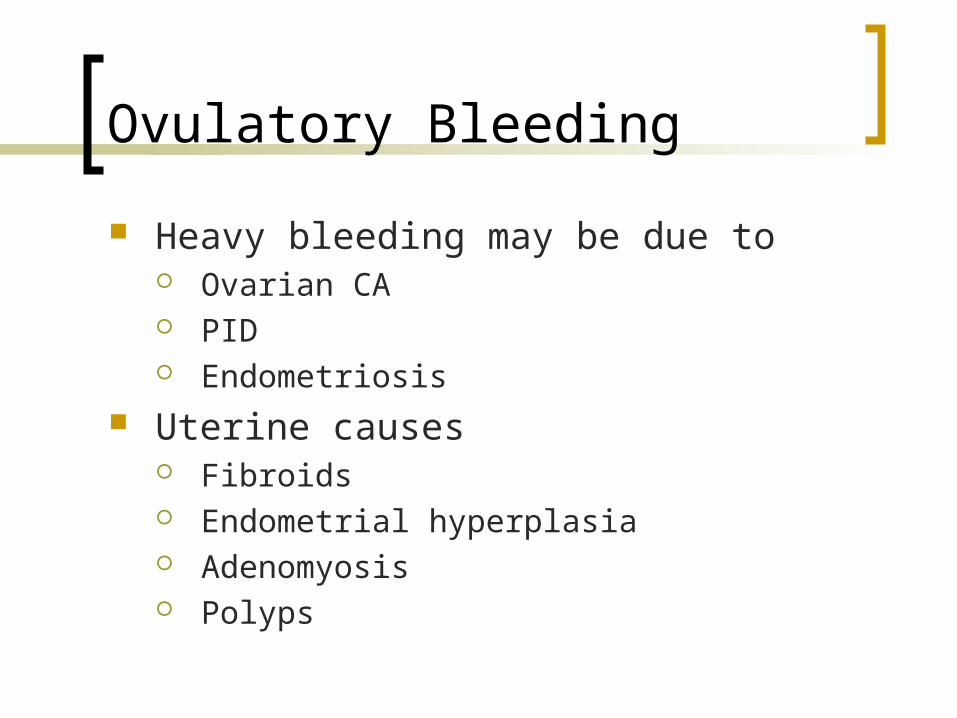
Ovulatory Bleeding
Heavy bleeding may be due to Ovarian CA PID Endometriosis
Uterine causes Fibroids Endometrial hyperplasia Adenomyosis Polyps
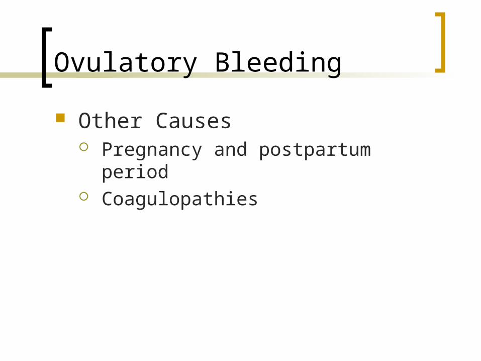
Ovulatory Bleeding
Other Causes Pregnancy and postpartum period Coagulopathies
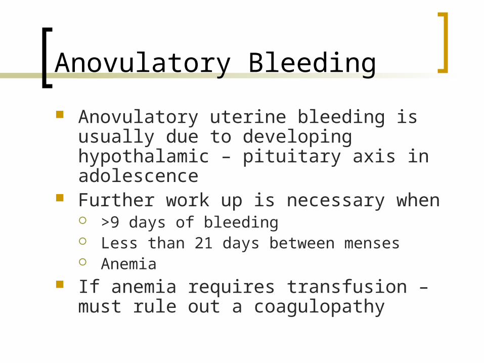
Anovulatory Bleeding
Anovulatory uterine bleeding is usually due to developing hypothalamic – pituitary axis in adolescence
Further work up is necessary when >9 days of bleeding Less than 21 days between menses Anemia
If anemia requires transfusion – must rule out a coagulopathy
Anovulatory Bleeding
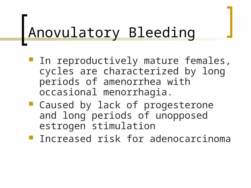
In reproductively mature females, cycles are characterized by long periods of amenorrhea with occasional menorrhagia.
Caused by lack of progesterone and long periods of unopposed estrogen stimulation
Increased risk for adenocarcinoma
Midcycle Bleeding
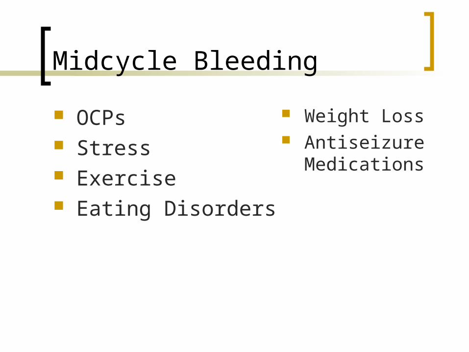
OCPs Stress Exercise Eating Disorders
Weight Loss Antiseizure
Medications
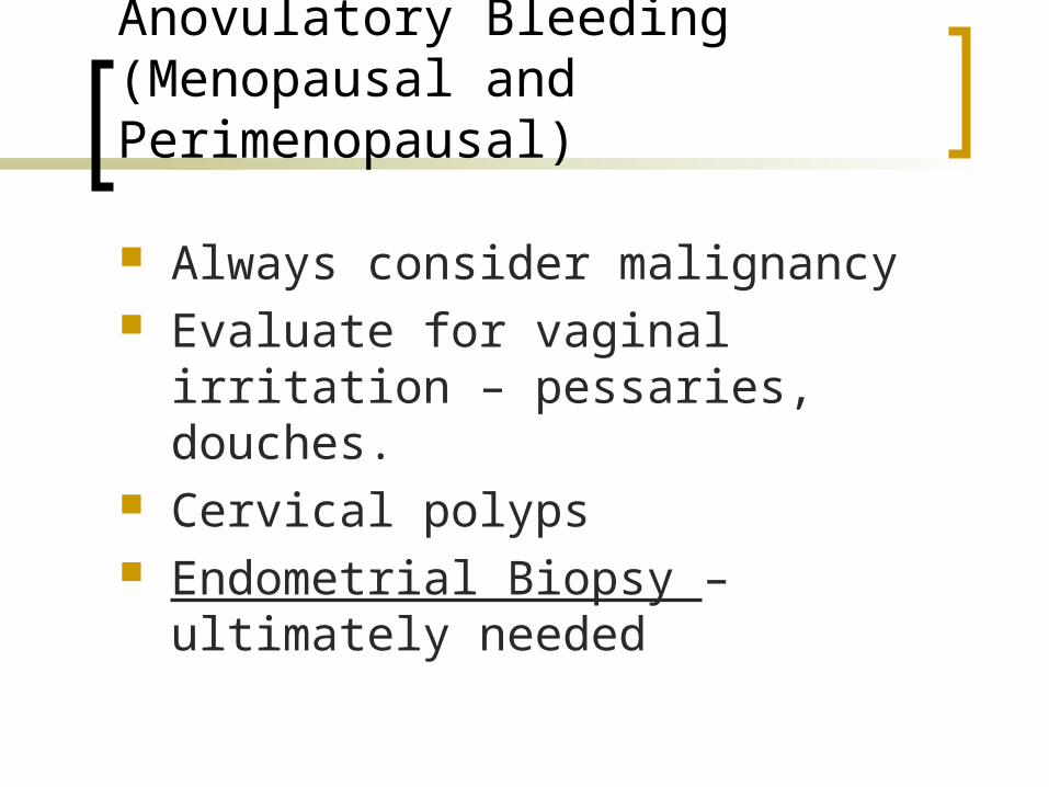
Anovulatory Bleeding (Menopausal and Perimenopausal)
Always consider malignancy Evaluate for vaginal irritation –
pessaries, douches. Cervical polyps Endometrial Biopsy – ultimately
needed
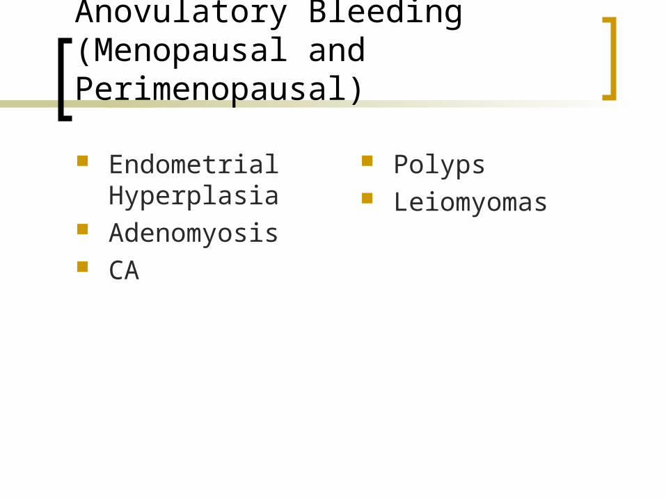
Anovulatory Bleeding (Menopausal and Perimenopausal)
Endometrial Hyperplasia
Adenomyosis CA
Polyps Leiomyomas
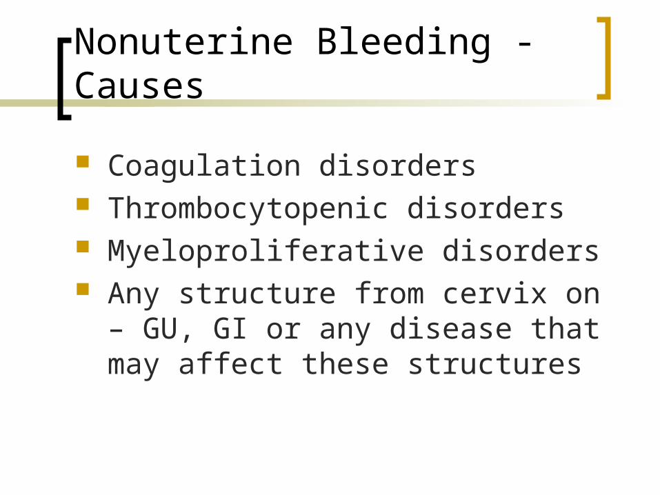
Nonuterine Bleeding - Causes
Coagulation disorders Thrombocytopenic disorders Myeloproliferative disorders Any structure from cervix on – GU, GI
or any disease that may affect these structures
Evaluation of Abnormal Vaginal Bleeding
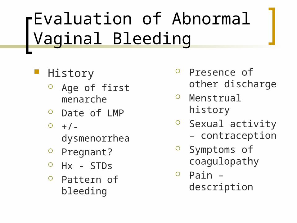
History Age of first
menarche Date of LMP +/- dysmenorrhea Pregnant? Hx - STDs Pattern of bleeding
Presence of other discharge
Menstrual history Sexual activity –
contraception Symptoms of
coagulopathy Pain – description
Evaluation of Abnormal Vaginal Bleeding
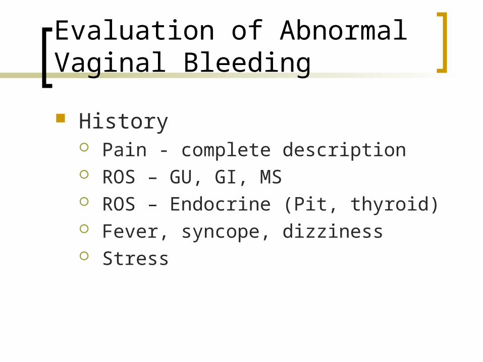
History Pain - complete description ROS – GU, GI, MS ROS – Endocrine (Pit, thyroid) Fever, syncope, dizziness Stress
Evaluation of Abnormal Vaginal Bleeding
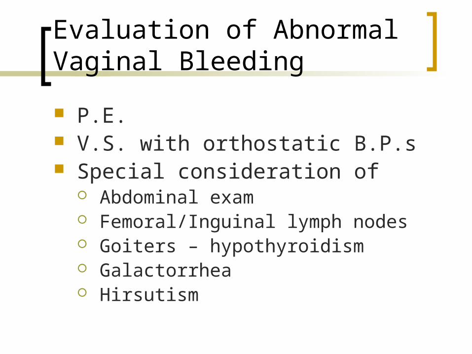
P.E. V.S. with orthostatic B.P.s Special consideration of
Abdominal exam Femoral/Inguinal lymph nodes Goiters – hypothyroidism Galactorrhea Hirsutism
Evaluation of Abnormal Vaginal Bleeding
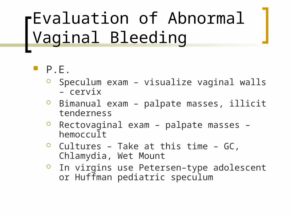
P.E. Speculum exam – visualize vaginal walls –
cervix Bimanual exam – palpate masses, illicit
tenderness Rectovaginal exam – palpate masses –
hemoccult Cultures – Take at this time – GC, Chlamydia,
Wet Mount In virgins use Petersen–type adolescent or
Huffman pediatric speculum
Evaluation of Abnormal Vaginal Bleeding
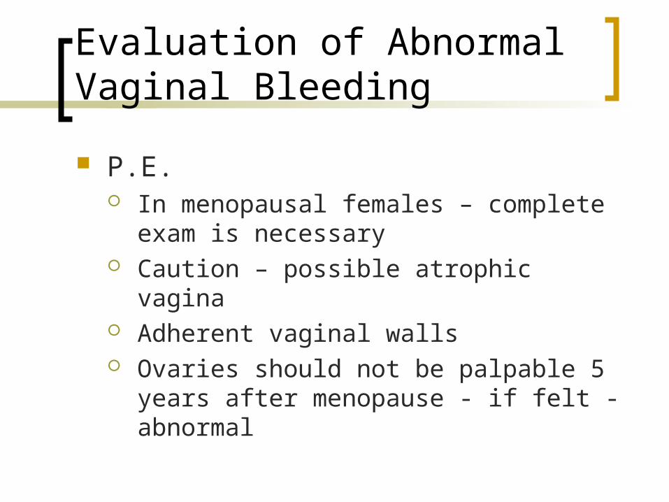
P.E. In menopausal females – complete exam
is necessary Caution – possible atrophic vagina Adherent vaginal walls Ovaries should not be palpable 5 years
after menopause - if felt - abnormal
Evaluation of Abnormal Vaginal Bleeding
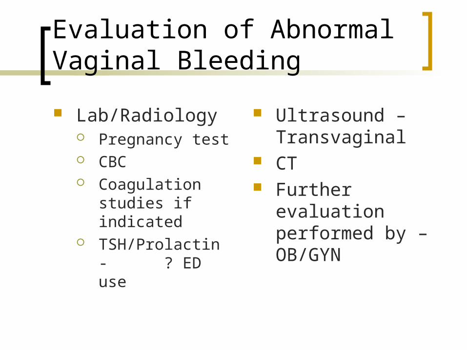
Lab/Radiology Pregnancy test CBC Coagulation studies
if indicated TSH/Prolactin
- ? ED use
Ultrasound – Transvaginal
CT Further evaluation
performed by – OB/GYN
Treatment – Abnormal Vaginal Bleeding (Non-Pregnant)
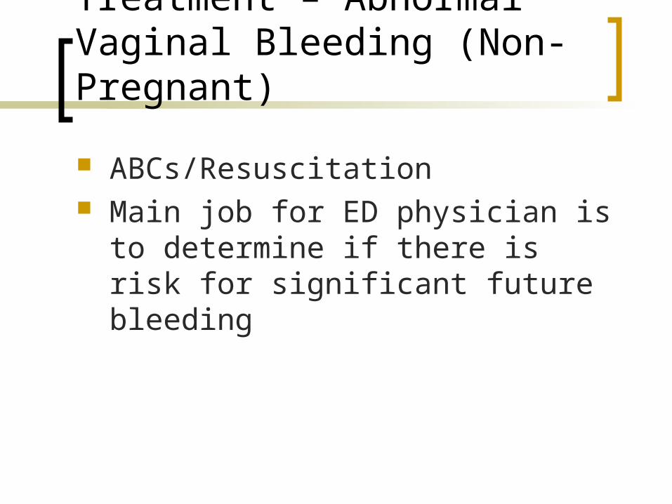
ABCs/Resuscitation Main job for ED physician is to
determine if there is risk for significant future bleeding
Treatment – Abnormal Vaginal Bleeding (Non-Pregnant)
If no hemodynamic compromise, only the following problems need to be ruled out/treated Pregnancy Trauma (Abuse) – injury Coagulopathy Infection Foreign bodies
If not one of the above – further outpatient evaluation
Treatment – Abnormal Vaginal Bleeding (Non-Pregnant)
Unstable Patient Resuscitation D&C may be needed for uterine bleeding Estrogens may be needed for bleeding
not caused by pregnancy or treatable with surgery
Treatment – Abnormal Vaginal Bleeding (Non-Pregnant)
Stable Patient Thin endometrium shown on ultrasound –
short term estrogen therapy useful See attached Table 101-3 for short-term
treatment regimens If diagnosis is cannot be made, patient
should be referred for further evaluation - OB/GYN
Long-Term Therapy
OCPs are very effective and provide contraception
NSAIDs aid in dysmenorrhea and help decrease bleeding
Other more uncommon therapies – progesterones, Danazol, hysteroscopy, endometrial ablation, and hysterectomy
Genital Trauma
Commonly due to vigorous voluntary/involuntary sexual activity
Posterior fornix is most common area injured
Adenomyosis
Caused by endometrial glands growing into myometrium
May cause menorrhagia and dysmenorrhea at the time of menstruation
Treatments are analgesics for pain – surgery may be needed for severe bleeding refectory to medical therapy
Leiomyomas
Fibroids – smooth muscle cell tumors - responsive to estrogen, usually multiple
Size increases in first part of pregnancy and at times with OCP use
Size decreases with menopause Fibroids are usually found during manual
exam or by ultrasound If acute degeneration or torsion occurs –
patients will present with acute abdomen symptoms on physical exam
Leiomyomas
Treatment is NSAIDs, progestins, GNRHs, or surgery if indicated
Uterine artery embolization is a new promising therapy
Blood Dyscrasias
Menstrual bleeding may be excessive and be the presenting symptom of a bleeding disorder
Treatment includes antifibrinolytics and OCPs. OCPs increase levels of factor VIII and vWF factor
Desmopressin (DDAVP) – increases release of factor VIII and vWF
In these groups NSAIDs are not helpful and may cause increased bleeding
Polycystic Ovary Syndrome
PCOS – caused by hyperandrogenism and anovulation without disease of adrenal or pituitary glands
Triad usually seen – obese, hirsutite, oligomenorrhea
Menses are heavy and prolonged Other characteristics – alopecia, increased
androgens, increased LH and FSH and acne Therapy – OCPs – low doses or cyclic progestins
Abdominal and Pelvic Pain in the Non-Pregnant Female
Classification of Pain
Visceral – caused by stretch of smooth muscle from obstruction of hollow organ. Ischemia and inflammation may also be involved.
Autonomic nerve fibers produce poorly localized abdominal pain – cramping in nature, midline.
Examples: Appendicitis Obstruction Nephrolithasis PID
Classification of Pain
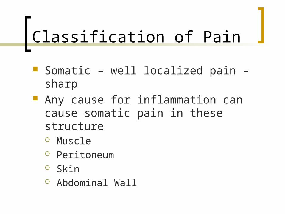
Somatic – well localized pain – sharp Any cause for inflammation can cause
somatic pain in these structure Muscle Peritoneum Skin Abdominal Wall
Classification of Pain
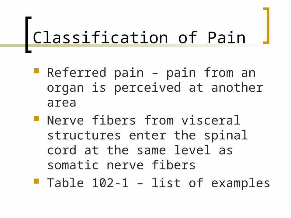
Referred pain – pain from an organ is perceived at another area
Nerve fibers from visceral structures enter the spinal cord at the same level as somatic nerve fibers
Table 102-1 – list of examples
Abdominal and Pelvic Pain in the Non-Pregnant Female
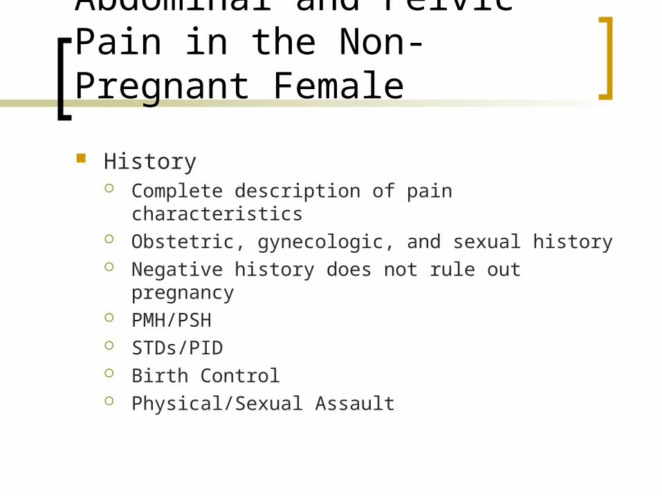
History Complete description of pain characteristics Obstetric, gynecologic, and sexual history Negative history does not rule out pregnancy PMH/PSH STDs/PID Birth Control Physical/Sexual Assault
Abdominal and Pelvic Pain in the Non-Pregnant Female
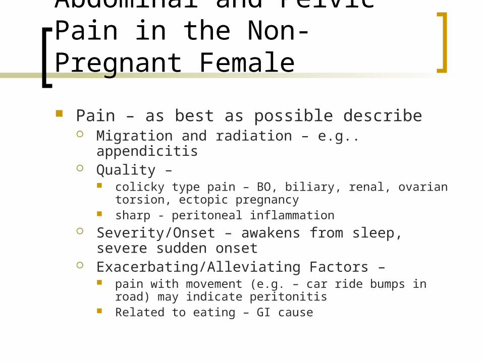
Pain – as best as possible describe Migration and radiation – e.g.. appendicitis Quality –
colicky type pain – BO, biliary, renal, ovarian torsion, ectopic pregnancy
sharp - peritoneal inflammation Severity/Onset – awakens from sleep, severe
sudden onset Exacerbating/Alleviating Factors –
pain with movement (e.g. – car ride bumps in road) may indicate peritonitis
Related to eating – GI cause
Associated Signs/Symptoms
Nausea Vomiting Constipation
Diarrhea Anorexia
Above symptoms are nonspecific
Associated Signs/Symptoms
Hematuria Dysuria Urgency Possible Pyleonephritis, UTI,
Nephrolithasis Above symptoms may also be caused
by a gynecologic cause
Flank Pain
Physical Exam
Vitals first – continue to monitor throughout ER stay
Orthostatics General appearance –
Peritoneal inflammation/Colicky Pain Involuntary/Voluntary guarding Mass Rebound Tenderness
Physical Exam
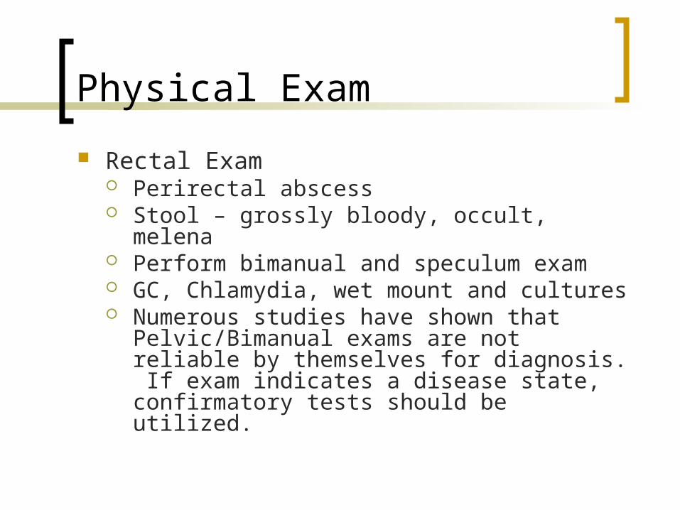
Rectal Exam Perirectal abscess Stool – grossly bloody, occult, melena Perform bimanual and speculum exam GC, Chlamydia, wet mount and cultures Numerous studies have shown that
Pelvic/Bimanual exams are not reliable by themselves for diagnosis. If exam indicates a disease state, confirmatory tests should be utilized.
Differential Diganosis of Nontraumatic Pelvic Pain in Non-Pregnant Adolescents and Adults
Table 102-2
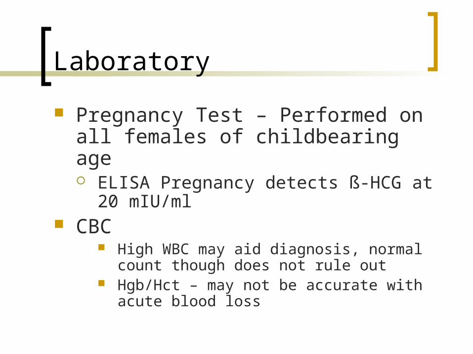
Laboratory
Pregnancy Test – Performed on all females of childbearing age ELISA Pregnancy detects ß-HCG at 20
mIU/ml CBC
High WBC may aid diagnosis, normal count though does not rule out
Hgb/Hct – may not be accurate with acute blood loss
Laboratory
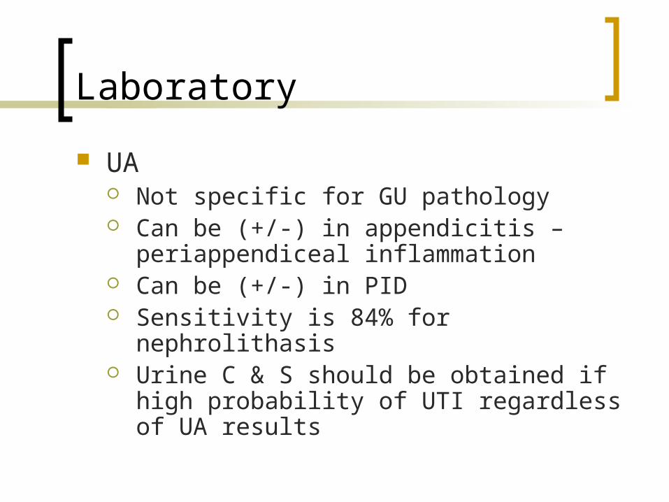
UA Not specific for GU pathology Can be (+/-) in appendicitis –
periappendiceal inflammation Can be (+/-) in PID Sensitivity is 84% for nephrolithasis Urine C & S should be obtained if high
probability of UTI regardless of UA results
Radiology
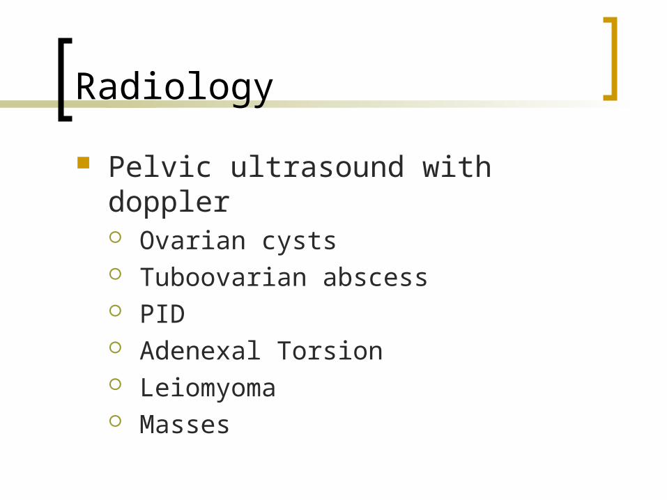
Pelvic ultrasound with doppler Ovarian cysts Tuboovarian abscess PID Adenexal Torsion Leiomyoma Masses
Radiology
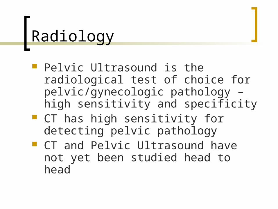
Pelvic Ultrasound is the radiological test of choice for pelvic/gynecologic pathology – high sensitivity and specificity
CT has high sensitivity for detecting pelvic pathology
CT and Pelvic Ultrasound have not yet been studied head to head
Laparoscopy
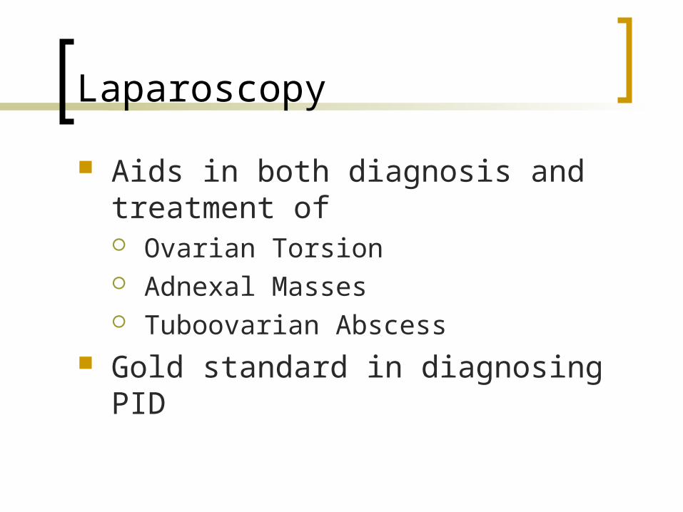
Aids in both diagnosis and treatment of Ovarian Torsion Adnexal Masses Tuboovarian Abscess
Gold standard in diagnosing PID
Treatment
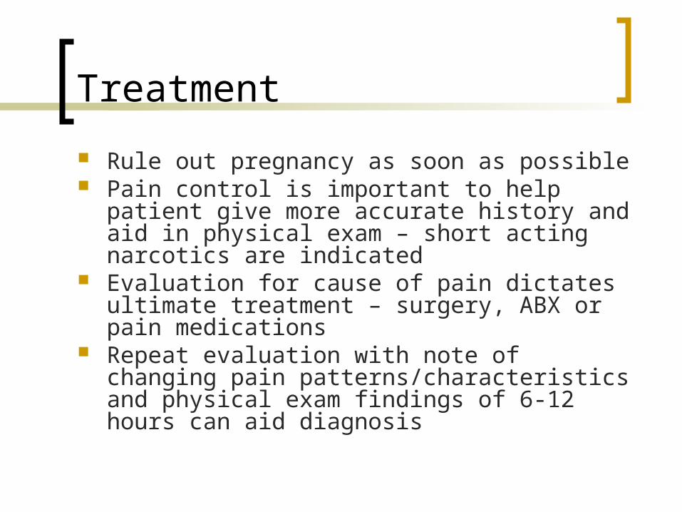
Rule out pregnancy as soon as possible Pain control is important to help patient give
more accurate history and aid in physical exam – short acting narcotics are indicated
Evaluation for cause of pain dictates ultimate treatment – surgery, ABX or pain medications
Repeat evaluation with note of changing pain patterns/characteristics and physical exam findings of 6-12 hours can aid diagnosis
Disposition
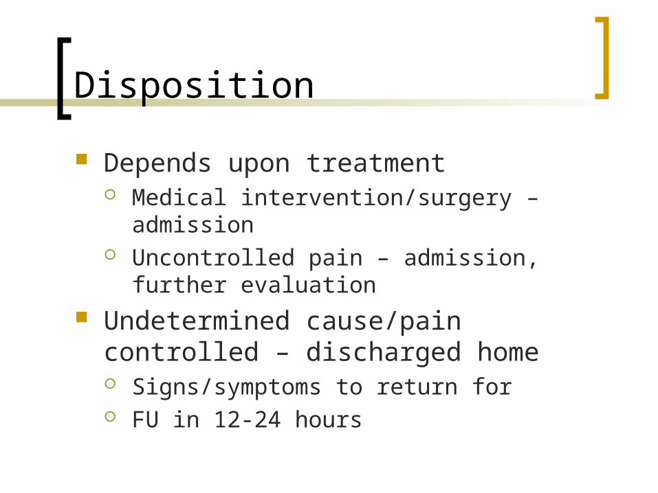
Depends upon treatment Medical intervention/surgery – admission Uncontrolled pain – admission, further
evaluation Undetermined cause/pain controlled –
discharged home Signs/symptoms to return for FU in 12-24 hours
Specific Diagnoses
Functional Ovarian Cysts - pain can result from one of the following Rupture Torsion Infection Hemorrhage
Specific Diagnoses
Tenderness/peritoneal signs may be present Hemorrhage may cause hemodynamic
compromise Ultrasound aids in diagnosis and helps
quantitate blood loss Unilocular, unilateral cysts less than 8 cm
can be observed. Usually resolve within 2 cycles
Specific Diagnoses
Multilocular, large >5 cm or solid cysts suggest another pathology that must be definitively diagnosed
Pelvic ultrasound must be used to confirm FOC
Endometriosis
Up to 15% of females may have – cause is undetermined
Usually present in 30s with pain associated with menses
Endometrium with glandular tissue may be located on ovaries, peritoneum or anywhere in abdominal/pelvic cavity
Endometriosis
Adhesions may form causing chronic pain
Physical exam may show diffuse or localized tenderness
Ultrasound may show endometriomas Diagnosis is made with laparoscopy Therapy is hormonal therapy,
analgesics
Adenomyosis
Caused by endometrial glands and stroma invading myometrium
Pt is typically in 40’s and presents with dysmenorrhea and menorrhagia
Physical exam may show enlarged uterus or mass Diagnosis rarely made in ED – endometrial biopsy
needed to rule out endometrial CA Therapy in ED is pain control Hormonal therapy and hysterectomy may be
needed
Adnexal Torsion
Surgical emergency – pain relief and for preservation of ovary
Torsion can be intermittent – can present with sudden onset of unrelenting pain or sharp intermittent pains with dull aching pain
Ovarian masses or cysts increase risk
Adnexal Torsion
PE may demonstrate involuntary guarding and rebound
Ultrasound with Doppler makes diagnosis
Consult surgery / OB/GYN early
Leiomyomas (Fibroids)
Most common pelvic tumor and need for surgery in females
Incidence increases after 40 More common in blacks Cause is unclear Cells are responsive to estrogen –
anything that increases estrogen may cause fibroid growth (pregnancy)
Leiomyomas (Fibroids)
Physical exam may reveal pelvic or abdominal masses
Fibroids can be located in all layers of uterus
Have a pseudocapsule – blood vessels rarely able to penetrate – fibroids often outgrew blood supply and degenerate causing pain
Leiomyomas (Fibroids)
Pedunculated fibroids can tourse causing acute pain. May have localized tenderness, involuntary guarding, rebound and fever
Ultrasound may be used to demonstrate size, location, and number of fibroids
ED intervention – analgesia Myomectomy/Hysterectomy for patients who
fail medical management