Embed Size (px)
Citation preview
PHYSIOLOGICAL RESEARCH • ISSN 0862-8408 (print) • ISSN 1802-9973 (online) 2017 Institute of Physiology of the Czech Academy of Sciences, Prague, Czech Republic Fax +420 241 062 164, e-mail: [email protected], www.biomed.cas.cz/physiolres
Physiol. Res. 66: 1021-1028, 2017
Vagotomy Decreases the Neuronal Activities of Medulla Oblongata and Alleviates Neurogenic Inflammation of Airways Induced by Repeated Intra-Esophageal Instillation of HCl in Guinea Pigs Zhe CHEN1*, Hui CHEN2*, Fagui CHEN1*, Dachuan GU2, Lejia SUN2, Weitao ZHANG2, Linfeng FAN2, Yong LIN3, Rong DONG2, Kefang LAI1
*These authors contributed equally to this work. 1State Key Laboratory of Respiratory Disease, The First Affiliated Hospital of Guangzhou Medical University, Guangzhou Institute of Respiratory Disease, Guangzhou, China, 2Medical School of Southeast University, Nanjing, China, 3Nanjing Chest Hospital, Nanjing, China
Received December 15, 2016 Accepted May 26, 2017 On-line September 22, 2017 Summary Neuronal activity in the medulla oblongata and neurogenic inflammation of airways were investigated in a guinea pig model induced by repeated intra-esophageal instillation of hydrochloric acid (HCl) after vagotomy. Unilateral vagotomy was performed in the vagotomy group, while a sham-operation was performed in the sham group. Operation was not conducted in sham control group. Airway inflammation was observed with hematoxylin and eosin (HE) staining. C-fos protein was measured by immunohistochemistry (IHC) and Western blot (WB). Substance P was examined by IHC and enzyme-linked immuno sorbent assay (ELISA). Airway microvascular permeability was detected by evans blue dye (EBD) fluorescence. Inflammation of airway was observed in the trachea and bronchi after chronic HCl perfusion into the lower esophagus, and was alleviated after unilateral vagotomy. C-fos expression in the medulla oblongata was lower in the vagotomy group compared to the sham control and sham groups. Substance P-like immunoreactivity (SP-li), concentration and microvascular leakage in airway were lower in the vagotomy group than that in the other groups. Our results suggest that vagotomy improved neurogenic inflammation of airways and decreased neuronal activities, the afferent nerves and neurons in medulla oblongata may be involved in neurogenic inflammation of airways mediated by esophageal-bronchial reflex. Key words Gastroesophageal reflux • Airway inflammation • Substance P • C-fos • Vagotomy
Corresponding authors K. Lai, State Key Laboratory of Respiratory Disease, The First Affiliated Hospital of Guangzhou Medical University, Guangzhou Institute of Respiratory Disease, 151 Yanjiang Rd., Guangzhou, China. E-mail: [email protected] and R. Dong, Medical School of Southeast University, 87 Dingjiaqiao, Nanjing 210009. E-mail: [email protected] Introduction
Gastroesophageal reflux (GER) is related to chronic cough as well as asthma, and the pathophysiological mechanism is involved in reflux and reflex. It is believed traditionally that the gastric contents reflux into larynx, even airway result in cough; however, esophageal 24 h pH monitoring showed that only distal reflux was observed in many patients with chronic cough. The neurogenic inflammation mediated by esophageal bronchial reflex has been recognized (Klauser et al. 1990, Irwin et al. 1993, Castell and Schnatz 1995, Lai et al. 2013). Previous studies have suggested that neurogenic inflammatory mediators such as substance P (SP) are increased by HCl perfusion into the lower esophagus of guinea pigs (Hamamoto et al. 1997, Liu et al. 2013). The neurogenic inflammatory mediators could be released via sensory nerve terminals, and cause airway microvascular leakage, infiltration of inflammatory cells, cough reflex hypersensitivity. The terminal of vagus nerve is located
1022 Chen et al. Vol. 66 on the medulla oblongata, and the medulla neurons are involved in the respiratory, cardiovascular and digest functions. It is unclear whether vagus nerve may play an important role in the neurogenic inflammation of airway associated with gastroesophageal reflux. We aimed to investigate the effects of vagotomy on neurogenic inflammation in the airways and neuronal activities of the medulla oblongata by repeated intra-esophageal instillation of HCl in guinea pigs. Methods
Animals
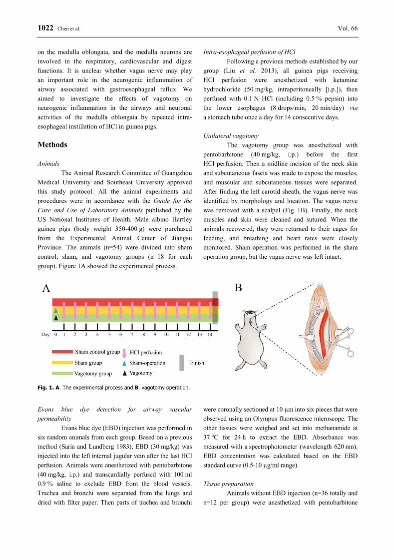
The Animal Research Committee of Guangzhou Medical University and Southeast University approved this study protocol. All the animal experiments and procedures were in accordance with the Guide for the Care and Use of Laboratory Animals published by the US National Institutes of Health. Male albino Hartley guinea pigs (body weight 350-400 g) were purchased from the Experimental Animal Center of Jiangsu Province. The animals (n=54) were divided into sham control, sham, and vagotomy groups (n=18 for each group). Figure 1A showed the experimental process.
Intra-esophageal perfusion of HCl Following a previous methods established by our
group (Liu et al. 2013), all guinea pigs receiving HCl perfusion were anesthetized with ketamine hydrochloride (50 mg/kg, intraperitoneally [i.p.]), then perfused with 0.1 N HCl (including 0.5 % pepsin) into the lower esophagus (8 drops/min, 20 min/day) via a stomach tube once a day for 14 consecutive days.
Unilateral vagotomy
The vagotomy group was anesthetized with pentobarbitone (40 mg/kg, i.p.) before the first HCl perfusion. Then a midline incision of the neck skin and subcutaneous fascia was made to expose the muscles, and muscular and subcutaneous tissues were separated. After finding the left carotid sheath, the vagus nerve was identified by morphology and location. The vagus nerve was removed with a scalpel (Fig. 1B). Finally, the neck muscles and skin were cleaned and sutured. When the animals recovered, they were returned to their cages for feeding, and breathing and heart rates were closely monitored. Sham-operation was performed in the sham operation group, but the vagus nerve was left intact.
Fig. 1. A. The experimental process and B. vagotomy operation. Evans blue dye detection for airway vascular permeability
Evans blue dye (EBD) injection was performed in six random animals from each group. Based on a previous method (Saria and Lundberg 1983), EBD (30 mg/kg) was injected into the left internal jugular vein after the last HCl perfusion. Animals were anesthetized with pentobarbitone (40 mg/kg, i.p.) and transcardially perfused with 100 ml 0.9 % saline to exclude EBD from the blood vessels. Trachea and bronchi were separated from the lungs and dried with filter paper. Then parts of trachea and bronchi
were coronally sectioned at 10 µm into six pieces that were observed using an Olympus fluorescence microscope. The other tissues were weighed and set into methanamide at 37 °C for 24 h to extract the EBD. Absorbance was measured with a spectrophotometer (wavelength 620 nm). EBD concentration was calculated based on the EBD standard curve (0.5-10 µg/ml range).
Tissue preparation
Animals without EBD injection (n=36 totally and n=12 per group) were anesthetized with pentobarbitone
2017 Vagotomy Decreases Neuronal Activity and Airway Inflammation 1023
(40 mg/kg, i.p.). Six random animals in each group were transcardially perfused with 0.3 % phosphate buffered saline (PBS). The bronchi and lungs were removed for ELISA, and the brainstem was removed for WB. The other six animals in each group were transcardially perfused with 0.3 % PBS followed by 4 % paraformaldehyde in PBS. The lungs and brainstem were removed for HE staining and IHC. The lung tissues were embedded in paraffin and sectioned at 5 µm, stained with hematoxylin and eosin, and observed using an Olympus light microscope.
ELISA
The bronchi and lungs were weighed, boiled (100 °C) for 10 min in 1 M acetic acid (1:10, wt/vol), then diluted with 0.1 M PBS and homogenized. Homogenates were transferred to polypropylene tubes and centrifuged (40,000 × g, 4 °C, 20 min). Before measurement, the supernatant was centrifuged again (40,000 × g, 4 °C, 20 min). SP concentration was measured with an ELISA kit following the instructions.
Western blot
The brainstem (total thickness 2 mm from rostral and caudal to obex) samples were placed in lysis buffer containing protease inhibitors, homogenized, and then centrifuged. The protein concentrations were measured using a BCA protein assay kit. Fifty micrograms of total protein was separated by SDS-PAGE, then transferred by electro-blotting onto a PVDF membrane. The membranes were blocked with 3 % BSA in Tris-buffered saline containing 0.1 % Tween-20 (TBST) for 1 h, followed by incubation overnight at 4 °C with the primary antibody (rabbit anti-Fos, 1:1,000, Santa Cruz, USA). The membranes were washed with TBST and incubated for 1 h with the appropriate horseradish peroxidase (HRP)-conjugated secondary antibody (1:5,000, Invitrogen). The protein blots were detected using an enhanced chemiluminescent substrate kit, and exposed to CL-XPosure film.
Immunohistochemistry
Brainstems and lung tissues were placed in 4 % paraformaldehyde at 4 °C for 4 h, and the brainstem samples were cryoprotected in 30 % sucrose at 4 °C overnight. Tissues were rapidly frozen with OCT and coronally sectioned at 40 µm (brainstem tissues at 20 µm) using a Leica freezing microtome. The thickness of total brainstem sections was 2 mm from rostral and caudal to obex. Tissue sections were incubated with 3 % H2O2 for
15 min to block endogenous peroxidase activity, washed with 0.3 % PBS (3 × 5 min), incubated for 1 h at room temperature with a blocking solution (10 % goat serum), and incubated overnight with the primary antibody (mouse anti-SP, 1:200, Abcam or rabbit anti-Fos, 1:500, Santa Cruz, USA). The tissue was washed with 0.3 % PBS (3 × 5 min), followed by incubation for 1 h at room temperature with a biotinylated secondary antibody (goat anti-mouse or goat anti-rabbit; 1:500; Abcam). After washing with 0.3 % PBS (3 × 5 min), sections were incubated for 30 min with avidin/biotinylated horseradish peroxidase, then washed with 0.3 % PBS (3 × 5 min) and reacted with 0.05 % 3,3’-diaminobenzidine (DAB, 5 min) (lung tissues) or 0.03 % 3-amino-9-ethylcarbazole (AEC, 10 min) (brainstem tissues) staining as a chromogen. Reaction time of tissue sections should be monitored under microscope, and sections were observed using an Olympus light microscope.
Statistical analysis
Data are expressed as means ± standard deviations and were analyzed for statistical differences using an analysis of variance (ANOVA) in SPSS 17.0 software. A P<0.05 was considered statistically significant. The mean EBD density, IHC immune-reactivity, and WB density were determined using Image-Pro Plus. The SP density (mean of density) we measured was mainly located on bronchial surrounding, which SP expression was mainly distributed on. One section in every six consecutive pieces of brain sections was selected, and total eight brain sections for immunohistochemistry were selected for statistics in each experimental animal. One section in every six consecutive pieces of trachea, bronchia or lung sections was selected, and total six sections in each tissue for immunohistochemistry or evans blue dye staining were selected for statistics in each experimental animal. Results
Animals that received left vagotomy breathed slower after surgery, but most animals recovered within 2 days. No animals died due to surgery. Pathologic changes in the airway
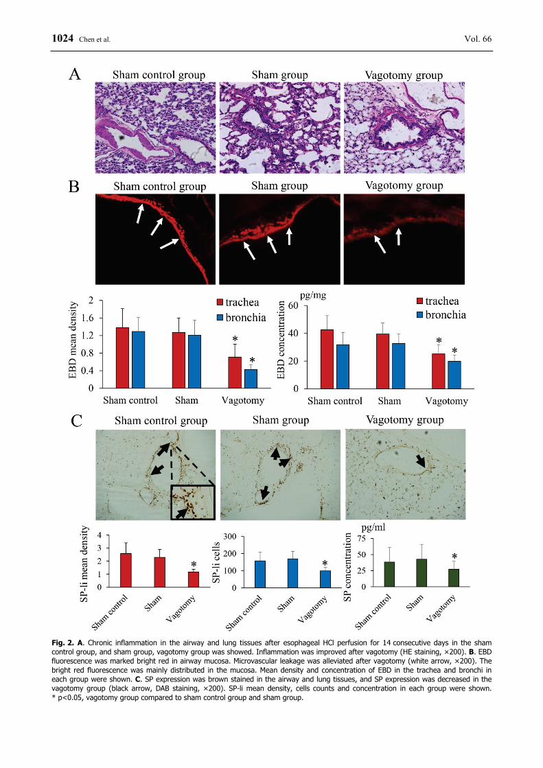
The bronchi of guinea pigs with 14 days of intra-esophagus perfusion with HCl were significantly inflamed as observed under a light microscope, but inflammation was alleviated by vagotomy (Fig. 2A).
1024 Chen et al. Vol. 66
Fig. 2. A. Chronic inflammation in the airway and lung tissues after esophageal HCl perfusion for 14 consecutive days in the sham control group, and sham group, vagotomy group was showed. Inflammation was improved after vagotomy (HE staining, ×200). B. EBD fluorescence was marked bright red in airway mucosa. Microvascular leakage was alleviated after vagotomy (white arrow, ×200). The bright red fluorescence was mainly distributed in the mucosa. Mean density and concentration of EBD in the trachea and bronchi in each group were shown. C. SP expression was brown stained in the airway and lung tissues, and SP expression was decreased in the vagotomy group (black arrow, DAB staining, ×200). SP-li mean density, cells counts and concentration in each group were shown. * p<0.05, vagotomy group compared to sham control group and sham group.
2017 Vagotomy Decreases Neuronal Activity and Airway Inflammation 1025
Vascular permeability in airway was decreased by vagotomy
The mean density of EBD fluorescence in trachea was lower in the vagotomy group than in the sham control and sham groups (0.71±0.29 versus 1.38±0.43 and 1.27±0.33, p<0.05), also in bronchi (0.43±0.11 versus 1.29±0.32 and 1.21±0.34, p<0.05). The concentration of EBD in the trachea was lower in the vagotomy group than in the other groups (25.17±6.53 versus 42.53±10.19 and 39.48±7.91, p<0.05), also in bronchi (19.91±4.29 versus 31.78±8.83 and 32.67±6.82, p<0.05). Chronic perfusion induced an increase in airway vascular permeability, and sham-operation had no effect on vascular permeability (p>0.05 compared to sham control group), but vagotomy resulted in a decrease in perme- ability compared to the other groups (p<0.05).
EBD fluorescence and concentration are shown in Figure 2B.
SP expression in airway was inhibited by vagotomy
SP expression was mainly located on the airway and lungs, particularly around the bronchi. SP density in bronchial surrounding was measured. SP expression was lower in the vagotomy group (mean density 1.17±0.21, positive cells 99.23±20.67) than in the sham control (mean density 2.58±0.82, positive cells 158.17±49.61) and sham groups (mean density 2.29±0.61, positive cells 169.67±42.23) (both p<0.05). SP concentration was also lower in the vagotomy group (27.34±12.31 versus 38.26±22.71 in sham control group and 42.76±23.09 in sham group; p<0.05). There were no differences between the sham control and sham groups (p>0.05). The data are shown in Figure 2C.
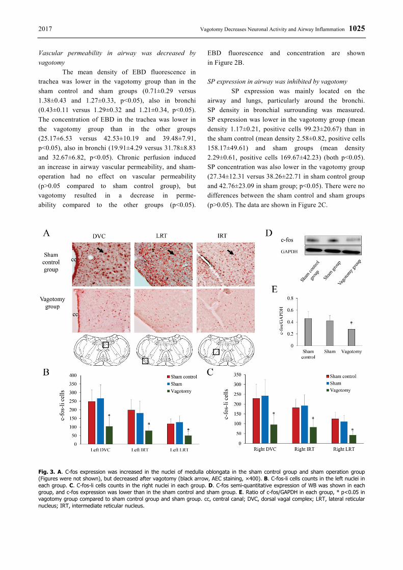
Fig. 3. A. C-fos expression was increased in the nuclei of medulla oblongata in the sham control group and sham operation group (Figures were not shown), but decreased after vagotomy (black arrow, AEC staining, ×400). B. C-fos-li cells counts in the left nuclei in each group. C. C-fos-li cells counts in the right nuclei in each group. D. C-fos semi-quantitative expression of WB was shown in each group, and c-fos expression was lower than in the sham control and sham group. E. Ratio of c-fos/GAPDH in each group, * p<0.05 in vagotomy group compared to sham control group and sham group. cc, central canal; DVC, dorsal vagal complex; LRT, lateral reticular nucleus; IRT, intermediate reticular nucleus.
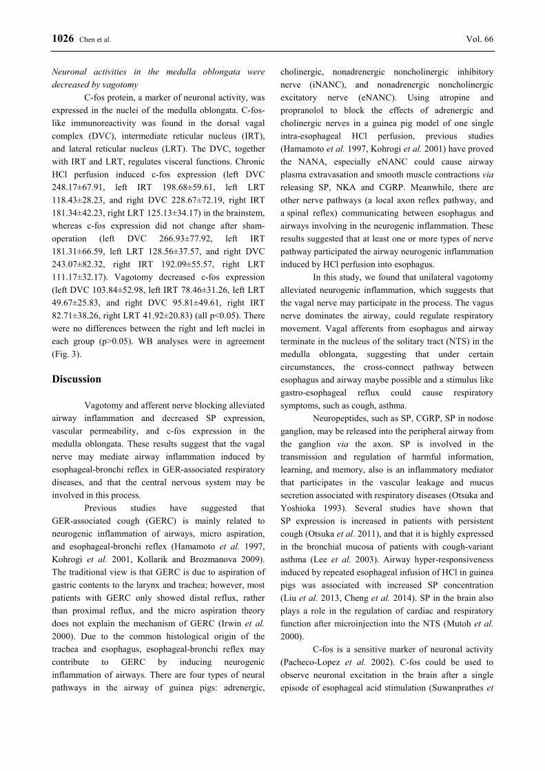
1026 Chen et al. Vol. 66 Neuronal activities in the medulla oblongata were decreased by vagotomy
C-fos protein, a marker of neuronal activity, was expressed in the nuclei of the medulla oblongata. C-fos-like immunoreactivity was found in the dorsal vagal complex (DVC), intermediate reticular nucleus (IRT), and lateral reticular nucleus (LRT). The DVC, together with IRT and LRT, regulates visceral functions. Chronic HCl perfusion induced c-fos expression (left DVC 248.17±67.91, left IRT 198.68±59.61, left LRT 118.43±28.23, and right DVC 228.67±72.19, right IRT 181.34±42.23, right LRT 125.13±34.17) in the brainstem, whereas c-fos expression did not change after sham-operation (left DVC 266.93±77.92, left IRT 181.31±66.59, left LRT 128.56±37.57, and right DVC 243.07±82.32, right IRT 192.09±55.57, right LRT 111.17±32.17). Vagotomy decreased c-fos expression (left DVC 103.84±52.98, left IRT 78.46±31.26, left LRT 49.67±25.83, and right DVC 95.81±49.61, right IRT 82.71±38.26, right LRT 41.92±20.83) (all p<0.05). There were no differences between the right and left nuclei in each group (p>0.05). WB analyses were in agreement (Fig. 3). Discussion
Vagotomy and afferent nerve blocking alleviated airway inflammation and decreased SP expression, vascular permeability, and c-fos expression in the medulla oblongata. These results suggest that the vagal nerve may mediate airway inflammation induced by esophageal-bronchi reflex in GER-associated respiratory diseases, and that the central nervous system may be involved in this process.
Previous studies have suggested that GER-associated cough (GERC) is mainly related to neurogenic inflammation of airways, micro aspiration, and esophageal-bronchi reflex (Hamamoto et al. 1997, Kohrogi et al. 2001, Kollarik and Brozmanova 2009). The traditional view is that GERC is due to aspiration of gastric contents to the larynx and trachea; however, most patients with GERC only showed distal reflux, rather than proximal reflux, and the micro aspiration theory does not explain the mechanism of GERC (Irwin et al. 2000). Due to the common histological origin of the trachea and esophagus, esophageal-bronchi reflex may contribute to GERC by inducing neurogenic inflammation of airways. There are four types of neural pathways in the airway of guinea pigs: adrenergic,
cholinergic, nonadrenergic noncholinergic inhibitory nerve (iNANC), and nonadrenergic noncholinergic excitatory nerve (eNANC). Using atropine and propranolol to block the effects of adrenergic and cholinergic nerves in a guinea pig model of one single intra-esophageal HCl perfusion, previous studies (Hamamoto et al. 1997, Kohrogi et al. 2001) have proved the NANA, especially eNANC could cause airway plasma extravasation and smooth muscle contractions via releasing SP, NKA and CGRP. Meanwhile, there are other nerve pathways (a local axon reflex pathway, and a spinal reflex) communicating between esophagus and airways involving in the neurogenic inflammation. These results suggested that at least one or more types of nerve pathway participated the airway neurogenic inflammation induced by HCl perfusion into esophagus.
In this study, we found that unilateral vagotomy alleviated neurogenic inflammation, which suggests that the vagal nerve may participate in the process. The vagus nerve dominates the airway, could regulate respiratory movement. Vagal afferents from esophagus and airway terminate in the nucleus of the solitary tract (NTS) in the medulla oblongata, suggesting that under certain circumstances, the cross-connect pathway between esophagus and airway maybe possible and a stimulus like gastro-esophageal reflux could cause respiratory symptoms, such as cough, asthma.
Neuropeptides, such as SP, CGRP, SP in nodose ganglion, may be released into the peripheral airway from the ganglion via the axon. SP is involved in the transmission and regulation of harmful information, learning, and memory, also is an inflammatory mediator that participates in the vascular leakage and mucus secretion associated with respiratory diseases (Otsuka and Yoshioka 1993). Several studies have shown that SP expression is increased in patients with persistent cough (Otsuka et al. 2011), and that it is highly expressed in the bronchial mucosa of patients with cough-variant asthma (Lee et al. 2003). Airway hyper-responsiveness induced by repeated esophageal infusion of HCl in guinea pigs was associated with increased SP concentration (Liu et al. 2013, Cheng et al. 2014). SP in the brain also plays a role in the regulation of cardiac and respiratory function after microinjection into the NTS (Mutoh et al. 2000).
C-fos is a sensitive marker of neuronal activity (Pacheco-Lopez et al. 2002). C-fos could be used to observe neuronal excitation in the brain after a single episode of esophageal acid stimulation (Suwanprathes et
2017 Vagotomy Decreases Neuronal Activity and Airway Inflammation 1027
al. 2003). Neurons in the cNTS (a subnucleus of the NTS), the location of central cough receptor terminals, were critical components involved in cough gating (Canning and Mori 2010). In this study, expression of c-fos protein was found in the DVC, IRT, and LRT. The NTS has fiber communications with the dorsal motor nucleus of the vagus (DMV) and area postrema (AP), and thus is called the dorsal vagal complex (DVC). The DMV directly receives vagal sensory fiber projections, and innervates the airway and digestive tract via efferent fibers. The DVC, together with the IRT, nucleus ambiguus, and ventrolateral medulla, form the medullary visceral zone (MVZ). The MVZ plays a key role in visceral functions. We found that c-fos expression was reduced after vagotomy, and these nuclei may be involved in the regulation of airway inflammation. The medulla oblongata nuclei were activated after HCl perfusion into esophagus, which was alleviated by vagotomy, indicating that the central neuronal sensitization may be increased in this process related to gastroesophageal reflux, and brain activities may regulate airway, as CNS activities “enlarge” inflammations of lung during asthma attack (Mazzone and Canning 2002, Widdicombe 2003). Higher brain nuclei involving in the regulation of respiratory sensations have been proved in an animal model (McGovern et al. 2015), whether higher brain nuclei regulate airway inflammation was unclear. In
our previous study, we observed that the SP expressions were increased in the nodose ganglion, the first sensory neurons in the vagal afferent nerves, also previous study (Mutoh et al. 2000) have proved that SP microinjection into NTS could enhance C-fiber output, and regulate respiratory movement. We would observe whether airway inflammation could be regulated by the changes of nuclei activities and neurotransmitter concentration, and also to explore the activities of nuclei in the high brain.
In conclusion, vagotomy alleviated neurogenic inflammation of airways and neuronal activity in the medulla oblongata in guinea pigs that received intra-esophagus perfusion of HCl. Afferent nerves and brainstem neurons may be involved in the process of inflammation. Conflict of Interest There is no conflict of interest. Acknowledgements This work was supported by Grants 81770098, 30370621 and 30871000 from the National Natural Science Foundation of China, and Grants 2016OP015 and 2007DA780154F0905 from Open Project of State Key Laboratory of Respiratory Disease. Authors would like to thank Dr. Chunli Liu and Dr. Ruchong Chen for their assistance of animal model.
References CANNING BJ, MORI N: An essential component to brainstem cough gating identified in anesthetized guinea pigs.
FASEB J 24: 3916-3926, 2010. CASTELL DO, SCHNATZ PF: Gastroesophageal reflux disease and asthma. Reflux or reflex. Chest 108: 1186-1187,
1995. CHENG YM, CAO AL, ZHENG JP, WANG HW, SUN YS, LIU CF, ZHANG BB, WANG Y, ZHU SL, WU DZ:
Airway hyperresponsiveness induced by repeated esophageal infusion of HCl in guinea pigs. Am J Respir Cell Mol Biol 51: 701-708, 2014.
HAMAMOTO J, KOHROGI H, KAWANO O, IWAGOE H, FUJII K, HIRATA N, ANDO M: Esophageal stimulation by hydrochloric acid causes neurogenic inflammation in the airways in guinea pigs. J Appl Physiol (1985) 82: 738-745, 1997.
IRWIN RS, FRENCH CL, CURLEY FJ, ZAWACKI JK, BENNETT FM: Chronic cough due to gastroesophageal reflux. Clinical, diagnostic, and pathogenetic aspects. Chest 104: 1511-1517, 1993.
IRWIN RS, MADISON JM, FRAIRE AE: The cough reflex and its relation to gastroesophageal reflux. Am J Med 108 (Suppl 4a): 73S-78S, 2000.
KLAUSER AG, SCHINDLBECK NE, MULLER-LISSNER SA: Symptoms in gastro-oesophageal reflux disease. Lancet 335: 205-208, 1990.
KOHROGI H, HAMAMOTO J, KAWANO O, IWAGOE H, FUJII K, HIRATA N, ANDO M: The role of substance P release in the lung with esophageal acid. Am J Med 111 (Suppl 8A): 25S-30S, 2001.
1028 Chen et al. Vol. 66 KOLLARIK M, BROZMANOVA M: Cough and gastroesophageal reflux: insights from animal models. Pulm
Pharmacol Ther 22: 130-134, 2009. LAI K, CHEN R, LIN J, HUANG K, SHEN H, KONG L, ZHOU X, LUO Z, YANG L, WEN F, ZHONG N:
A prospective, multicenter survey on causes of chronic cough in China. Chest 143: 613-620, 2013. LEE SY, KIM MK, SHIN C, SHIM JJ, KIM HK, KANG KH, YOO SH, IN KH: Substance P-immunoreactive nerves
in endobronchial biopsies in cough-variant asthma and classic asthma. Respiration 70: 49-53, 2003. LIU C, CHEN R, LUO W, LAI K, ZHONG N: Neurogenic airway inflammation induced by repeated intra-esophageal
instillation of HCl in guinea pigs. Inflammation 36: 493-500, 2013. MAZZONE SB, CANNING BJ: Central nervous system control of the airways: pharmacological implications. Curr
Opin Pharmacol 2: 220-228, 2002. MCGOVERN AE, DRIESSEN AK, SIMMONS DG, POWELL J, DAVIS-POYNTER N, FARRELL MJ, MAZZONE
SB: Distinct brainstem and forebrain circuits receiving tracheal sensory neuron inputs revealed using a novel conditional anterograde transsynaptic viral tracing system. J Neurosci 35: 7041-7055, 2015.
MUTOH T, BONHAM AC, JOAD JP: Substance P in the nucleus of the solitary tract augments bronchopulmonary C fiber reflex output. Am J Physiol Regul Integr Comp Physiol 279: R1215-R1223, 2000.
OTSUKA M, YOSHIOKA K: Neurotransmitter functions of mammalian tachykinins. Physiol Rev 73: 229-308, 1993. OTSUKA K, NIIMI A, MATSUMOTO H, ITO I, YAMAGUCHI M, MATSUOKA H, JINNAI M, OGUMA T,
TAKEDA T, NAKAJI H, CHIN K, SASAKI K, AOYAMA N, MISHIMA M: Plasma substance P levels in patients with persistent cough. Respiration 82: 431-438, 2011.
PACHECO-LOPEZ G, ESPINOSA E, ZAMORANO-ROJAS HM, RAMIREZ-AMAYA V, BERMUDEZ-RATTONI F: Peripheral protein immunization induces rapid activation of the CNS, as measured by c-Fos expression. J Neuroimmunol 131: 50-59, 2002.
SARIA A, LUNDBERG JM: Evans blue fluorescence: quantitative and morphological evaluation of vascular permeability in animal tissues. J Neurosci Methods 8: 41-49, 1983.
SUWANPRATHES P, NGU M, ING A, HUNT G, SEOW F: c-Fos immunoreactivity in the brain after esophageal acid stimulation. Am J Med 115 (Suppl 3A): 31S-38S, 2003.
WIDDICOMBE JG: Overview of neural pathways in allergy and asthma. Pulm Pharmacol Ther 16: 23-30, 2003.