Embed Size (px)
Citation preview
VALIDATION OF A QUANTITATIVE DETERMINATION METHOD
OF DICLOFENAC FOR IN VITRO BIOEQUIVALENCE
EVALUATION OF TRANSDERMAL GEL PREPARATIONS
S. I. Guseva,1 M. V. Karlina,2 O. N. Pozharitskaya,2 A. N. Shikov,1, 2 and N. M. Faustova3
Translated from Khimiko-Farmatsevticheskii Zhurnal, Vol. 44, No. 1, pp. 46 – 49, January, 2010.
Original article submitted January 1, 2009.
A quantitative determination method of diclofenac for studying the bioequivalence of transdermal gel prepa-
rations of this drug has been validated. It was established that the proposed method is precise, reproducible,
and linear in a certain concentration range. The release of diclofenac from transdermal gels with different vis-
cosities and from the reference preparation was studied using the paddle-over-disk technique. Drug release
from the transdermal gel preparation was modeled using various mathematical laws including the Higuchi
law, first-order kinetics, cube root, and Weibull function. An analysis of the experimental data showed that the
release of diclofenac from gels proceeds by diffusion and obeys the Higuchi law. It was found that the rate of
drug release from the gel depends significantly on the viscosity. Similarity coefficients were calculated in or-
der to compare diclofenac dissolution profiles and to determine their equivalence. It was concluded that ex-
perimental samples of diclofenac gel with medium and high viscosity were equivalent to the reference prepa-
ration.
Key words: diclofenac, transdermal gels, bioequivalence testing in vitro.
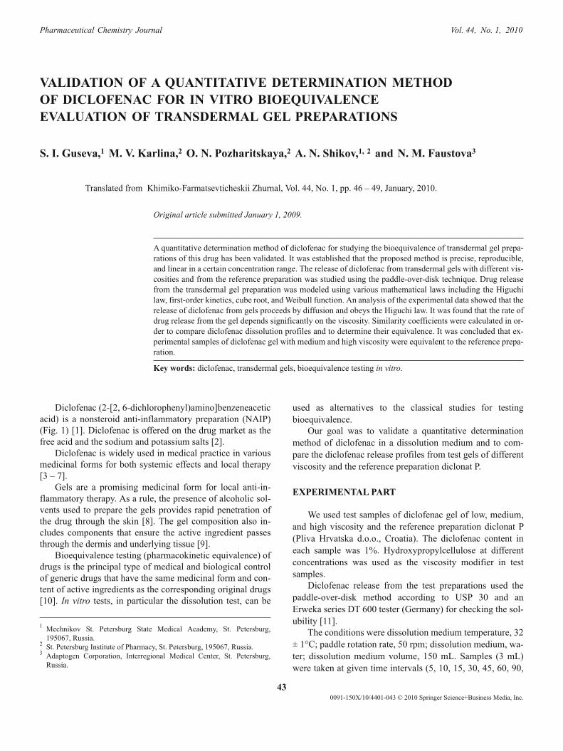
Diclofenac (2-[2, 6-dichlorophenyl)amino]benzeneacetic
acid) is a nonsteroid anti-inflammatory preparation (NAIP)
(Fig. 1) [1]. Diclofenac is offered on the drug market as the
free acid and the sodium and potassium salts [2].
Diclofenac is widely used in medical practice in various
medicinal forms for both systemic effects and local therapy
[3 – 7].
Gels are a promising medicinal form for local anti-in-
flammatory therapy. As a rule, the presence of alcoholic sol-
vents used to prepare the gels provides rapid penetration of
the drug through the skin [8]. The gel composition also in-
cludes components that ensure the active ingredient passes
through the dermis and underlying tissue [9].
Bioequivalence testing (pharmacokinetic equivalence) of
drugs is the principal type of medical and biological control
of generic drugs that have the same medicinal form and con-
tent of active ingredients as the corresponding original drugs
[10]. In vitro tests, in particular the dissolution test, can be
used as alternatives to the classical studies for testing
bioequivalence.
Our goal was to validate a quantitative determination
method of diclofenac in a dissolution medium and to com-
pare the diclofenac release profiles from test gels of different
viscosity and the reference preparation diclonat P.
EXPERIMENTAL PART
We used test samples of diclofenac gel of low, medium,
and high viscosity and the reference preparation diclonat P
(Pliva Hrvatska d.o.o., Croatia). The diclofenac content in
each sample was 1%. Hydroxypropylcellulose at different
concentrations was used as the viscosity modifier in test
samples.
Diclofenac release from the test preparations used the
paddle-over-disk method according to USP 30 and an
Erweka series DT 600 tester (Germany) for checking the sol-
ubility [11].
The conditions were dissolution medium temperature, 32
± 1°C; paddle rotation rate, 50 rpm; dissolution medium, wa-
ter; dissolution medium volume, 150 mL. Samples (3 mL)
were taken at given time intervals (5, 10, 15, 30, 45, 60, 90,
43
0091-150X/10/4401-043 © 2010 Springer Science+Business Media, Inc.
Pharmaceutical Chemistry Journal Vol. 44, No. 1, 2010
1Mechnikov St. Petersburg State Medical Academy, St. Petersburg,
195067, Russia.2
St. Petersburg Institute of Pharmacy, St. Petersburg, 195067, Russia.3
Adaptogen Corporation, Interregional Medical Center, St. Petersburg,
Russia.
120, 180 min) and filtered through a 0.45-�m filter. The dis-
solution medium volume was restored using water.
Quantitative analysis of diclofenac used a spectrophoto-
metric method at 275 nm. Measurements were made on a
PharmaSpec 1700 UV-Vis spectrophotometer (Shimadzu, Ja-
pan).
The diclofenac dissolution process was modeled using
the following methods: Higuchi equation, first-order kinetic
equation, cube-root law, and Weibull model [12].
Dissolution rate constants were calculated by
least-squares methods [13].
Dissolution profiles were compared by calculating the
similarity coefficient f2
[14, 15] using the formula:
fn
R Tj j
j
n
2
2
1
0 5
50 11
100� � �
�
�
�
�
�
�
�
�
�
�
�
�
��
�
�
�
��
�
log | |
.
�
,
where n is the number of time points and Rj
and Tj
are the
percent dissolved compound from the preparation at each
time point j.
RESULTS AND DISCUSSION
Validation of quantitative determination method
Process validation is an important part of a quality assur-
ance and control system and is obligatory in practice for
quality manufacture of a medical product.
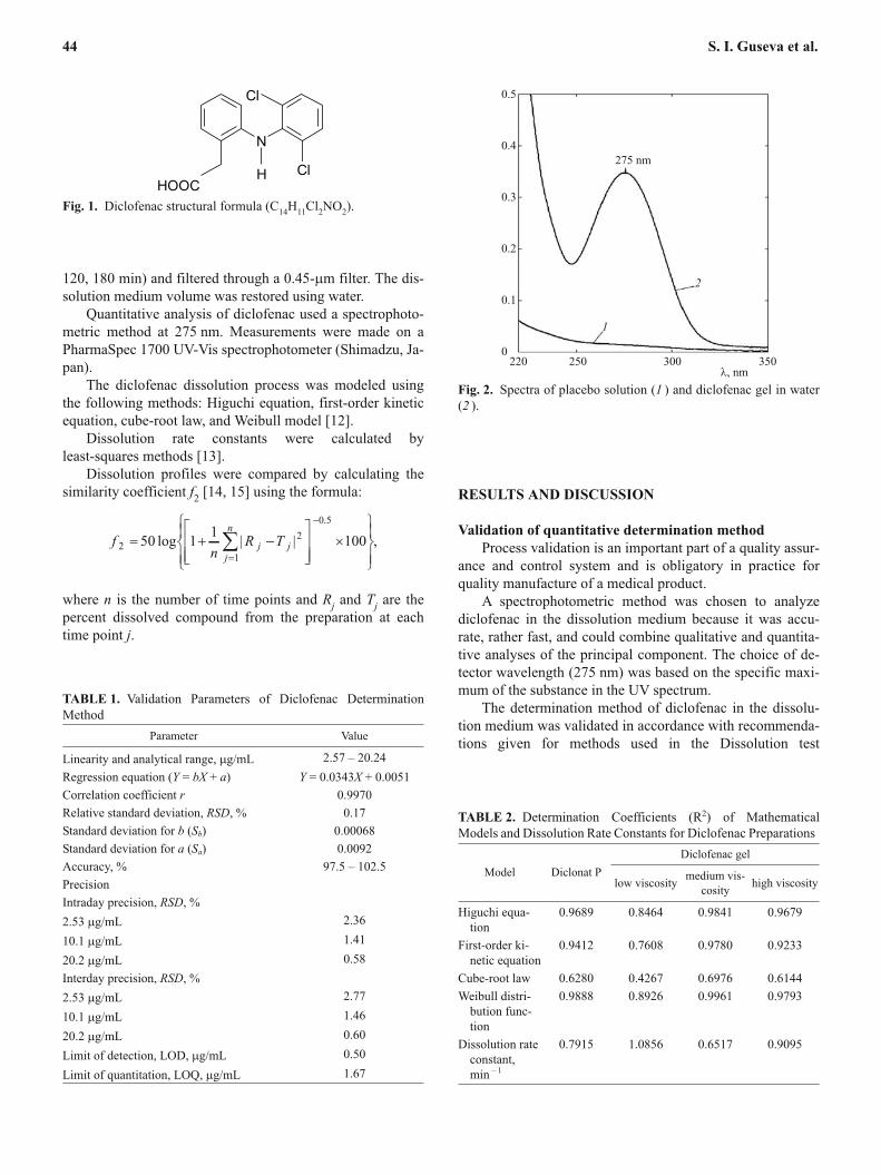
A spectrophotometric method was chosen to analyze
diclofenac in the dissolution medium because it was accu-
rate, rather fast, and could combine qualitative and quantita-
tive analyses of the principal component. The choice of de-
tector wavelength (275 nm) was based on the specific maxi-
mum of the substance in the UV spectrum.
The determination method of diclofenac in the dissolu-
tion medium was validated in accordance with recommenda-
tions given for methods used in the Dissolution test
44 S. I. Guseva et al.
N
HOOC
Cl
ClH
Fig. 1. Diclofenac structural formula (C14
H11
Cl2NO
2).
0.5
0.4
0.3
0.2
0.1
0
220 250 300 350
�, nm
275 nm
1
2
Fig. 2. Spectra of placebo solution (1 ) and diclofenac gel in water
(2 ).
TABLE 1. Validation Parameters of Diclofenac Determination
Method
Parameter Value
Linearity and analytical range, �g/mL 2.57 – 20.24
Regression equation (Y = bX + a) Y = 0.0343X + 0.0051
Correlation coefficient r 0.9970
Relative standard deviation, RSD, % 0.17
Standard deviation for b (Sb) 0.00068
Standard deviation for a (Sa) 0.0092
Accuracy, % 97.5 – 102.5
Precision
Intraday precision, RSD, %
2.53 �g/mL 2.36
10.1 �g/mL 1.41
20.2 �g/mL 0.58
Interday precision, RSD, %
2.53 �g/mL 2.77
10.1 �g/mL 1.46
20.2 �g/mL 0.60
Limit of detection, LOD, �g/mL 0.50
Limit of quantitation, LOQ, �g/mL 1.67
TABLE 2. Determination Coefficients (R2) of Mathematical
Models and Dissolution Rate Constants for Diclofenac Preparations
Model Diclonat P
Diclofenac gel
low viscositymedium vis-
cosityhigh viscosity
Higuchi equa-
tion
0.9689 0.8464 0.9841 0.9679
First-order ki-
netic equation
0.9412 0.7608 0.9780 0.9233
Cube-root law 0.6280 0.4267 0.6976 0.6144
Weibull distri-
bution func-
tion
0.9888 0.8926 0.9961 0.9793
Dissolution rate
constant,
min– 1
0.7915 1.0856 0.6517 0.9095
[16 – 18]. The validation method parameters were the speci-
ficity, linearity and analytical range of the method, accuracy
(or correctness), limit of detection, and limit of quantitation.
Table 1 lists the validation parameters for the method.
Spectra of a model mixture of excipients included in the
preparation (placebo) were recorded in order to check the
specificity of the method. It was found that the excipients did
not interfere with the diclofenac determination in the dissolu-
tion medium (Fig. 2).
The resulting values of the statistical criteria for the ex-
amined concentration range satisfied the requirements [18].
Therefore, the validated method can be used for the Dissolu-
tion test.
The stability of diclofenac in the dissolution medium was
also evaluated. It was shown that the solutions were stable
for 1 d at room temperature and for 2 d at 8 – 10°C.
Diclofenac release rate
Topical application was modeled using the pad-
dle-over-disk method that is applicable for transdermal me-
dicinal forms [11]. Distilled water was chosen as the dissolu-
tion medium. A total of 12 determinations were made for
each preparation. Figure 3 shows the results.
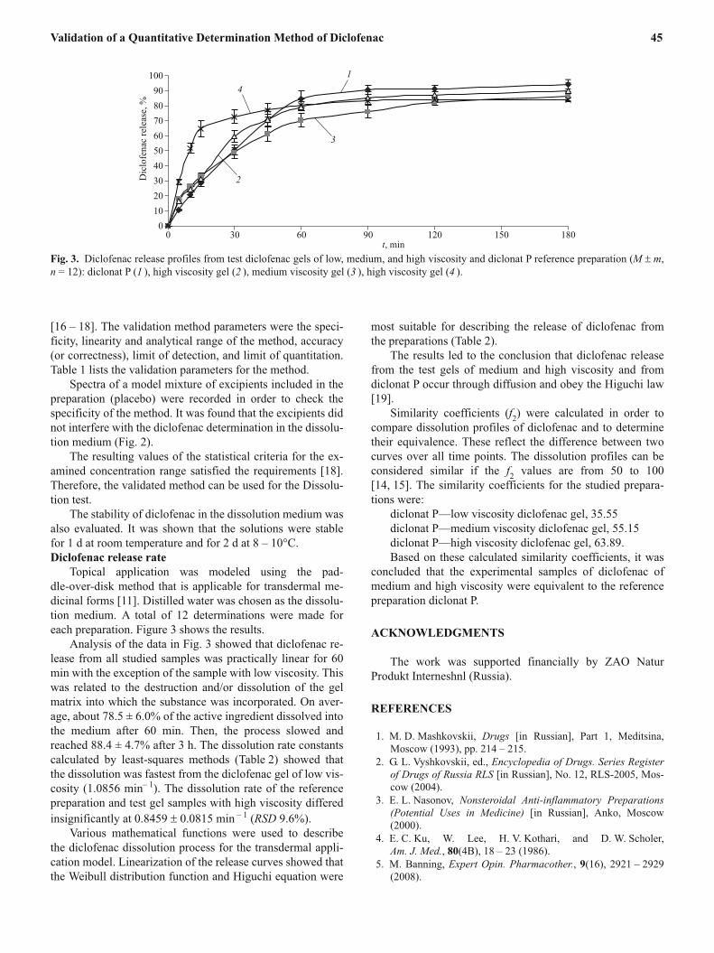
Analysis of the data in Fig. 3 showed that diclofenac re-
lease from all studied samples was practically linear for 60
min with the exception of the sample with low viscosity. This
was related to the destruction and/or dissolution of the gel
matrix into which the substance was incorporated. On aver-
age, about 78.5 ± 6.0% of the active ingredient dissolved into
the medium after 60 min. Then, the process slowed and
reached 88.4 ± 4.7% after 3 h. The dissolution rate constants
calculated by least-squares methods (Table 2) showed that
the dissolution was fastest from the diclofenac gel of low vis-
cosity (1.0856 min– 1
). The dissolution rate of the reference
preparation and test gel samples with high viscosity differed
insignificantly at 0.8459 � 0.0815 min– 1
(RSD 9.6%).
Various mathematical functions were used to describe
the diclofenac dissolution process for the transdermal appli-
cation model. Linearization of the release curves showed that
the Weibull distribution function and Higuchi equation were
most suitable for describing the release of diclofenac from
the preparations (Table 2).
The results led to the conclusion that diclofenac release
from the test gels of medium and high viscosity and from
diclonat P occur through diffusion and obey the Higuchi law
[19].
Similarity coefficients (f2) were calculated in order to
compare dissolution profiles of diclofenac and to determine
their equivalence. These reflect the difference between two
curves over all time points. The dissolution profiles can be
considered similar if the f2
values are from 50 to 100
[14, 15]. The similarity coefficients for the studied prepara-
tions were:
diclonat P—low viscosity diclofenac gel, 35.55
diclonat P—medium viscosity diclofenac gel, 55.15
diclonat P—high viscosity diclofenac gel, 63.89.
Based on these calculated similarity coefficients, it was
concluded that the experimental samples of diclofenac of
medium and high viscosity were equivalent to the reference
preparation diclonat P.
ACKNOWLEDGMENTS
The work was supported financially by ZAO Natur
Produkt Interneshnl (Russia).
REFERENCES
1. M. D. Mashkovskii, Drugs [in Russian], Part 1, Meditsina,
Moscow (1993), pp. 214 – 215.
2. G. L. Vyshkovskii, ed., Encyclopedia of Drugs. Series Register
of Drugs of Russia RLS [in Russian], No. 12, RLS-2005, Mos-
cow (2004).
3. E. L. Nasonov, Nonsteroidal Anti-inflammatory Preparations
(Potential Uses in Medicine) [in Russian], Anko, Moscow
(2000).
4. E. C. Ku, W. Lee, H. V. Kothari, and D. W. Scholer,
Am. J. Med., 80(4B), 18 – 23 (1986).
5. M. Banning, Expert Opin. Pharmacother., 9(16), 2921 – 2929
(2008).
Validation of a Quantitative Determination Method of Diclofenac 45
100
90
80
70
60
50
40
30
20
10
0
0 30 60 90 120 150 180
t, min
4
1
3
2Dic
lofenac
rele
ase,%
Fig. 3. Diclofenac release profiles from test diclofenac gels of low, medium, and high viscosity and diclonat P reference preparation (M � m,
n = 12): diclonat P (1 ), high viscosity gel (2 ), medium viscosity gel (3 ), high viscosity gel (4 ).
6. P. S. Tugwell, G. A. Wells, and J. Z. Shainhouse, J. Rheumatol.,
31(10), 2002 – 2012 (2004).
7. N. V. Chichasova, Consilium Med., 3(5), (2001).
8. S. Baboota, F. Shakeel, and K. Kohli, Methods Find. Exp. Clin.
Pharmacol., 28(2), 109 – 114 (2006).
9. A. Arellano, S. Santoyo, C. Martin, and P. Ygartua, Eur. J.
Pharm. Sci., 7(2), 129 – 135 (1999).
10. Note for Guidance on the Investigation of Bioavailablity and
Bioequivalence, The Eurpoean Agency for the Evaluation of
Medical Products, London (2001).
11. The United States Pharmacopoeia, The National Formulary 30 /
25 (2007), Article .
12. P. Costa and L. J. M. Sousa, Drug. Dev. Ind. Pharm., 29(1),
89 – 97 (2003).
13. V. D. Ponomarev, V. G. Belikov, and N. I. Kokovkin-Shcher-
bak, Mathematical Methods in Pharmacy [in Russian], Medi-
tsina, Moscow (1983).
14. P. Costa and L. J. M. Sousa, Eur. J. Pharm. Sci., 13, 123 – 133
(2001).
15. Performance of Qualitative Bioequivalence Studies of Drugs.
Methodical Instructions [in Russian], Min. Health and Soc. De-
velopment of the RF, Moscow (2004).
16. A. P. Arzamastsev, N. P. Sadchikova, and Yu. Ya. Kharitonov,
Bulletin of the Scientific Center for Expertise and State Control
of Drugs, No. 1, 28 – 29 (2001).
17. The United States Pharmacopoeia, The National Formulary
30/25 (2007), Article .
18. Guideline for Validation of Analytical Methods for Drugs [in
Russian], Min. Health and Soc. Development of the RF, Mos-
cow (2007).
19. W. I. Higuchi, J. Pharm. Sci., 51(8), 802 – 804 (1962).
46 S. I. Guseva et al.