Embed Size (px)
Citation preview

FocseptAlto
C
Validation of Functional State of Coronary Tandem Lesions UsingComputational Flow Dynamics
Seung-Jung Park, MD, PhDa,*, Jung-Min Ahn, MDa, Nico H.J. Pijls, MD, PhDb,Bernard De Bruyne, MD, PhDc, Eun Bo Shim, PhDd, Young-Tae Kim, PhDd,
Soo-Jin Kang, MD, PhDa, Haegeun Song, MDa, Jong-Young Lee, MDa, Won-Jang Kim, MDa,Duk-Woo Park, MD, PhDa, Seung-Whan Lee, MD, PhDa, Young-Hak Kim, MD, PhDa,
Cheol Whan Lee, MD, PhDa, and Seong-Wook Park, MD, PhDa
Functional lesion assessment for coronary tandem lesions and its clinical applications havenot been thoroughly studied. The aim of this study was to test the hypothesis that thefractional flow reserve (FFR) gradient across an individual stenosis (�FFR) during pres-sure-wire pullback is a surrogate of the relative functional severity of each stenosis incoronary tandem lesions. For in vitro validation, computational flow dynamic modeling ofcoronary tandem lesion with various degree of stenosis was constructed. For clinicalvalidation, a total of 52 patients (104 lesions) with coronary tandem lesions (2 stenosesalong 1 coronary artery) were consecutively enrolled, and tailored stent procedures basedon �FFR was performed, at first treating the lesion with large �FFR and then subsequentlyreassessing the FFR for the remaining lesion. The coronary stenosis was consideredfunctionally significant and stenting was performed when the FFR of a lesion was <0.80.Using in vitro computational flow dynamic modeling, the lesion with the large �FFR of thecoronary tandem lesion was indicated as the lesion with the greater degree of simulateddiameter stenosis. In the clinical cohort, 28 patients (53.8%) had only single-lesion treat-ment, and stent implantation for 28 lesions (26.9%) was deferred according to the proposedstrategy. During the 9-month follow-up period, only 1 repeat revascularization occurredamong the deferred lesions. In conclusion, for the treatment of coronary tandem lesions,�FFR may be a useful index for prioritizing the treatment sequence and optimizing thestenting procedure. In this way, unnecessary stent implantation can be avoided, with theachievement of favorable functional and clinical outcomes. © 2012 Elsevier Inc. All rights
reserved. (Am J Cardiol 2012;110:1578–1584)tl
M
msmAontsftbcfltitpt
Fractional flow reserve (FFR) is a reliable functionalindex for epicardial coronary stenosis.1 However, a simple
FR measurement does not predict the functional severityf an individual stenosis in a coronary tandem lesion, be-ause of the complex hydromechanic interaction betweentenoses.2,3 We therefore hypothesized that the FFR gradi-nt across an individual stenosis (designated �FFR) duringressure-wire pullback is a surrogate of the relative func-ional severity of each stenosis in a coronary tandem lesion.ccordingly, we proposed the strategy of first treating the
esion with a large �FFR and then subsequently reassessinghe FFR for the remaining lesion. This concept of the “rulef big delta” FFR has been validated by means of compu-
aUniversity of Ulsan College of Medicine, Asan Medical Center, Seoul,Korea; bCatharina Hospital, Eindhoven, The Netherlands; cCardiovascular
enter Aalst, Aalst, Belgium; and dDepartment of Mechanical andBiomedical Engineering, Kangwon National University, Chuncheon,Kanwon-do, Korea. Manuscript received June 1, 2012; revised manuscriptreceived and accepted July 13, 2012.
This study was funded by Grant A102065 from the Korea HealthcareTechnology R&D Project, Ministry of Health and Welfare, Republic ofKorea, and by the Cardiovascular Research Foundation, Seoul, Korea.
*Corresponding author: Tel: 82-2-3010-4812; fax: 82-2-475-6898.
hE-mail address: [email protected] (S.-J.Park).0002-9149/12/$ – see front matter © 2012 Elsevier Inc. All rights reserved.http://dx.doi.org/10.1016/j.amjcard.2012.07.023
ational flow dynamic (CFD) modeling of coronary tandemesions as well as in a prospective clinical cohort.
ethods
To validate the study hypothesis, we developed CFDodeling for coronary tandem lesions. Figure 1 shows a
chematic figure of a simulated coronary tandem lesion. Weade a total of 147 combinations of the proximal (stenosis, 30% to 90% diameter stenosis, increasing in incrementsf 10%) and distal (stenosis B, 30% to 90% diameter ste-osis, increasing in increments of 10%) stenoses with dis-ances of 10, 20, and 30 mm. Herein, percentage diametertenosis created in the simulation is equivalent to the trueunctional severity of an individual stenosis. In the simula-ion, we assumed that the downstream coronary vasculareds were maximally dilated and used the commercial CFDode (ANSYS Inc., Pennsylvania) Fluent to simulate theow around the tandem lesion in several stenotic condi-
ions. A detailed explanation of the approach is representedn the Supplemental Methods. We also externally validatedhe results of CFD modeling using historical data from arevious study by Pijls et al2 to obtain the formula neededo assess the individual FFR of coronary tandem stenoses in
uman (Supplemental Table 1 and Supplemental Figure 1).www.ajconline.org

saetart
adaKIeSt
aast
distal-
1579Coronary Artery Disease/Functional Assessment for Tandem Stenosis
Between July 2009 and April 2011, a total of 52 patientswith coronary tandem lesion for which the FFR value was�0.80 at a position distal to the distal stenosis were prospec-tively enrolled in the present analysis of clinical cohort. Acoronary tandem lesion was defined as 2 separate stenoses with�50% diameter stenosis determined by visual estimation,within 1 epicardial coronary artery, separated by an angio-graphically normal appearing segment.3 Lesions with largeide branches between the stenoses and the left main coronaryrtery stenosis were excluded. Patients with left ventricularjection fractions �40%, bypass graft lesions, thrombus-con-aining lesions, and any contraindications to adenosine werelso excluded. This study was approved by the institutionaleview board of our hospital, and informed consent was ob-ained from all patients before the study.
Catheterization is performed through the femoral routend using standard catheters. Coronary angiograms wereigitally recorded and assessed off-line in a quantitativengiographic core laboratory (Asan Medical Center, Seoul,orea), using an automated edge detection system (CAAS
I; Pie Medical, Maastricht, The Netherlands) operated byxperienced personnel who were unaware of the study aims.tandard qualitative and quantitative analyses and defini-
ions were used for angiographic analysis.4
FFR measurements were performed using 0.014-inchpressure wires (St. Jude Medical, St. Paul, Minnesota), asdescribed previously.2,5 Briefly, under fluoroscopic guid-nce, the pressure wire was advanced into the coronaryrtery to a position distal to the most distal lesion, andteady-state maximum hyperemia was induced by the con-inuous administration of 140 to 200 �g/kg/min adenosine
into the large antecubital vein or central vein. During max-imum hyperemia, the pressure wire was slowly pulled backfrom the distal coronary artery to the ostium of the coronaryartery, thereby recording the mean aortic pressure (Pa),mean coronary pressure between the 2 lesions (Pm), andmean coronary pressure distal to the most distal lesion (Pd).Corresponding FFR values (FFRa � Pa/Pa � 1, FFRm �Pm/Pa, and FFRd � Pd/Pa) and FFR gradients (�FFR [A] �FFRa � FFRm, and �FFR [B] � FFRm � FFRd) at each
Figure 1. Schematic figure of a coronary tandem lesion. Stenoses A and B aeach spot of the proximal-to-proximal stenosis, between stenoses, and the
point were also calculated (Figure 1). Finally, the pressure wire
was completely pulled back into the guiding catheter, and weverified that no drift had occurred during the procedure.
After completion of the FFR measurement along theentire coronary tandem lesion, the treatment strategy wasdetermined on the basis of the measured FFR value. Acoronary stenosis was considered functionally significantwhen the FFR of the lesion was �0.80. Therefore, all thecoronary tandem lesions included in the present study werejustified to be revascularized. Percutaneous coronary inter-vention (PCI) was first performed for any lesion thatshowed a large �FFR between 2 stenoses, as seen during thepullback of the pressure wire. Thereafter, FFR was reas-sessed for the remaining stenosis. If the FFR was �0.80,PCI was performed, and if the FFR was �0.80, PCI wasdeferred. PCI was performed using standard methods.6
Drug-eluting stent implantation was adopted as a defaultstrategy under intravascular ultrasound guidance.
Clinical follow-up was performed at 1 month after theprocedure and every 3 months thereafter. Adverse cardiacevents were defined as death, myocardial infarction, andtarget vessel revascularization during the follow-up period.
Continuous variables are expressed as mean � SD andcategorical variables as numbers and percentages. Continu-ous variables were compared using Student’s t tests orMann-Whitney U tests, and categorical variables were com-pared using chi-square or Fisher’s exact tests, as appropri-ate. Binary logistic regression analysis was performed tofind the predictors of dual-lesion treatment. Among thehemodynamic and angiographic parameters, only the vari-ables with p values �1.00 in univariate analysis were en-tered into the multivariate model, and backward steppingwas used to determine the independent predictors. All pvalues were 2 sided, and p values �0.05 were consideredstatistically significant. All statistical analyses were per-formed using SPSS version 12.0 for Windows (SPSS, Inc.,Chicago, Illinois).
Results
A total of 147 combinations of stenosis A and stenosis B
mal and distal stenoses, respectively, and FFRa, FFRm, and FFRd indicateto-distal stenosis, respectively.
re proxi
were created. The change in FFR according to the differing
as Pm/Pa and FFR (B) as Pd/Pm.
mtmstenosis.
AMDHH
1580 The American Journal of Cardiology (www.ajconline.org)
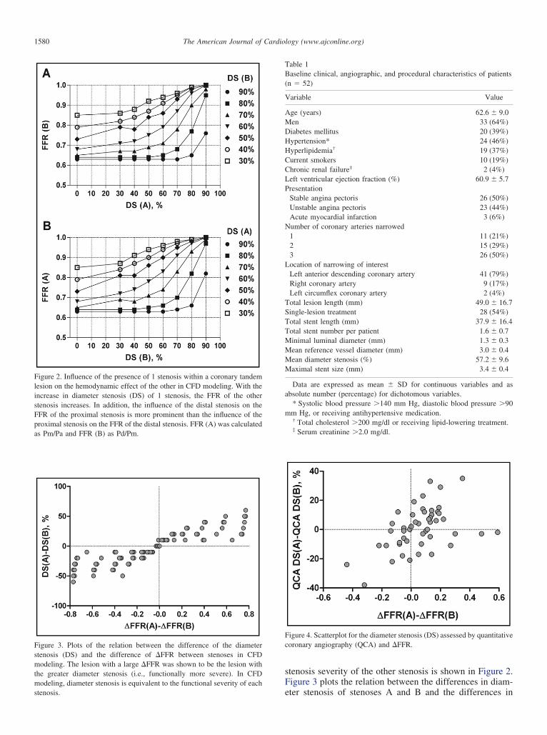
stenosis severity of the other stenosis is shown in Figure 2.Figure 3 plots the relation between the differences in diam-
Figure 4. Scatterplot for the diameter stenosis (DS) assessed by quantitativecoronary angiography (QCA) and �FFR.
Table 1Baseline clinical, angiographic, and procedural characteristics of patients(n � 52)
Variable Value
ge (years) 62.6 � 9.0en 33 (64%)iabetes mellitus 20 (39%)ypertension* 24 (46%)yperlipidemia† 19 (37%)
Current smokers 10 (19%)Chronic renal failure‡ 2 (4%)Left ventricular ejection fraction (%) 60.9 � 5.7Presentation
Stable angina pectoris 26 (50%)Unstable angina pectoris 23 (44%)Acute myocardial infarction 3 (6%)
Number of coronary arteries narrowed1 11 (21%)2 15 (29%)3 26 (50%)
Location of narrowing of interestLeft anterior descending coronary artery 41 (79%)Right coronary artery 9 (17%)Left circumflex coronary artery 2 (4%)
Total lesion length (mm) 49.0 � 16.7Single-lesion treatment 28 (54%)Total stent length (mm) 37.9 � 16.4Total stent number per patient 1.6 � 0.7Minimal luminal diameter (mm) 1.3 � 0.3Mean reference vessel diameter (mm) 3.0 � 0.4Mean diameter stenosis (%) 57.2 � 9.6Maximal stent size (mm) 3.4 � 0.4
Data are expressed as mean � SD for continuous variables and asabsolute number (percentage) for dichotomous variables.
* Systolic blood pressure �140 mm Hg, diastolic blood pressure �90mm Hg, or receiving antihypertensive medication.
† Total cholesterol �200 mg/dl or receiving lipid-lowering treatment.‡ Serum creatinine �2.0 mg/dl.
Figure 2. Influence of the presence of 1 stenosis within a coronary tandemlesion on the hemodynamic effect of the other in CFD modeling. With theincrease in diameter stenosis (DS) of 1 stenosis, the FFR of the otherstenosis increases. In addition, the influence of the distal stenosis on theFFR of the proximal stenosis is more prominent than the influence of theproximal stenosis on the FFR of the distal stenosis. FFR (A) was calculated
Figure 3. Plots of the relation between the difference of the diameterstenosis (DS) and the difference of �FFR between stenoses in CFD
odeling. The lesion with a large �FFR was shown to be the lesion withhe greater diameter stenosis (i.e., functionally more severe). In CFDodeling, diameter stenosis is equivalent to the functional severity of each
eter stenosis of stenoses A and B and the differences in

oa�s
plgpycwTc
n�a
(
ca
F erefore
1581Coronary Artery Disease/Functional Assessment for Tandem Stenosis
�FFR (A) and �FFR (B). Furthermore, the experimentalresults of Pijls et al2 were plotted for the external validationf our simulated results in Supplemental Figure 1. Figure 2nd Supplemental Figure 1 show that the lesion with a largeFFR is considered the lesion with the more functionally
evere stenotic lesion.From July 2009 to April 2011, a total of 52 consecutive
atients with angiographically confirmed coronary tandemesions were consecutively enrolled. The baseline demo-raphic, angiographic, and procedural characteristics of allatients are listed in Table 1. The mean patient age was 63ears, and 64% of our study patients were men. Mostoronary tandem lesions (79%) included in our analysisere located in the left anterior descending coronary artery.he angiographically determined entire lesion length of theoronary lesions was approximately 49 mm.
The correlation between the differences in diameter ste-osis assessed by quantitative coronary angiography and theFFR of the proximal and distal stenosis showed poor
Figure 5. Representative case of �FFR and its application: single-lesion treto mid left anterior descending coronary artery (A). FFR measurement usinsuggested that stenosis B was functionally more stenotic than stenosis A (
FR of the remaining stenosis (stenosis A) was measured at 0.82 (D). Th
greement (� � 0.41). Disagreement between the 2 param- h
eters was observed in 31% of patients with coronary tandemlesions (Figure 4).
All FFR measurements were successfully performed.Coronary stents were sequentially implanted according tothe results of the FFR measurements as described earlier.Figures 5 and 6 demonstrate representative cases for single-lesion and dual-lesion treatment, respectively. The treat-ment strategy and results are summarized in Figure 7. Theproximal lesion was treated first in 32 patients. Among atotal of 104 stenoses, only 76 lesions (73.1%) were treatedby 84 stent implantations. Revascularization for the remain-ing 28 lesions (26.9%) was deferred on the basis of FFR�0.80. Therefore, 28 patients (53.2%) were treated only bysingle-lesion treatment, after which the FFR of coronarytandem lesion recovered from 0.70 � 0.05 to 0.86 � 0.04p �0.001).
When we compared the hemodynamic and quantitativeoronary angiographic parameters between single-lesionnd dual-lesion treatment, the dual-lesion treatment group
Coronary angiogram showing the coronary tandem lesion in the proximalure-wire pullback showed that �FFR (B) was larger than FFR (A), whichrefore, stenosis B was treated first with stent implantation (C). Thereafter,, PCI for stenosis A was deferred.
atment.g press
B). The
ad lower FFRd and larger �FFR of the remaining lesion
1582 The American Journal of Cardiology (www.ajconline.org)
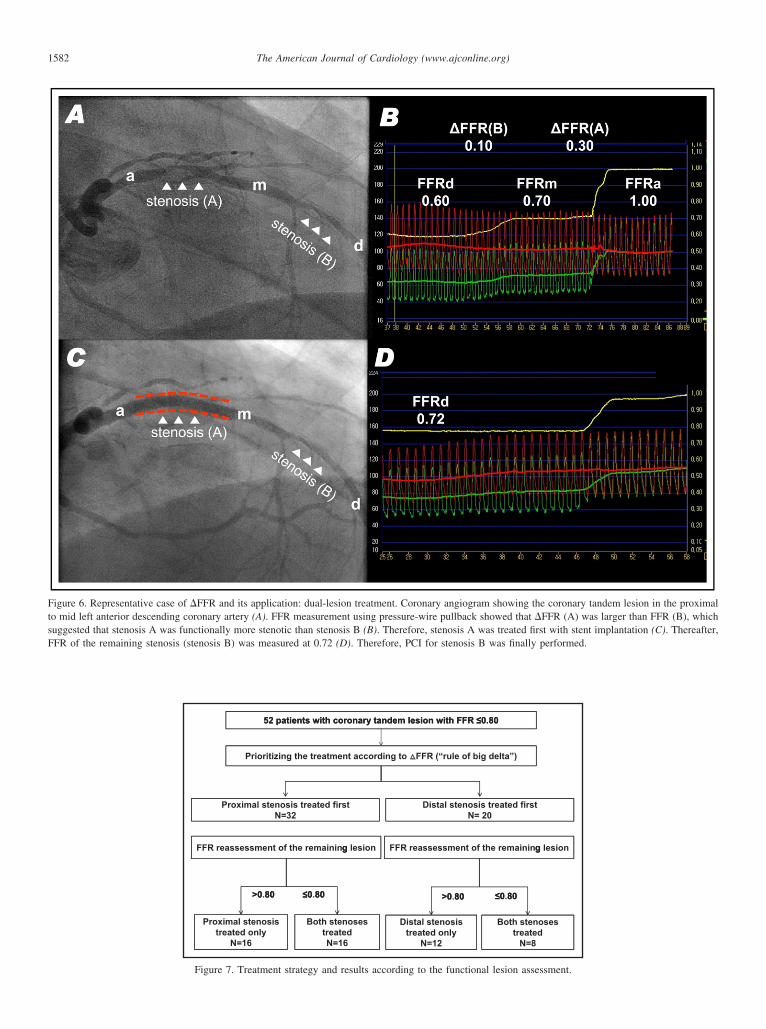
Figure 6. Representative case of �FFR and its application: dual-lesion treatment. Coronary angiogram showing the coronary tandem lesion in the proximalto mid left anterior descending coronary artery (A). FFR measurement using pressure-wire pullback showed that �FFR (A) was larger than FFR (B), whichsuggested that stenosis A was functionally more stenotic than stenosis B (B). Therefore, stenosis A was treated first with stent implantation (C). Thereafter,
FFR of the remaining stenosis (stenosis B) was measured at 0.72 (D). Therefore, PCI for stenosis B was finally performed.52 patients with coronary tandem lesion with FFR ≤0.8052 patients with coronary tandem lesion with FFR ≤0.80
Prioritizing the treatment according to FFR (“rule of big delta”)
Proximal stenosis treated first Distal stenosis treated firstN=32 N= 20
FFR reassessment of the remaining lesion FFR reassessment of the remaining lesiong g
>0 80 ≤0 80 >0 80 ≤0 80
Proximal stenosistreated only
Both stenosestreated
Distal stenosistreated only
Both stenosestreated
>0.80 ≤0.80 >0.80 ≤0.80
N=16 N=16 N=12 N=8
Figure 7. Treatment strategy and results according to the functional lesion assessment.
fav
t
wabwwiaFst
ds
inbmptbd
omoo
TTT
1583Coronary Artery Disease/Functional Assessment for Tandem Stenosis
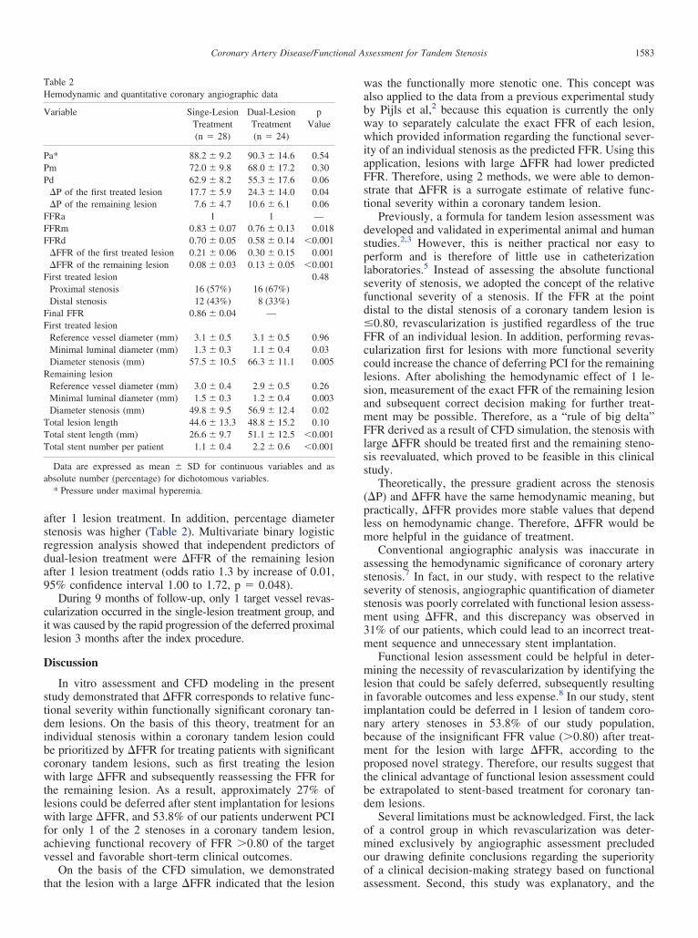
after 1 lesion treatment. In addition, percentage diameterstenosis was higher (Table 2). Multivariate binary logisticregression analysis showed that independent predictors ofdual-lesion treatment were �FFR of the remaining lesionafter 1 lesion treatment (odds ratio 1.3 by increase of 0.01,95% confidence interval 1.00 to 1.72, p � 0.048).
During 9 months of follow-up, only 1 target vessel revas-cularization occurred in the single-lesion treatment group, andit was caused by the rapid progression of the deferred proximallesion 3 months after the index procedure.
Discussion
In vitro assessment and CFD modeling in the presentstudy demonstrated that �FFR corresponds to relative func-tional severity within functionally significant coronary tan-dem lesions. On the basis of this theory, treatment for anindividual stenosis within a coronary tandem lesion couldbe prioritized by �FFR for treating patients with significantcoronary tandem lesions, such as first treating the lesionwith large �FFR and subsequently reassessing the FFR forthe remaining lesion. As a result, approximately 27% oflesions could be deferred after stent implantation for lesionswith large �FFR, and 53.8% of our patients underwent PCIor only 1 of the 2 stenoses in a coronary tandem lesion,chieving functional recovery of FFR �0.80 of the targetessel and favorable short-term clinical outcomes.
On the basis of the CFD simulation, we demonstrated
Table 2Hemodynamic and quantitative coronary angiographic data
Variable Singe-LesionTreatment
Dual-LesionTreatment
pValue
(n � 28) (n � 24)
Pa* 88.2 � 9.2 90.3 � 14.6 0.54Pm 72.0 � 9.8 68.0 � 17.2 0.30Pd 62.9 � 8.2 55.3 � 17.6 0.06
�P of the first treated lesion 17.7 � 5.9 24.3 � 14.0 0.04�P of the remaining lesion 7.6 � 4.7 10.6 � 6.1 0.06
FFRa 1 1 —FFRm 0.83 � 0.07 0.76 � 0.13 0.018FFRd 0.70 � 0.05 0.58 � 0.14 �0.001
�FFR of the first treated lesion 0.21 � 0.06 0.30 � 0.15 0.001�FFR of the remaining lesion 0.08 � 0.03 0.13 � 0.05 �0.001
First treated lesion 0.48Proximal stenosis 16 (57%) 16 (67%)Distal stenosis 12 (43%) 8 (33%)
Final FFR 0.86 � 0.04 —First treated lesion
Reference vessel diameter (mm) 3.1 � 0.5 3.1 � 0.5 0.96Minimal luminal diameter (mm) 1.3 � 0.3 1.1 � 0.4 0.03Diameter stenosis (mm) 57.5 � 10.5 66.3 � 11.1 0.005
Remaining lesionReference vessel diameter (mm) 3.0 � 0.4 2.9 � 0.5 0.26Minimal luminal diameter (mm) 1.5 � 0.3 1.2 � 0.4 0.003Diameter stenosis (mm) 49.8 � 9.5 56.9 � 12.4 0.02otal lesion length 44.6 � 13.3 48.8 � 15.2 0.10otal stent length (mm) 26.6 � 9.7 51.1 � 12.5 �0.001otal stent number per patient 1.1 � 0.4 2.2 � 0.6 �0.001
Data are expressed as mean � SD for continuous variables and asabsolute number (percentage) for dichotomous variables.
* Pressure under maximal hyperemia.
hat the lesion with a large �FFR indicated that the lesion a
as the functionally more stenotic one. This concept waslso applied to the data from a previous experimental studyy Pijls et al,2 because this equation is currently the onlyay to separately calculate the exact FFR of each lesion,hich provided information regarding the functional sever-
ty of an individual stenosis as the predicted FFR. Using thispplication, lesions with large �FFR had lower predictedFR. Therefore, using 2 methods, we were able to demon-trate that �FFR is a surrogate estimate of relative func-ional severity within a coronary tandem lesion.
Previously, a formula for tandem lesion assessment waseveloped and validated in experimental animal and humantudies.2,3 However, this is neither practical nor easy to
perform and is therefore of little use in catheterizationlaboratories.5 Instead of assessing the absolute functionalseverity of stenosis, we adopted the concept of the relativefunctional severity of a stenosis. If the FFR at the pointdistal to the distal stenosis of a coronary tandem lesion is�0.80, revascularization is justified regardless of the trueFFR of an individual lesion. In addition, performing revas-cularization first for lesions with more functional severitycould increase the chance of deferring PCI for the remaininglesions. After abolishing the hemodynamic effect of 1 le-sion, measurement of the exact FFR of the remaining lesionand subsequent correct decision making for further treat-ment may be possible. Therefore, as a “rule of big delta”FFR derived as a result of CFD simulation, the stenosis withlarge �FFR should be treated first and the remaining steno-sis reevaluated, which proved to be feasible in this clinicalstudy.
Theoretically, the pressure gradient across the stenosis(�P) and �FFR have the same hemodynamic meaning, butpractically, �FFR provides more stable values that dependless on hemodynamic change. Therefore, �FFR would bemore helpful in the guidance of treatment.
Conventional angiographic analysis was inaccurate inassessing the hemodynamic significance of coronary arterystenosis.7 In fact, in our study, with respect to the relativeseverity of stenosis, angiographic quantification of diameterstenosis was poorly correlated with functional lesion assess-ment using �FFR, and this discrepancy was observed in31% of our patients, which could lead to an incorrect treat-ment sequence and unnecessary stent implantation.
Functional lesion assessment could be helpful in deter-mining the necessity of revascularization by identifying thelesion that could be safely deferred, subsequently resultingin favorable outcomes and less expense.8 In our study, stentmplantation could be deferred in 1 lesion of tandem coro-ary artery stenoses in 53.8% of our study population,ecause of the insignificant FFR value (�0.80) after treat-ent for the lesion with large �FFR, according to the
roposed novel strategy. Therefore, our results suggest thathe clinical advantage of functional lesion assessment coulde extrapolated to stent-based treatment for coronary tan-em lesions.
Several limitations must be acknowledged. First, the lackf a control group in which revascularization was deter-ined exclusively by angiographic assessment precluded
ur drawing definite conclusions regarding the superiorityf a clinical decision-making strategy based on functional
ssessment. Second, this study was explanatory, and the
1584 The American Journal of Cardiology (www.ajconline.org)
study population in the clinical cohort was small. Therefore,a larger study with long-term clinical follow-up will berequired. Third, we did not consider the effect of the inter-position of a side branch between stenoses, which maymodify the hemodynamic influence of the relative signifi-cance of 2 stenoses. Finally, our CFD study was conductedunder the simple assumption of maximal hyperemia. There-fore, our model has some limitations in terms of directclinical application, and more complex modeling or in vivostudy would be necessary.
Supplementary data
Supplementary data associated with this article can befound, in the online version, at http://dx.doi.org/10.1016/j.amjcard.2012.07.023.
1. Pijls NH, De Bruyne B, Peels K, Van Der Voort PH, Bonnier HJ,Bartunek JKJJ, Koolen JJ. Measurement of fractional flow reserve toassess the functional severity of coronary-artery stenoses. N Engl J Med1996;334:1703–1708.
2. Pijls NH, De Bruyne B, Bech GJ, Liistro F, Heyndrickx GR, BonnierHJ, Koolen JJ. Coronary pressure measurement to assess the hemody-namic significance of serial stenoses within one coronary artery: vali-dation in humans. Circulation 2000;102:2371–2377.
3. De Bruyne B, Pijls NH, Heyndrickx GR, Hodeige D, Kirkeeide R,Gould KL. Pressure-derived fractional flow reserve to assess serial
epicardial stenoses: theoretical basis and animal validation. Circulation2000;101:1840–1847.
4. Popma JJ, Leon MB, Moses JW, Holmes DR Jr, Cox N, Fitzpatrick M,Douglas J, Lambert C, Mooney M, Yakubov S, Kuntz RE. Quantitativeassessment of angiographic restenosis after sirolimus-eluting stent im-plantation in native coronary arteries. Circulation 2004;110:3773–3780.
5. Kern MJ, Lerman A, Bech JW, De Bruyne B, Eeckhout E, Fearon WF,Higano ST, Lim MJ, Meuwissen M, Piek JJ, Pijls NH, Siebes M, SpaanJA. Physiological assessment of coronary artery disease in the cardiaccatheterization laboratory: a scientific statement from the AmericanHeart Association Committee on Diagnostic and Interventional CardiacCatheterization, Council on Clinical Cardiology. Circulation 2006;114:1321–1341.
6. Smith SC Jr, Dove JT, Jacobs AK, Kennedy JW, Kereiakes D, Kern MJ,Kuntz RE, Popma JJ, Schaff HV, Williams DO, Gibbons RJ, Alpert JP,Eagle KA, Faxon DP, Fuster V, Gardner TJ, Gregoratos G, Russell RO.ACC/AHA guidelines of percutaneous coronary interventions (revisionof the 1993 PTCA guidelines)—executive summary. A report of theAmerican College of Cardiology/American Heart Association TaskForce on Practice Guidelines (Committee to Revise the 1993 Guidelinesfor Percutaneous Transluminal Coronary Angioplasty). J Am Coll Car-diol 2001;37:2215–2239.
7. Tonino PA, Fearon WF, De Bruyne B, Oldroyd KG, Leesar MA, VerLee PN, Maccarthy PA, Van’t Veer M, Pijls NH. Angiographic versusfunctional severity of coronary artery stenoses in the FAME studyfractional flow reserve versus angiography in multivessel evaluation.J Am Coll Cardiol 2010;55:2816–2821.
8. Tonino PA, De Bruyne B, Pijls NH, Siebert U, Ikeno F, van’ t Veer M,Klauss V, Manoharan G, Engstrom T, Oldroyd KG, Ver Lee PN,MacCarthy PA, Fearon WF. Fractional flow reserve versus angiography
for guiding percutaneous coronary intervention. N Engl J Med 2009;360:213–224.