Embed Size (px)
Citation preview
RESEARCH ARTICLE Open Access
Validity and reliability of fluoroscopy fordigital radiography: a new way to evaluatediaphragmatic mobilityBruna Estima Leal1, Márcia Aparecida Gonçalves1, Liseane Gonçalves Lisboa2, Larissa Martins Schmitz Linné2,Michelle Gonçalves de Souza Tavares3, Wellington Pereira Yamaguti4 and Elaine Paulin1,5*
Abstract
Background: Fluoroscopy is considered the most accurate method to evaluate the diaphragm, yet most existingmethods for measuring diaphragmatic mobility using fluoroscopy are complex. To assess the validity and reliabilityof a new evaluation method of diaphragmatic motion using fluoroscopy by digital radiography of healthy adults.
Methods: Twenty-six adults were evaluated, according to the parameters: anthropometry and pulmonary function test.The evaluation of diaphragm mobility by means of fluoroscopy by digital radiography method was randomly conductedby two raters (A and B). The Pearson correlation coefficient and the intraclass correlation coefficient (ICC) were used toassess the concurrent validity. The inter-rater and intra-rater reliability of the measurement of diaphragmatic motion wasdetermined using ICC and a confidence interval of 95%.
Results: There was a relationship in the assessment of the concurrent validity. There was good inter-rater reliability forright hemidiaphragm mobility and moderate reliability for left hemidiaphragm in the first assessment. In the secondassessment, there was good reliability for the mobility of both hemidiaphragms. There was good intra-rater reliability inthe mobility of both hemidiaphragms for raters A and B.
Conclusion: The evaluation of diaphragmatic motion using fluoroscopy by digital radiography proved to be a valid andreliable method of healthy adults.
Keywords: Diaphragm, Fluoroscopy, Validity, Reproducibility of results
BackgroundSpecifically evaluating the mobility of the diaphragm isimportant for understanding and diagnosing possible alter-ations in the muscle, which can be compromised in severalways: due to central or peripheral nervous system dysfunc-tion, muscular disease and thoracic or abdominal disease,resulting in a reduction of mobility or paralysis [1–6].In clinical practice it is essential to use valid and reliable
methods for assessing diaphragmatic dysfunction, becausethe use of subjective methods may compromise the results.
Therefore, it is extremely important that any assessmentmethod have its validity and reliability tested to ensure thatthe error in the measurement is reduced [7–9].There are several imaging methods that assess diaphrag-
matic mobility: fluoroscopy [10–13], ultrasound [14, 15],computed tomography [16], magnetic resonance [17–19],and chest radiography [20, 21]. Each technique has itsparticularities in the observation of the diaphragm, con-sidering cost, radiation exposure and method availabilityin the study environment [5, 22].Considering all the methods for evaluating diaphragmatic
mobility, the fluoroscopy is the most accurade method forassessing the diaphragm muscle because it provides dynamicimages of the diaphragm and direct visualization of dia-phragmatic movements in real time [5]. However, there areno studies confirming the validity and reliability of digitalradiography fluoroscopy to assess diaphragmatic excursion.
* Correspondence: [email protected] Therapy Department,Santa Catarina State University (UDESC),Florianopolis, SC, Brazil5Santa Catarina State University (UDESC), Rua Pascoal Simone, 358,Coqueiros, Florianópolis, SC, BrazilCEP: 88080-350Full list of author information is available at the end of the article
© The Author(s). 2017 Open Access This article is distributed under the terms of the Creative Commons Attribution 4.0International License (http://creativecommons.org/licenses/by/4.0/), which permits unrestricted use, distribution, andreproduction in any medium, provided you give appropriate credit to the original author(s) and the source, provide a link tothe Creative Commons license, and indicate if changes were made. The Creative Commons Public Domain Dedication waiver(http://creativecommons.org/publicdomain/zero/1.0/) applies to the data made available in this article, unless otherwise stated.
Leal et al. BMC Pulmonary Medicine (2017) 17:62 DOI 10.1186/s12890-017-0402-x
Diaphragmatic mobility can be measured by the fluoros-copy method in various ways, but those forms reported inthe literature are not simple to be obtained [10–13]. Somemeasures require radiographic impression for analysis [13],others, those recorded on video [10, 11], are not alwaysavailable on fluoroscopy devices, and in other measure-ments, image calculations involve several complex lines forobtaining the value of diaphragmatic mobility [13].Since fluoroscopy assesses diaphragm motion in real
time [5], and the ways reported in the literature formeasuring diaphragm mobility by means of fluoroscopyare complex [10–13], and there is no literature studiesinvestigating its validity and reliability, we propose creat-ing a new method and a new measurement procedurethat is much more easily obtained in clinical practice,using fluoroscopy by digital radiography.Thus, the aim of this study was to assess the validity
and reliability (inter- and intra-rate) of a new method ofevaluation of diaphragmatic mobility using fluoroscopyby digital radiography.
MethodsSampleIn this study, 26 apparently healthy adults were included ina convenience sample. They were recruited among studentsand employees of the Universidade do Estado de SantaCatarina (Brazil), as well as their relatives. The sample sizecalculation was based on Bonett [23] and Fleiss [24] studies.According to these authors, to assess test reliability, a sam-ple can vary between 15 and 20 participants.Inclusion criteria for the study were: non-smoking
healthy subjects with normal pulmonary function (FVCand FEV1 ≥ 80% predicted and FEV1/FVC ≥ 0.7) withoutcardiorespiratory or neurological diseases, women whowere not pregnant or with suspected pregnancy, partici-pants without a diagnosis of cancer, or disease history orany other alteration that could impair the evaluations.Exclusion criteria were: participants who presented clinicalcomplications of respiratory nature, inability to performany of the procedures used in the study (lack of under-standing or collaboration), clinical complications of therespiratory system and those who requested exclusion fromthe study. This study was approved (16696413.8.0000.0118)by the Research Ethics Committee of the Universidade doEstado de Santa Catarina (UDESC), Brazil, and all partici-pants provided written informed consent.
Study designThis is a cross-sectional study, with validity and reliabilitytest, which assessed the agreement degree of diaphrag-matic mobility by means of X-ray digital fluoroscopy [25].Initially, anthropometric and pulmonary parameters
were evaluated. Following, digital X-rays were scheduledin order to measure diaphragmatic excursion. Before the
fluoroscopy examination, training of the diaphragmthrough diaphragmatic breathing exercise was performed,slow vital capacity (SVC) was also measured before andduring the examination.The examination of diaphragmatic motion by digital
radiography fluoroscopy was randomly performed bytwo radiologists (raters A and B), and subsequentlydigital diaphragmatic mobility (DMdig) was measured byboth raters, aiming to evaluate the intra-rater and inter-rater reliability. Diaphragmatic mobility by distance(DMdist) [20] was measured by rater A, to evaluate thevalidity of the method.
Collection proceduresAnthropometryFor measurement of body mass and height, a previouslycalibrated scale and a stadiometer (Welmy® model W200/5)were used respectively. Once the anthropometric valueswere obtained (weight and height), the body mass index(BMI) was calculated using the equation: body mass/(height)2 (kg/m2). Subjects were classified according to BMIas underweight (≤18.5 kg/m2), normal (18.5–24.9 kg/m2),overweight (25–29.9 kg/m2) and obese (≥ 30 kg/m2) [26].
Pulmonary function testThe pulmonary function test was performed using a previ-ously calibrated spirometer in accordance with themethods and criteria recommended by the AmericanThoracic Society [27]. For assessment of forced vital cap-acity (FVC), forced expiratory volume in the first second(FEV1), and the FEV1/FVC ratio, a portable digital Easy-One® spirometer of the ndd brand was used. The criteriafor normal lung test consisted in FVC and FEV1 ≥ 80%predicted and FEV1/FVC ≥ 0.7. Spirometric variables wereexpressed as absolute values and as percentages of pre-dicted normal values, according to Pereira et al. [28].Before performing the pulmonary function test, pulse
oxygen saturation (SpO2) and heart rate (HR) were mea-sured with the participant in the supine position and atrest with a pulse oximeter (Oximeter, Model MD300C11).
Assessment of diaphragmatic mobility by digitalradiography fluoroscopyDiaphragmatic mobility was assessed through examinationof digital X-ray fluoroscopy in anteroposterior incidence(AP) by two raters. To perform the test, a Siemens fluoros-copy device, model Lumino RF Classic was used at a dis-tance of 1.15 m from the image intensifier and X-ray tube.Subjects were placed on a radioscopic table in a supine
position with their feet supported to restrict their move-ment on the table, and a radiopaque graduation ruler wasplaced under the subjects’ trunks in a longitudinal direc-tion and in the craniocaudal direction. Before performingthe fluoroscopy by digital radiography to evaluate the
Leal et al. BMC Pulmonary Medicine (2017) 17:62 Page 2 of 10
diaphragmatic motion, training in diaphragmaticbreathing was provided. The goal was to develop dia-phragmatic proprioception movement and enable theevaluation of diaphragm maximum amplitude duringfluoroscopic digital X-ray examination. Afterwards, wemeasured slow vital capacity (SVC) using a WrightRespirometer Brit.® UK ventilometer, before and dur-ing the examination of digital X-ray fluoroscopy withsubjects in a supine position. Three manoeuvers wereperformed before radiographic exposure, and the high-est value was recorded for later comparison with whatwould be assessed during the examination of dia-phragmatic mobility. During the examination of digitalX-ray fluoroscopy, each rater asked the subjects, whileexhaling, to breathe using TLC (total lung capacity)until they approached RV (residual volume), and then,upon exhaling, to breathe from RV until approachingTLC. The values of the SVC manoeuvers obtainedwere compared to each other (before and during theexam), to determine whether the subjects performedthe same respiratory effort before and during theevaluation of diaphragmatic mobility. If there was adifference greater than 10% from the previouslyobtained value, the examination would be repeated onthe same day.Subjects were evaluated randomly, in a simple raffle,
by two expert radiologists (raters A and B) who guidedsubjects in a standardized way. The raters viewed thediaphragm movement through the display on the fluor-oscopy device, which was positioned on the central partof the thorax, viewing both hemidiaphragms at thesame time. The images of maximum expiration andinspiration were recorded on the same film, whichremained motionless during exams.
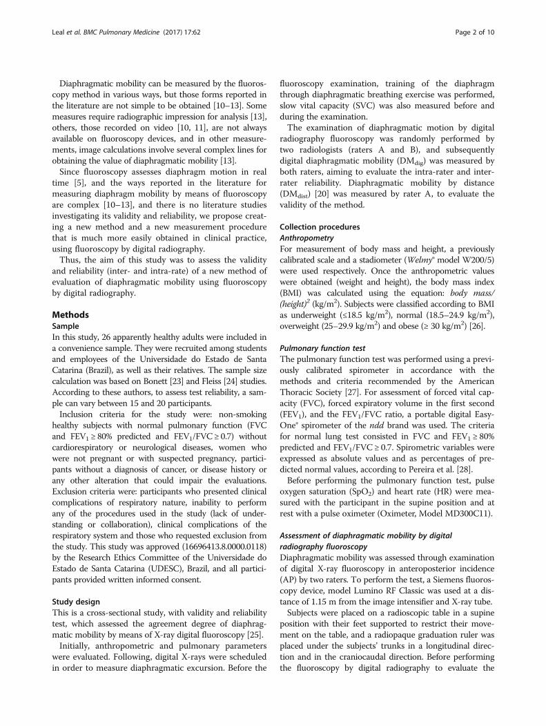
Digital measurement of diaphragmatic mobility (DMdig)Diaphragmatic mobility (DMdig) was measured by cal-culating the distance between the diaphragmatic domein expiration and inspiration for the right and lefthemidiaphragms, based on the method described bySaltiel et al. [20]. Initially, the line of MediWorks8.4.215 software was calibrated using a radiopaque rulerto correct the magnification caused by the divergenceof the rays. This calibration was performed by drawinga line with the computer cursor over a distance of10 mm on the image of the ruler in the digital radiog-raphy, thus determining the actual distance. Then, tomeasure diaphragmatic mobility, the highest point onthe hemidiaphragm in expiration was found and astraight perpendicular line was drawn with the cursoruntil it met the hemidiaphragm in inspiration findingthe distance between the diaphragmatic domes (Fig. 1).This measurement was performed for both right andleft hemidiaphragms (RH) and (LH).
Measurement of diaphragmatic mobility by distance(DMdist)To measure DMdist, rater A identified, on the printedchest radiography, the highest point of the diaphragmduring expiration of each hemidiaphragmatic dome,and from this point a line was drawn with a blackmarker until the hemidiaphragm during inspiration wasfound. The diaphragm mobility was determined by thedistance between the diaphragm dome on expirationand inspiration by the calliper, both on the right sideand the left side. To correct the image magnificationcaused by the divergence of X-rays, a correction for-mula was used: Corrected mobility (mm) = mobilitymeasure (mm) × 10/graduation on the ruler (mm) [20].
Analysis of the validityTo assess the validity of the method (criterion validity),we analysed the concurrent validity. The competingmethod was the method of Saltiel et al. [20], which eval-uates diaphragm mobility on the printed radiograph bydistance (DMdist) by means of a calliper. Validity wasassessed by relating the first measurement of DMdig ob-tained by rater A with the measurement using theDMdist method by the same rater. We evaluated bothright and left hemidiaphragms.
Analysis of reliabilityMeasurements of diaphragmatic mobility were assessedsoon after scanning the radiograph. The first measure-ment was used for the analysis of inter-rater reliabilityof the method (measurements A1 and B1) in the fol-lowing manner: a measurement taken by rater A (A1)was correlated with that taken by rater B (B1). Then, asecond measurement was performed for the analysis ofinter-rater reliability of the measure as follows: rater Ameasured the digital radiography fluoroscopy examperformed by rater B, obtaining measure AB, and raterB measured the digital radiography fluoroscopy examperformed by rater A obtaining measure BA. The ana-lysis was performed by correlating measures AB withB1 and BA with A1.The intra-rater reliability of the measurement was
assessed by measuring diaphragmatic mobility from theprevious examination, after a minimum interval of7 days and maximum of 20 days from the first meas-urement25. Both raters A and B measured the digitalradiographs one more time (measures A2 and B2) per-formed at the beginning of the study. Inter-rater reli-ability was also examined in the second assessment.Raters A and B did not know who conducted the ra-
diographs and did not have access to the values of theother’s assessments. The results were analysed aftercompletion of all assessments.
Leal et al. BMC Pulmonary Medicine (2017) 17:62 Page 3 of 10
Statistical analysisData were analysed using SPSS for Windows, version20.0 (IBM SPSS Statistics, IBM, Armonk, NY, USA)and GraphPad Prism 5.1 program and treated withdescriptive analysis (mean and standard deviation) andinferential analysis. Shapiro-Wilk test was used to ana-lyse data normality.Pearson correlation coefficient and intraclass correl-
ation coefficient (two-way random model, with abso-lute agreement - ICC[2.1]) were used to assess thecorrelation between the digital method (DMdig) andthe method of distance (DMdist.). The analyses ofinter-rater and intra-rater reproducibility were deter-mined using intraclass correlation coefficient (two-wayrandom model, with absolute agreement - ICC[2.1]) andconfidence interval (CI) of 95% [8]. Reliability wasinterpreted as the magnitude of Portney and Watkinscoefficient of reliability [8]: ‘poor’ for coefficientsunder 0.50, ‘moderate’ for coefficients between 0.50and 0.75, and ‘good’ for coefficients above 0.75. ICCvaries from 0.00 to 1.00, and values close to 1.00 showstrong reliability. Bland-Altman plot was also used toallow better visualization of agreement between theindividual measures [29].Wilcoxon test was used to compare the values of slow
vital capacity before and during radiographic exposuresfor each rater. Paired T test was used to compare themobility of the right and left hemidiaphragms. The Ttest for independent samples was used to compare thevalues of diaphragmatic mobility between male and fe-male participants. The significance level for statisticaltreatment was 5% (p < 0.05).
ResultsTwenty-six healthy adults, 17 women (65.4%) and 9 men(34.6%) were evaluated, with a mean age of 28.19 ± 6.1 years,mean BMI 23.89 ± 4.2, classified as normal, healthy andwith normal pulmonary function (Table 1). No subjectrefused to participate or desisted during assessments.A high correlation was found between DMdig and DMdist
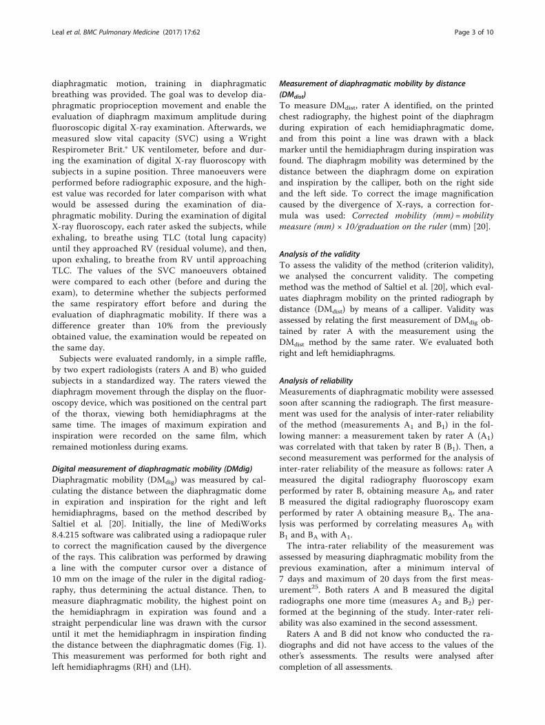
for the mobility of the right hemidiaphragm (RH) (r = 0.97,p = 0.00) and the left one (LH) (r = 0.88, p = 0. 00). Therewas good reliability for mobility in both hemidiaphragms(RH: ICC[1, 2] = 0.98, 95% CI = 0.96 to 0.99; LH: ICC[2,1] =0.93, 95% CI = 0.84 to 0.97) (Fig. 2).In the inter-raters analysis, in the first assessment
there was good reliability for RH and moderate in LH.
Fig. 1 Measurement of diaphragm mobility obtained by the software. Digital radiography of the chest in anteroposterior view (AP) during maximalexpiration and maximal inspiration conducted on the same film. Measurements of the mobility of right and left hemidiaphragms were obtained by thesoftware of the device using the ruler on the image for calibration. Source: author's own production
Table 1 Anthropometric and cardiopulmonary characteristics ofthe study participants
Variables Average ± standard deviation variables (n = 26)
Age (years) 28.19 ± 6.1
Body mass (kg) 68.14 ± 16.7
Height (m) 1.68 ± 0.1
BMI (kg.m- 2) 23.89 ± 4.2
HR (bpm) 72.61 ± 9.7
SpO2 (%) 98.35 ± 0.6
FVC (% predicted) 92.85 ± 7.7
FEV1 (% predicted) 94.73 ± 7.1
FEV1/FVC (L) 0.91 ± 0.2
Values were express as mean and standard deviationn number of subjetcs, kg lbs, m meters, BMI body mass index, HR heart rate,bpm: beats per minute, SpO2 oxygen saturation by pulse, FVC (%predicted):Estimated percentage of FVC, FEV1 (%predicted): Estimated percentage offorced expiratory volume in one second, L liters
Leal et al. BMC Pulmonary Medicine (2017) 17:62 Page 4 of 10
In the second assessment, there was good reliability formobility in both hemidiaphragms. There was good intra-rater reliability of mobility in both RH and LH for rater A.Similar results were found in the measurements obtainedby rater B in assessing RH and LH. The analyses of inter-rater and intra-rater reliability for digital diaphragmaticmobility of RH and LH are described in Table 2.To demonstrate higher reliability, we also evaluated
inter-rater reproducibility of the measurement, whererater A measured the examination performed by rater B,yielding measure AB and we compared this with meas-ure B1. For this analysis there was good reliability for theratings of RH and LH (ICC[2,1] = 0.98, 95% CI = 0.96 to0.99; ICC[2,1] = 0.95, 95% CI = 0.90 to 0.98, respectively).
Rater B also measured the examination performed byrater A to obtain measure BA. When comparing it withmeasure A1, there was good reliability for RH (ICC[2,1] =0.98, 95% CI = 0.96 to 0.99) and for LH (ICC[2,1] = 0.86,95% CI = 0.72 to 0.94).According to Bland-Altman plot, there was good con-
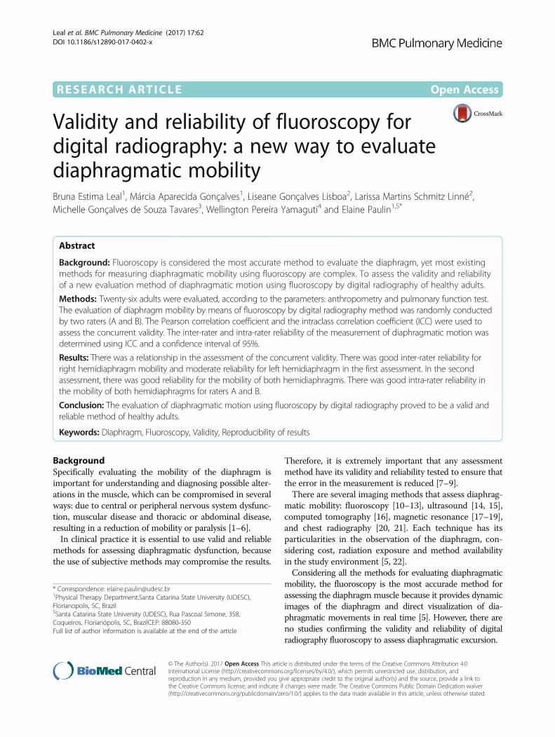
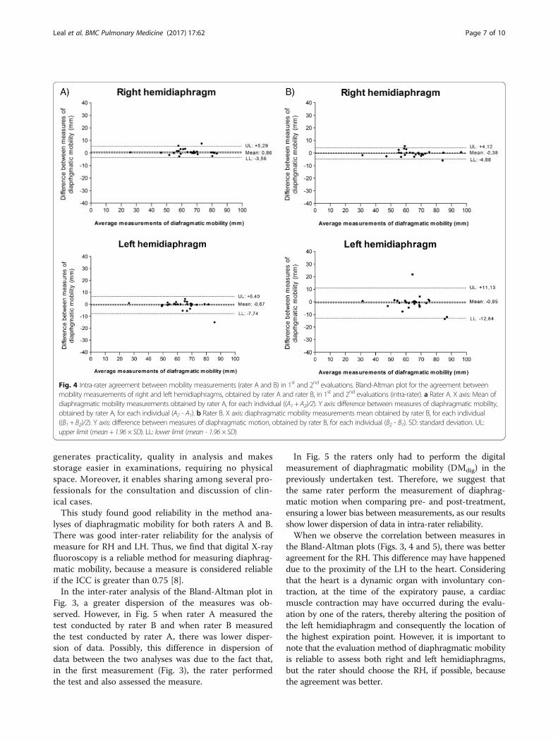
cordance between measures of mobility of both RH andLH, obtained by raters A and B (inter-rater agreement)(Fig. 3), and good concordance between measures ofmobility of RH and LH, obtained by each of the raters(Fig. 4), at two different times (intra-rater agreement).Figure 5 shows that there was good concordance be-tween measures of mobility of RH and LH, obtained byraters A and B when rater A measured the test con-ducted by rater B and when rater B measured the testconducted by rater A. There was good concordance be-cause the difference between the measures is the limitsof agreement (upper and lower limits). The mean of themeasures between raters in all analyses is close to zero,which indicates reproducibility of the measurements.However, when analysing the inter-rater graphics, therewas greater dispersion of the means obtained.There were no differences between the manoeuvers
of slow vital capacity (SVC) performed before and dur-ing fluoroscopy examinations by radiography by ratersA and B, respectively (before: 3.92 ± 1.5 mm; during:4.07 ± 1.5 mm, p = 0.57; before: 3.92 ± 1.5 mm; during :4.12 ± 1.4 mm, p = 0.46).The mobility measured in the right (RH) and left (LH)
hemidiaphragms by the two raters (A and B) showed nostatistically significant difference. The mean values of
Fig. 2 Correlation between DMdig and DMdist. Correlation between methods DMdig and DMdist to assess the validity of the method (concurrent validity).The concurrent validity by relating the first measurement of DMdig obtained by rater A with the measurement using the DMdist method by the samerater. a Right hemidiaphragm. b Left hemidiaphragm. DMdig: digital diaphragmatic mobility. DMdist: diaphragmatic mobility by distance
Table 2 Inter-rater and intra-rater reliability of the mobilitymeasurement of the right and left hemidiaphragms method
Variables ICC[2,1] CI 95%
Right hemidiaphragm
Inter-rater reliability 1ª assess 0.89 0.76–0.95
2ª assess 0.84 0.68–0.93
Intra-rater reliability rater A 0.83 0.66–0.92
rater B 0.89 0.76–0.95
Left hemidiaphragm
Inter-rater reliability 1ª assess 0.73 0.48–0.87
2ª assess 0.78 0.56–0.89
Intra-rater reliability rater A 0.86 0.70–0.93
rater B 0.83 0.65–0.92
ICC [2,1] the intraclass correlation coefficient (two-way random model, withabsolute and agreement), CI 95% confidence interval of 95%
Leal et al. BMC Pulmonary Medicine (2017) 17:62 Page 5 of 10
RH and LH mobility when analysed by rater A were64.8 ± 12.6 mm (30.3 to 96.7 mm) and 64.1 ± 10.8 mm(31.7 to 92.3 mm), respectively and when analysed byrater B, 64.7 ± 12.5 mm (26.1 to 83.3 mm) and 62.9 ±11.3 mm (27.0 to 81.5 mm), respectively. There was no sta-tistically significant difference in the mobility of RH and LHmeasured by the two raters: A (p = 0.69) and B (p = 0.41).
DiscussionThis study demonstrated that the digital X-ray fluoros-copy method is valid and reliable for measuring mobil-ity of the left and right hemidiaphragms in healthyadults. In clinical practice, the use of reliable instru-ments is essential for ensuring reliable results [9].Fluoroscopy is the most accurate method for evalu-
ating diaphragm dysfunction as it assesses the dia-phragmatic motion in real time [5], but somemeasures require a video be made of the image [10,11], which is not always available in fluoroscopyequipment, or they require calculations involving
complex procedures to determine diaphragmatic mo-tion [13].Thus, the proposal of this study is innovative be-
cause it verifies the validity and the reliability of a newmethod of fluoroscopy and a new way of measuringdiaphragmatic movement. For this, we applied a com-mon resource used in clinical practice, which is the X-ray, and we innovated by using fluoroscopy associatedwith digital radiography. This method is easy to applyand measure, has a low cost because it is not necessaryto print the radiography, and it can be another tool forprofessionals in the health assessment of patient’s dia-phragmatic mobility.Our proposal was to perform a digital measurement
using software that is routinely used in medical radio-logical practice, but this is a unique way for evaluatingdiaphragmatic mobility in the scientific community.The measurement of diaphragmatic motion using thescanned image (DMdig) is simple and practical to per-form, and proved to be reliable. We emphasize thatthe digitization of the exam is a technology that
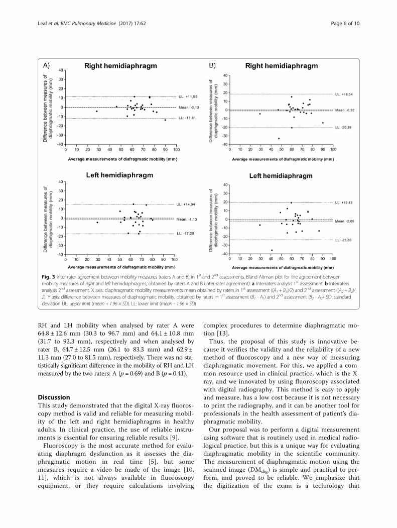
Fig. 3 Inter-rater agreement between mobility measures (raters A and B) in 1st and 2nd assessments. Bland-Altman plot for the agreement betweenmobility measures of right and left hemidiaphragms, obtained by raters A and B (inter-rater agreement). a Interraters analysis 1st assessment. b Interratersanalysis 2nd assessment. X axis: diaphragmatic mobility measurements mean obtained by raters in 1st assessment ((A1 + B1)/2) and 2nd assessment ((A2 + B2)/2). Y axis: difference between measures of diaphragmatic mobility, obtained by raters in 1st assessment (B1 - A1) and 2nd assessment (B2 - A2). SD: standarddeviation UL: upper limit (mean + 1.96 × SD). LL: lower limit (mean - 1.96 × SD)
Leal et al. BMC Pulmonary Medicine (2017) 17:62 Page 6 of 10
generates practicality, quality in analysis and makesstorage easier in examinations, requiring no physicalspace. Moreover, it enables sharing among several pro-fessionals for the consultation and discussion of clin-ical cases.This study found good reliability in the method ana-
lyses of diaphragmatic mobility for both raters A and B.There was good inter-rater reliability for the analysis ofmeasure for RH and LH. Thus, we find that digital X-rayfluoroscopy is a reliable method for measuring diaphrag-matic mobility, because a measure is considered reliableif the ICC is greater than 0.75 [8].In the inter-rater analysis of the Bland-Altman plot in
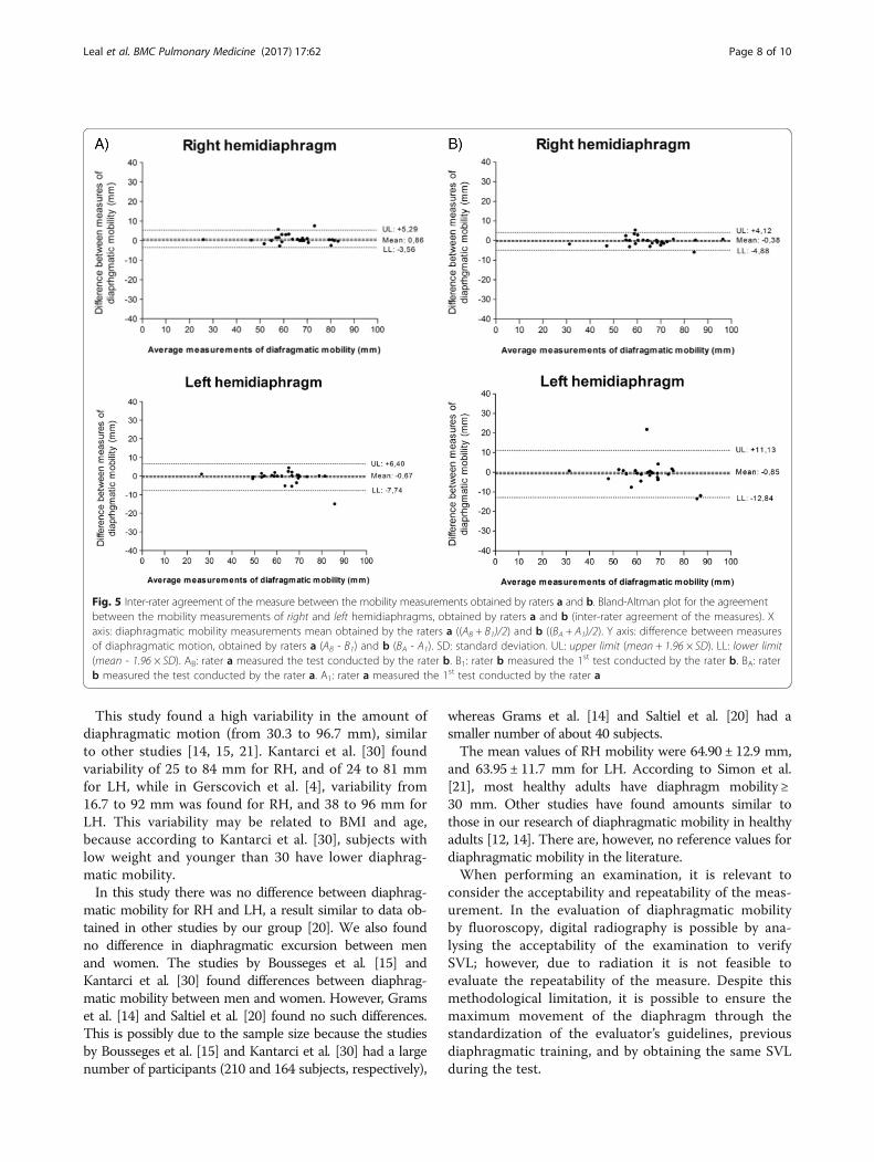
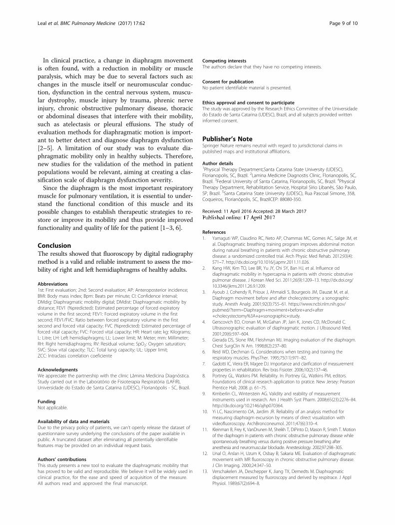
Fig. 3, a greater dispersion of the measures was ob-served. However, in Fig. 5 when rater A measured thetest conducted by rater B and when rater B measuredthe test conducted by rater A, there was lower disper-sion of data. Possibly, this difference in dispersion ofdata between the two analyses was due to the fact that,in the first measurement (Fig. 3), the rater performedthe test and also assessed the measure.
In Fig. 5 the raters only had to perform the digitalmeasurement of diaphragmatic mobility (DMdig) in thepreviously undertaken test. Therefore, we suggest thatthe same rater perform the measurement of diaphrag-matic motion when comparing pre- and post-treatment,ensuring a lower bias between measurements, as our resultsshow lower dispersion of data in intra-rater reliability.When we observe the correlation between measures in
the Bland-Altman plots (Figs. 3, 4 and 5), there was betteragreement for the RH. This difference may have happeneddue to the proximity of the LH to the heart. Consideringthat the heart is a dynamic organ with involuntary con-traction, at the time of the expiratory pause, a cardiacmuscle contraction may have occurred during the evalu-ation by one of the raters, thereby altering the position ofthe left hemidiaphragm and consequently the location ofthe highest expiration point. However, it is important tonote that the evaluation method of diaphragmatic mobilityis reliable to assess both right and left hemidiaphragms,but the rater should choose the RH, if possible, becausethe agreement was better.
Fig. 4 Intra-rater agreement between mobility measurements (rater A and B) in 1st and 2nd evaluations. Bland-Altman plot for the agreement betweenmobility measurements of right and left hemidiaphragms, obtained by rater A and rater B, in 1st and 2nd evaluations (intra-rater). a Rater A. X axis: Mean ofdiaphragmatic mobility measurements obtained by rater A, for each individual ((A1 + A2)/2). Y axis: difference between measures of diaphragmatic mobility,obtained by rater A, for each individual (A2 - A1). b Rater B. X axis: diaphragmatic mobility measurements mean obtained by rater B, for each individual((B1 + B2)/2). Y axis: difference between measures of diaphragmatic motion, obtained by rater B, for each individual (B2 - B1). SD: standard deviation. UL:upper limit (mean + 1.96 × SD). LL: lower limit (mean - 1.96 × SD)
Leal et al. BMC Pulmonary Medicine (2017) 17:62 Page 7 of 10
This study found a high variability in the amount ofdiaphragmatic motion (from 30.3 to 96.7 mm), similarto other studies [14, 15, 21]. Kantarci et al. [30] foundvariability of 25 to 84 mm for RH, and of 24 to 81 mmfor LH, while in Gerscovich et al. [4], variability from16.7 to 92 mm was found for RH, and 38 to 96 mm forLH. This variability may be related to BMI and age,because according to Kantarci et al. [30], subjects withlow weight and younger than 30 have lower diaphrag-matic mobility.In this study there was no difference between diaphrag-
matic mobility for RH and LH, a result similar to data ob-tained in other studies by our group [20]. We also foundno difference in diaphragmatic excursion between menand women. The studies by Bousseges et al. [15] andKantarci et al. [30] found differences between diaphrag-matic mobility between men and women. However, Gramset al. [14] and Saltiel et al. [20] found no such differences.This is possibly due to the sample size because the studiesby Bousseges et al. [15] and Kantarci et al. [30] had a largenumber of participants (210 and 164 subjects, respectively),
whereas Grams et al. [14] and Saltiel et al. [20] had asmaller number of about 40 subjects.The mean values of RH mobility were 64.90 ± 12.9 mm,
and 63.95 ± 11.7 mm for LH. According to Simon et al.[21], most healthy adults have diaphragm mobility ≥30 mm. Other studies have found amounts similar tothose in our research of diaphragmatic mobility in healthyadults [12, 14]. There are, however, no reference values fordiaphragmatic mobility in the literature.When performing an examination, it is relevant to
consider the acceptability and repeatability of the meas-urement. In the evaluation of diaphragmatic mobilityby fluoroscopy, digital radiography is possible by ana-lysing the acceptability of the examination to verifySVL; however, due to radiation it is not feasible toevaluate the repeatability of the measure. Despite thismethodological limitation, it is possible to ensure themaximum movement of the diaphragm through thestandardization of the evaluator’s guidelines, previousdiaphragmatic training, and by obtaining the same SVLduring the test.
Fig. 5 Inter-rater agreement of the measure between the mobility measurements obtained by raters a and b. Bland-Altman plot for the agreementbetween the mobility measurements of right and left hemidiaphragms, obtained by raters a and b (inter-rater agreement of the measures). Xaxis: diaphragmatic mobility measurements mean obtained by the raters a ((AB + B1)/2) and b ((BA + A1)/2). Y axis: difference between measuresof diaphragmatic motion, obtained by raters a (AB - B1) and b (BA - A1). SD: standard deviation. UL: upper limit (mean + 1.96 × SD). LL: lower limit(mean - 1.96 × SD). AB: rater a measured the test conducted by the rater b. B1: rater b measured the 1st test conducted by the rater b. BA: raterb measured the test conducted by the rater a. A1: rater a measured the 1st test conducted by the rater a
Leal et al. BMC Pulmonary Medicine (2017) 17:62 Page 8 of 10
In clinical practice, a change in diaphragm movementis often found, with a reduction in mobility or muscleparalysis, which may be due to several factors such as:changes in the muscle itself or neuromuscular conduc-tion, dysfunction in the central nervous system, muscu-lar dystrophy, muscle injury by trauma, phrenic nerveinjury, chronic obstructive pulmonary disease, thoracicor abdominal diseases that interfere with their mobility,such as atelectasis or pleural effusions. The study ofevaluation methods for diaphragmatic motion is import-ant to better detect and diagnose diaphragm dysfunction[2–5]. A limitation of our study was to evaluate dia-phragmatic mobility only in healthy subjects. Therefore,new studies for the validation of the method in patientpopulations would be relevant, aiming at creating a clas-sification scale of diaphragm dysfunction severity.Since the diaphragm is the most important respiratory
muscle for pulmonary ventilation, it is essential to under-stand the functional condition of this muscle and itspossible changes to establish therapeutic strategies to re-store or improve its mobility and thus provide improvedfunctionality and quality of life for the patient [1–3, 6].
ConclusionThe results showed that fluoroscopy by digital radiographymethod is a valid and reliable instrument to assess the mo-bility of right and left hemidiaphragms of healthy adults.
Abbreviations1st: First evaluation; 2nd: Second evaluation; AP: Anteroposterior incidence;BMI: Body mass index; Bpm: Beats per minute; CI: Confidence interval;DMdig: Diaphragmatic mobility digital; DMdist: Diaphragmatic mobility bydistance; FEV1 (%predicted): Estimated percentage of forced expiratoryvolume in the first second; FEV1: Forced expiratory volume in the firstsecond; FEV1/FVC: Ratio between forced expiratory volume in the firstsecond and forced vital capacity; FVC (%predicted): Estimated percentage offorced vital capacity; FVC: Forced vital capacity; HR: Heart rate; kg: Kilograms;L: Litre; LH: Left hemidiaphragms; LL: Lower limit; M: Meter; mm: Millimeter;RH: Right hemidiaphragms; RV: Residual volume; SpO2: Oxygen saturation;SVC: Slow vital capacity; TLC: Total lung capacity; UL: Upper limit;ZCC: Intraclass correlation coeficiente
AcknowledgmentsWe appreciate the partnership with the clinic Lâmina Medicina Diagnóstica.Study carried out in the Laboratório de Fisioterapia Respiratória (LAFIR),Universidade do Estado de Santa Catarina (UDESC), Florianópolis - SC, Brazil.
FundingNot applicable.
Availability of data and materialsDue to the privacy policy of patients, we can’t openly release the dataset ofquestionnaire survey underlying the conclusions of the paper available inpublic. A truncated dataset after eliminating all potentially identifiablefeatures may be provided on an individual request basis.
Authors’ contributionsThis study presents a new tool to evaluate the diaphragmatic mobility thathas proved to be valid and reproducible. We believe it will be widely used inclinical practice, for the ease and speed of acquisition of the measure.All authors read and approved the final manuscript.
Competing interestsThe authors declare that they have no competing interests.
Consent for publicationNo patient identifiable material is presented.
Ethics approval and consent to participateThe study was approved by the Research Ethics Committee of the Universidadedo Estado de Santa Catarina (UDESC), Brazil, and all subjects provided writteninformed consent.
Publisher’s NoteSpringer Nature remains neutral with regard to jurisdictional claims inpublished maps and institutional affiliations.
Author details1Physical Therapy Department,Santa Catarina State University (UDESC),Florianopolis, SC, Brazil. 2Lamina Medicine Diagnostis Clinic, Florianopolis, SC,Brazil. 3Federal University of Santa Catarina, Florianopolis, SC, Brazil. 4PhysicalTherapy Department, Rehabilitation Service, Hospital Sírio Libanês, São Paulo,SP, Brazil. 5Santa Catarina State University (UDESC), Rua Pascoal Simone, 358,Coqueiros, Florianópolis, SC, BrazilCEP: 88080-350.
Received: 11 April 2016 Accepted: 28 March 2017
References1. Yamaguti WP, Claudino RC, Neto AP, Chammas MC, Gomes AC, Salge JM, et
al. Diaphragmatic breathing training program improves abdominal motionduring natural breathing in patients with chronic obstructive pulmonarydisease: a randomized controlled trial. Arch Physic Med Rehab. 2012;93(4):571–7. http://dx.doi.org/10.1016/j.apmr.2011.11.026.
2. Kang HW, Kim TO, Lee BR, Yu JY, Chi SY, Ban HJ, et al. Influence oddiaphragmatic mobility in hypercapnia in patients with chronic obstrutivepulmonar disease. J Korean Med Sci. 2011;26(9):1209–13. http://dx.doi.org/10.3346/jkms.2011.26.9.1209.
3. Ayoub J, Cohendy R, Prioux J, Ahmaidi S, Bourgeois JM, Dauzat M, et al.Diaphragm moviment before and after cholecystectomy: a sonographicstudy. Anesth Analg. 2001;92(3):755–61. https://www.ncbi.nlm.nih.gov/pubmed/?term=Diaphragm+moviment+before+and+after+cholecystectomy%3A+a+sonographic+study.
4. Gerscovich EO, Cronan M, McGahan JP, Jain K, Jones CD, McDonald C.Ultrasonographic evaluation of diaphragmatic motion. J Ultrasound Med.2001;20(6):597–604.
5. Gierada DS, Slone RM, Fleishman MJ. Imaging evaluation of the diaphragm.Chest SurgClin N Am. 1998;8(2):237–80.
6. Reid WD, Dechman G. Considerations when testing and training therespiratory muscles. PhysTher. 1995;75(11):971–82.
7. Gadotti IC, Vieira ER, Magee DJ. Importance and clarification of measurementproperties in rehabilitation. Rev bras Fisioter. 2006;10(2):137–46.
8. Portney GL, Watkins PM. Reliability. In: Portney GL, Watkins PM, editors.Foundations of clinical research application to pratice. New Jersey: PearsonPrentice Hall; 2008. p. 61–75.
9. Kimberlin CL, Winterstein AG. Validity and reability of measurementinstruments used in research. Am J Health Syst Pharm. 2008;65(23):2276–84.http://dx.doi.org/10.2146/ajhp070364.
10. Yi LC, Nascimento OA, Jardim JR. Reliability of an analysis method formeasuring diaphragm excursion by means of direct visualization withvideofluoroscopy. ArchBronconeumol. 2011;47(6):310–4.
11. Kleinman B, Frey K, VanDrunen M, Sheikh T, DiPinto D, Mason R, Smith T. Motionof the diaphragm in patients with chronic obstructive pulmonary disease whilespontaneously breathing versus during positive pressure breathing afteranesthesia and neuromuscular blockade. Anestesiology. 2002;97:298–305.
12. Unal O, Arslan H, Uzum K, Osbay B, Sakaria ME. Evaluation of diaphragmaticmovement with MR fluoroscopy in chronic obstructive pulmonary disease.J Clin Imaging. 2000;24:347–50.
13. Verschakelen JA, Deschepper K, Jiang TX, Demedts M. Diaphragmaticdisplacement measured by fluoroscopy and derived by respitrace. J ApplPhysiol. 1989;67(2):694–8.
Leal et al. BMC Pulmonary Medicine (2017) 17:62 Page 9 of 10
14. Grams ST, Saltiel RV, Pedrini A, Mayer AF, Schivinski CIS, Paulin E. Assesmentof the reproducibility of the indirect ultrasound method of measuringdiaphragm mobility. Clin Physiol Funct Imaging. 2014;34:18–25.
15. Boussuges A, Gole Y, Blanc P. Diaphragmatic motion studied by m-modeultrasonography: methods, reproducibility, and normal values. Chest. 2009;135(2):391–400. http://dx.doi.org/10.1378/chest.08-1541.
16. Leung JC, Nance ML, Schwab CW, Miller Jr WT. Thickening of thediaphragm: a new computed tomography sign of diaphragm injury.J Thorac Imaging. 1999;14(2):126–9.
17. Iwasawa T, Yoshiike Y, Saito K, Kagei S, Gotoh T, Matsubara S. Paradoxicalmotion of the hemidiaphragm in patients with emphysema. J ThoracImaging. 2000;15(3):191–5.
18. Plathow C, Ley S, Fink C, Puderbach M, Heilmann M, Zuna I, Kauczor HU.Evaluation of chest motion and volumetry during the breathing cycle bydynamic MRI in healthy subjects: comparison with pulmonary functiontests. Investig Radiol. 2004;39(4):202–9.
19. Kotani T, Minami S, Takahashi K, Isobe K, Nakata Y, Takaso M, Inoue M, MarutaT. An analysis of chest wall and diaphragm motions in patients with idiopathicscoliosis using dynamic breathing MRI. Revista Spine. 2004;29(3):298–302.
20. Saltiel RV, Grams ST, Pedrini A, Paulin E. High reliability of measure ofdiaphragmatic mobility by radiographic method in healthy individuals. BrazJ PhysTher. 2013;17(2):128–36. doi: 10.1590/S1413-35552012005000076.
21. Simon G, Bonnell J, Kazantzis G, Waller RE. Some radiological observationson the range of movement of the diaphragm. Clin Radiol. 1969;20(2):231–3.http://dx.doi.org/10.1016/S0009-9260(69)80181-9.
22. Roberts HC. Imaging the diaphragm. ThoracSurgClin. 2009;19(4):431–50.http://dx.doi.org/10.1016/j.thorsurg.2009.08.008.
23. Bonett DG. Sample size requirements for testing and estimating coefficientalpha. J Educ Behav Stat Winter. 2002;27(4):335–40.
24. Fleiss JL. Design and analysis of clinical experiments. New York: Wiley; 1999. p. 2–27.25. Hulley SB. Delineando a pesquisa clínica: uma abordagem epidemiológica.
Porto Alegre: Artmed; 2008. p. 23–4.26. World Health Organization. WHO obesity technical report series. obesity:
preventing and managing the global epidemic. report of a world healthorganization consultation. Geneva: World Health Organization; 2000. p. 284–56.
27. Miller MR. Series “ATS/ERS task force: standardisation of lung functiontesting”. Standardisations of spirometry. Eur Respir J. 2005;26:319–38.http://dx.doi.org/10.1183/09031936.05.00034805.
28. Pereira CAC, Rodrigues SC, Sato T. Novos valores de referência paraespirometria forçada em brasileiros adultos de raça branca. J Bras Pneumol.2007;33(4):397–406. http://dx.doi.org/10.1590/S1806-37132007000400008.
29. Bland JM, Altman DG. Statistical methods for assessing agreement betweentwo methods of clinical measurement. Lancet. 1986;327(8476):307–10.http://dx.doi.org/10.1016/S0140-6736(86)90837-8.
30. Kantarci F, Mihmanli I, DemireL MK, Harmanci K, Akman C, Aydogan F, et al. Normaldiaphragmatic motion and the effects of body composition: determination withm-mode sonography. J Ultrasound Med. 2004;23(2):255–60.
• We accept pre-submission inquiries
• Our selector tool helps you to find the most relevant journal
• We provide round the clock customer support
• Convenient online submission
• Thorough peer review
• Inclusion in PubMed and all major indexing services
• Maximum visibility for your research
Submit your manuscript atwww.biomedcentral.com/submit
Submit your next manuscript to BioMed Central and we will help you at every step:
Leal et al. BMC Pulmonary Medicine (2017) 17:62 Page 10 of 10