Embed Size (px)
Citation preview
2
TITLE: VALIGA - EUROPEAN VALIDATION STUDY OF THE OXFORD CLASSIFICATION OF IGA NEPHROPATHY Summary of proposed research
IgA nephropathy (IgAN), the most common glomerular disease worldwide, is potentially
progressive to renal failure. In individual patients its course is unpredictable before development of
severe proteinuria, hypertension, reduced glomerular filtration rate and renal fibrosis. There is a
need to detect progressive cases in early stages, when a therapeutic intervention is more likely to be
effective.
A breakthrough report just published from an International Consensus - based on a
retrospective analysis of 265 adults and children with IgAN from four continents - focuses on
prognostic information provided by renal biopsy. According to this Oxford Classification of IgAN,
four pathological features (mesangial hypercellularity, segmental glomerulosclerosis, endocapillary
hypercellularity, and tubular atrophy/interstitial fibrosis) predict renal outcome independently from
all clinical indicators at the time of biopsy and during follow up (Kidney International
2009;76:534-45; and Kidney International 2009;76:546-56). The limited number of patients and
their heterogeneous origin indicate a need for validation studies involving large cohorts of patients.
The proposed study will investigate European patients and the results will be complementary to
those from similar studies in North America and Asia, allowing a global perspective on the value
of these predictive factors
Biopsy-proven IgAN with long follow-up or rapidly progressive course (about 500 cases)
will be enrolled by 26 Centers of Nephrology and Renal Pathology from 9 Countries. Renal
biopsies will be scored by the local pathologist and centrally reviewed in Oxford. Clinical data at
renal biopsy and during the follow-up will be provided by local Nephrologists to the Coordinating
Center. Statistical analysis will be performed by Canadian experts.
This multicenter, multinational study supported by the ERA-EDTA Working Group of
Immunonephrology will provide information beyond the validation of the Oxford classification of
IgAN, aiming at detecting for each lesion the most effective treatment and the “point of no return”
when no treatment is effective.
5 Key words: 1) glomerular diseases
2) IgA nephropathy
3) risk factors for progression
4) renal pathology
5) chronic kidney disease
3
Relevance in Europe
1) The relevance for Europe is because IgAN is a common cause of CKD, progressing to need
of renal replacement treatment. It is the most dominant glomerular disease in Europe,
accounting for 20-30% of renal biopsies and representing half of all cases of
glomerulonephritis. The 20-year cumulative renal surviving rate ranges from 14 to 39%. Its
progression to end-stage renal disease occurs over a wide time range from a few months to
more than 50 years. Hence, IgAN is likely to be underdiagnosed particularly in elderly
patients with hypertension found to have CKD of unknown origin. The ERA-EDTA registry
reported in 1991 that 67% of patients with IgAN enter a chronic dialysis program as young
adults (24-54 year-old). Knowing the slow function decline in IgAN, it is likely that many of
these progressive cases began in childhood. Thus a broad spectrum of patients are affected
by this disease and therefore the study has a potential of benefit in all age groups by its focus
on detecting new risk factors for progression.
2) The study proposed will place European nephrologists and renal pathologists in a worldwide
network of scientist, complementing/augmenting similar validation studies ongoing in other
continents, including North America and Asia. This proposal will also provide updated
information for European patients.
3) This study will provide a template for collaboration among European nephrologists and renal
pathologists in methodology related to scoring renal biopsies, collecting clinical data and
participating in data analysis (in collaboration with experts from Canada familiar with the
methodology used in the original Oxford classification).
4) This study, proposed by the newly formed ERA-EDTA Working Group of
Immunonephrology, will provide added value by establishing a European infrastructure
network of nephrologists and pathologists which can act as a nidus for future Europe-wide
investigations on glomerular and immunological renal disorders.
Proposed Start Date: 01/01/2010 (dd/mm/year)
Proposed duration: 18 months
4
PROPOSED RESEARCH
1. Purpose.
IgA nephropathy (IgAN), the most common glomerular disease worldwide, and progresses to
end stage renal disease in about 20% of cases by 10 years. Its clinical course is unpredictable before
development of severe proteinuria, hypertension, reduced glomerular filtration rate and extensive
interstitial fibrosis. There has been continuing debate whether pathological features seen on renal
biopsy contribute additional prognostic information particularly in early stages, when a therapeutic
intervention is more likely to be effective.
A breakthrough report just published from an International Consensus - based on a
retrospective analysis of 265 adults and children with IgAN from four continents - focuses on
prognostic information provided by renal biopsy. According to this Oxford Classification of IgAN,
four pathological features (mesangial hypercellularity, segmental glomerulosclerosis, endocapillary
hypercellularity, and tubular atrophy/interstitial fibrosis) predict renal outcome independently from all
clinical indicators at the time of biopsy and during follow up (Kidney International 2009;76:534-45;
and Kidney International 2009;76: 546-56).
Since that work was done on a limited number of patients of rather heterogeneous origin, there
is a need for validation studies which are now being planned in North America, Asia, and Europe.
The purposes of this European study are:
1) to validate the Oxford Classification of IgAN in an European cohort from a multinational
network of nephrology and renal pathology centers which will provide biopsy material and
clinical data of patients encompassing all clinical and histological features of IgAN,
2) to further improve the pathology classification detecting the value of selected associations
of readily identified pathology lesions and clinical features, aiming at detecting the most
effective treatment and the “point of no return” when no treatment is effective.
3) to improve the research network among European Countries collaborating with a Canadian
center performing the necessary statistics, meanwhile establishing connections with
complementary studies in North America and Asia, in a global research team.
2. Background.
IgA nephropathy [IgAN], the most common glomerular disease worldwide, progresses to ESRD
in approximately 20% of cases within 10 years (1-3). Its course is not easily predicted using clinical
data until advanced disease is confirmed by the development of severe proteinuria, hypertension and
reduced GFR (1-6). Recent work has confirmed the prognostic importance of reduction in proteinuria
during follow up, allowing increasing refinement of the prognostic information (7). Pathologists have
produced several classifications of IgAN over the last 25 years (8-15), each has strengths and
5
limitations in predicting prognosis, and none has gained pre-eminence. There has been continuing
debate whether pathological features seen on renal biopsy contribute additional prognostic information
beyond that provided by clinical features (16). This lack of consensus on classifications based on
pathology has curtailed and weakened the investigation of this disease.
An international consensus involving nephrologists and pathologists from many parts of the
world, from 8 countries and 4 continents, has been established (17) and has developed a new
classification for IgAN, called the Oxford Classification, (18,19). This classification is based on
detailed analysis of retrospective clinical data obtained on 265 adults and children with IgAN, followed
for a median of 5 years in concert with intense detailed review of their renal biopsy tissue (Pathology
definitions are detailed in Appendix 1).
Six pathology variables were identified on the basis of reproducibility among pathologists, least
susceptibility to sampling error, and ease of scoring in routine practice while avoiding strong
colinearity (mesangial hypercellularity score, segmental glomerulosclerosis or adhesion, endocapillary
hypercellularity, cellular or fibrocellular crescents, tubular atrophy/interstitial fibrosis, artery score.
Four of them (mesangial hypercellularity score, segmental glomerulosclerosis or adhesion,
endocapillary hypercellularity, and tubular atrophy/interstitial fibrosis) were shown to have
independent value in predicting renal outcome even after taking into account all clinical indicators
available at the time of biopsy as well as during follow up (Table 1) . The value of crescents could not
be addressed due to their low prevalence in the enrolled cohort which did not include rapidly
progressive cases.
The Oxford classification of IgAN needs validation, and studies are being planned in North
America, Asia, and Europe. The observational study proposed will provide validation of the Oxford
Classification in a European cohort, as well as testing extension of the classification, for example to
those with more severe, rapidly progressive IgAN.
We seek ERA-EDTA support for this study involving several countries across Europe. It will
necessitate European nephrologists and renal pathologists to work together for scoring renal biopsies,
for collecting clinical data as well as participating in the analysis in collaboration with the experts
from Canada familiar with the methodology used in the original Oxford classification .
This study is proposed by the newly formed ERA-EDTA Working Group of Immunonephrology.
As well as the immediate benefits of the IgAN study, this proposal will also provide added value by
establishing a European infrastructure network of nephrologists and pathologists which can act as a
nidus for future Europe-wide investigations on glomerular and immunological renal disorders.
6
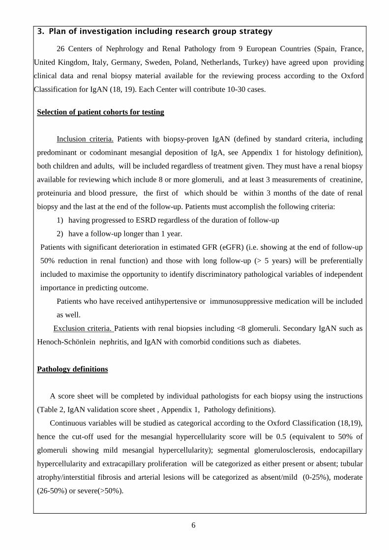
3. Plan of investigation including research group strategy
26 Centers of Nephrology and Renal Pathology from 9 European Countries (Spain, France,
United Kingdom, Italy, Germany, Sweden, Poland, Netherlands, Turkey) have agreed upon providing
clinical data and renal biopsy material available for the reviewing process according to the Oxford
Classification for IgAN (18, 19). Each Center will contribute 10-30 cases.
Selection of patient cohorts for testing
Inclusion criteria.
1) having progressed to ESRD regardless of the duration of follow-up
Patients with biopsy-proven IgAN (defined by standard criteria, including
predominant or codominant mesangial deposition of IgA, see Appendix 1 for histology definition),
both children and adults, will be included regardless of treatment given. They must have a renal biopsy
available for reviewing which include 8 or more glomeruli, and at least 3 measurements of creatinine,
proteinuria and blood pressure, the first of which should be within 3 months of the date of renal
biopsy and the last at the end of the follow-up. Patients must accomplish the following criteria:
2) have a follow-up longer than 1 year.
Patients with significant deterioration in estimated GFR (eGFR) (i.e. showing at the end of follow-up
50% reduction in renal function) and those with long follow-up (> 5 years) will be preferentially
included to maximise the opportunity to identify discriminatory pathological variables of independent
importance in predicting outcome.
Patients who have received antihypertensive or immunosuppressive medication will be included
as well.
Exclusion criteria.
Patients with renal biopsies including <8 glomeruli. Secondary IgAN such as
Henoch-Schönlein nephritis, and IgAN with comorbid conditions such as diabetes.
Pathology definitions
A score sheet will be completed by individual pathologists for each biopsy using the instructions
(Table 2, IgAN validation score sheet , Appendix 1, Pathology definitions).
Continuous variables will be studied as categorical according to the Oxford Classification (18,19),
hence the cut-off used for the mesangial hypercellularity score will be 0.5 (equivalent to 50% of
glomeruli showing mild mesangial hypercellularity); segmental glomerulosclerosis, endocapillary
hypercellularity and extracapillary proliferation will be categorized as either present or absent; tubular
atrophy/interstitial fibrosis and arterial lesions will be categorized as absent/mild (0-25%), moderate
(26-50%) or severe(>50%).
7
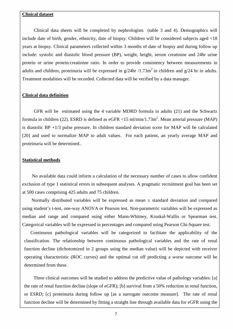
Clinical dataset
Clinical data sheets will be completed by nephrologists (table 3 and 4). Demographics will
include date of birth, gender, ethnicity, date of biopsy. Children will be considered subjects aged <18
years at biopsy. Clinical parameters collected within 3 months of date of biopsy and during follow up
include: systolic and diastolic blood pressure (BP), weight, height, serum creatinine and 24hr urine
protein or urine protein:creatinine ratio. In order to provide consistency between measurements in
adults and children, proteinuria will be expressed in g/24hr /1.73m2 in children and g/24 hr in adults.
Treatment modalities will be recorded. Collected data will be verified by a data manager.
Clinical data definition
GFR will be estimated using the 4 variable MDRD formula in adults (21) and the Schwartz
formula in children (22). ESRD is defined as eGFR <15 ml/min/1.73m2. Mean arterial pressure (MAP)
is diastolic BP +1/3 pulse pressure. In children standard deviation score for MAP will be calculated
[20] and used to normalize MAP to adult values. For each patient, an yearly average MAP and
proteinuria will be determined..
Statistical methods
No available data could inform a calculation of the necessary number of cases to allow confident
exclusion of type 1 statistical errors in subsequent analyses. A pragmatic recruitment goal has been set
at 500 cases comprising 425 adults and 75 children.
Normally distributed variables will be expressed as mean ± standard deviation and compared
using student’s t-test, one-way ANOVA or Pearson test. Non-parametric variables will be expressed as
median and range and compared using either Mann-Whitney, Kruskal-Wallis or Spearman test.
Categorical variables will be expressed in percentages and compared using Pearson Chi-Square test.
Continuous pathological variables will be categorized to facilitate the applicability of the
classification. The relationship between continuous pathological variables and the rate of renal
function decline (dichotomized in 2 groups using the median value) will be depicted with receiver
operating characteristic (ROC curves) and the optimal cut off predicting a worse outcome will be
determined from these.
Three clinical outcomes will be studied to address the predictive value of pathology variables: [a]
the rate of renal function decline (slope of eGFR); [b] survival from a 50% reduction in renal function,
or ESRD; [c] proteinuria during follow up [as a surrogate outcome measure]. The rate of renal
function decline will be determined by fitting a straight line through available data for eGFR using the
8
principle of least squares.
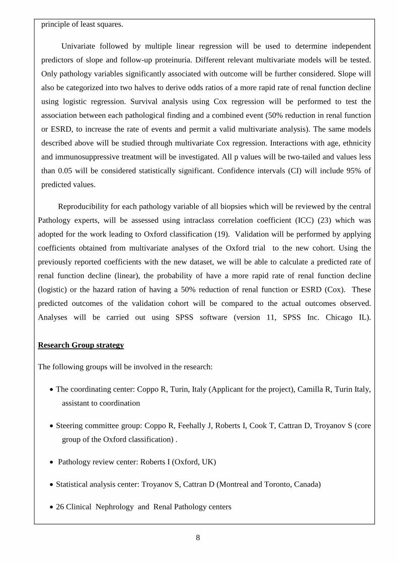
Univariate followed by multiple linear regression will be used to determine independent
predictors of slope and follow-up proteinuria. Different relevant multivariate models will be tested.
Only pathology variables significantly associated with outcome will be further considered. Slope will
also be categorized into two halves to derive odds ratios of a more rapid rate of renal function decline
using logistic regression. Survival analysis using Cox regression will be performed to test the
association between each pathological finding and a combined event (50% reduction in renal function
or ESRD, to increase the rate of events and permit a valid multivariate analysis). The same models
described above will be studied through multivariate Cox regression. Interactions with age, ethnicity
and immunosuppressive treatment will be investigated. All p values will be two-tailed and values less
than 0.05 will be considered statistically significant. Confidence intervals (CI) will include 95% of
predicted values.
Reproducibility for each pathology variable of all biopsies which will be reviewed by the central
Pathology experts, will be assessed using intraclass correlation coefficient (ICC) (23) which was
adopted for the work leading to Oxford classification (19). Validation will be performed by applying
coefficients obtained from multivariate analyses of the Oxford trial to the new cohort. Using the
previously reported coefficients with the new dataset, we will be able to calculate a predicted rate of
renal function decline (linear), the probability of have a more rapid rate of renal function decline
(logistic) or the hazard ration of having a 50% reduction of renal function or ESRD (Cox). These
predicted outcomes of the validation cohort will be compared to the actual outcomes observed.
Analyses will be carried out using SPSS software (version 11, SPSS Inc. Chicago IL).
Research Group strategy
The following groups will be involved in the research:
• The coordinating center: Coppo R, Turin, Italy (Applicant for the project), Camilla R, Turin Italy,
assistant to coordination
• Steering committee group: Coppo R, Feehally J, Roberts I, Cook T, Cattran D, Troyanov S (core
group of the Oxford classification) .
• Pathology review center: Roberts I (Oxford, UK)
• Statistical analysis center: Troyanov S, Cattran D (Montreal and Toronto, Canada)
• 26 Clinical Nephrology and Renal Pathology centers
9
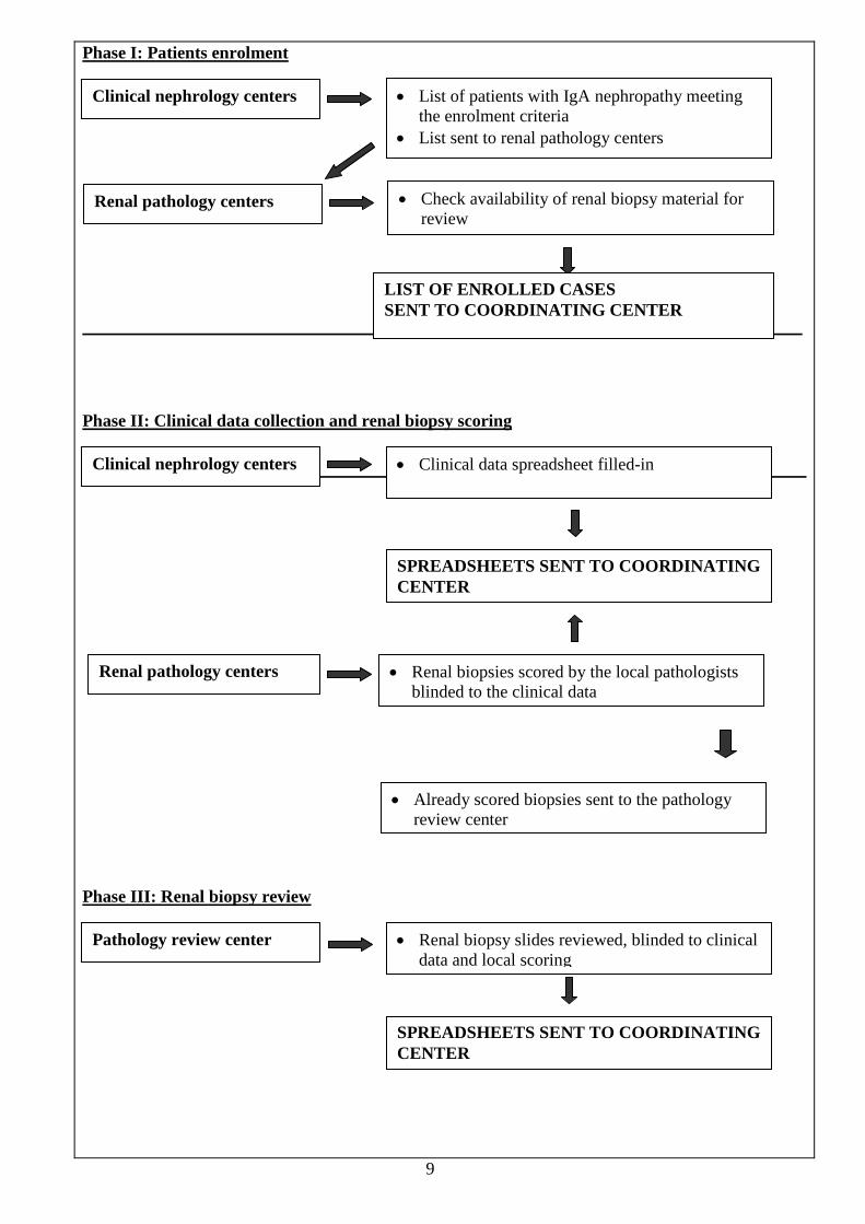
Phase I: Patients enrolment
Phase II: Clinical data collection and renal biopsy scoring
Phase III: Renal biopsy review
Clinical nephrology centers • List of patients with IgA nephropathy meeting the enrolment criteria
• List sent to renal pathology centers
Clinical nephrology centers • Clinical data spreadsheet filled-in
• Renal biopsies scored by the local pathologists blinded to the clinical data
SPREADSHEETS SENT TO COORDINATING CENTER
Renal pathology centers
• Already scored biopsies sent to the pathology review center
Pathology review center • Renal biopsy slides reviewed, blinded to clinical data and local scoring
SPREADSHEETS SENT TO COORDINATING CENTER
Renal pathology centers • Check availability of renal biopsy material for review
LIST OF ENROLLED CASES SENT TO COORDINATING CENTER
10
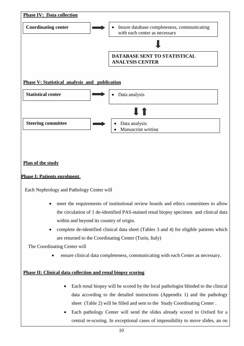
Phase IV: Data collection
Phase V: Statistical analysis and publication
Plan of the study
Phase I: Patients enrolment.
Each Nephrology and Pathology Center will
• meet the requirements of institutional review boards and ethics committees to allow
the circulation of 1 de-identified PAS-stained renal biopsy specimen and clinical data
within and beyond its country of origin.
• complete de-identified clinical data sheet (Tables 3 and 4) for eligible patients which
are returned to the Coordinating Center (Turin, Italy)
The Coordinating Center will
• ensure clinical data completeness, communicating with each Center as necessary.
Phase II: Clinical data collection and renal biopsy scoring
• Each renal biopsy will be scored by the local pathologist blinded to the clinical
data according to the detailed instructions (Appendix 1) and the pathology
sheet (Table 2) will be filled and sent to the Study Coordinating Center .
• Each pathology Center will send the slides already scored to Oxford for a
central re-scoring. In exceptional cases of impossibility to move slides, an on
Coordinating center • Insure database completeness, communicating with each center as necessary
DATABASE SENT TO STATISTICAL ANALYSIS CENTER
Statistical center • Data analysis
Steering committee • Data analysis • Manuscript writing
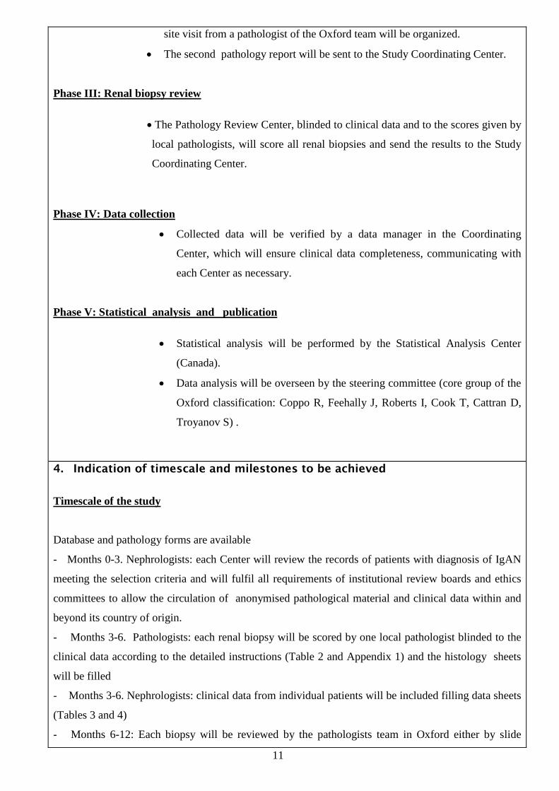
11
site visit from a pathologist of the Oxford team will be organized.
• The second pathology report will be sent to the Study Coordinating Center.
Phase III: Renal biopsy review
• The Pathology Review Center, blinded to clinical data and to the scores given by
local pathologists, will score all renal biopsies and send the results to the Study
Coordinating Center.
Phase IV: Data collection
• Collected data will be verified by a data manager in the Coordinating
Center, which will ensure clinical data completeness, communicating with
each Center as necessary.
Phase V: Statistical analysis and publication
• Statistical analysis will be performed by the Statistical Analysis Center
(Canada).
• Data analysis will be overseen by the steering committee (core group of the
Oxford classification: Coppo R, Feehally J, Roberts I, Cook T, Cattran D,
Troyanov S) .
4. Indication of timescale and milestones to be achieved
Timescale of the study
Database and pathology forms are available
- Months 0-3. Nephrologists: each Center will review the records of patients with diagnosis of IgAN
meeting the selection criteria and will fulfil all requirements of institutional review boards and ethics
committees to allow the circulation of anonymised pathological material and clinical data within and
beyond its country of origin.
- Months 3-6. Pathologists: each renal biopsy will be scored by one local pathologist blinded to the
clinical data according to the detailed instructions (Table 2 and Appendix 1) and the histology sheets
will be filled
- Months 3-6. Nephrologists: clinical data from individual patients will be included filling data sheets
(Tables 3 and 4)
- Months 6-12: Each biopsy will be reviewed by the pathologists team in Oxford either by slide
12
circulation or by on-site visits. Both original and reviewed pathology reports will be sent to the Study
Coordinating Center.
- Months 3-12 The data sheets will be collected by the Study Coordinating Center (Turin), which
will thoroughly check the database completeness
- Months 12-16 Statistical analysis will be performed by the Statistical Analysis Center (S. Troyanov,
Canada).
- Months 15-18 Data will be discussed within the steering committee, then draft results will be
circulated among all the participating Centers. Final report will be completed and a manuscript
submitted.
Milestones to be achieved
This multicenter collaborative study has several relevant milestones:
1) To establish working principles for the ERA-EDTA working group of “Immunonephrology” with a
high quality multicenter study, which offers the opportunity to implement the newly created
network.
2) To involve ERA-EDTA in this Collaborative International Consensus group (as far as Europe in
concerned) initiated under the auspices of the International Society of Nephrology, the Renal
Pathology Society and the International IgAN Network.
3) To test the reproducibility of the new Oxford classification for IgAN in different European Centers,
aiming at facilitating an improvement in data exchange
4) To validate the Oxford classification on a large European cohort encompassing all ages and different
clinical and pathological features, aiming at facilitating networking of Centers.
This process will finally lead to other relevant milestones:
1) It will accelerate progress in developing a prognostic system with the sensitivity and specificity to
predict outcome for individual patients.
2) It will increase the capacity to make international comparisons between different outcome studies,
3) It will enhance new opportunities to refine the stratification of risk for progression
4) It will eventually allow the design of more informative clinical intervention trials. A slowly
progressive disease like IgAN needs large studies of long duration to evaluate new interventions
unless patients with a high risk of progression can be better defined early in the course of the
disease. The validation study on a large cohort of IgAN patients from various European countries
will provide the suitable basis for this aim.
List of Collaborating Researchers and Institutions:
13
1) John Feehally Division of Nephrology, Department of Infection, Immunity & Inflammation University of Leicester, Leicester General Hospital , Gwendolen Road, LE5 4PW, Leicester, UK [email protected] Patologists - Ian SD Roberts Department of Cellular Pathology, John Radcliffe Hospital Headington, OX3 9DU Oxford, UK [email protected] - H.Terence Cook. Department of Histopathology ,Imperial College,Hammersmith Hospita Du Cane Road, W120NN London [email protected] 2) Colin C Geddes Western Infirmary, Dumbarton Road, G11 6NT, Glasgow,UK. [email protected] 3) Jurgen Floege and Frank Eitner Division of Nephrology and Immunology University of Aachen, Aachen, Germany. Pathologist: Hermann-Josef Groene, Deutsches Krebsforschungszentrum, Im Nauenheimer Feld 280, 69120 Heidelberg, Germany [email protected] 4) Borje Haraldsson Department of Nephrology,The Sahlgrenska Academy and University Hospital SE-413 45 Gothenburg, Sweden [email protected] 5) Bengt Fellstron Department of Nephrology University Hospital SE-75185 Uppsala, Sweden [email protected] 6) Andrzej Wiecek Department of Nephrology, Endocrinology and Metabolic Diseases Silesian University School of Medicine PL-40-027 Francuska 20-24,
14
Katowice, Poland email: [email protected] 7) Rezan Topaloglu and Aysin Bakkaloglu Department of Pediatric Nephrology and Rhematology Dicle Orhan , Pathology Center Hacettepe University Faculty of Medicine 06100 Ankara Turkey E-mail: [email protected] 8) Jose Ballarin Fundación Puigvert Nephrology Department Fundation Puigvert Pathologist:Yolanda Arce Renal Pathology Unit Cartagena 340,08025 Barcelone, Spain [email protected] 9) Manuel Praga Servicio de Nefrología, Hospital 12 de Octubre, Madrid, Spain [email protected] 10) Jesus Egido Fundación Jiménez Díaz. Universidad Autónoma de Madrid. Madrid. España. [email protected] 11) Josep Maria Grinyó Xavier Fulladosa Oliveras Nephrology Department Hospital Universitari de Bellvitge. C/ Feixa Llarga s/n. 08907. L'Hospitalet, Barcelona. [email protected] [email protected] 12) François Berthoux Nephrology, Dialysis and Renal Transplantation, North University Hospital - CHU de St. Etienne, 42055 ,Cedex 2, Saint-Etienne ,France [email protected] 13) Sandrine Florquin and Raymond Krediet Academic Medical Centre, Pathology NL-1100 DD Amsterdam, The Netherlands [email protected]
15
14) Ton J Rabelink and Cees Van Kooten Department of Nephrology, Pathologist . JA de Bruijn Leiden University Medical Center, Albinusdreef 2, 2300 RC Leiden, The Netherlands. [email protected] 15) Alessandro Amore and Roberta Camilla Nefrology, Dialysis and Transplantation Regina Margherita University Hospital Piazza Polonia 94, 10126 Torino, Italy [email protected] Pathologist: Gianna Mazzucco Department of Medical Sciences and Human Oncology University of Torino Torino Italy gianna.mazzucco@unito,it 16) Piero Stratta and Giuseppe Segoloni Nephrology, Dialysis and Transplantation Centers University of Turin and Novara Pathologist: Gianna Mazzucco Department of Medical Sciences and Human Oncology University of Torino Torino, Italy [email protected] [email protected] 17) Antonio Dal Canton and Ciro Esposito University of Pavia, IRCCS Policlinico San Matteo viale Camillo Golgi 19, 27100 Pavia, Italy [email protected] 18) Franco Ferrario Nephrology and Dialysis Ospedale San Carlo Milano, Italy [email protected] 19) Pier Giorgio Messa Ospedale Maggiore Policlinico Mangiagalli e Regina Elena Via della Commenda 15, 20122 Milano, Italy [email protected] 20) Giovanni Cancarini A.O. Spedali Civili di Brescia Piazza Spedali Civili 1 25123 Brescia, Italy [email protected]
16
21) Giuseppe Remuzzi and Mauro Abbate Clinical Research Centre for rare diseases “Aldo e Cele Daccò” Ranica Italy [email protected] 22) Lucia Del Vecchio and Claudio Pozzi Nephrology and Dialysis Lecco, Italy [email protected] Hospital E. Bassini, via Gorki 50, 20092 Cinisello Balsamo (Milano), Italy. [email protected] 23) Antonio Lupo University of Verona Ospedale Civile Maggiore Piazzale Stefani 1 37126 Verona, Italy [email protected] 24) Maurizio Salvadori and Lino Cerami Nephrology, Dialysis and Transplantation A.O. Careggi-Villa Monnatessa Viale Pieraccioni 18, 50139, Firenze, Italy [email protected] 25) Paolo Schena Nephrology, Dialysis and Transplantation, University of Bari,Policlinico di Bari Piazza Giulio Cesare 11 70124 Bari, Italy [email protected] 26) Loreto Gesualdo Renal Pathologist: Anna Maria Di Palma Nephrology,Dialysis and Transplantation Department (DIAN) Azienda Ospedaliero-Universitaria “OO.RR” Viale Pinto 253 71100 Foggia, Italy [email protected] 27) Statistical Analysis Center: Stéphan Troyanov Hôpital du Sacré-Coeur de Montréal 5400 Gouin boulevard west Montreal, QC, CANADA - H4J 1C5 [email protected] 28) Statistical Analysis Center Daniel Cattran University Health Network Toronto general Hospital Suite 1256,11floor CSB
18
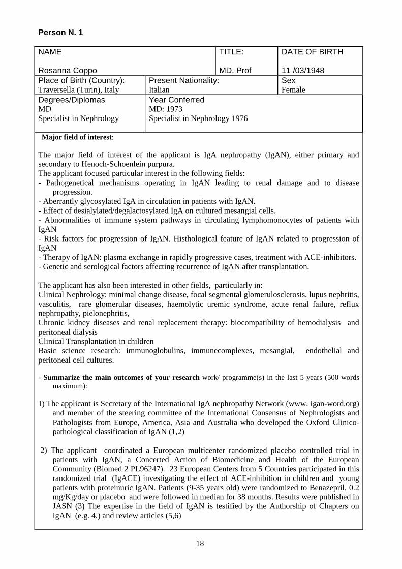
Person N. 1 NAME Rosanna Coppo
TITLE: MD, Prof
DATE OF BIRTH 11 /03/1948
Place of Birth (Country): Traversella (Turin), Italy
Present Nationality: Italian
Sex Female
Degrees/Diplomas MD Specialist in Nephrology
Year Conferred MD: 1973 Specialist in Nephrology 1976
Major field of interest:
The major field of interest of the applicant is IgA nephropathy (IgAN), either primary and secondary to Henoch-Schoenlein purpura. The applicant focused particular interest in the following fields: - Pathogenetical mechanisms operating in IgAN leading to renal damage and to disease
progression. - Aberrantly glycosylated IgA in circulation in patients with IgAN. - Effect of desialylated/degalactosylated IgA on cultured mesangial cells. - Abnormalities of immune system pathways in circulating lymphomonocytes of patients with IgAN - Risk factors for progression of IgAN. Histhological feature of IgAN related to progression of IgAN - Therapy of IgAN: plasma exchange in rapidly progressive cases, treatment with ACE-inhibitors. - Genetic and serological factors affecting recurrence of IgAN after transplantation. The applicant has also been interested in other fields, particularly in: Clinical Nephrology: minimal change disease, focal segmental glomerulosclerosis, lupus nephritis, vasculitis, rare glomerular diseases, haemolytic uremic syndrome, acute renal failure, reflux nephropathy, pielonephritis, Chronic kidney diseases and renal replacement therapy: biocompatibility of hemodialysis and peritoneal dialysis Clinical Transplantation in children Basic science research: immunoglobulins, immunecomplexes, mesangial, endothelial and peritoneal cell cultures. - Summarize the main outcomes of your research work/ programme(s) in the last 5 years (500 words
maximum): 1) The applicant is Secretary of the International IgA nephropathy Network (www. igan-word.org)
and member of the steering committee of the International Consensus of Nephrologists and Pathologists from Europe, America, Asia and Australia who developed the Oxford Clinico-pathological classification of IgAN (1,2)
2) The applicant coordinated a European multicenter randomized placebo controlled trial in
patients with IgAN, a Concerted Action of Biomedicine and Health of the European Community (Biomed 2 PL96247). 23 European Centers from 5 Countries participated in this randomized trial (IgACE) investigating the effect of ACE-inhibition in children and young patients with proteinuric IgAN. Patients (9-35 years old) were randomized to Benazepril, 0.2 mg/Kg/day or placebo and were followed in median for 38 months. Results were published in JASN (3) The expertise in the field of IgAN is testified by the Authorship of Chapters on IgAN (e.g. 4,) and review articles (5,6)
19
3) The applicant succeeded in performing a Multicenter collaborative Italian Study in transplanted patients having had IgAN as renal disease leading to renal replacement treatment. Five Italian Centers participated in the study. Genetic factors, including genes encoding for cytokines and chemokynes as well as for the renin angiotensin system and serologic factors, including aberrantly glycosylated IgA and macromolecular IgA were detected and correlated with recurrence of IgAN in the grafted kidney. Results were published in Ref 7.
4) The applicant chaired a Multicenter collaborative Italian Study of the Immunopathology Group
of the Italian Society of nephrology investigating the predictive value of clinical and histological features on the progression of IgAN secondary to Henoch-Shoenlein Purpura: 43 Italian Centers participated in the study Results were published in Ref 8 . The expertise in the field is testified by the Authorship of Chapters on IgAN secondary to Henoch-Schoenlein Purpura (e.g.9)
5) The applicant participated in several research supported by the Italian National Health Ministry
which investigated the presence of aberrantly glycostlated IgA in IgAN and some immune abnormalities of circulating lymphomonocytes in patients with IgAN, namely the switch from proteasome to immuneproteasome, which may favour the antigen presentation and the immune response (Chair A.Dal Canton, 2004; and Chair PA Tovo, 2005) Results were published in several papers, including Ref 10 and 11
References 1) Cattran D, Coppo R, Cook TH, Feehally J, Roberts ISD, Troyanov S, Alpers CE, Amore A, Barratt
J, Berthoux F, Bonsib S, Bruijn JA, D’Agati V, D’Amico G, Emancipator S, Emma F, Ferrario F, Fervenza FC, Florquin S, Fogo A, Geddes CC, Groene HJ, Haas M, AM Herzenberg, Hill P, Hogg RJ, Hsu S, Jennette JC, Joh K, Julian BA, Kawamura T, Lai F, Li LS, Li P, Liu ZH, Mezzano S, Schena FP, Tomino Y, Walker P, Wang H, Weening JJ, Yoshikawa N, Zhang H.
The Oxford Classification of IgA Nephropathy: Pathology definitions, correlations and reproducibility. Kidney International 2009; 76(5):546-56.
2) Roberts ISD, Cook HT, Troyanov S, Alpers CE, Amore A, Barratt J, Berthoux F, Bonsib S, Bruijn JA, Cattran DC, CoppoR, D’Agati V, D’AmicoG, Emancipator S, Emma F, Feehally J, Ferrario F, Fervenza FC, Florquin S, Fogo A, Geddes CC, Groene HJ, Haas M, Herzenberg AM, Hill P, Hogg RJ, Hsu S, Jennette CJ, Joh K, Julian BA, Kawamura T, Lai F, Li LS, Li PKT, Liu ZH, Mackinnon B, MezzanoS, Schena FP, TominoY, Walker P, Wang H, Weening JJ, Yoshikawa N and Zhang H, A Working Group of the International IgA Nephropathy Network and the Renal Pathology Society. The Oxford Classification of IgA Nephropathy: Rationale, clinicopathological correlations, and proposal for classification. Kidney International 2009; 76:534-45.
3) Coppo R, Peruzzi L, Amore A, Piccoli A, Cochat P, Stone R, Kirschstein M, Linné T. IgACE: a placebo-controlled, randomized trial of angiotensin-converting enzyme inhibitors in children and young people with IgA nephropathy and moderate proteinuria. J Am Soc Nephrol. 2007;18:1880-8.
4) Schena FP and Coppo R: IgA Nephropathies Nephrology [eds. Davison AM, Ritz E, Cameron JS, Oxford textbook of Clinical Nephrology Winearls C] 3rd edition, Oxford University Press 2005
5) Coppo R. Pediatric IgA nephropathy: clinical and therapeutic perspectives.Semin Nephrol. 2008;28:18-26.
6) Coppo R, Amore A, Peruzzi L, Mancuso D, Camilla R. Angiotensin antagonists and fish oil for treating IgA nephropathy . Contrib Nephrol. 2007;157:27-36.
7) Coppo R, Amore A, Chiesa M, Lombardo F, Cirina P, Andrulli S, Passerini P, Conti G, Peruzzi L, Giraudi R, Messina M, Segoloni G, Ponticelli C. Serological and genetic factors in early recurrence of IgA nephropathy after renal transplantation. Clin Transplant. 2007;21728-37.
8) Coppo R, Andrulli S, Amore A, Gianoglio B, Conti G, Peruzzi L, Locatelli F, Cagnoli L. Predictors of outcome in Henoch-Schönlein nephritis in children and adults.Am J Kidney Dis. 2006;47:993-1003
9) Coppo R and Amore A.: Henoch Schoenlein Purpura nephritis (eds Avner E, Harmon W, Niaudet P). Pediatric Nephrology, 5th edition Lippincott 2004
10) Coppo R, Amore A. Aberrant glycosylation in IgA nephropathy. Kidney Int. 2004;65:1544-7. 11) Coppo R, Camilla R, Alfarano A, Balegno S, Mancuso D, Peruzzi L, Amore A, Dal Canton A, Sepe
V, Tovo P. Upregulation of the immunoproteasome in peripheral blood mononuclear cells of patients with IgA nephropathy. Kidney Int. 2009;75:536-41
20
References : Total number of peer reviewed publications in Pub Med: 206 Publications (List up to 5 recent publications relevant to proposed project)
1. Cattran D, Coppo R, Cook TH, Feehally J, Roberts ISD, Troyanov S, Alpers CE, Amore A, Barratt J, Berthoux F, Bonsib S, Bruijn JA, D’Agati V, D’Amico G, Emancipator S, Emma F, Ferrario F, Fervenza FC, Florquin S, Fogo A, Geddes CC, Groene HJ, Haas M, AM Herzenberg, Hill P, Hogg RJ, Hsu S, Jennette JC, Joh K, Julian BA, Kawamura T, Lai F, Li LS, Li P, Liu ZH, Mezzano S, Schena FP, Tomino Y, Walker P, Wang H, Weening JJ, Yoshikawa N, Zhang H. The Oxford Classification of IgA Nephropathy: Pathology definitions, correlations and reproducibility. Kidney International 2009; 76:546-56.
2. Roberts ISD, Cook HT, Troyanov S, Alpers CE, Amore A, Barratt J, Berthoux F, Bonsib S, Bruijn
JA, Cattran DC, CoppoR, D’Agati V, D’AmicoG, Emancipator S, Emma F, Feehally J, Ferrario F, Fervenza FC, Florquin S, Fogo A, Geddes CC, Groene HJ, Haas M, Herzenberg AM, Hill P, Hogg RJ, Hsu S, Jennette CJ, Joh K, Julian BA, Kawamura T, Lai F, Li LS, Li PKT, Liu ZH, Mackinnon B, MezzanoS, Schena FP, TominoY, Walker P, Wang H, Weening JJ, Yoshikawa N and Zhang H, A Working Group of the International IgA Nephropathy Network and the Renal Pathology Society. The Oxford Classification of IgA Nephropathy: Rationale, clinicopathological correlations, and proposal for classification. Kidney International 2009; 76:534-45.
3. IgACE: a placebo-controlled, randomized trial of angiotensin-converting enzyme inhibitors in
children and young people with IgA nephropathy and moderate proteinuria. Coppo R, Peruzzi L, Amore A, Piccoli A, Cochat P, Stone R, Kirschstein M, Linné T. J Am Soc Nephrol. 2007 Jun;18(6):1880-8. Epub 2007 May 18.
4. International IgA nephropathy network clinico-pathological classification of IgA nephropathy.
Feehally J, Barratt J, Coppo R, Cook T, Roberts I; International IgA Nephropathy Network. Contrib Nephrol. 2007;157:13-8.
5. Factors predicting progression of IgA nephropathies. Coppo R, D'Amico G. J Nephrol. 2005 Sep-
Oct;18(5):503-12.
21
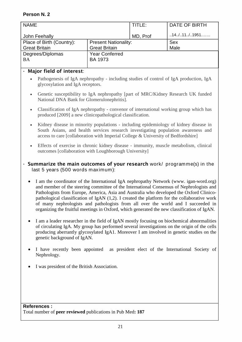
Person N. 2 NAME John Feehally
TITLE: MD, Prof
DATE OF BIRTH ..14../..11../..1951.……
Place of Birth (Country): Great Britain
Present Nationality: Great Britain
Sex Male
Degrees/Diplomas BA
Year Conferred BA 1973
- Major field of interest: • Pathogenesis of IgA nephropathy - including studies of control of IgA production, IgA
glycosylation and IgA receptors.
• Genetic susceptibility to IgA nephropathy [part of MRC/Kidney Research UK funded National DNA Bank for Glomerulonephritis].
• Classification of IgA nephropathy - convenor of international working group which has produced [2009] a new clinicopathological classification.
• Kidney disease in minority populations - including epidemiology of kidney disease in South Asians, and health services research investigating population awareness and access to care [collaboration with Imperial College & University of Bedfordshire]
• Effects of exercise in chronic kidney disease - immunity, muscle metabolism, clinical outcomes [collaboration with Loughborough University]
- Summarize the main outcomes of your research work/ programme(s) in the
last 5 years (500 words maximum): • I am the coordinator of the International IgA nephropathy Network (www. igan-word.org)
and member of the steering committee of the International Consensus of Nephrologists and Pathologists from Europe, America, Asia and Australia who developed the Oxford Clinico-pathological classification of IgAN (1,2). I created the platform for the collaborative work of many nephrologists and pathologists from all over the world and I succeeded in organizing the fruitful meetings in Oxford, which generated the new classification of IgAN.
• I am a leader researcher in the field of IgAN mostly focusing on biochemical abnormalities
of circulating IgA. My group has performed several investigations on the origin of the cells producing aberrantly glycosylated IgA1. Moreover I am involved in genetic studies on the genetic background of IgAN.
• I have recently been appointed as president elect of the International Society of
Nephrology. • I was president of the British Association.
References : Total number of peer reviewed publications in Pub Med: 187
22
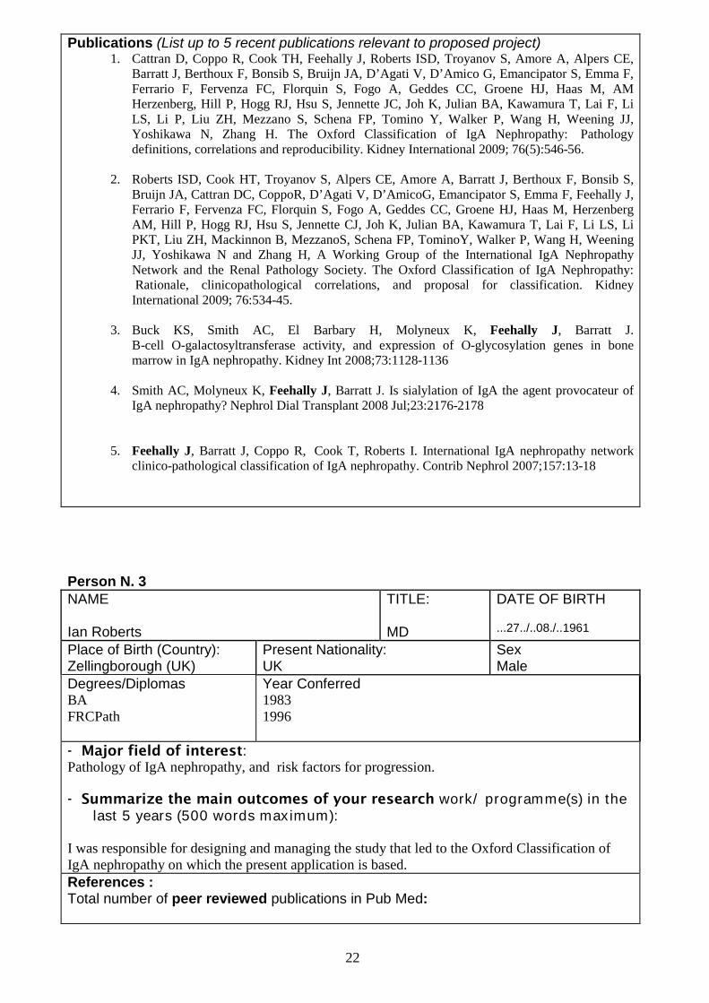
Publications (List up to 5 recent publications relevant to proposed project) 1. Cattran D, Coppo R, Cook TH, Feehally J, Roberts ISD, Troyanov S, Amore A, Alpers CE,
Barratt J, Berthoux F, Bonsib S, Bruijn JA, D’Agati V, D’Amico G, Emancipator S, Emma F, Ferrario F, Fervenza FC, Florquin S, Fogo A, Geddes CC, Groene HJ, Haas M, AM Herzenberg, Hill P, Hogg RJ, Hsu S, Jennette JC, Joh K, Julian BA, Kawamura T, Lai F, Li LS, Li P, Liu ZH, Mezzano S, Schena FP, Tomino Y, Walker P, Wang H, Weening JJ, Yoshikawa N, Zhang H. The Oxford Classification of IgA Nephropathy: Pathology definitions, correlations and reproducibility. Kidney International 2009; 76(5):546-56.
2. Roberts ISD, Cook HT, Troyanov S, Alpers CE, Amore A, Barratt J, Berthoux F, Bonsib S,
Bruijn JA, Cattran DC, CoppoR, D’Agati V, D’AmicoG, Emancipator S, Emma F, Feehally J, Ferrario F, Fervenza FC, Florquin S, Fogo A, Geddes CC, Groene HJ, Haas M, Herzenberg AM, Hill P, Hogg RJ, Hsu S, Jennette CJ, Joh K, Julian BA, Kawamura T, Lai F, Li LS, Li PKT, Liu ZH, Mackinnon B, MezzanoS, Schena FP, TominoY, Walker P, Wang H, Weening JJ, Yoshikawa N and Zhang H, A Working Group of the International IgA Nephropathy Network and the Renal Pathology Society. The Oxford Classification of IgA Nephropathy: Rationale, clinicopathological correlations, and proposal for classification. Kidney International 2009; 76:534-45.
3. Buck KS, Smith AC, El Barbary H, Molyneux K, Feehally J, Barratt J.
B-cell O-galactosyltransferase activity, and expression of O-glycosylation genes in bone marrow in IgA nephropathy. Kidney Int 2008;73:1128-1136
4. Smith AC, Molyneux K, Feehally J, Barratt J. Is sialylation of IgA the agent provocateur of
IgA nephropathy? Nephrol Dial Transplant 2008 Jul;23:2176-2178
5. Feehally J, Barratt J, Coppo R, Cook T, Roberts I. International IgA nephropathy network clinico-pathological classification of IgA nephropathy. Contrib Nephrol 2007;157:13-18
Person N. 3 NAME Ian Roberts
TITLE: MD
DATE OF BIRTH ...27../..08./..1961
Place of Birth (Country): Zellingborough (UK)
Present Nationality: UK
Sex Male
Degrees/Diplomas BA FRCPath
Year Conferred 1983 1996
- Major field of interest: Pathology of IgA nephropathy, and risk factors for progression. - Summarize the main outcomes of your research work/ programme(s) in the
last 5 years (500 words maximum): I was responsible for designing and managing the study that led to the Oxford Classification of IgA nephropathy on which the present application is based. References : Total number of peer reviewed publications in Pub Med:
23
Publications (List up to 5 recent publications relevant to proposed project) 1. Cattran D, Coppo R, Cook TH, Feehally J, Roberts ISD, Troyanov S, Alpers CE, Amore A,
Barratt J, Berthoux F, Bonsib S, Bruijn JA, D’Agati V, D’Amico G, , Emancipator S, Emma F, Ferrario F, Fervenza FC, Florquin S, Fogo A, Geddes CC, Groene HJ, Haas M, AM Herzenberg, Hill P, Hogg RJ, Hsu S, Jennette JC, Joh K, Julian BA, Kawamura T, Lai F, Li LS, Li P, Liu ZH, Mezzano S, Schena FP, Tomino Y, Walker P, Wang H, Weening JJ, Yoshikawa N, Zhang H. The Oxford Classification of IgA Nephropathy: Pathology definitions, correlations and reproducibility. Kidney International 2009; 76(5):546-56.
2. Roberts ISD, Cook HT, Troyanov S, Alpers CE, Amore A, Barratt J, Berthoux F, Bonsib S,
Bruijn JA, Cattran DC, CoppoR, D’Agati V, D’AmicoG, Emancipator S, Emma F, Feehally J, Ferrario F, Fervenza FC, Florquin S, Fogo A, Geddes CC, Groene HJ, Haas M, Herzenberg AM, Hill P, Hogg RJ, Hsu S, Jennette CJ, Joh K, Julian BA, Kawamura T, Lai F, Li LS, Li PKT, Liu ZH, Mackinnon B, MezzanoS, Schena FP, TominoY, Walker P, Wang H, Weening JJ, Yoshikawa N and Zhang H, A Working Group of the International IgA Nephropathy Network and the Renal Pathology Society. The Oxford Classification of IgA Nephropathy: Rationale, clinicopathological correlations, and proposal for classification. Kidney International 2009; 76:534-45.
3. Feehally J, Barratt J, Coppo R, Cook T, Roberts I; International IgA Nephropathy Network. International
IgA nephropathy network clinico-pathological classification of IgA nephropathy.Contrib Nephrol. 2007;157:13-8.
4. Solez K, Colvin RB, Racusen LC, Haas M, Sis B, Mengel M, Halloran PF, Baldwin W, Banfi G, Collins
AB, Cosio F, David DS, Drachenberg C, Einecke G, Fogo AB, Gibson IW, Glotz D, Iskandar SS, Kraus E, Lerut E, Mannon RB, Mihatsch M, Nankivell BJ, Nickeleit V, Papadimitriou JC, Randhawa P, Regele H, Renaudin K, Roberts I, Seron D, Smith RN, Valente M. Banff 07 classification of renal allograft pathology: updates and future directions. Am J Transplant. 2008;8:753-60.
5. Ballardie FW, Roberts IS. Controlled prospective trial of prednisolone and cytotoxics in progressive IgA
nephropathy.J Am Soc Nephrol. 2002 ;13:142-8.
24
Person N. 4 NAME H Terence Cook
TITLE: MD, Prof
DATE OF BIRTH ..04./.04../.1954
Place of Birth (Country): Great Britain
Present Nationality: Great Britain
Sex Male
Degrees/Diplomas BA MBBS MRCP FRCPath FMedSci
Year Conferred 1975 1980 1983 1989 2008
- Major field of interest: Renal Pathology - Summarize the main outcomes of your research work/ programme(s) in the
last 5 years (500 words maximum):
• My research is into pathological mechanisms in glomerulonephritis. I have studied animal models to elucidate genes that are associated with susceptibility to glomerulonephritis and have shown that in a strain of rat susceptible to crescentic glomerulonephritis there are seven genetic loci that control susceptibility. I have identified genes responsible for susceptibility at two of these loci.
• My clinical position is as a renal pathologist at the West London Renal and Transplant
Centre. My research in human glomerulonephritis has centred on the classification of glomerulonephritis and reproducibility of the assessment of histological features. My work has particularly focused on SLE and IgA nephropathy. I was responsible for designing and managing the study that led to the Oxford Classification of IgA nephropathy on which the present application is based.
References : Total number of peer reviewed publications in Pub Med: 133 Publications (List up to 5 recent publications relevant to proposed project)
1. Cattran D, Coppo R, Cook TH, Feehally J, Roberts ISD, Troyanov S, Alpers CE, Amore A, Barratt J, Berthoux F, Bonsib S, Bruijn JA, D’Agati V, D’Amico G, , Emancipator S, Emma F, Ferrario F, Fervenza FC, Florquin S, Fogo A, Geddes CC, Groene HJ, Haas M, AM Herzenberg, Hill P, Hogg RJ, Hsu S, Jennette JC, Joh K, Julian BA, Kawamura T, Lai F, Li LS, Li P, Liu ZH, Mezzano S, Schena FP, Tomino Y, Walker P, Wang H, Weening JJ, Yoshikawa N, Zhang H. The Oxford Classification of IgA Nephropathy: Pathology definitions, correlations and reproducibility. Kidney International 2009; 76(5):546-56.
2. Roberts ISD, Cook HT, Troyanov S, Alpers CE, Amore A, Barratt J, Berthoux F, Bonsib S,
Bruijn JA, Cattran DC, CoppoR, D’Agati V, D’AmicoG, Emancipator S, Emma F, Feehally J, Ferrario F, Fervenza FC, Florquin S, Fogo A, Geddes CC, Groene HJ, Haas M, Herzenberg AM, Hill P, Hogg RJ, Hsu S, Jennette CJ, Joh K, Julian BA, Kawamura T, Lai F, Li LS, Li PKT, Liu ZH, Mackinnon B, MezzanoS, Schena FP, TominoY, Walker P, Wang H, Weening JJ, Yoshikawa N and Zhang H, A Working Group of the International IgA Nephropathy Network and the Renal Pathology Society. The Oxford Classification of IgA Nephropathy: Rationale, clinicopathological correlations, and proposal for classification. Kidney International 2009; 76:534-45.
3. Roufosse CA; Cook HT. (May 2009). Pathological predictors of prognosis in immunoglobulin
A nephropathy: a review. Curr Opin Nephrol Hypertens. 18:212-219
25
4. Philibert D; Cattran D; Cook T. (Jan 2008). Clinicopathologic correlation in IgA nephropathy.
SEMIN NEPHROL. 28:10-17
5. Cook HT. (2007). Interpretation of renal biopsies in IgA nephropathy. Contrib Nephrol. 157:44-49
Person N. 5 NAME Sthéphan Troyanov
TITLE: MD
DATE OF BIRTH .25./.09./1973…
Place of Birth (Country): Montreal, Canada
Present Nationality: Canadian
Sex Male
Degrees/Diplomas MD FRCPC (int med) FRCPC (nephrol)
Year Conferred 1997 2002 2002
- Major field of interest: Glomerulonephritis - Summarize the main outcomes of your research work/ programme(s) in the
last 5 years (500 words maximum): My fields of interest included glomerular disease as well as biostatistics and clinical epidemiology. In the previous 5 years, my research work has addressed: - The risk assessment of glomerular disease. In particular, with a focus on quantifying the predictive values of proteinuria and gender in primary glomerular disease and compared them to other known risk factors. - I have studied the predictive value of inflammatory urine biomarkers in the progression of diabetic and non-diabetics glomerular disease. In particular, we have shown that urinary monocyte chemotactic protein 1 predicts the rate of renal function decline independently and additively to proteinuria. - I have also studied the predictive value of pathology in membranous nephropathy. - I participated in the completion of the “International Classification of IgA Nephropathy”. Specifically, I was responsible for the statistical analysis of the data collected. - I leaded a study on the risk factor of acute kidney injury using starch-based volume replacement in the intensive care patients using propensity scoring methodology References : Total number of peer reviewed publications in Pub Med: 20 Publications (List up to 5 recent publications relevant to proposed project)
1. Cattran D, Coppo R, Cook TH, Feehally J, Roberts ISD, Troyanov S, Alpers CE, Amore A, Barratt J, Berthoux F, Bonsib S, Bruijn JA, D’Agati V, D’Amico G, , Emancipator S, Emma F, Ferrario F, Fervenza FC, Florquin S, Fogo A, Geddes CC, Groene HJ, Haas M, AM Herzenberg, Hill P, Hogg RJ, Hsu S, Jennette JC, Joh K, Julian BA, Kawamura T, Lai F, Li LS, Li P, Liu ZH, Mezzano S, Schena FP, Tomino Y, Walker P, Wang H, Weening JJ, Yoshikawa N, Zhang H. The Oxford Classification of IgA Nephropathy: Pathology definitions, correlations and reproducibility. Kidney International 2009; 76(5):546-56.
2. Roberts ISD, Cook HT, Troyanov S, Alpers CE, Amore A, Barratt J, Berthoux F, Bonsib S, Bruijn JA, Cattran DC, CoppoR, D’Agati V, D’AmicoG, Emancipator S, Emma F, Feehally J, Ferrario F, Fervenza FC, Florquin S, Fogo A, Geddes CC, Groene HJ, Haas M, Herzenberg AM, Hill P, Hogg
26
RJ, Hsu S, Jennette CJ, Joh K, Julian BA, Kawamura T, Lai F, Li LS, Li PKT, Liu ZH, Mackinnon B, MezzanoS, Schena FP, TominoY, Walker P, Wang H, Weening JJ, Yoshikawa N and Zhang H, A Working Group of the International IgA Nephropathy Network and the Renal Pathology Society. The Oxford Classification of IgA Nephropathy: Rationale, clinicopathological correlations, and proposal for classification. Kidney International 2009; 76:534-45.
3. Remission of proteinuria improves prognosis in IgA nephropathy. Reich HN, Troyanov S, Scholey
JW, Cattran DC; Toronto Glomerulonephritis Registry. J Am Soc Nephrol. 2007; 18:3177-83.
4. Hladunewich MA, Troyanov S, Calafati J, Cattran DC; for the Metropolitan Toronto The Natural History of the Non-Nephrotic Membranous Nephropathy Patient.Glomerulonephritis Registry. Clin J Am Soc Nephrol. 2009 Aug 6.
5. Cattran DC, Reich HN, Beanlands HJ, Miller JA, Scholey JW, Troyanov S; Genes, Gender and
Glomerulonephritis Group. The impact of sex in primary glomerulonephritis. Nephrol Dial Transplant. 2008;23:2247-53.
27
Person N. 6 NAME Daniel C Cattran
TITLE: MD. Prof
DATE OF BIRTH 01/07/1941
Place of Birth (Country): Canada
Present Nationality: Canadian
Sex Male
Degrees/Diplomas M.D. FRCP (C) Specialist license in nephrology FACP
Year Conferred 1966 1972 1983 1983
- Major field of interest: • Glomerular diseases • IgA nephropathy • Proteinuric renal diseases • Risk factors for progression
- Summarize the main outcomes of your research work/ programme(s) in the
last 5 years (500 words maximum): My focus through my career has been in clinical research in glomerulonephritis. I created within the University of Toronto framework a network of connected nephrologists and nephropathologist interested in these disorders over 25 years ago. This platform has been maintained and continues to collect information on the natural history, and the effects of treatment of patients with glomerulonephritis in order to help identify factors related to progression and to develop treatment strategies to slow or prevent end-stage renal disease. Although initially limited to the Toronto area it has expanded to both national and international centers. I recognized, early on, the critical need for better predictors of those who would progress. My predictive algorithm developed for use in patients with membranous nephropathy clearly demonstrated improved sensitivity and specificity as well as better negative and positive predictive values compared to current practice at that time (Cattran, et al. KI, 1993). I was the first to show a specific long-term renal protective benefit of angiotensin converting enzyme inhibition in preventing progression in patients with glomerulonephritis, specifically IgA nephropathy. This therapy is now considered a standard of care in this disease I also developed a predictive algorithms for outcome in patients with IgAN (Cattran et al. AJKD 2001) recently validated in collaboration with a group from Scotland (Geddes et al NDT 2008). It was with this paper that I realized how limited are our current methods of predicting outcome. Using all known clinical, laboratory and histologic predictors, I was only able to explain 1/3 of the variation in disease progression rate. This led to the partnership that developed and subsequently published, the Oxford classification of IGA nephropathy (K. I. 2009) Later, I recognized that RCTs in glomerulonephritis, required a broader base in order to acquire the proper sample size and still be feasible. I established the North American Nephrotic Syndrome Study Group involving multiple centres in both Canada and the United States. I was the organizer and senior author of multi-centre RCT’s one in membranous nephropathy and the other in focal and segmental glomerulosclerosis focusing on the use of Cyclosporine, a calcineurin inhibitor in patients with persistent nephrotic range proteinuria. Both studies showed a positive benefit that often persisted for years beyond the treatment period. Evaluation of new therapeutic agents in glomerular diseases remains one of my major objectives. Most recently culminating in senior authorship in a pilot trial of Rituximab in patients with membranous nephropathy. (KI, 2007). A recent thrust, (CIHR net grant), has laid the groundwork for the integration of translational physiology, basic biology and the psychosocial elements of disease related to progression of glomerulonephritis. This grant has opened up new avenues of investigation in the evaluation and treatment of patients with these glomerular disorders. This includes looking for new predictors of
28
outcome in IgA nephropathy and other types of glomerulonephritis using genomic and proteomic techniques, part of this current proposed grant to the CIHR References : Total number of peer reviewed publications in Pub Med: 187 Publications (List up to 5 recent publications relevant to proposed project)
1. Cattran D, Coppo R, Cook TH, Feehally J, Roberts ISD, Troyanov S, Alpers CE, Amore A, Barratt J, Berthoux F, Bonsib S, Bruijn JA, D’Agati V, D’Amico G, , Emancipator S, Emma F, Ferrario F, Fervenza FC, Florquin S, Fogo A, Geddes CC, Groene HJ, Haas M, AM Herzenberg, Hill P, Hogg RJ, Hsu S, Jennette JC, Joh K, Julian BA, Kawamura T, Lai F, Li LS, Li P, Liu ZH, Mezzano S, Schena FP, Tomino Y, Walker P, Wang H, Weening JJ, Yoshikawa N, Zhang H. The Oxford Classification of IgA Nephropathy: Pathology definitions, correlations and reproducibility. Kidney International 2009; 76(5):546-56.
2. Roberts ISD, Cook HT, Troyanov S, Alpers CE, Amore A, Barratt J, Berthoux F, Bonsib S,
Bruijn JA, Cattran DC, CoppoR, D’Agati V, D’AmicoG, Emancipator S, Emma F, Feehally J, Ferrario F, Fervenza FC, Florquin S, Fogo A, Geddes CC, Groene HJ, Haas M, Herzenberg AM, Hill P, Hogg RJ, Hsu S, Jennette CJ, Joh K, Julian BA, Kawamura T, Lai F, Li LS, Li PKT, Liu ZH, Mackinnon B, MezzanoS, Schena FP, TominoY, Walker P, Wang H, Weening JJ, Yoshikawa N and Zhang H, A Working Group of the International IgA Nephropathy Network and the Renal Pathology Society. The Oxford Classification of IgA Nephropathy: Rationale, clinicopathological correlations, and proposal for classification. Kidney International 2009; 76:534-45.
3. Continental variations in IgA nephropathy among Asians. Prakash S, Kanjanabuch T, Austin
PC, Croxford R, Hsu CY, Choi AI, Cattran DC. Clin Nephrol. 2008 Nov;70(5):377-84.
4. Validation of the Toronto formula to predict progression in IgA nephropathy. Mackinnon B, Fraser EP, Cattran DC, Fox JG, Geddes CC. Nephron Clin Pract. 2008;109(3):c148-53
5. Remission of proteinuria improves prognosis in IgA nephropathy. Reich HN, Troyanov S,
Scholey JW, Cattran DC; Toronto Glomerulonephritis Registry. J Am Soc Nephrol. 2007; 18:3177-83.
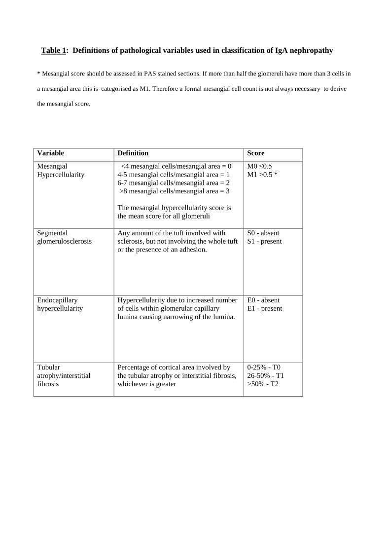
Table 1
: Definitions of pathological variables used in classification of IgA nephropathy
* Mesangial score should be assessed in PAS stained sections. If more than half the glomeruli have more than 3 cells in
a mesangial area this is categorised as M1. Therefore a formal mesangial cell count is not always necessary to derive
the mesangial score.
Variable Definition Score
Mesangial Hypercellularity
<4 mesangial cells/mesangial area = 0 4-5 mesangial cells/mesangial area = 1 6-7 mesangial cells/mesangial area = 2 >8 mesangial cells/mesangial area = 3 The mesangial hypercellularity score is the mean score for all glomeruli
M0 ≤0.5 M1 >0.5 *
Segmental glomerulosclerosis
Any amount of the tuft involved with sclerosis, but not involving the whole tuft or the presence of an adhesion.
S0 - absent S1 - present
Endocapillary hypercellularity
Hypercellularity due to increased number of cells within glomerular capillary lumina causing narrowing of the lumina.
E0 - absent E1 - present
Tubular atrophy/interstitial fibrosis
Percentage of cortical area involved by the tubular atrophy or interstitial fibrosis, whichever is greater
0-25% - T0 26-50% - T1 >50% - T2
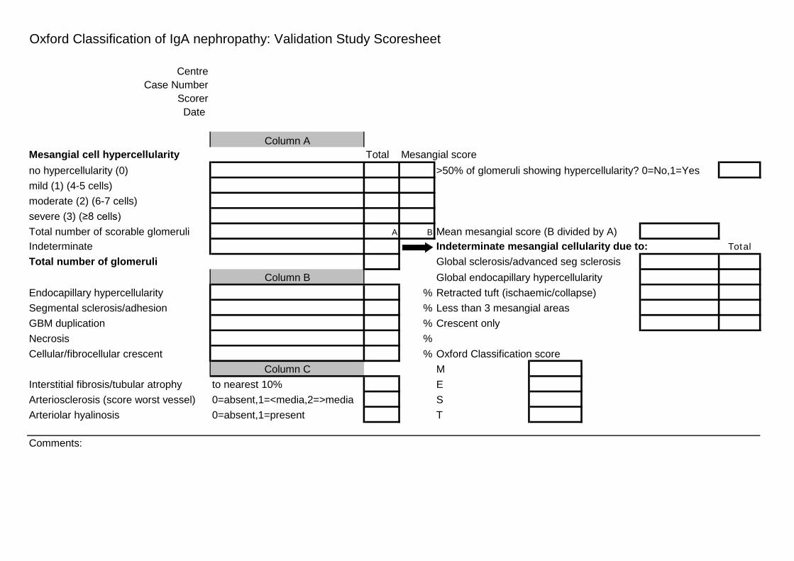
Oxford Classification of IgA nephropathy: Validation Study Scoresheet
CentreCase Number
ScorerDate
Column AMesangial cell hypercellularity Total Mesangial scoreno hypercellularity (0) >50% of glomeruli showing hypercellularity? 0=No,1=Yesmild (1) (4-5 cells)moderate (2) (6-7 cells)severe (3) (≥8 cells)Total number of scorable glomeruli A B Mean mesangial score (B divided by A)Indeterminate Indeterminate mesangial cellularity due to: TotalTotal number of glomeruli Global sclerosis/advanced seg sclerosis
Column B Global endocapillary hypercellularityEndocapillary hypercellularity % Retracted tuft (ischaemic/collapse)Segmental sclerosis/adhesion % Less than 3 mesangial areasGBM duplication % Crescent only Necrosis %Cellular/fibrocellular crescent % Oxford Classification score
Column C M Interstitial fibrosis/tubular atrophy to nearest 10% EArteriosclerosis (score worst vessel) 0=absent,1=<media,2=>media SArteriolar hyalinosis 0=absent,1=present T
Comments:
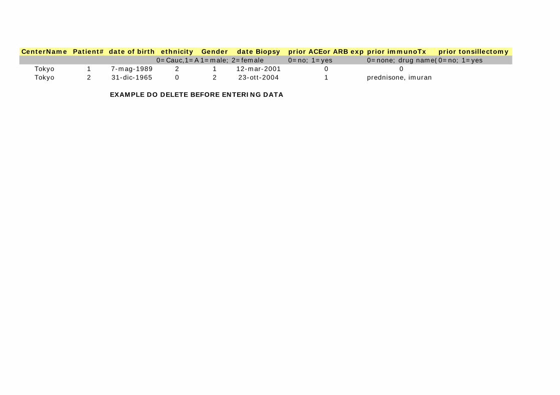
CenterName Patient# date of birth ethnicity Gender date Biopsy prior ACEor ARB exp prior immunoTx prior tonsillectomy0=Cauc,1=A 1=male; 2=female 0=no; 1=yes 0=none; drug name(0=no; 1=yes
Tokyo 1 7-mag-1989 2 1 12-mar-2001 0 0Tokyo 2 31-dic-1965 0 2 23-ott-2004 1 prednisone, imuran
EXAMPLE DO DELETE BEFORE ENTERING DATA
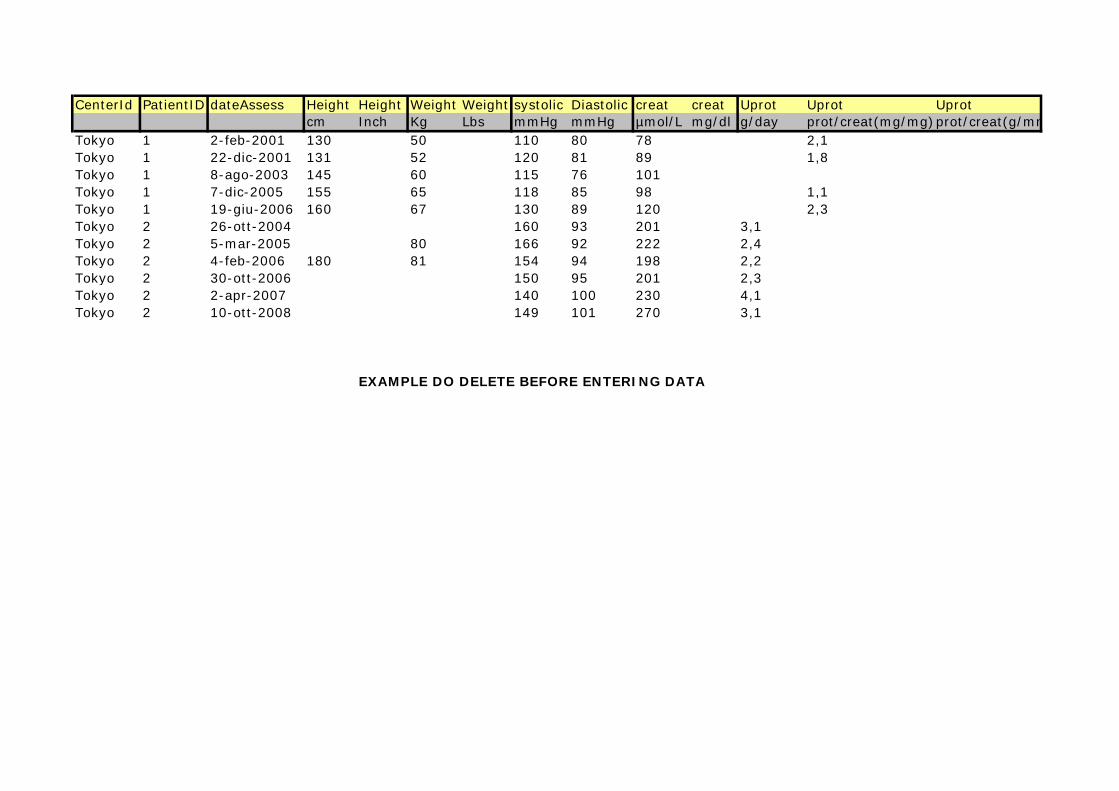
CenterId PatientID dateAssess Height Height Weight Weight systolic Diastolic creat creat Uprot Uprot Uprotcm Inch Kg Lbs mmHg mmHg µmol/L mg/dl g/day prot/creat(mg/mg)prot/creat(g/mm
Tokyo 1 2-feb-2001 130 50 110 80 78 2,1Tokyo 1 22-dic-2001 131 52 120 81 89 1,8Tokyo 1 8-ago-2003 145 60 115 76 101Tokyo 1 7-dic-2005 155 65 118 85 98 1,1Tokyo 1 19-giu-2006 160 67 130 89 120 2,3Tokyo 2 26-ott-2004 160 93 201 3,1Tokyo 2 5-mar-2005 80 166 92 222 2,4Tokyo 2 4-feb-2006 180 81 154 94 198 2,2Tokyo 2 30-ott-2006 150 95 201 2,3Tokyo 2 2-apr-2007 140 100 230 4,1Tokyo 2 10-ott-2008 149 101 270 3,1
EXAMPLE DO DELETE BEFORE ENTERING DATA
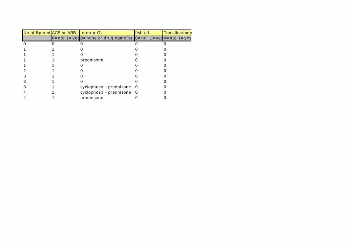
Nb of BpmedsACE or ARB ImmunoTx fish oil Tonsillectomy 0=no; 1=yes 0=none or drug name(s) 0=no; 1=yes 0=no; 1=yes0 0 0 0 01 1 0 0 01 1 0 0 01 1 prednisone 0 01 1 0 0 02 1 0 0 03 1 0 0 03 1 0 0 03 1 cyclophosp +prednisone 0 04 1 cyclophosp +prednisone 0 04 1 prednisone 0 0
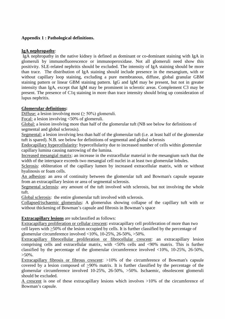
Appendix 1 : Pathological definitions.
IgA nephropathy IgA nephropathy in the native kidney is defined as dominant or co-dominant staining with IgA in glomeruli by immunofluorescence or immunoperoxidase. Not all glomeruli need show this positivity. SLE-related nephritis should be excluded. The intensity of IgA staining should be more than trace. The distribution of IgA staining should include presence in the mesangium, with or without capillary loop staining, excluding a pure membranous, diffuse, global granular GBM staining pattern or linear GBM staining pattern. IgG and IgM may be present, but not in greater intensity than IgA, except that IgM may be prominent in sclerotic areas. Complement C3 may be present. The presence of C1q staining in more than trace intensity should bring up consideration of lupus nephritis.
:
Glomerular definitions: Diffuse: a lesion involving most (≥ 50%) glomeruli. Focal: a lesion involving <50% of glomeruli. Global: a lesion involving more than half of the glomerular tuft (NB see below for definitions of segmental and global sclerosis). Segmental: a lesion involving less than half of the glomerular tuft (i.e. at least half of the glomerular tuft is spared). N.B. see below for definitions of segmental and global sclerosis Endocapillary hypercellularity: hypercellularity due to increased number of cells within glomerular capillary lumina causing narrowing of the lumina. Increased mesangial matrix: an increase in the extracellular material in the mesangium such that the width of the interspace exceeds two mesangial cell nuclei in at least two glomerular lobules. Sclerosis: obliteration of the capillary lumen by increased extracellular matrix, with or without hyalinosis or foam cells. An adhesion: an area of continuity between the glomerular tuft and Bowman's capsule separate from an extracapillary lesion or area of segmental sclerosis. Segmental sclerosis: any amount of the tuft involved with sclerosis, but not involving the whole tuft. Global sclerosis: the entire glomerular tuft involved with sclerosis. Collapsed/ischaemic glomerulus
: A glomerulus showing collapse of the capillary tuft with or without thickening of Bowman’s capsule and fibrosis in Bowman’s space
Extracapillary lesions are subclassified as follows: Extracapillary proliferation or cellular crescent: extracapillary cell proliferation of more than two cell layers with >50% of the lesion occupied by cells. It is further classified by the percentage of glomerular circumference involved <10%, 10-25%, 26-50%, >50%. Extracapillary fibrocellular proliferation or fibrocellular crescent: an extracapillary lesion comprising cells and extracellular matrix, with <50% cells and <90% matrix. This is further classified by the percentage of the glomerular circumference involved <10%, 10-25%, 26-50%, >50%. Extracapillary fibrosis or fibrous crescent: >10% of the circumference of Bowman's capsule covered by a lesion composed of >90% matrix. It is further classified by the percentage of the glomerular circumference involved 10-25%, 26-50%, >50%. Ischaemic, obsolescent glomeruli should be excluded. A crescent
is one of these extracapillary lesions which involves >10% of the circumference of Bowman’s capsule.
Mesangial hypercellularityIf <4 mesangial cells/mesangial area = normal,
is subclassified as follows:
4-5 mesangial cells/mesangial area = mild mesangial hypercellularity, 6-7 mesangial cells/mesangial area = moderate mesangial hypercellularity, 8 or more mesangial cells/mesangial area = severe mesangial hypercellularity. Note: This is scored for each glomerulus by assessing the most cellular mesangial area. Mesangial areas immediately adjacent to the vascular stalk should not be scored. Individual mesangial areas showing hypercellularity are separated by areas of narrowing to the width of less than 2 mesangial cell nuclei (ie count clusters, not files of mesangial cell nuclei). Tubulointerstitial definitions: Tubular atrophy: is defined by thick irregular tubular basement membranes with decreased diameter of tubules. It is scored according to the percent of cortical area involvement with 1-5% rounded to 5% and other values rounded to the closest 10%. Interstitial fibrosis: is defined as increased extracellular matrix separating tubules in the cortical area. It is scored as percentage involvement with 1-5% rounded to 5% and other values rounded to the closest 10% Interstitial inflammation: is defined as inflammatory cells within the cortical interstitium in excess. It is scored as percentage involvement with 1-5% rounded to 5% and other values rounded to the closest 10%. It should be noted whether the inflammation is confined to areas of interstitial fibrosis or not. Additional tubular lesions are noted as follows: The presence of numerous red blood cells, defined as tubules completely filled with red blood cells with or without casts, is noted as a lesion when it involves >20% of tubules. Acute tubular injury
of the proximal tubular epithelium is defined by simplification of the epithelium without tubular basement membrane thickening.
Vascular definitions: Arterial lesions are scored based on the most severe lesions. Interlobular and larger arteries are scored separately. An interlobular artery is one surrounded by cortex; an arcuate artery is one at the corticomedullary junction. Intimal thickening is scored by comparing the thickness of the intima to that of the media in the same segment of vessel. Score the intima variously as normal, and thickened to more or less than the thickness of the media. Arteriolar hyaline
is noted as the proportion of arterioles affected (0, 1-25%, 26-50%, >50%).