Embed Size (px)
Citation preview
Valvular Heart Disease for the General Internist
Nishant K. Sekaran, M.D., M.Sc.
Intermountain Heart Institute
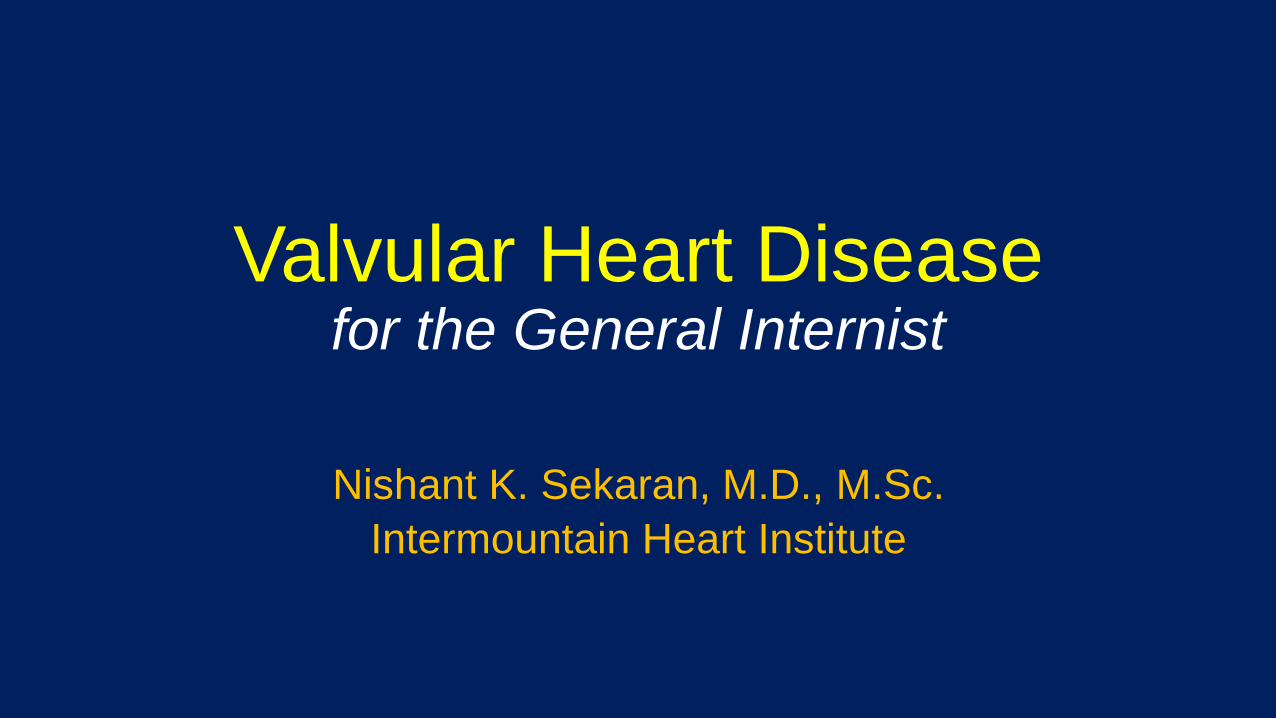
Objectives:
• Describe the Etiology and natural history of common valve disorders, including mitral valve stenosis and regurgitation, aortic valve stenosis and regurgitation
• Outline key aspects of pathophysiology for common valve disorders
• Assess a patient with suspected valve disease and manage subsequent surveillance after diagnosis
• Construct an evidence-based management plan for the patient with a valve disorder
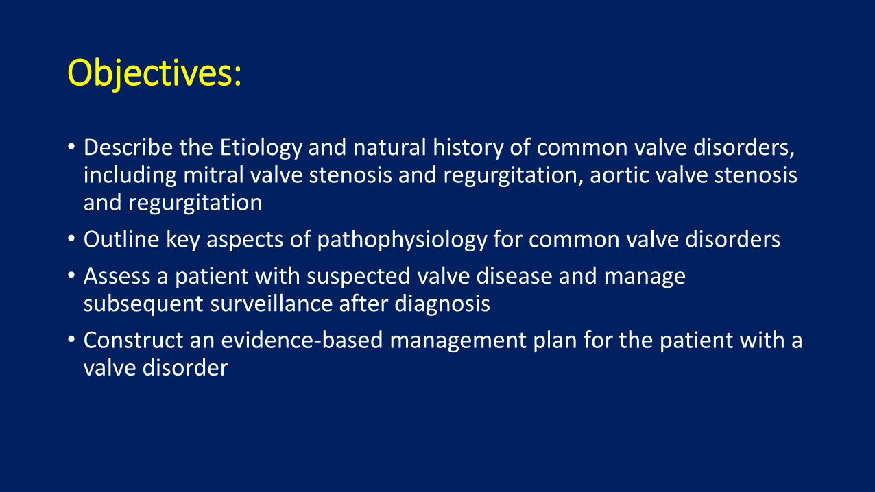
Valvular Heart Disease
Topics
• Aortic valve (stenosis, regurgitation)
• Mitral valve (stenosis, regurgitation)
• Special (tricuspid, bicuspid, endocarditis, prosthetic)
Valvular Heart Disease
Goals
• Etiology, natural history
• Pathophysiology highlights
• Diagnosis, surveillance
• Treatment options
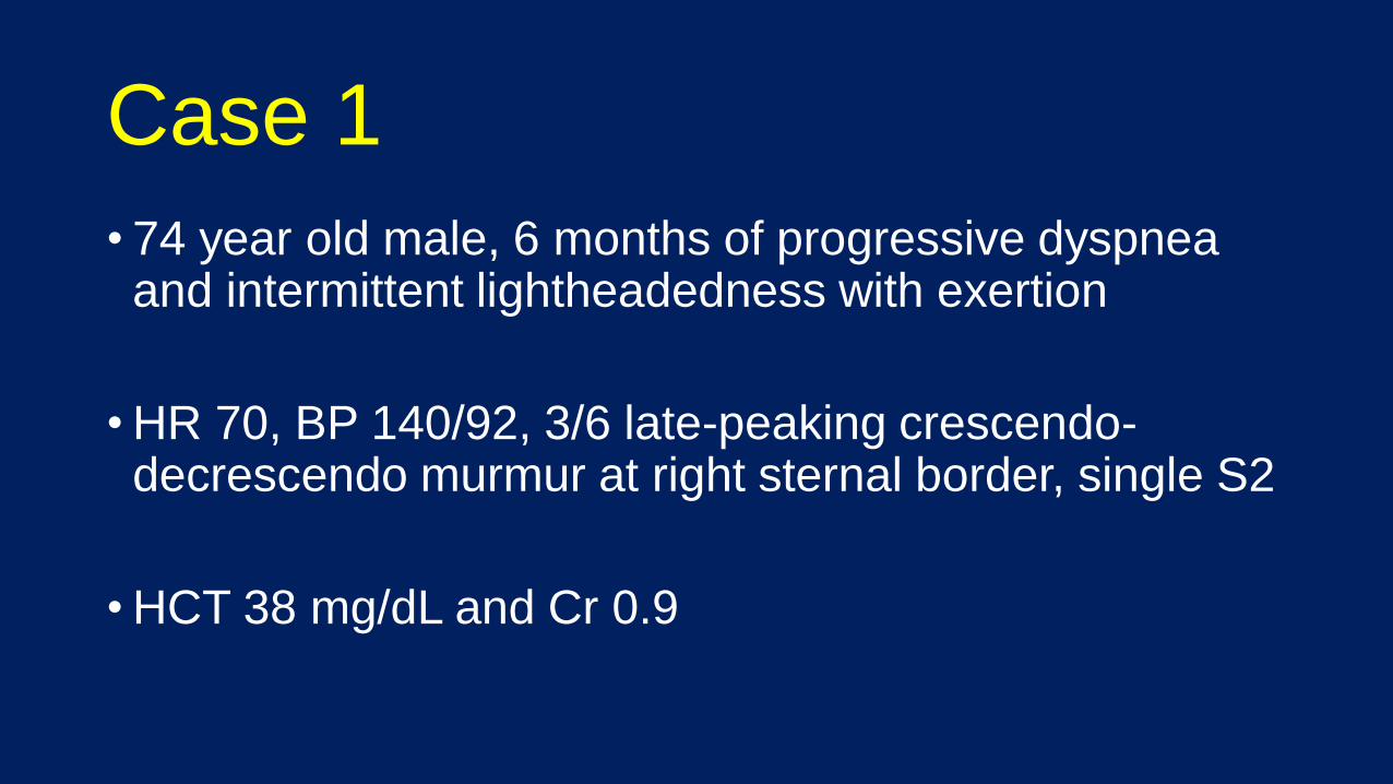
Case 1
• 74 year old male, 6 months of progressive dyspnea and intermittent lightheadedness with exertion
• HR 70, BP 140/92, 3/6 late-peaking crescendo-decrescendo murmur at right sternal border, single S2
• HCT 38 mg/dL and Cr 0.9

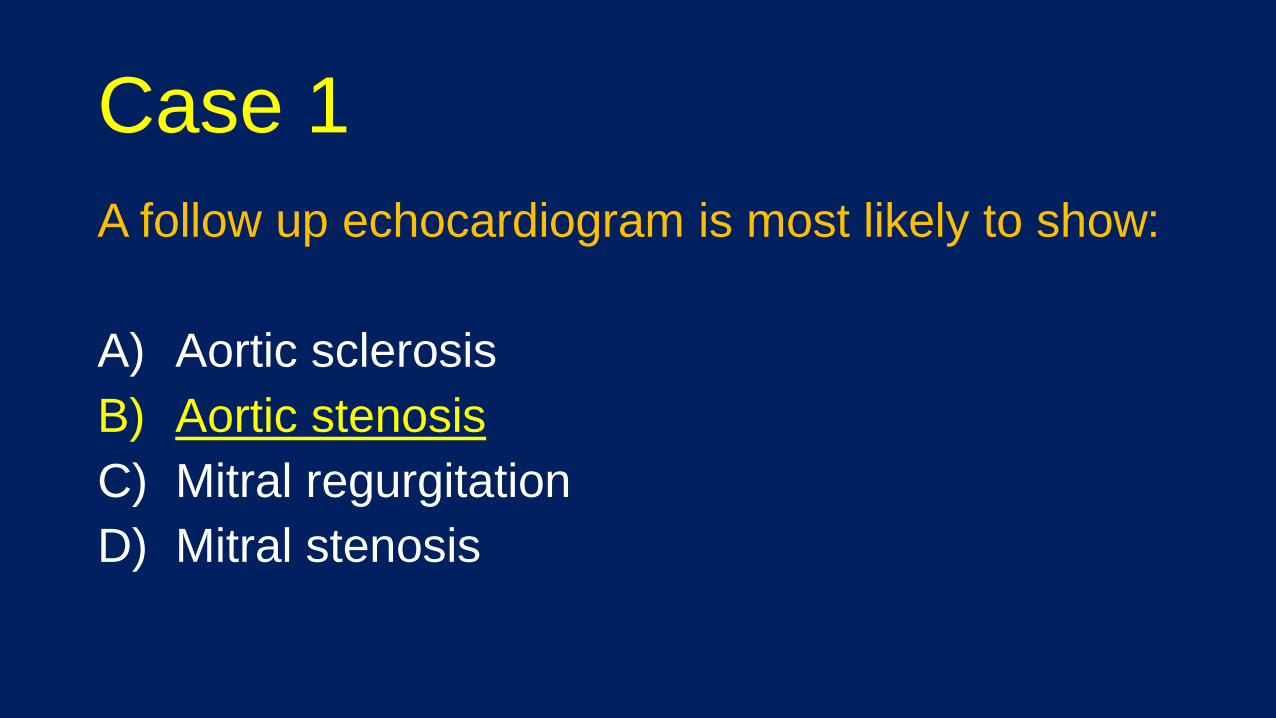
Case 1
A follow up echocardiogram is most likely to show:
A) Aortic sclerosis
B) Aortic stenosis
C) Mitral regurgitation
D) Mitral stenosis
Case 1
A follow up echocardiogram is most likely to show:
A) Aortic sclerosis
B) Aortic stenosis
C) Mitral regurgitation
D) Mitral stenosis
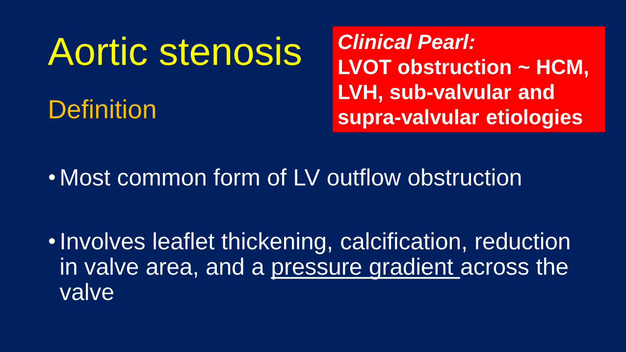
Aortic stenosis
Definition
• Most common form of LV outflow obstruction
• Involves leaflet thickening, calcification, reduction in valve area, and a pressure gradient across the valve
Clinical Pearl:
LVOT obstruction ~ HCM,
LVH, sub-valvular and
supra-valvular etiologies
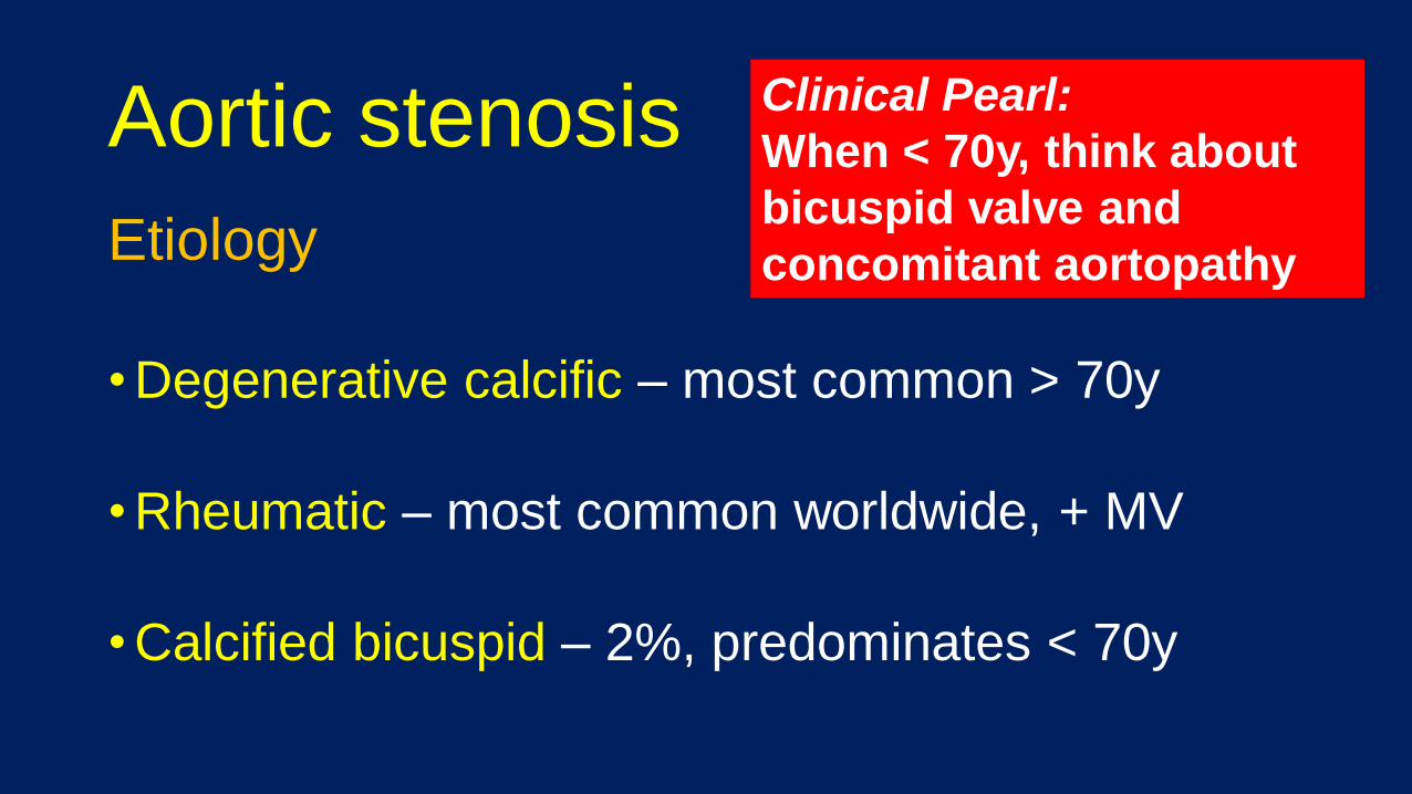
Aortic stenosis
Etiology
• Degenerative calcific – most common > 70y
• Rheumatic – most common worldwide, + MV
• Calcified bicuspid – 2%, predominates < 70y
Clinical Pearl:
When < 70y, think about
bicuspid valve and
concomitant aortopathy
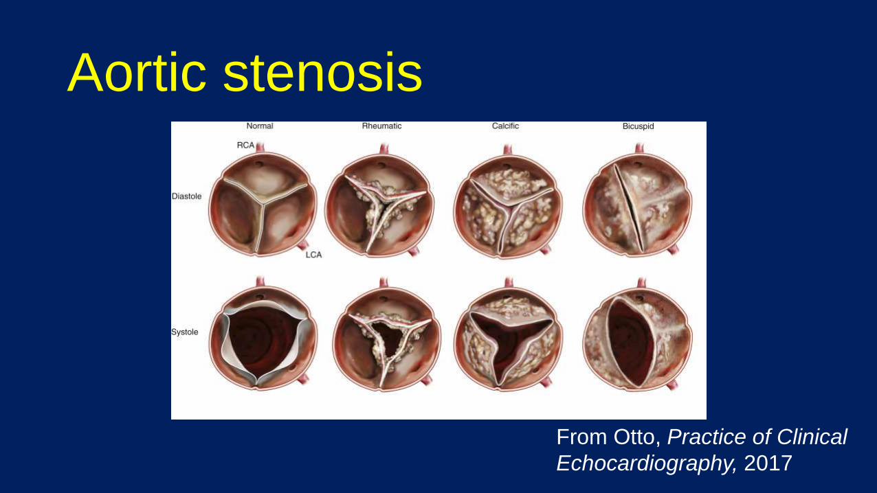
Aortic stenosis
From Otto, Practice of Clinical
Echocardiography, 2017
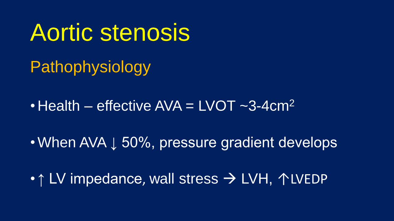
Aortic stenosis
Pathophysiology
• Health – effective AVA = LVOT ~3-4cm2
• When AVA ↓ 50%, pressure gradient develops
• ↑ LV impedance, wall stress LVH, ↑LVEDP
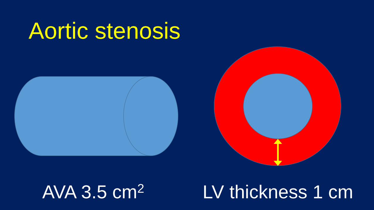
Aortic stenosis
AVA 3.5 cm2 LV thickness 1 cm
Your text here
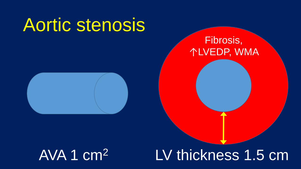
Aortic stenosis
AVA 1 cm2 LV thickness 1.5 cm
Fibrosis,
↑LVEDP, WMA
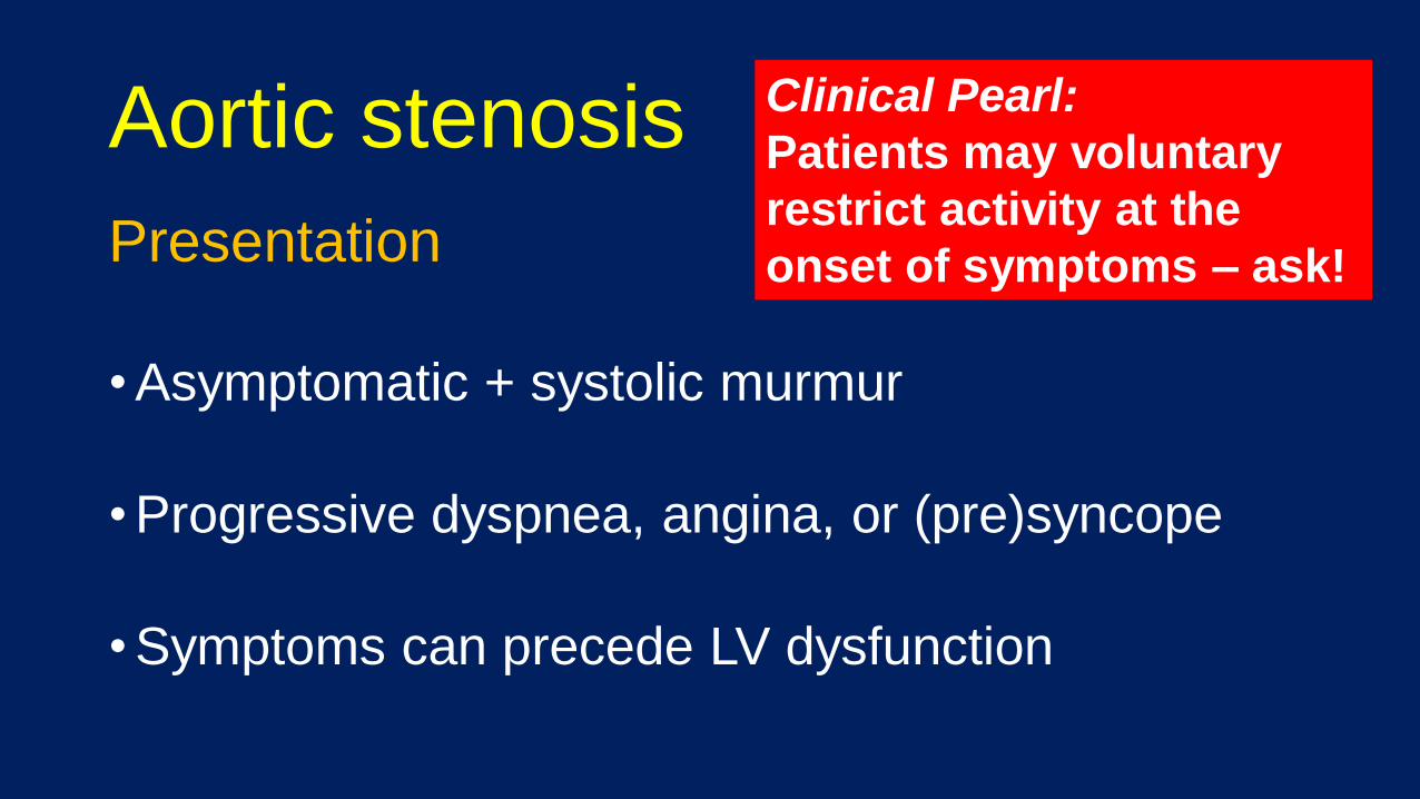
Aortic stenosis
Presentation
• Asymptomatic + systolic murmur
• Progressive dyspnea, angina, or (pre)syncope
• Symptoms can precede LV dysfunction
Clinical Pearl:
Patients may voluntary
restrict activity at the
onset of symptoms – ask!
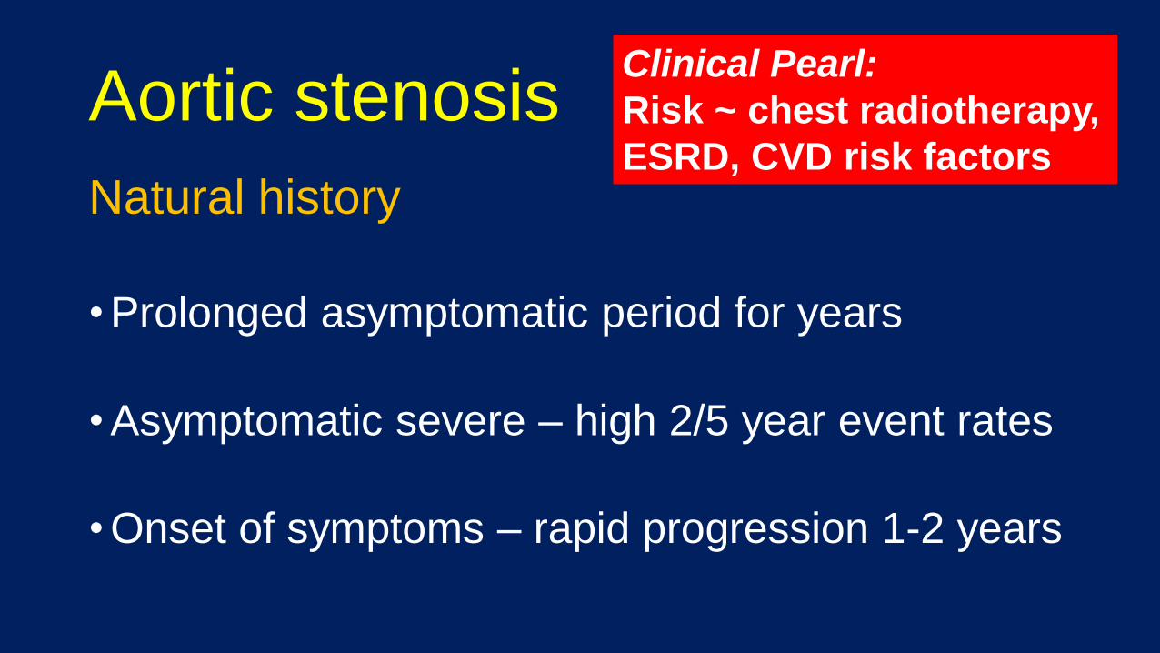
Aortic stenosis
Natural history
• Prolonged asymptomatic period for years
• Asymptomatic severe – high 2/5 year event rates
• Onset of symptoms – rapid progression 1-2 years
Clinical Pearl:
Risk ~ chest radiotherapy,
ESRD, CVD risk factors
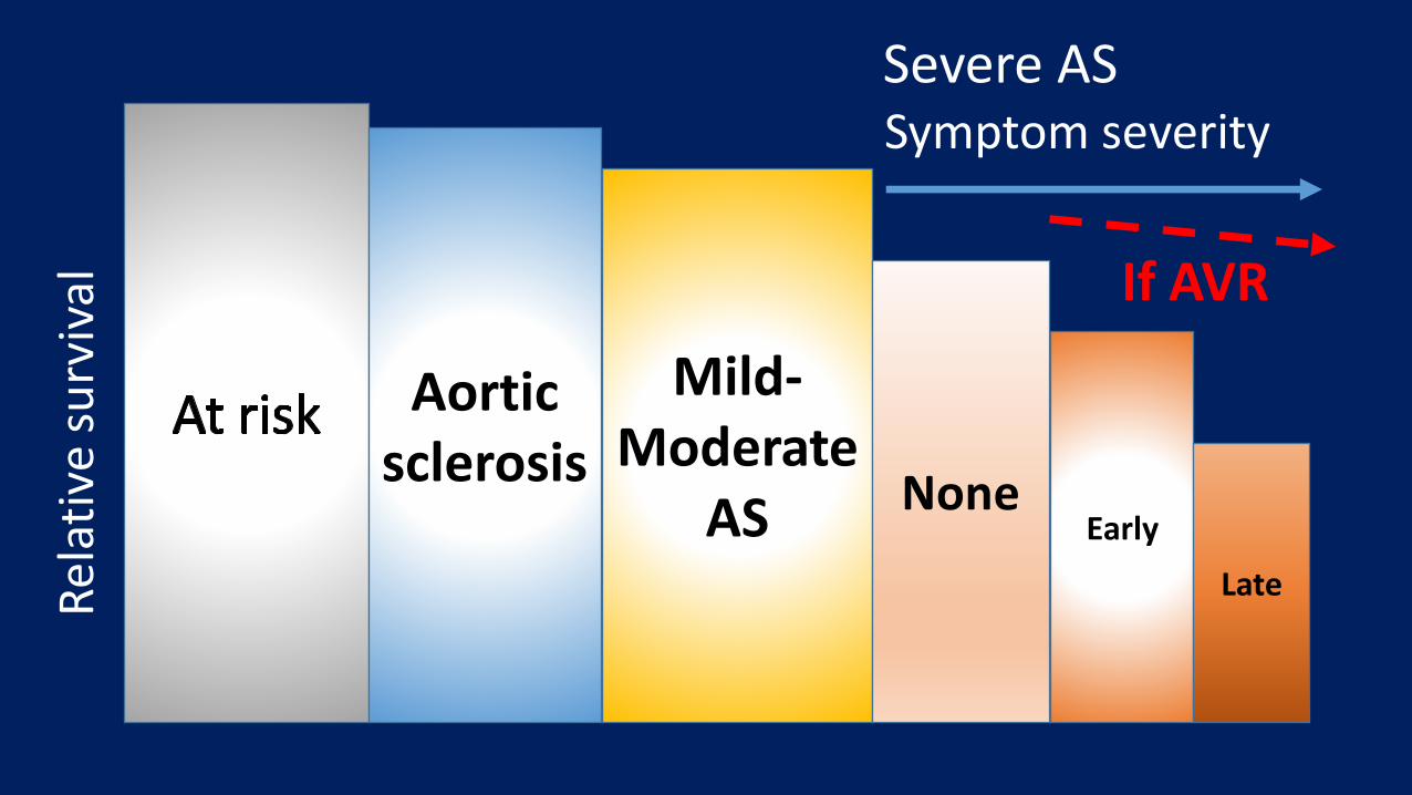
Aortic sclerosis
Mild-Moderate
AS NoneEarly
LateRel
ativ
e su
rviv
alSevere ASSymptom severity
If AVR
Aortic stenosis
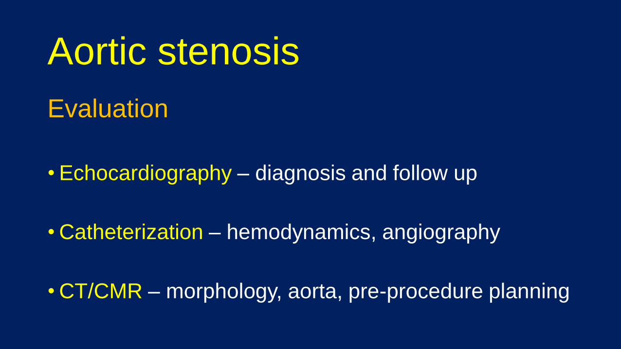
Evaluation
• Echocardiography – diagnosis and follow up
• Catheterization – hemodynamics, angiography
• CT/CMR – morphology, aorta, pre-procedure planning
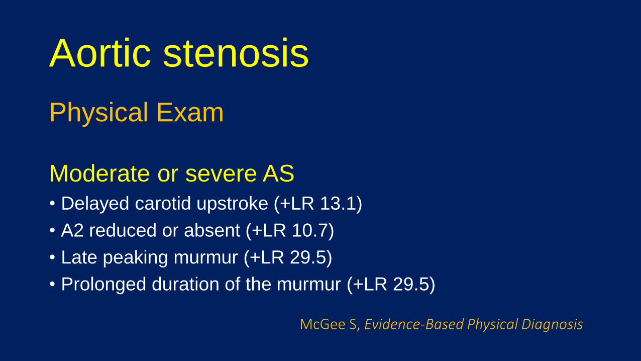
Aortic stenosis
Physical Exam
Moderate or severe AS• Delayed carotid upstroke (+LR 13.1)
• A2 reduced or absent (+LR 10.7)
• Late peaking murmur (+LR 29.5)
• Prolonged duration of the murmur (+LR 29.5)
McGee S, Evidence-Based Physical Diagnosis
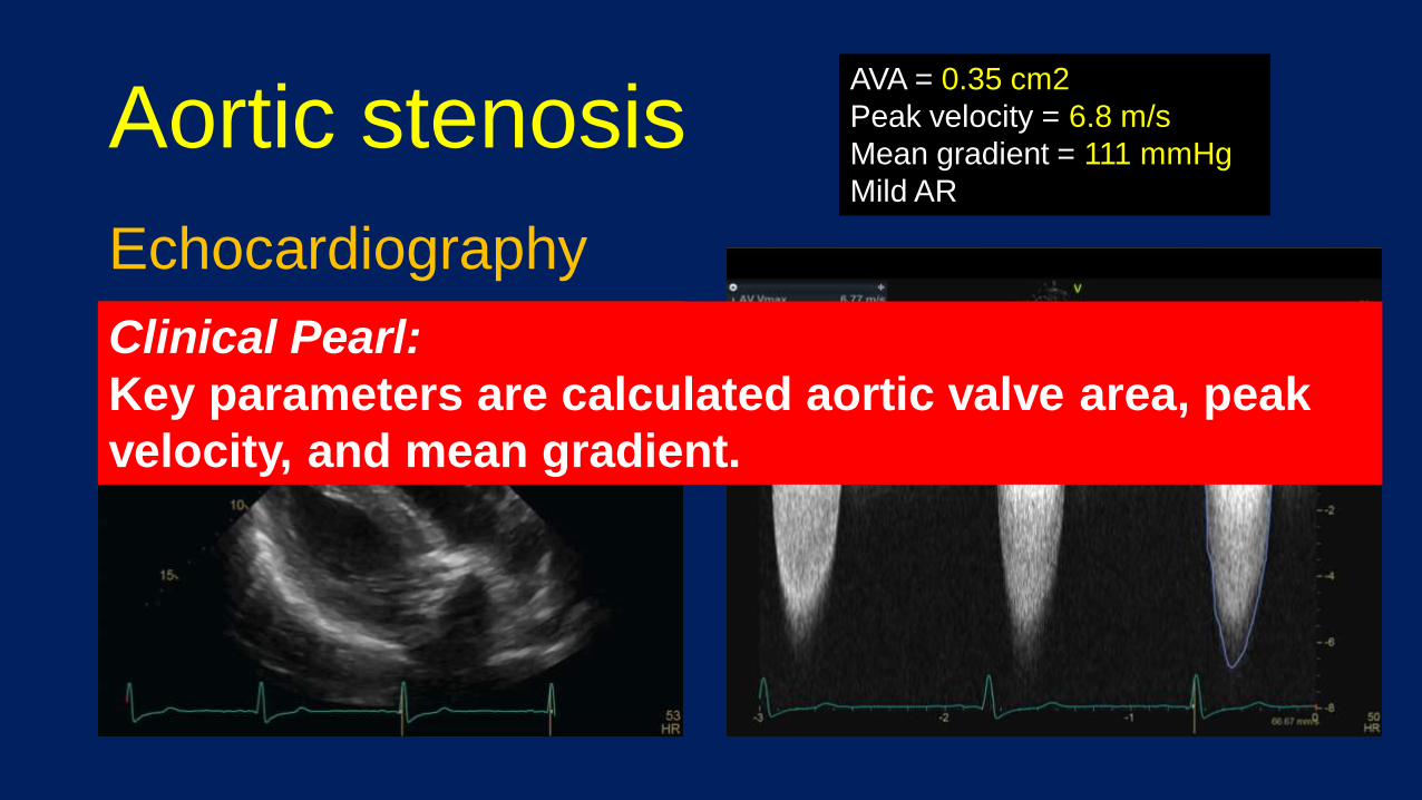
Aortic stenosis
Echocardiography
AVA = 0.35 cm2
Peak velocity = 6.8 m/s
Mean gradient = 111 mmHg
Mild AR
Clinical Pearl:
Key parameters are calculated aortic valve area, peak
velocity, and mean gradient.
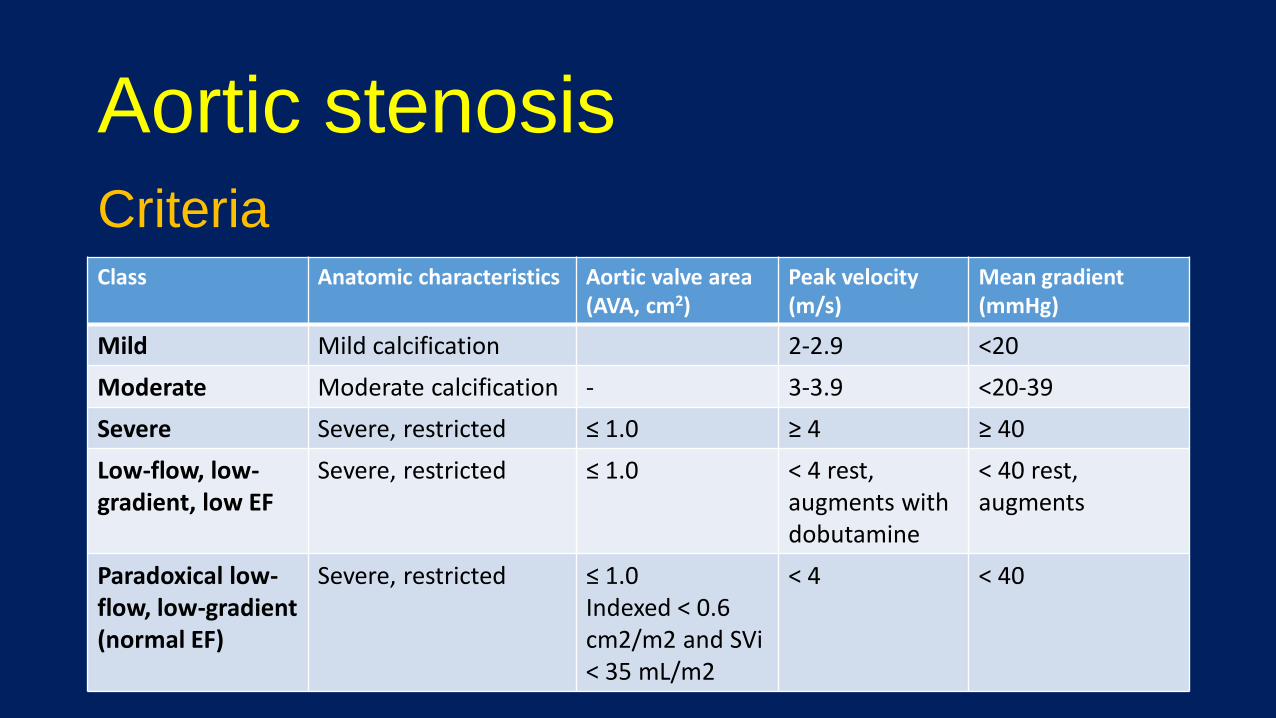
Aortic stenosis
CriteriaClass Anatomic characteristics Aortic valve area
(AVA, cm2)Peak velocity (m/s)
Mean gradient (mmHg)
Mild Mild calcification 2-2.9 <20
Moderate Moderate calcification - 3-3.9 <20-39
Severe Severe, restricted ≤ 1.0 ≥ 4 ≥ 40
Low-flow, low-gradient, low EF
Severe, restricted ≤ 1.0 ˂ 4 rest,augments with dobutamine
< 40 rest, augments
Paradoxical low-flow, low-gradient (normal EF)
Severe, restricted ≤ 1.0 Indexed < 0.6 cm2/m2 and SVi< 35 mL/m2
< 4 < 40
Aortic stenosis
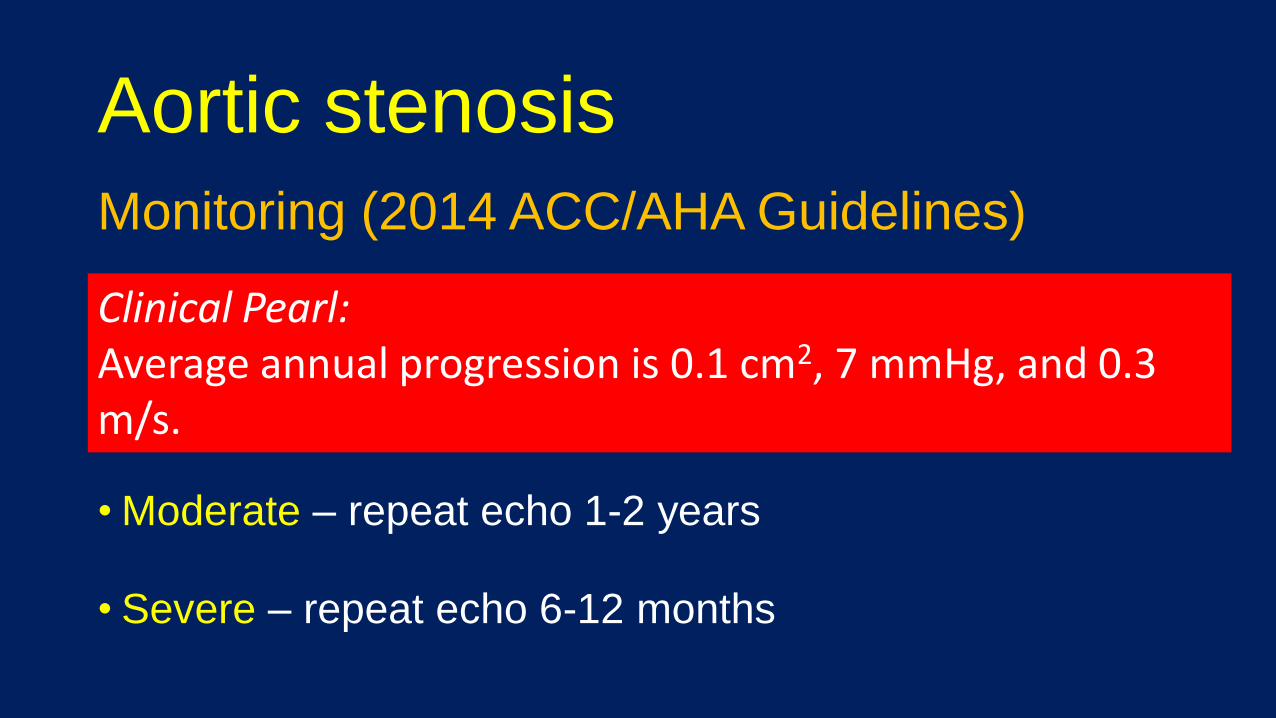
Monitoring (2014 ACC/AHA Guidelines)
• Annual history and physical
• Mild – repeat echo 3-5 years
• Moderate – repeat echo 1-2 years
• Severe – repeat echo 6-12 months
Clinical Pearl: Average annual progression is 0.1 cm2, 7 mmHg, and 0.3 m/s.
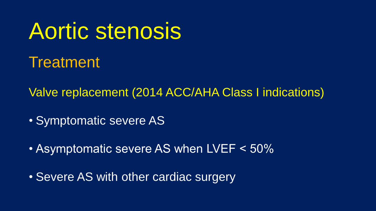
Aortic stenosis
Treatment
Valve replacement (2014 ACC/AHA Class I indications)
• Symptomatic severe AS
• Asymptomatic severe AS when LVEF ˂ 50%
• Severe AS with other cardiac surgery
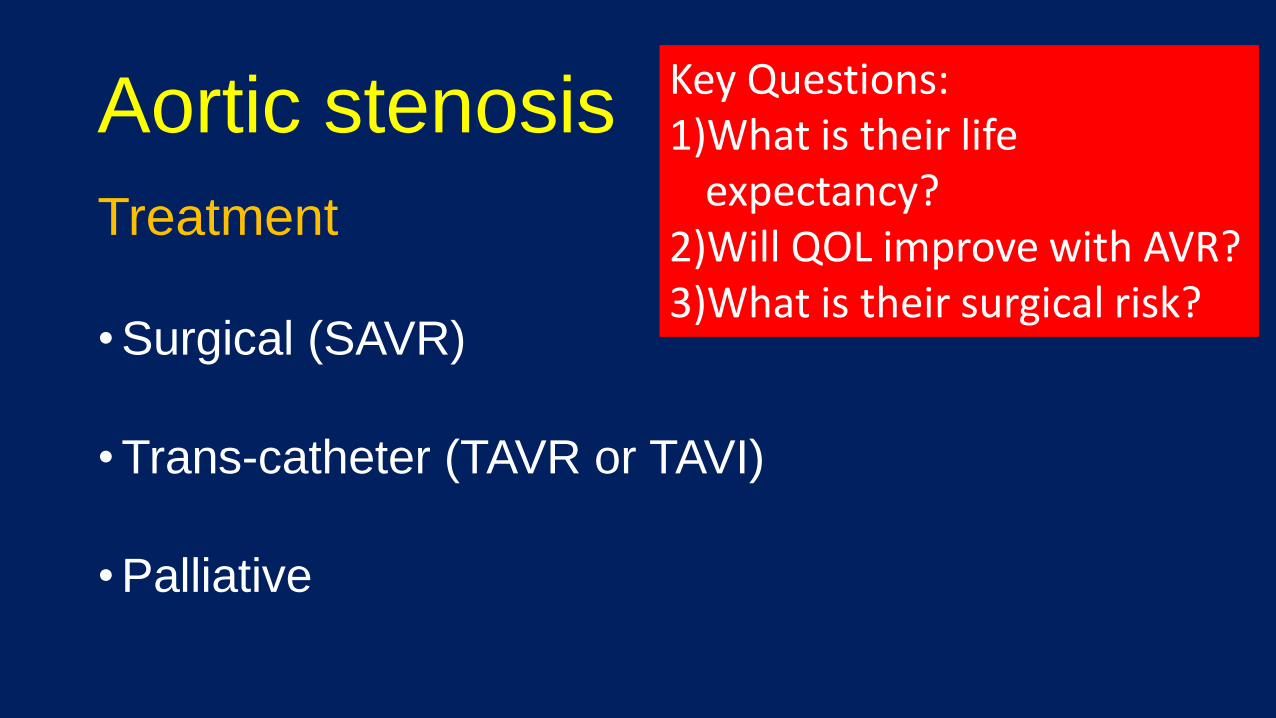
Aortic stenosis
Treatment
• Surgical (SAVR)
• Trans-catheter (TAVR or TAVI)
• Palliative
Key Questions:1)What is their life
expectancy?2)Will QOL improve with AVR?3)What is their surgical risk?
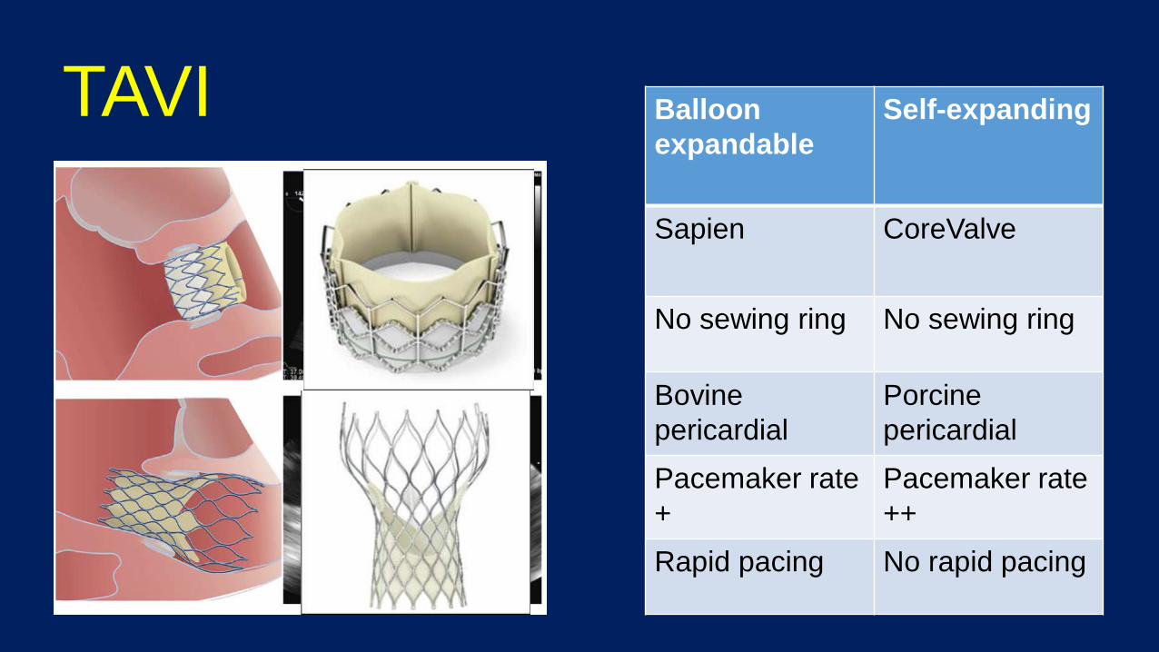
TAVI Balloon
expandable
Self-expanding
Sapien CoreValve
No sewing ring No sewing ring
Bovine
pericardial
Porcine
pericardial
Pacemaker rate
+
Pacemaker rate
++
Rapid pacing No rapid pacing
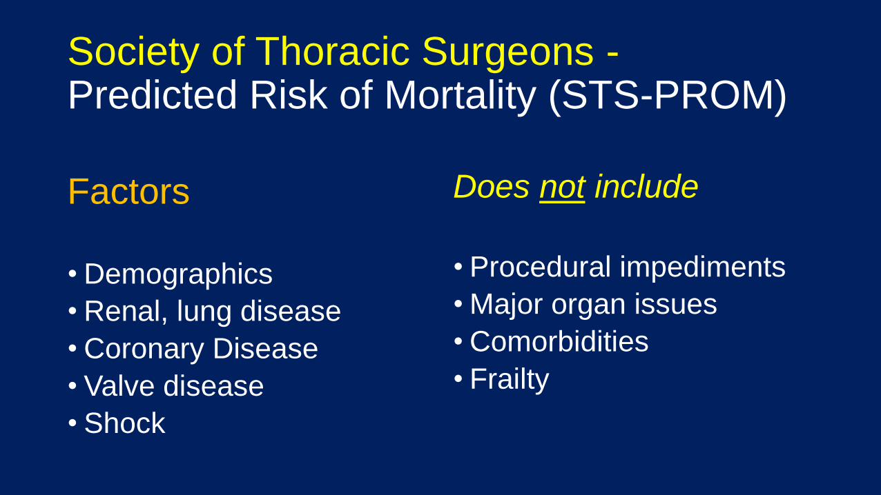
Society of Thoracic Surgeons -Predicted Risk of Mortality (STS-PROM)
Factors
• Demographics
• Renal, lung disease
• Coronary Disease
• Valve disease
• Shock
Does not include
• Procedural impediments
• Major organ issues
• Comorbidities
• Frailty
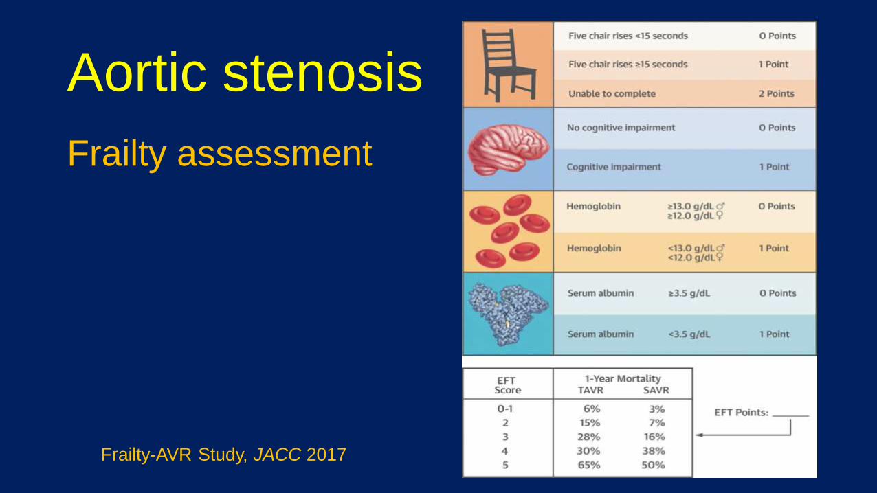
Aortic stenosis
Frailty assessment
Frailty-AVR Study, JACC 2017
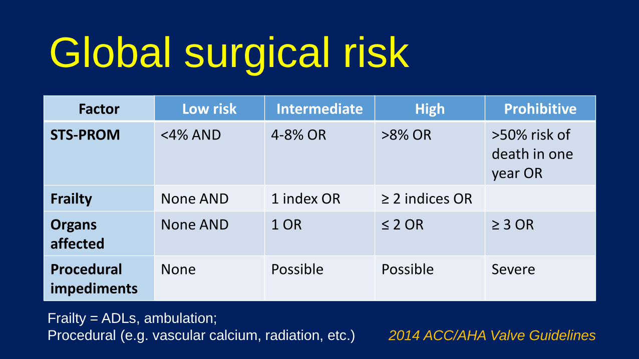
Global surgical riskFactor Low risk Intermediate High Prohibitive
STS-PROM <4% AND 4-8% OR >8% OR >50% risk of death in one year OR
Frailty None AND 1 index OR ≥ 2 indices OR
Organsaffected
None AND 1 OR ≤ 2 OR ≥ 3 OR
Procedural impediments
None Possible Possible Severe
Frailty = ADLs, ambulation;
Procedural (e.g. vascular calcium, radiation, etc.) 2014 ACC/AHA Valve Guidelines
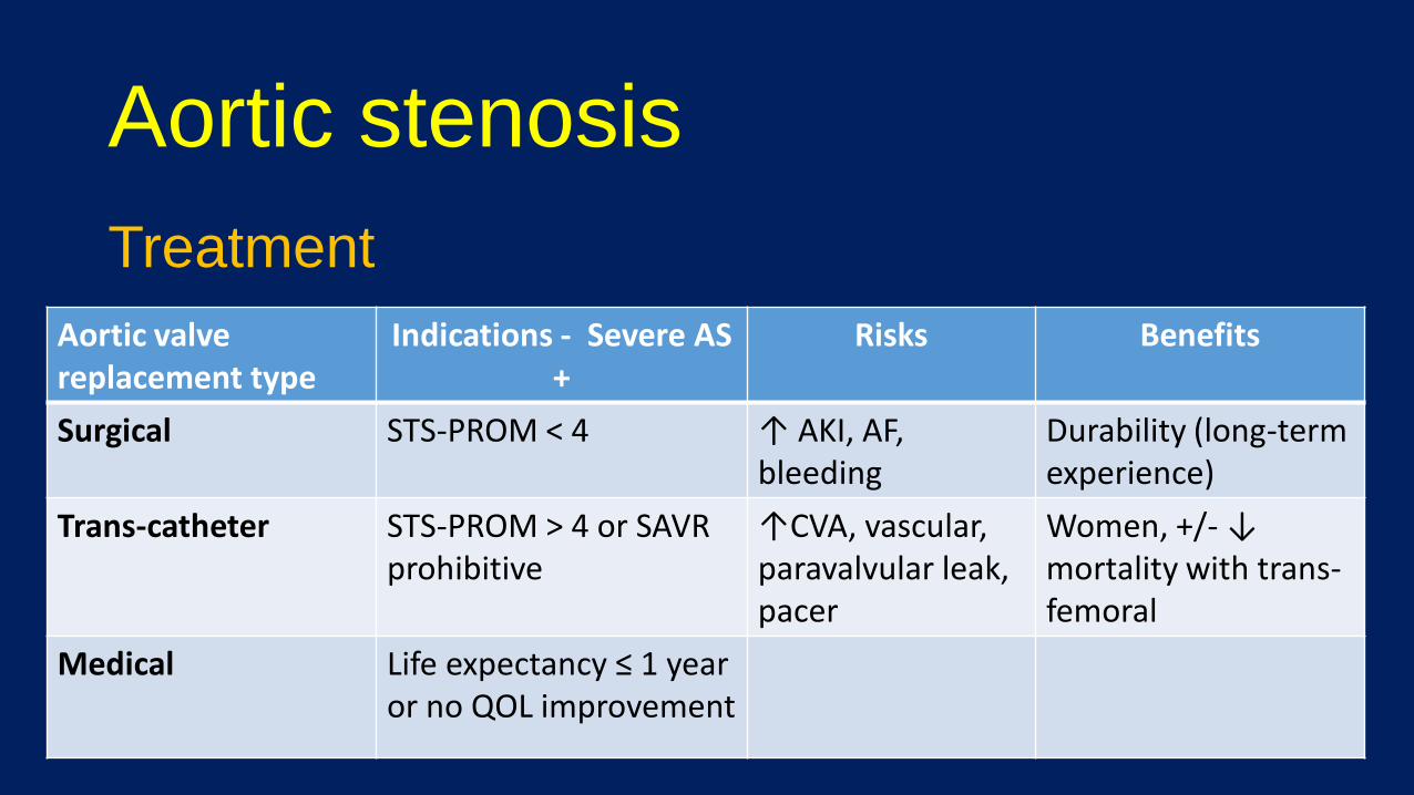
Aortic stenosis
Treatment
Aortic valve replacement type
Indications - Severe AS +
Risks Benefits
Surgical STS-PROM < 4 ↑ AKI, AF, bleeding
Durability (long-term experience)
Trans-catheter STS-PROM > 4 or SAVR prohibitive
↑CVA, vascular, paravalvular leak, pacer
Women, +/- ↓ mortality with trans-femoral
Medical Life expectancy ≤ 1 year or no QOL improvement
Aortic regurgitation
Case 2• 74 year old male presenting with 6 months of progressive dyspnea and intermittent lightheadedness with exertion
• Exam notable for HR 70, BP 130/43, and 2/6 mid-peaking crescendo/decrescendo murmur at the right sternal border with 2/4 diastolic murmur and prominent carotid upstroke
• HCT 38 mg/dL and Cr 0.9
Case 2
A follow up echocardiogram is most likely to show:
A) Aortic regurgitation
B) Aortic stenosis
C) Mitral regurgitation
D) Mitral stenosis
Case 2
A follow up echocardiogram is most likely to show:
A) Aortic regurgitation
B) Aortic stenosis
C) Mitral regurgitation
D) Mitral stenosis
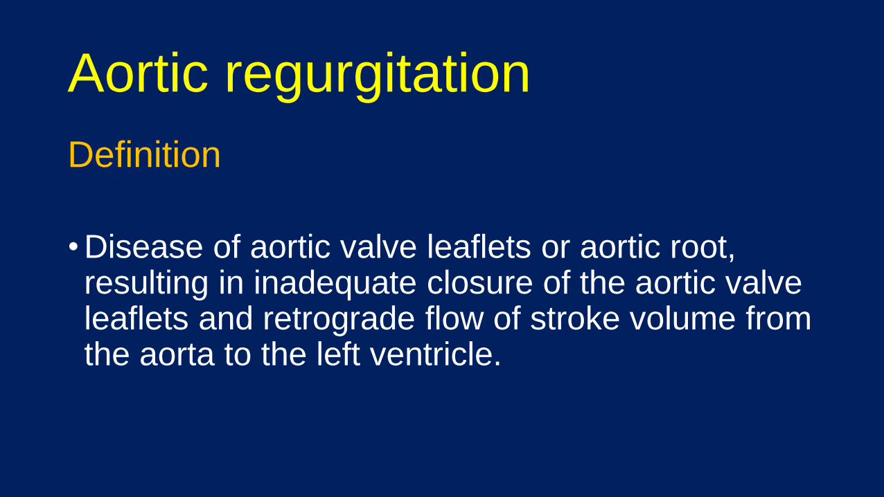
Aortic regurgitation
Definition
• Disease of aortic valve leaflets or aortic root, resulting in inadequate closure of the aortic valve leaflets and retrograde flow of stroke volume from the aorta to the left ventricle.
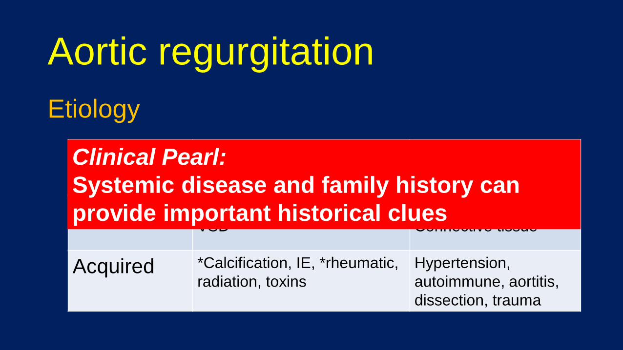
Aortic regurgitation
Etiology
Aortic leaflets Aortic root
Congenital *Abnormal cusp number
VSD
Annular ectasia
Connective tissue
Acquired *Calcification, IE, *rheumatic,
radiation, toxins
Hypertension,
autoimmune, aortitis,
dissection, trauma
Clinical Pearl:
Systemic disease and family history can
provide important historical clues
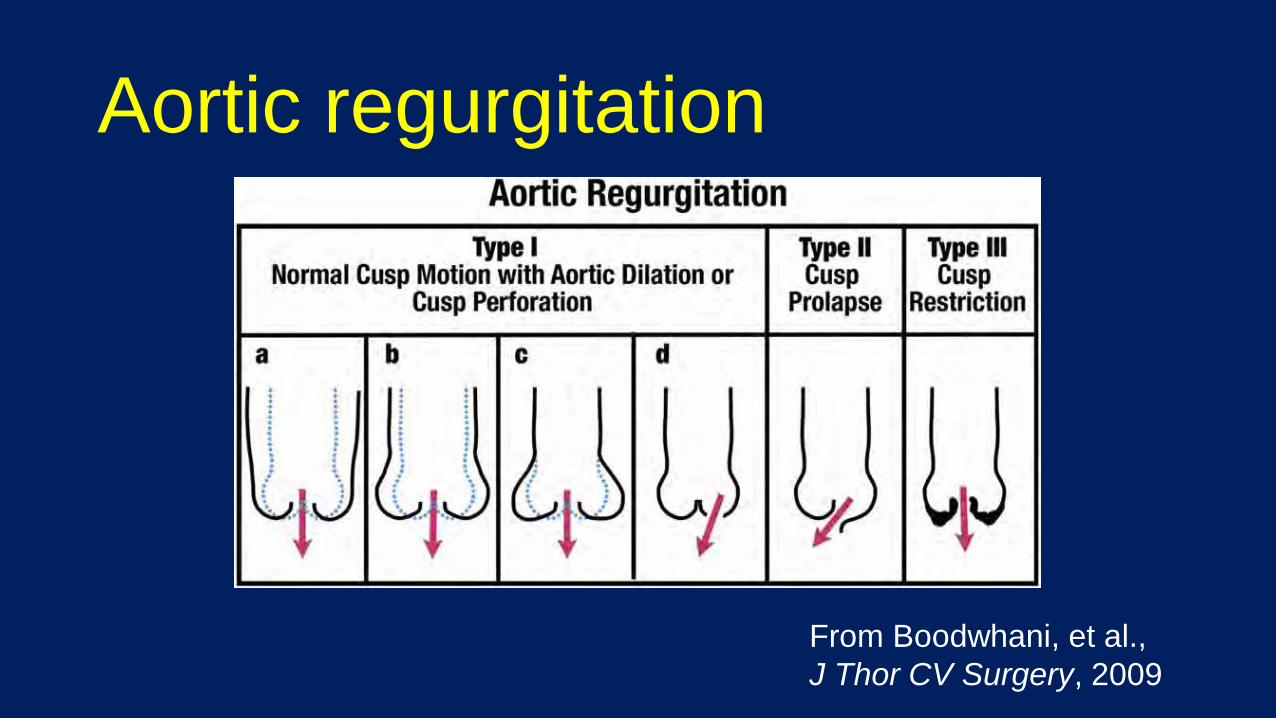
Aortic regurgitation
From Boodwhani, et al.,
J Thor CV Surgery, 2009
Aortic regurgitation
Pathophysiology
• In diastole – fraction of stroke volume leaks back
• LVEDV↑ wall stress ↑, eccentric hypertrophy
• ↑ SV, CO, pulse pressure (high SBP, low DBP)
Clinical Pearl:
Aortic regurgitation presents both a
pressure and volume problem, thereby
increasing wall stress.
Aortic regurgitation
Presentation
• Asymptomatic for years ventricular remodeling
• Left sided heart failure symptoms
• Angina (↓ diastolic flow), palpitations (LV enlargement)
Aortic regurgitation
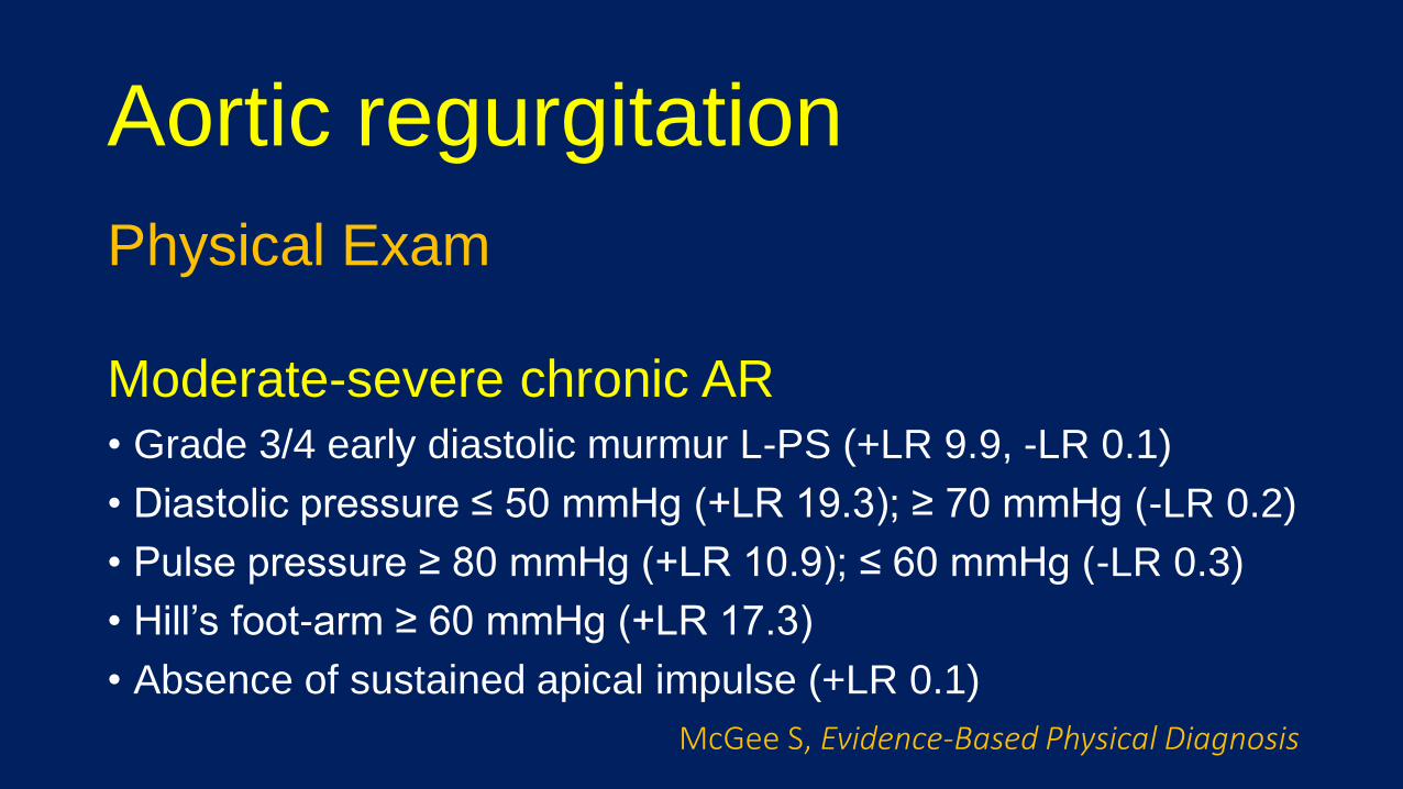
Physical Exam
Moderate-severe chronic AR• Grade 3/4 early diastolic murmur L-PS (+LR 9.9, -LR 0.1)
• Diastolic pressure ≤ 50 mmHg (+LR 19.3); ≥ 70 mmHg (-LR 0.2)
• Pulse pressure ≥ 80 mmHg (+LR 10.9); ≤ 60 mmHg (-LR 0.3)
• Hill’s foot-arm ≥ 60 mmHg (+LR 17.3)
• Absence of sustained apical impulse (+LR 0.1)
McGee S, Evidence-Based Physical Diagnosis
Aortic regurgitation
Evaluation
• Echocardiography – diagnosis and follow up
• Catheterization – hemodynamics, angiography
• CT/CMR – morphology, aorta, pre-procedure planning
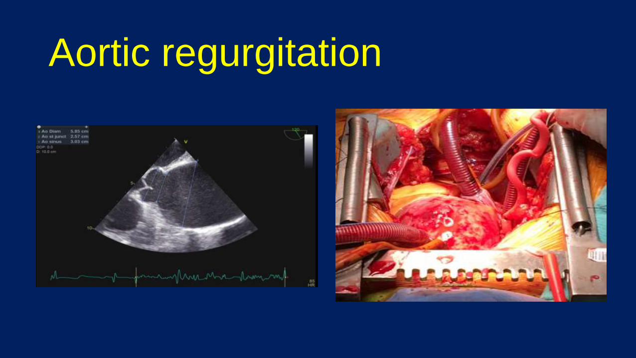
Aortic regurgitation
Aortic regurgitation
Monitoring (2014 ACC/AHA Guidelines)
• Annual history and physical
• Mild – repeat echo 3-5 years
• Moderate – repeat echo 1-2 years
• Severe – repeat echo 6-12 months
Aortic regurgitation
Treatment
Valve replacement (2014 ACC/AHA Class I indications)
• Symptomatic severe AR
• Asymptomatic severe AR with LVEF < 50%
• Severe AR if other cardiac surgery needed

Mitral stenosis
Case 3
• 70 year old woman presenting with 6 months of recurrent hospitalizations for heart failure with pulmonary edema.
• Exam notable for HR 96, BP 150/95, and 2/6 apical systolic murmur with 2/4 diastolic murmur with associated rumble and prominent P2 component at the apex. Lung crackles.
Case 3
A follow up echocardiogram is most likely to show:
A) Aortic sclerosis
B) Aortic stenosis
C) Mitral regurgitation
D) Mitral stenosis
Case 3
A follow up echocardiogram is most likely to show:
A) Aortic sclerosis
B) Aortic stenosis
C) Mitral regurgitation
D) Mitral stenosis
Mitral stenosis
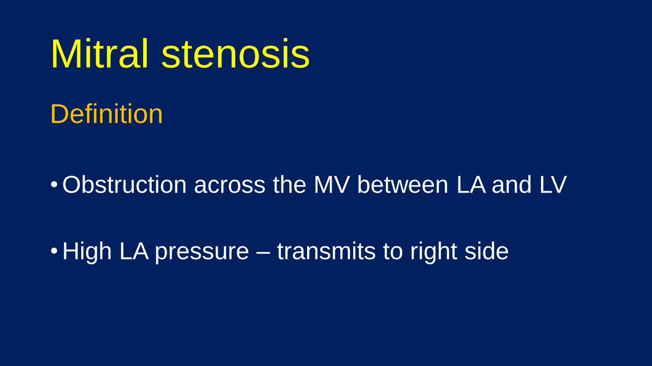
Definition
• Obstruction across the MV between LA and LV
• High LA pressure – transmits to right side
Mitral stenosis
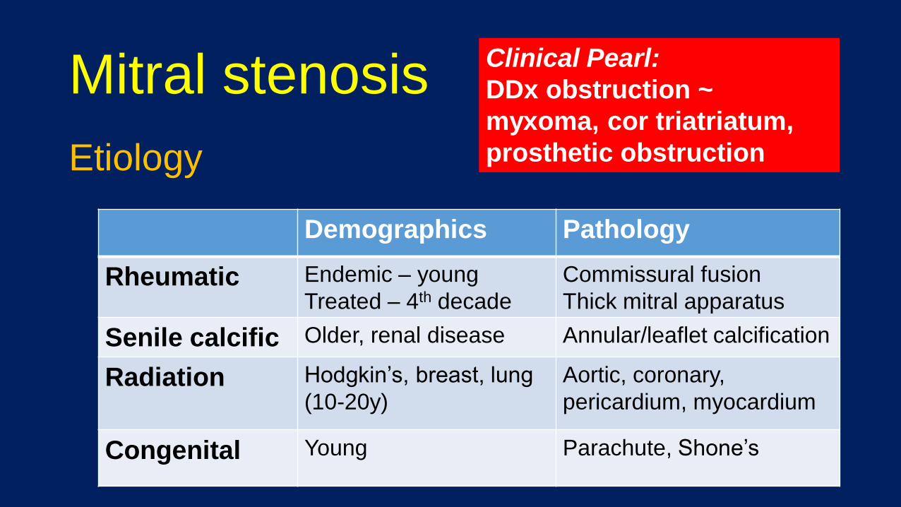
Etiology
Demographics Pathology
Rheumatic Endemic – young
Treated – 4th decade
Commissural fusion
Thick mitral apparatus
Senile calcific Older, renal disease Annular/leaflet calcification
Radiation Hodgkin’s, breast, lung
(10-20y)
Aortic, coronary,
pericardium, myocardium
Congenital Young Parachute, Shone’s
Clinical Pearl:
DDx obstruction ~
myxoma, cor triatriatum,
prosthetic obstruction
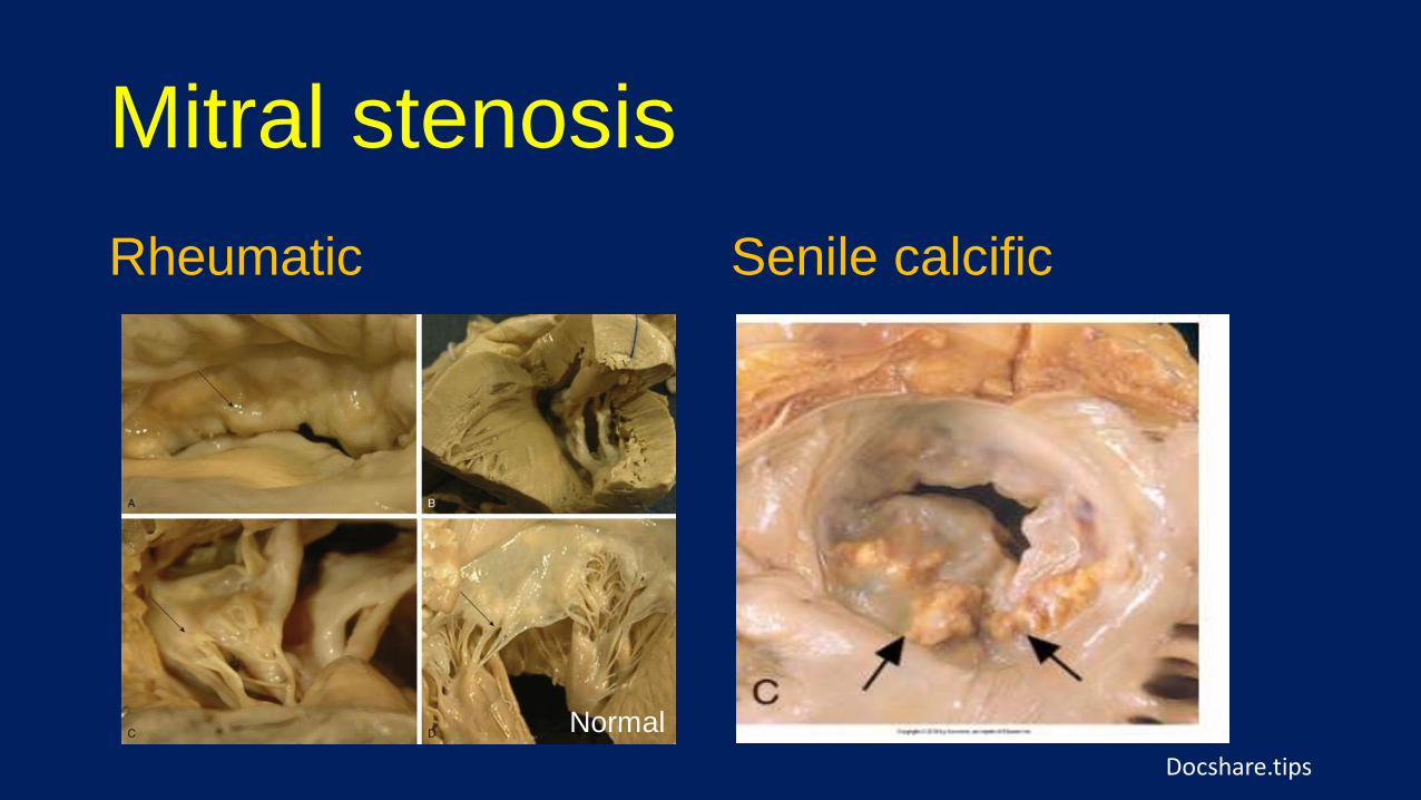
Mitral stenosis
Rheumatic Senile calcific
Normal
Docshare.tips
Mitral stenosis
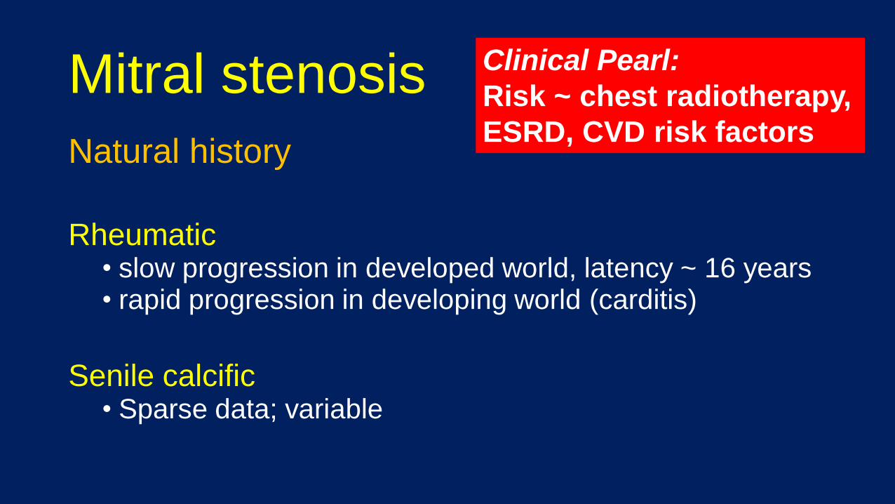
Natural history
Rheumatic• slow progression in developed world, latency ~ 16 years• rapid progression in developing world (carditis)
Senile calcific• Sparse data; variable
Clinical Pearl:
Risk ~ chest radiotherapy,
ESRD, CVD risk factors
Mitral stenosis
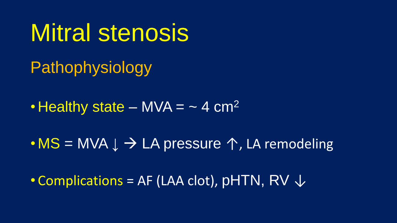
Pathophysiology
• Healthy state – MVA = ~ 4 cm2
• MS = MVA ↓ LA pressure ↑, LA remodeling
• Complications = AF (LAA clot), pHTN, RV ↓
Mitral stenosis
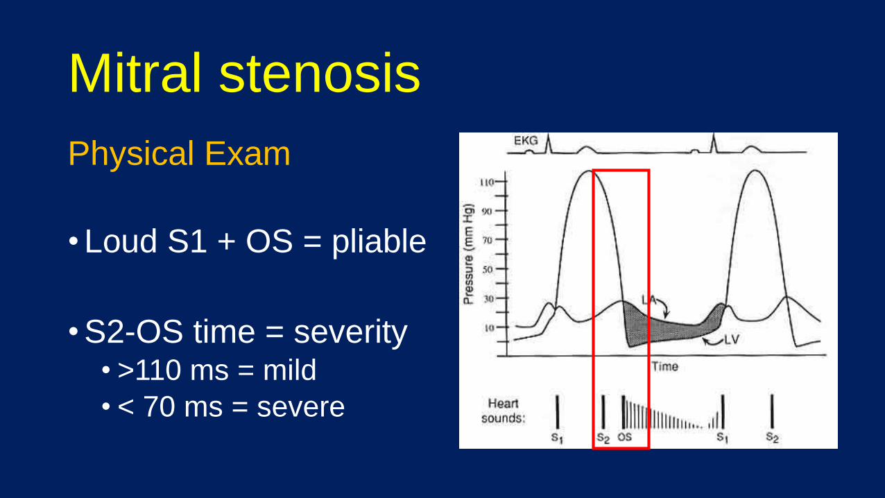
Physical Exam
• Loud S1 + OS = pliable
• S2-OS time = severity• >110 ms = mild
• < 70 ms = severe
Mitral stenosis
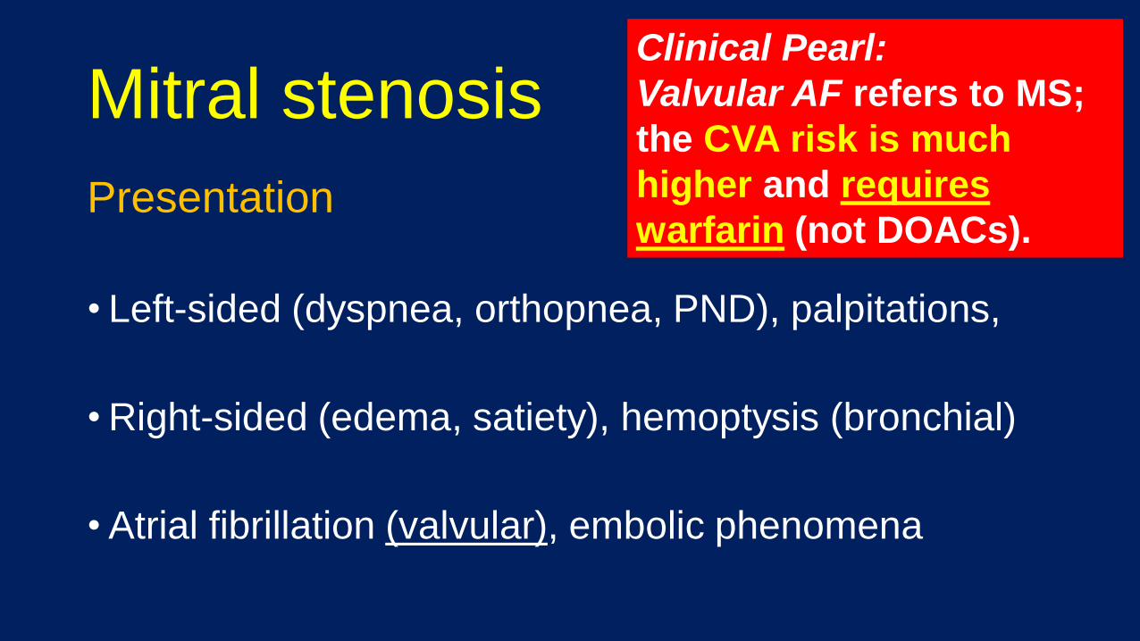
Presentation
• Left-sided (dyspnea, orthopnea, PND), palpitations,
• Right-sided (edema, satiety), hemoptysis (bronchial)
• Atrial fibrillation (valvular), embolic phenomena
Clinical Pearl:
Valvular AF refers to MS;
the CVA risk is much
higher and requires
warfarin (not DOACs).
Mitral stenosis
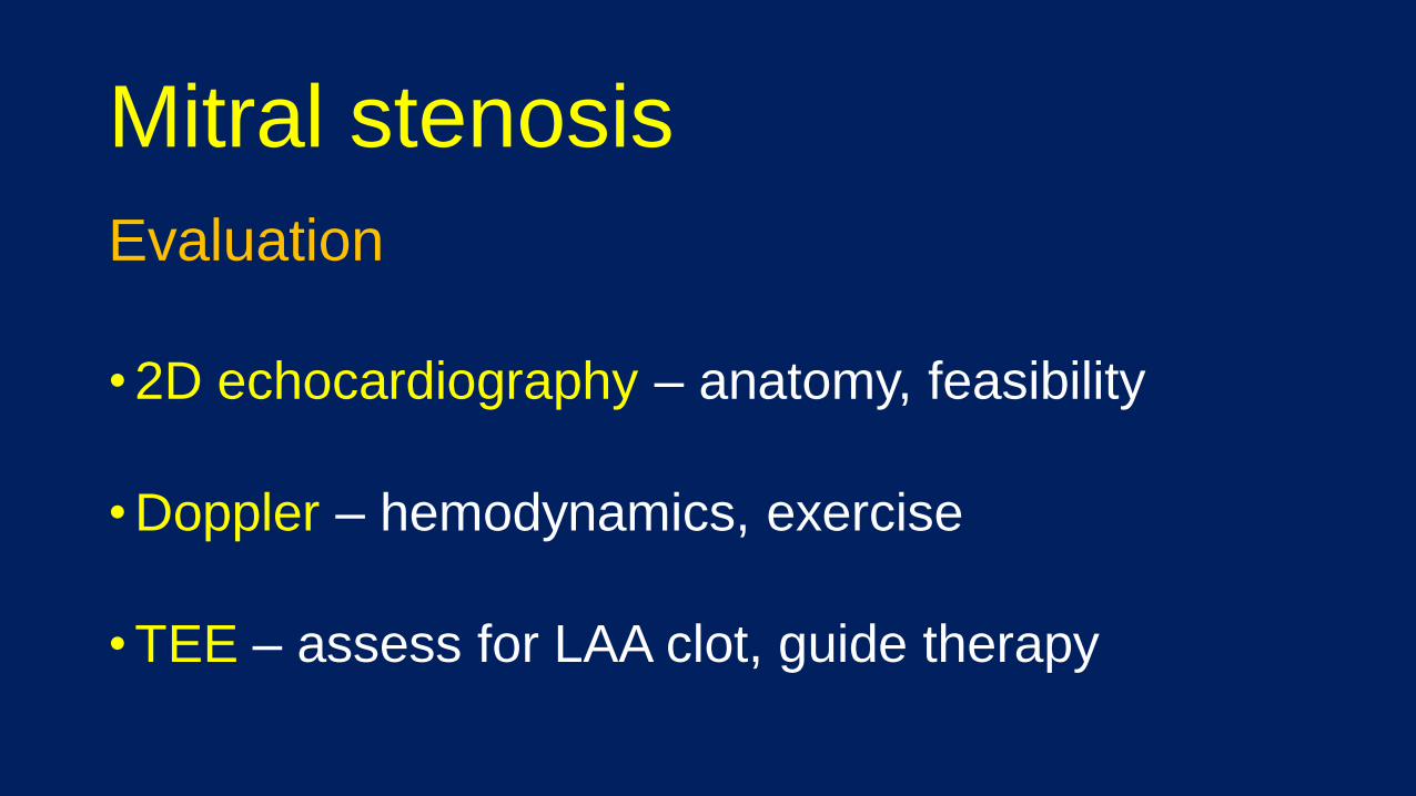
Evaluation
• 2D echocardiography – anatomy, feasibility
• Doppler – hemodynamics, exercise
• TEE – assess for LAA clot, guide therapy
Mitral stenosis
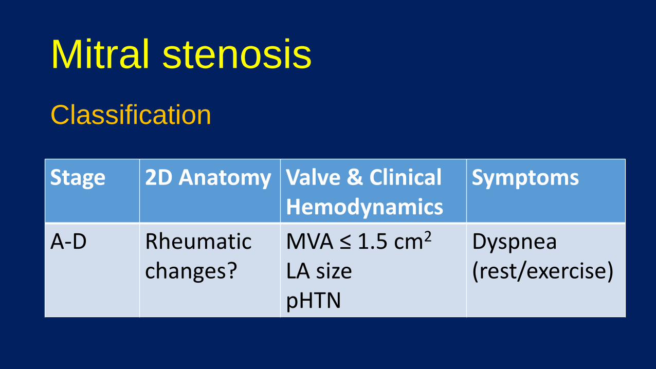
Classification
Stage 2D Anatomy Valve & Clinical Hemodynamics
Symptoms
A-D Rheumatic changes?
MVA ≤ 1.5 cm2
LA sizepHTN
Dyspnea (rest/exercise)
Mitral stenosis
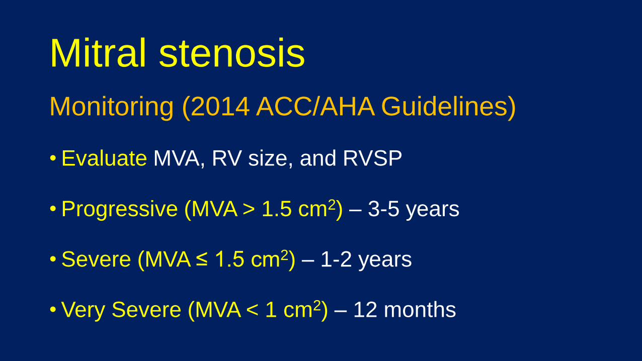
Monitoring (2014 ACC/AHA Guidelines)
• Evaluate MVA, RV size, and RVSP
• Progressive (MVA > 1.5 cm2) – 3-5 years
• Severe (MVA ≤ 1.5 cm2) – 1-2 years
• Very Severe (MVA < 1 cm2) – 12 months
Mitral stenosis
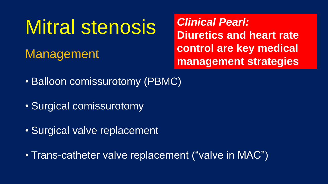
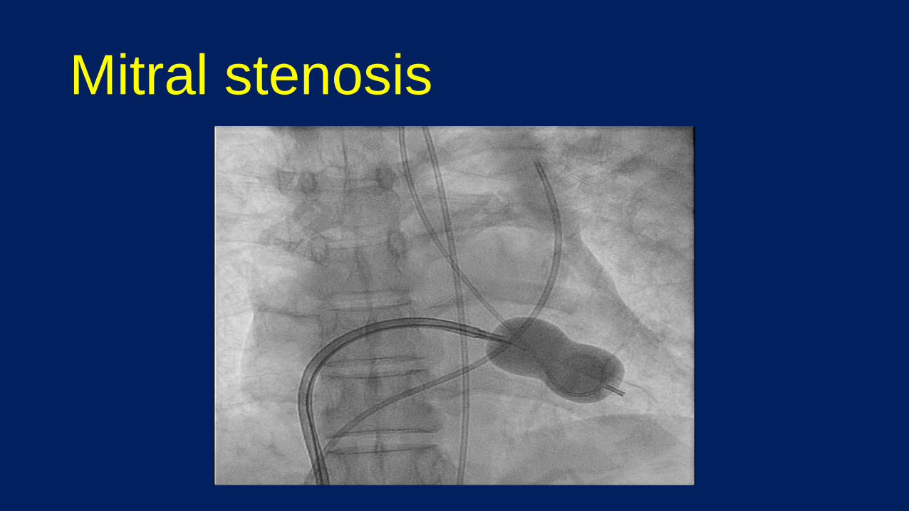
Management
• Balloon comissurotomy (PBMC)
• Surgical comissurotomy
• Surgical valve replacement
• Trans-catheter valve replacement (“valve in MAC”)
Clinical Pearl:
Diuretics and heart rate
control are key medical
management strategies
Mitral stenosis
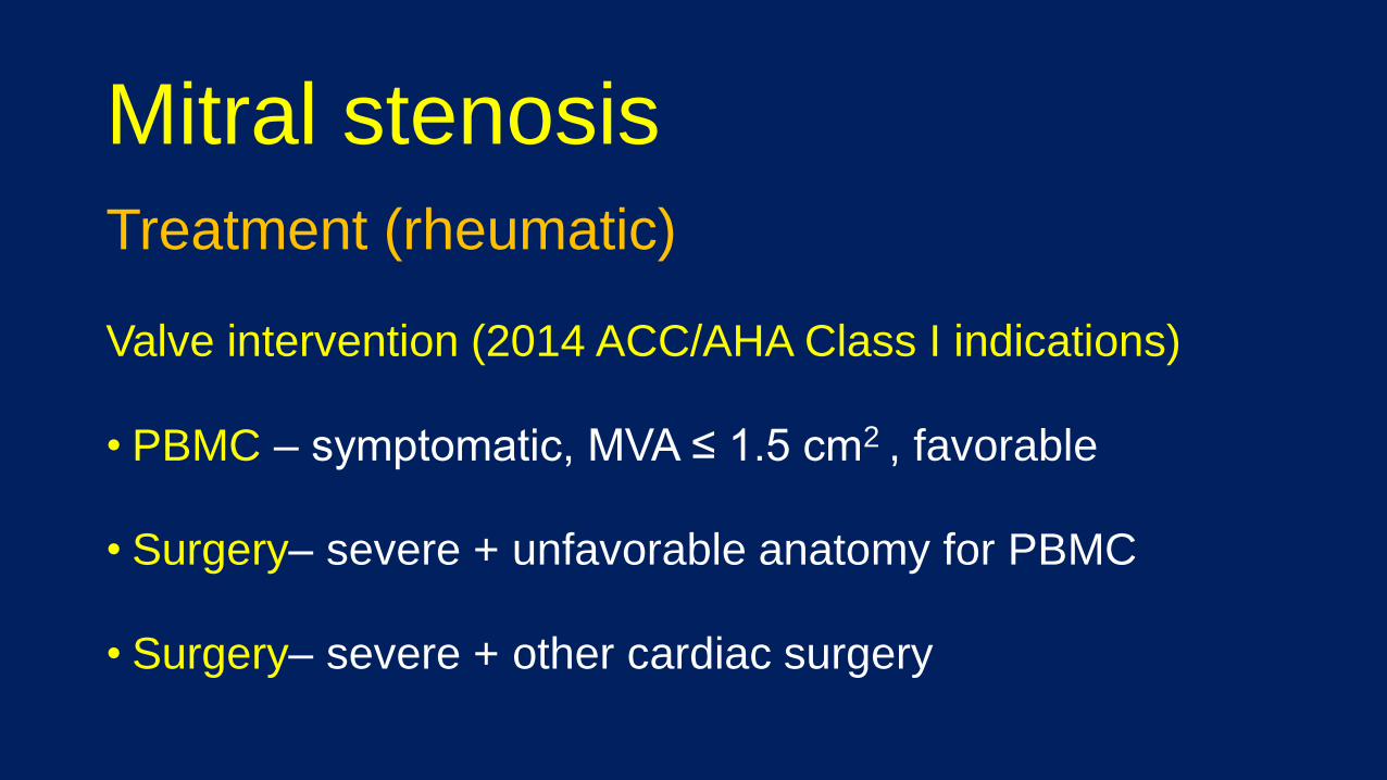
Treatment (rheumatic)
Valve intervention (2014 ACC/AHA Class I indications)
• PBMC – symptomatic, MVA ≤ 1.5 cm2 , favorable
• Surgery– severe + unfavorable anatomy for PBMC
• Surgery– severe + other cardiac surgery
Mitral stenosis
Mitral stenosis
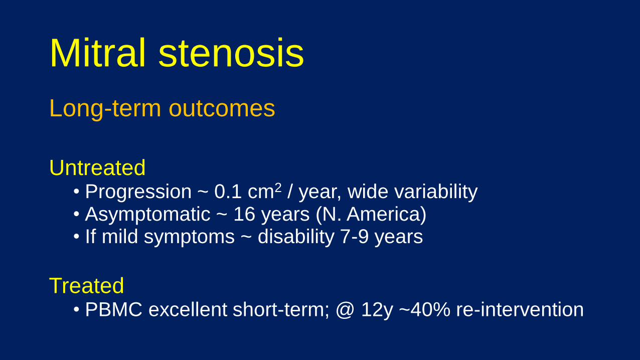
Long-term outcomes
Untreated • Progression ~ 0.1 cm2 / year, wide variability• Asymptomatic ~ 16 years (N. America)• If mild symptoms ~ disability 7-9 years
Treated• PBMC excellent short-term; @ 12y ~40% re-intervention
Mitral regurgitation
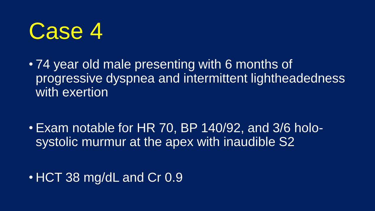
Case 4
• 74 year old male presenting with 6 months of progressive dyspnea and intermittent lightheadedness with exertion
• Exam notable for HR 70, BP 140/92, and 3/6 holo-systolic murmur at the apex with inaudible S2
• HCT 38 mg/dL and Cr 0.9

Case 4
A follow up echocardiogram is most likely to show:
A) Aortic sclerosis
B) Aortic stenosis
C) Mitral regurgitation
D) Mitral stenosis
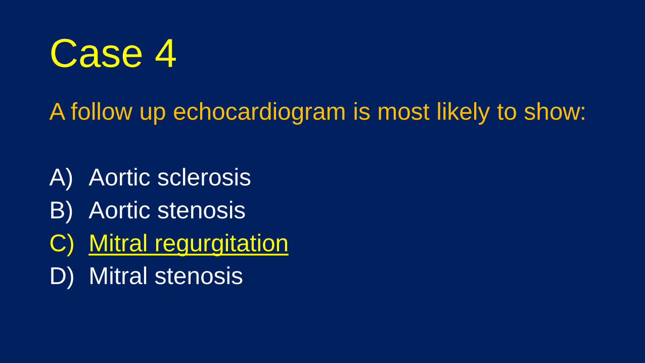
Case 4
A follow up echocardiogram is most likely to show:
A) Aortic sclerosis
B) Aortic stenosis
C) Mitral regurgitation
D) Mitral stenosis
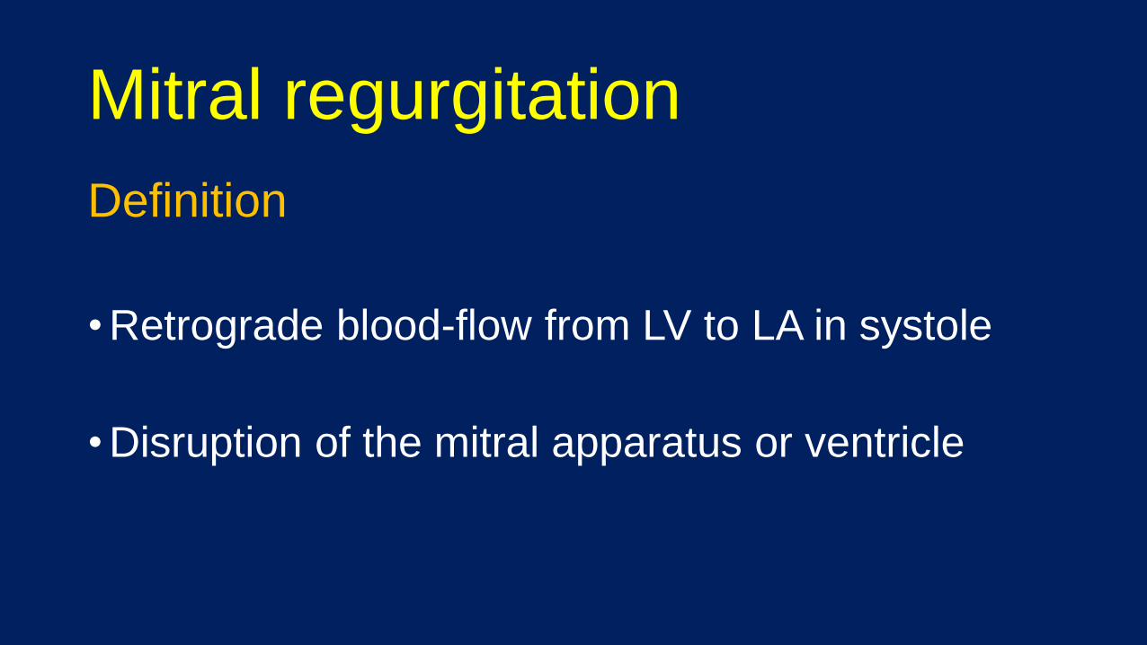
Mitral regurgitation
Definition
• Retrograde blood-flow from LV to LA in systole
• Disruption of the mitral apparatus or ventricle
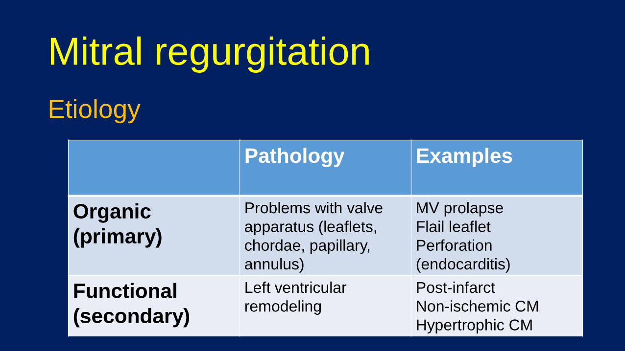
Mitral regurgitation
Etiology
Pathology Examples
Organic
(primary)
Problems with valve
apparatus (leaflets,
chordae, papillary,
annulus)
MV prolapse
Flail leaflet
Perforation
(endocarditis)
Functional
(secondary)
Left ventricular
remodeling
Post-infarct
Non-ischemic CM
Hypertrophic CM
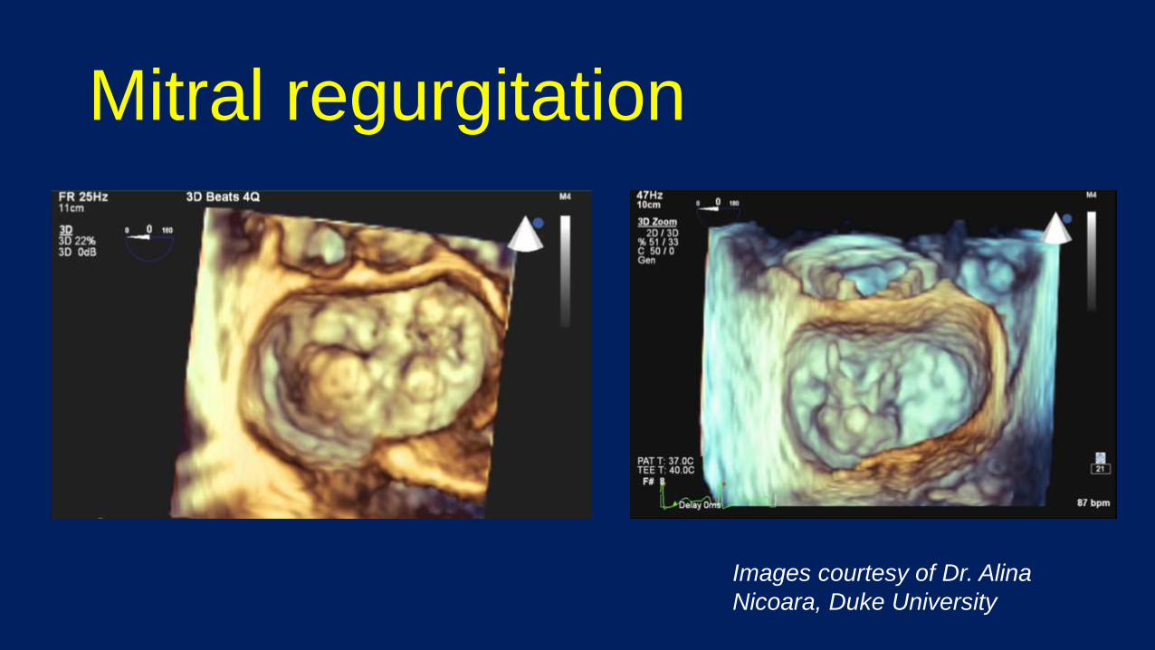
Mitral regurgitation
Images courtesy of Dr. Alina
Nicoara, Duke University

Mitral regurgitation
Pathophysiology
• Results in volume load to LV ↑ wall stress
• Eccentric LV remodeling (new sarcomeres)
• LV/LA dilation, ↓ contractility, ↑ LVESD, ↓ LVEF
Mitral regurgitation
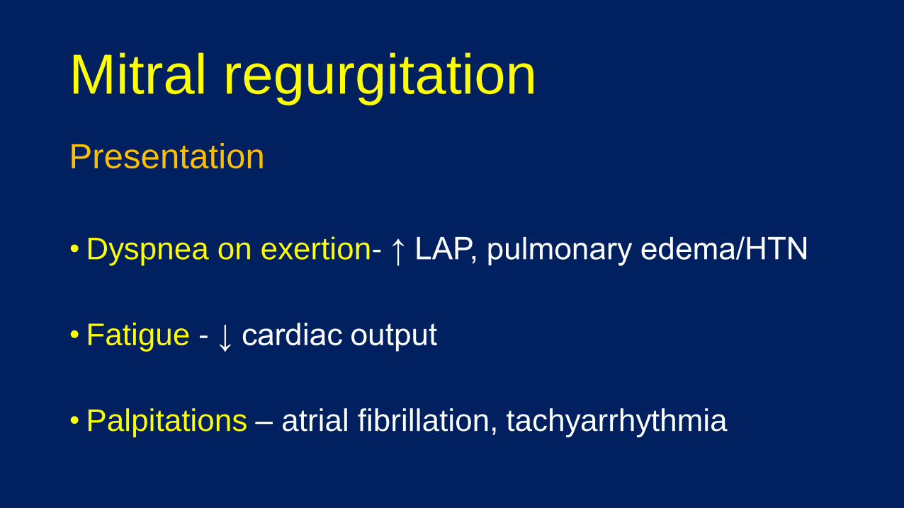
Presentation
• Dyspnea on exertion- ↑ LAP, pulmonary edema/HTN
• Fatigue - ↓ cardiac output
• Palpitations – atrial fibrillation, tachyarrhythmia
Mitral regurgitation
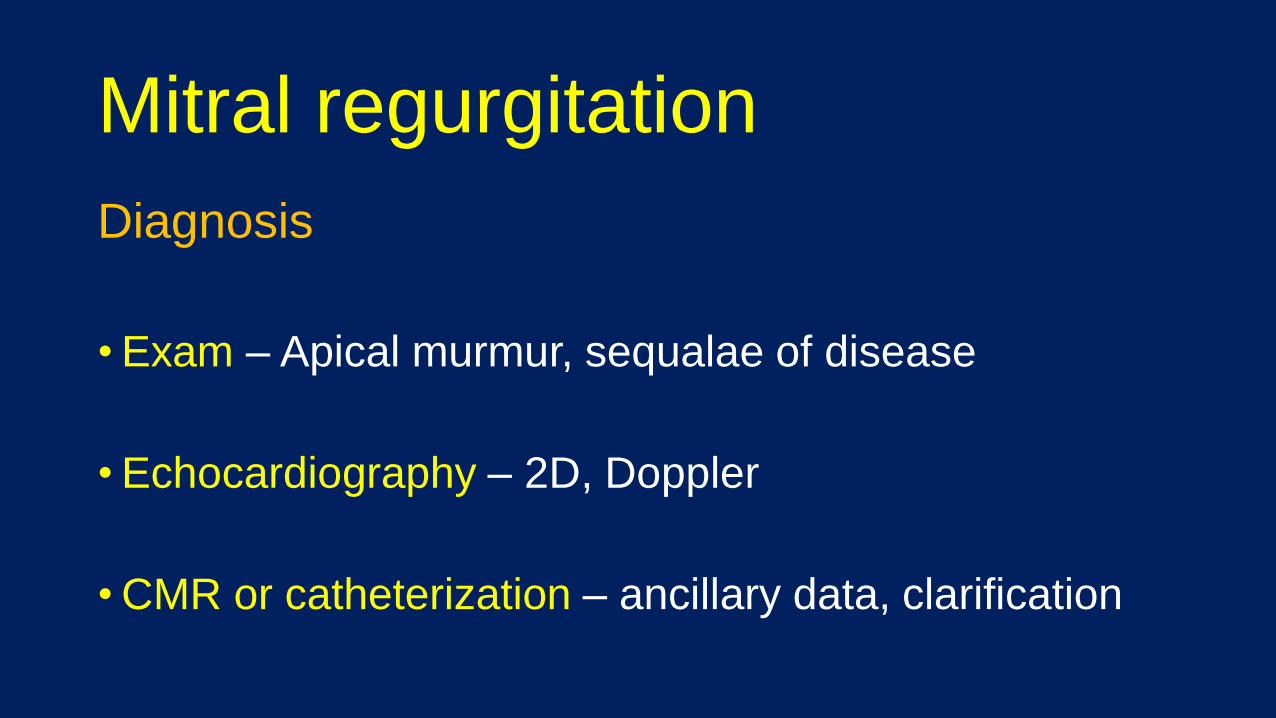
Diagnosis
• Exam – Apical murmur, sequalae of disease
• Echocardiography – 2D, Doppler
• CMR or catheterization – ancillary data, clarification
Mitral regurgitation
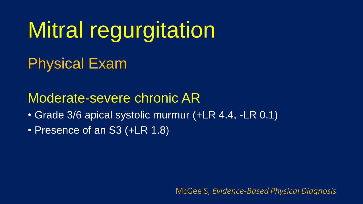
Physical Exam
Moderate-severe chronic AR• Grade 3/6 apical systolic murmur (+LR 4.4, -LR 0.1)
• Presence of an S3 (+LR 1.8)
McGee S, Evidence-Based Physical Diagnosis
Mitral regurgitation
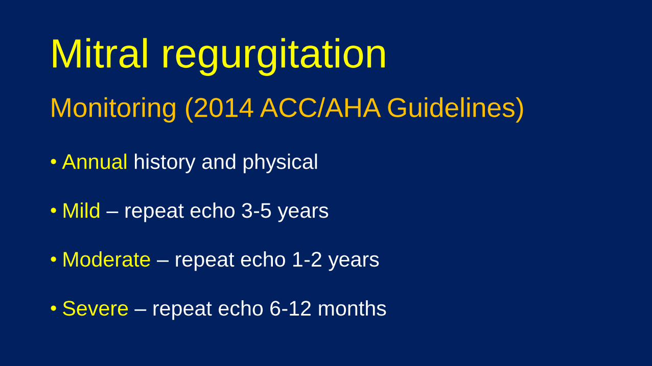
Monitoring (2014 ACC/AHA Guidelines)
• Annual history and physical
• Mild – repeat echo 3-5 years
• Moderate – repeat echo 1-2 years
• Severe – repeat echo 6-12 months
Mitral regurgitation
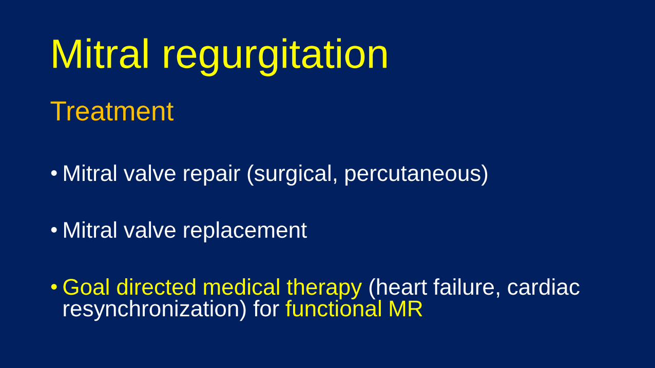
Treatment
• Mitral valve repair (surgical, percutaneous)
• Mitral valve replacement
• Goal directed medical therapy (heart failure, cardiac resynchronization) for functional MR
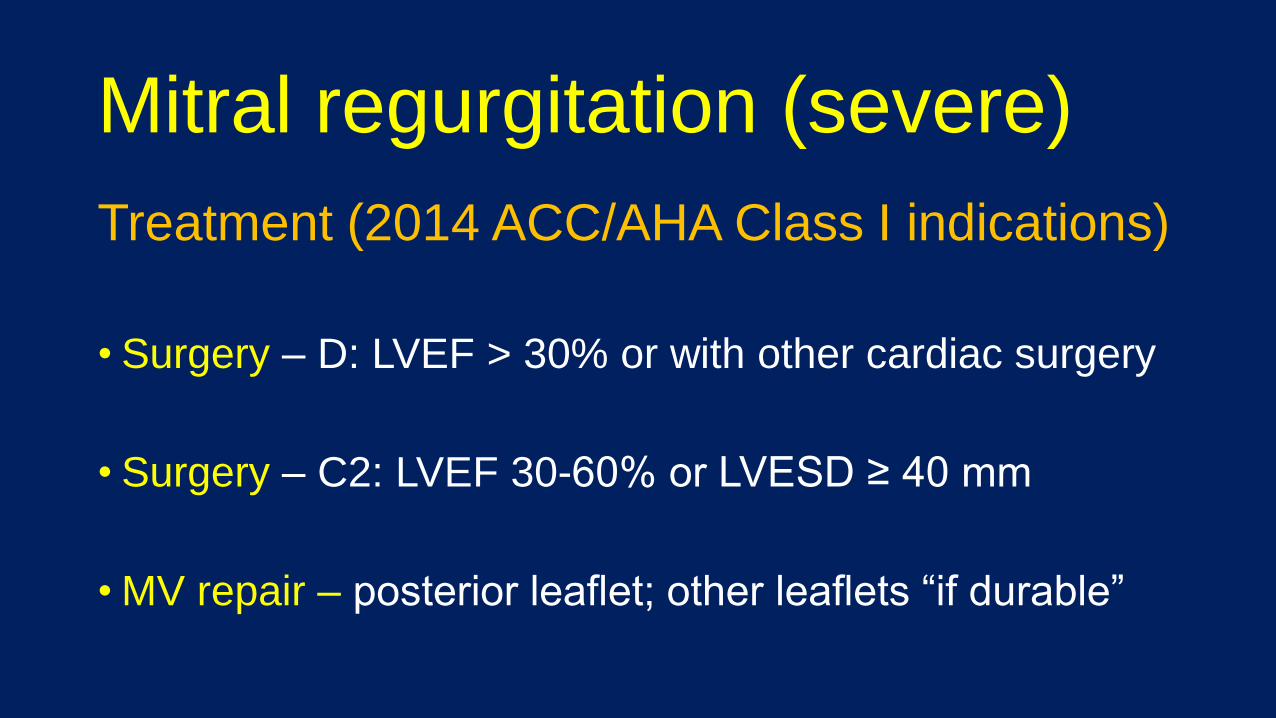
Mitral regurgitation (severe)
Treatment (2014 ACC/AHA Class I indications)
• Surgery – D: LVEF > 30% or with other cardiac surgery
• Surgery – C2: LVEF 30-60% or LVESD ≥ 40 mm
• MV repair – posterior leaflet; other leaflets “if durable”

Special situations
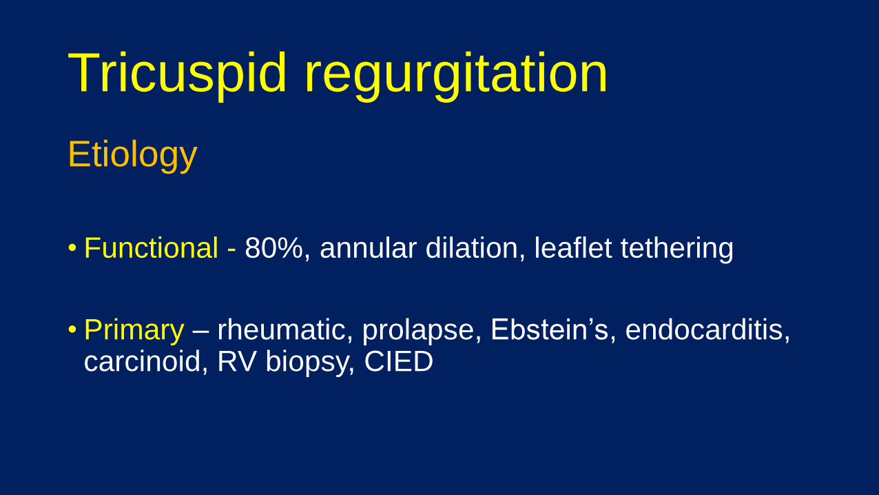
Tricuspid regurgitation
Etiology
• Functional - 80%, annular dilation, leaflet tethering
• Primary – rheumatic, prolapse, Ebstein’s, endocarditis, carcinoid, RV biopsy, CIED
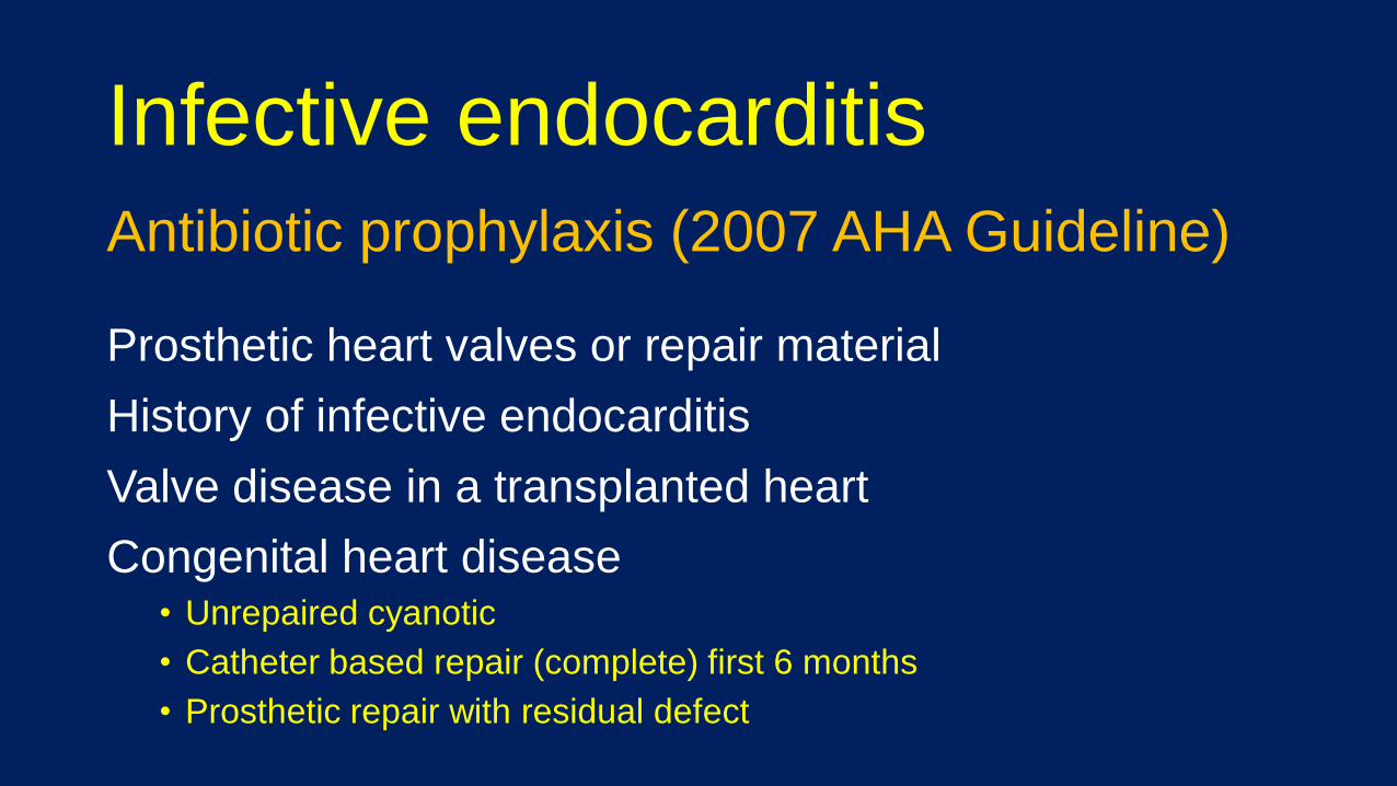
Infective endocarditis
Antibiotic prophylaxis (2007 AHA Guideline)
Prosthetic heart valves or repair material
History of infective endocarditis
Valve disease in a transplanted heart
Congenital heart disease• Unrepaired cyanotic
• Catheter based repair (complete) first 6 months
• Prosthetic repair with residual defect
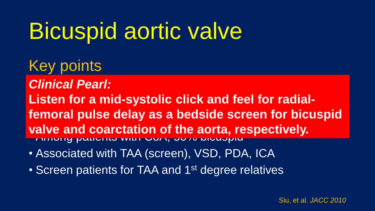
Bicuspid aortic valve
Key points
• Most common congenital defect - 0.5-2% adults
• Indications for repair include AS/AR +/- TAA
• Among patients with CoA, 50% bicuspid
• Associated with TAA (screen), VSD, PDA, ICA
• Screen patients for TAA and 1st degree relatives
Siu, et al. JACC 2010
Clinical Pearl:
Listen for a mid-systolic click and feel for radial-
femoral pulse delay as a bedside screen for bicuspid
valve and coarctation of the aorta, respectively.
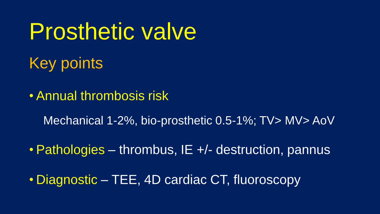
Prosthetic valve
Key points
• Annual thrombosis risk
Mechanical 1-2%, bio-prosthetic 0.5-1%; TV> MV> AoV
• Pathologies – thrombus, IE +/- destruction, pannus
• Diagnostic – TEE, 4D cardiac CT, fluoroscopy
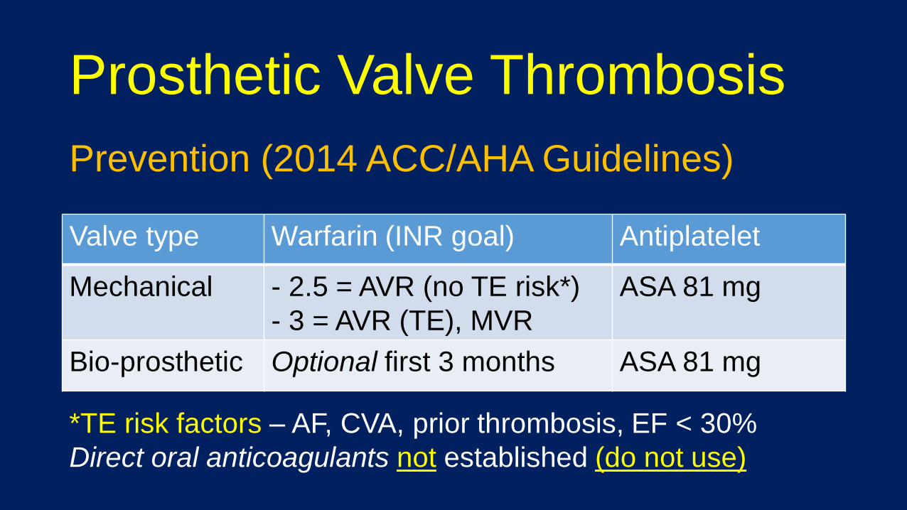
Prosthetic Valve Thrombosis
Prevention (2014 ACC/AHA Guidelines)
Valve type Warfarin (INR goal) Antiplatelet
Mechanical - 2.5 = AVR (no TE risk*)
- 3 = AVR (TE), MVR
ASA 81 mg
Bio-prosthetic Optional first 3 months ASA 81 mg
*TE risk factors – AF, CVA, prior thrombosis, EF < 30%
Direct oral anticoagulants not established (do not use)


![OPEN ACCESS Mini Review Real-Time Three-Dimensional ...stenosis, mitral stenosis and mitral regurgitation [22]. In this article, we intend to make a brief review of the importance](https://img.pdfslide.net/doc/110x75/610e7dfdec79fe75f71bd1eb/open-access-mini-review-real-time-three-dimensional-stenosis-mitral-stenosis.jpg)


![Rheumatic Aortic Valve Disease with Mitral Stenosis—A Case ... · 2005 [1]. RHD predominantly affects the left-sided cardiac valves, causing regurgitation, stenosis, or mixed hemodynamic](https://img.pdfslide.net/doc/110x75/5c941ea809d3f2c75a8c1b7c/rheumatic-aortic-valve-disease-with-mitral-stenosisa-case-2005-1-rhd.jpg)