Embed Size (px)
Citation preview
300
Bulgarian Journal of Agricultural Science, 21 (No 2) 2015, 300–304Agricultural Academy
VARIATIONS IN THE CHLOROPLAST ULTRASTRUCTURE IN IN VITRO-CULTURED HYPERICUM SPP. PLANTSM. STEFANOVA*, D. KOLEVA and T. GANEVASofi a University “St. Kliment Ohridski”, Faculty of Biology, Department of Botany, BG – 1164 Sofi a, Bulgaria
Abstract
STEFANOVA, M., D. KOLEVA and T. GANEVA, 2015. Variations in the chloroplast ultrastructure in in vitro-cultured Hypericum spp. plants. Bulg. J. Agric. Sci., 21: 300–304
Genus Hypericum comprises more than 480 species. Some of them have unique therapeutical properties, others are en-demic or endangered. These features make the species appropriate objects for biotechnological manipulations. In vitro condi-tions considerably affect organogenesis and hystogenesis of plants subjected to micropropagation. Structural organization of the plastid apparatus in the leaf mesophyll of in vitro-cultured plants is an indicator for evaluation of the regeneration potential of the explants.
The subject of the present study is the chloroplast ultrastructure of 7 in vitro-cultured Hypericum species – H. perforatum, H. humifusum, H. kalmianum, H. annulatum, H. tomentosum, H. pulchrum and H. rumeliacum. The aim is to analyze the mor-phological diversity of these vital for the process of regeneration cell compartments.
TEM-analysis allows us to identify the differences among species at subcellular level of organization in correlation to the in vitro conditions. The morphological diversity of chloroplast structure manifests as altered plastid shape, organization of the inter-nal membrane system, and starch content. The chloroplast shape of H. perforatum, H. annulatum, and H. pulchrum is elongated, which is typical for in vivo chloroplast. These species also are characterized with properly-structured thylakoid system. However, the grana height, the amount of stromal thylakolds, and the spatial orientation of the entire membrane system can vary. Signs of thylakoid destruction are observed in H. humifusum, H. kalmianum, H. tomentosum, and H. rumeliacum. Only three in vitro-cultured species (H. perforatum, H. annulatum and H. rumeliacum) develop chloroplasts with large amount of starch.
The results show that the chloroplasts in H. annulatum have the most proper structure while the H. tomentosum chloroplasts are the most atypical. The great morphological variability in the organization of the plastid apparatus in Hypericum species reveals autonomous structural response of each of them to the in vitro conditions despite their genetic similarity.
Key words: Hypericum sp., chloroplast ultrastructure, in vitro, transmission electron microscopyAbbreviations: TEM – transmission electron microscopy
*E-mail: [email protected]
Introduction
Structural organization of the chloroplasts, differentiated in vitro, is a very informative indicator for the regeneration capability of each species. A lot of TEM-studies examined the morphological variability in the organization of the plastid apparatus of in vitro-regenerated plants in search of favorable preconditions for a regular chloroplast structure.
Previous ultrastructural studies observed a wide variety in chloroplast structure of in vitro plants from different sys-tematic groups (Wetzstein and Sommer, 1982; Sudriá et al., 2001; Majada et al., 2002; Jausoro et al., 2010). It was found that in in vitro conditions were quite possible normal chloro-plasts to be structured (Laetsch and Stetler, 1965; Sudriá et al., 2001; Kapchina-Toteva and Stoyanova, 2003; Oliveira et al., 2008; Magyar-Tábori et al., 2010; Stoyanova-Koleva et
301Variations in the Chloroplast Ultrastructure in In vitro-Cultured Hypericum Spp. Plants
al., 2011). Quite often, however, chloroplasts with unusual shape and odd organization of the internal membrane system were noticed (Wetzstein and Sommer, 1982; Lee et al., 1985; Majada et al., 2002; Synková et al., 2003). Also chloroplasts with partially or entirely impaired membrane compartment and varying quantity of plastoglobuli and starch grains were observed (Lamhamedi et al., 2003; Lucchesini et al., 2006; Ladygin et al., 2008; Stefanova et al., 2013). It was shown that any change in the parameters of the culture medium occurred in ultrastructural level in a specifi c way for each species (Lee et al., 1985; Olmos and Hellín, 1998; Louro et al., 1999; Serret and Trillas, 2000; Oliveira et al., 2008; Stoyanova-Koleva et al., 2011; Stefanova et al., 2013). It has not yet been revealed to what extent the species specifi city is a limiting factor for a structuring of the plastid apparatus in vitro. Obtaining such information would be possible through a comparative ultrastructural study of genetically related species.
Subject of the present study were 7 in vitro-cultured un-der equal conditions Hypericum species – H. perforatum, H. humifusum, H. kalmianum, H. annulatum, H. tomentosum, H. pulchrum and H. rumeliacum. The aim was to examine the mesophyll chloroplast ultrastructure by TEM and to ana-lyze the morphological diversity of these vital for the process of regeneration cell compartments.
Materials and Methods
Plant materialStem explants from seven Hypericum species (Н. per-
foratum, Н. humifusum, Н. kalmianum, Н. annulatum, Н. tomentosum, Н. pulchrum, and H. rumeliacum) were intro-duced in vitro through direct plant regeneration and cultured on a standard full-strength medium (Murashige and Skoog, 1962), supplemented with 2% (m/v) sucrose and 8.0 g.dm–3 agar. The growth conditions were: temperature 22°C, 16-h photoperiod, photosynthetic photon fl ux density 60 μmol m–2 s-1 (white fl uorescent tubes).
TEM-analysis After 35 days of cultivation small leaf segments (1–2
mm2) of fully expanded leaves were taken from the 2nd or 3rd nodes and fi xed in 3% (m/v) glutaraldehyde in 0.1 M sodium phosphate buffer (pH 7.4) for 12 h at 4°C. The leaf segments were post fi xed in 1% (m/v) KMnO4 in the same buffer for 2 h at room temperature. After dehydration by increasing con-centrations of ethyl alcohol (from 25 to 100%), the samples were embedded in Durcupan (Fluka, Buchs, Switzerland). Ultra thin cross-sections were cut from the palisade paren-chyma with Reichert-Jung (Wien, Austria) ultramicrotome
and were contrasted with lead citrate (Reynolds, 1963). Ob-servation was performed by JEOL 1200 EX (Tokyo, Japan) electron microscope.
Results
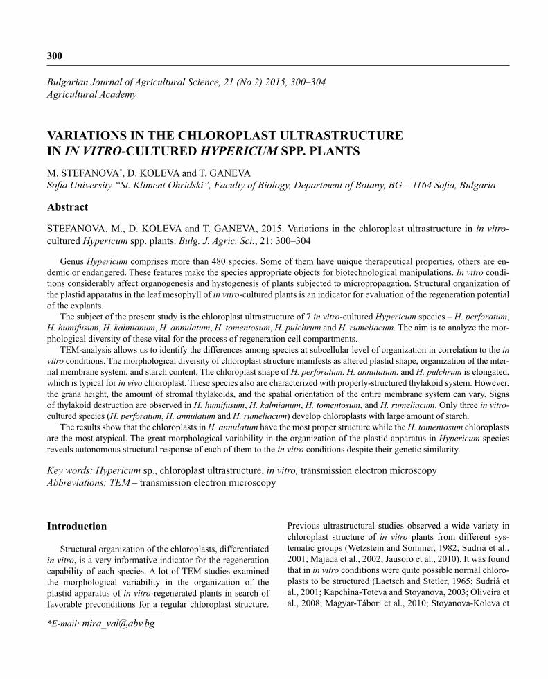
TEM-analysis of the seven Hypericum species showed a great diversity in the structural organization of the meso-phyll chloroplasts. In cross-section the chloroplasts of H. perforatum (Figure 1A) were elliptic and the area, occupied by the internal membrane system, was broad. The internal membrane system was structured from a lot of low grana (5–8 thylakoids) and less grana with average height (about 20 thylakoids). The grana were connected with long stromal thylakoids. In the stroma of each chloroplast relatively big starch grains were observed. They consisted of a starch, sur-rounded by a well-structured enzyme capsule and a very thin sugar zone.
The chloroplasts of H. humifusum (Figure 1B) in cross-section were semicircular, convex towards the vacuole and fl attened towards the cell wall. The internal membrane sys-tem was situated at the side of the vacuole. In each chloro-plast there was a large peristromium at the side of the cell wall. The majority of the grana were low (up to 10 thyla-koids). The stromal thylakoids were long and very dense. There were not starch grains in the stroma.
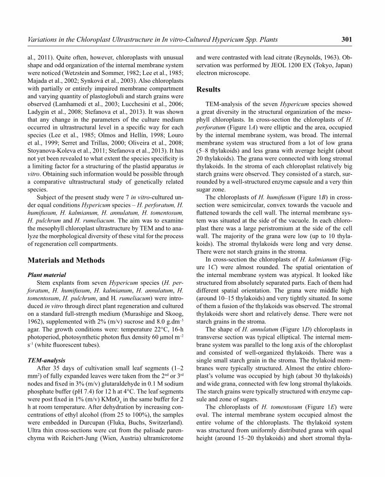
In cross-section the chloroplasts of H. kalmianum (Fig-ure 1C) were almost rounded. The spatial orientation of the internal membrane system was atypical. It looked like structured from absolutely separated parts. Each of them had different spatial orientation. The grana were middle high (around 10–15 thylakoids) and very tightly situated. In some of them a fusion of the thylakoids was observed. The stromal thylakoids were short and relatively dense. There were not starch grains in the stroma.
The shape of H. annulatum (Figure 1D) chloroplasts in transverse section was typical elliptical. The internal mem-brane system was parallel to the long axis of the chloroplast and consisted of well-organized thylakoids. There was a single small starch grain in the stroma. The thylakoid mem-branes were typically structured. Almost the entire chloro-plast’s volume was occupied by high (about 30 thylakoids) and wide grana, connected with few long stromal thylakoids. The starch grains were typically structured with enzyme cap-sule and zone of sugars.
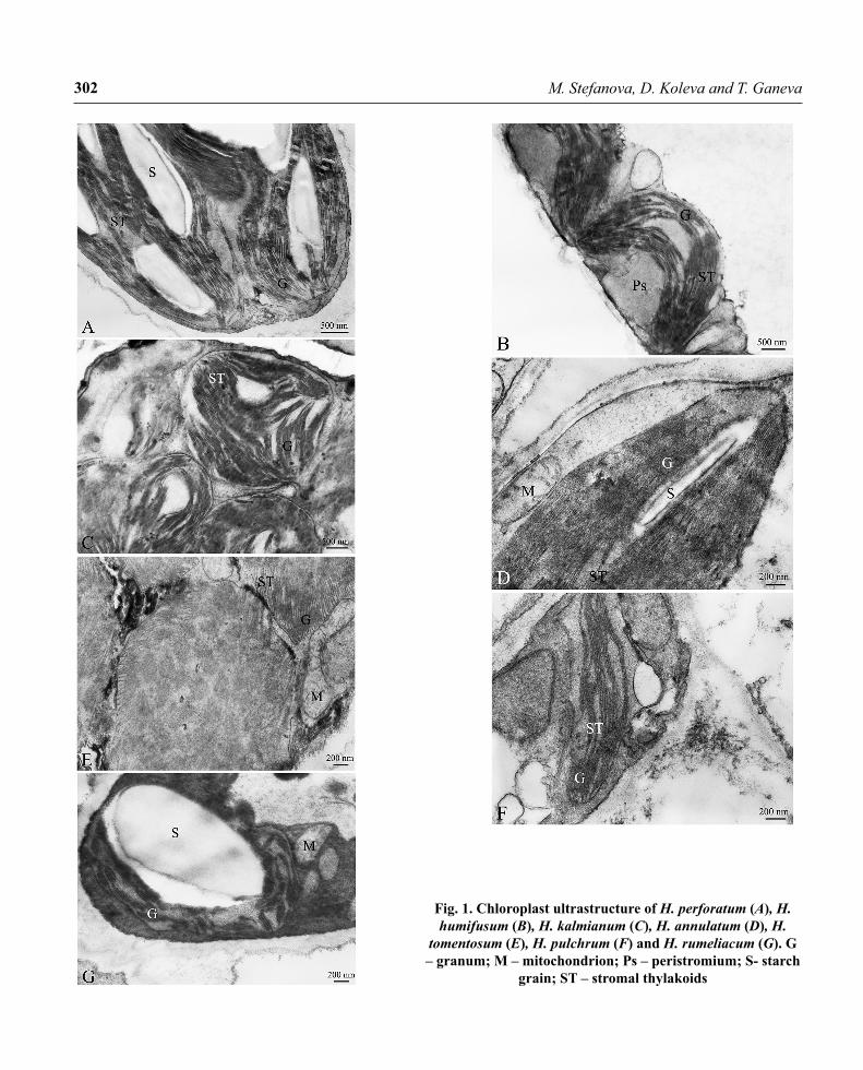
The chloroplasts of H. tomentosum (Figure 1E) were oval. The internal membrane system occupied almost the entire volume of the chloroplasts. The thylakoid system was structured from uniformly distributed grana with equal height (around 15–20 thylakoids) and short stromal thyla-
302 M. Stefanova, D. Koleva and T. Ganeva
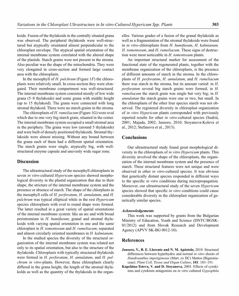
Fig. 1. Chloroplast ultrastructure of H. perforatum (A), H. humifusum (B), H. kalmianum (C), H. annulatum (D), H.
tomentosum (E), H. pulchrum (F) and H. rumeliacum (G). G – granum; M – mitochondrion; Ps – peristromium; S- starch
grain; ST – stromal thylakoids
303Variations in the Chloroplast Ultrastructure in In vitro-Cultured Hypericum Spp. Plants
koids. Fusion of the thylakoids in the centrally situated grana was observed. The peripheral thylakoids were well-struc-tured but atypically orientated almost perpendicular to the chloroplast envelope. The atypical spatial orientation of the internal membrane system correlated with the altered shape of the plastids. Starch grains were not present in the stroma. Also peculiar was the shape of the mitochondria. They were very elongated in cross-section and formed large contact area with the chloroplasts.
In the mesophyll of H. pulchrum (Figure 1F) the chloro-plasts were relatively small. In cross-section they were elon-gated. Their membrane compartment was well-structured. The internal membrane system consisted mostly of low wide grana (5–8 thylakoids) and some grana with middle height (up to 15 thylakoid). The grana were connected with long stromal thylakoid. There were no starch grains in the stroma.
The chloroplasts of H. rumeliacum (Figure 1G) were oval which due to one very big starch grain, situated in the center. The internal membrane system occupied a small stromal area in the periphery. The grana were low (around 5 thylakoids) and were built of densely positioned thylakoids. Stromal thy-lakoids were almost missing. Without any bound between the grana each of them had a different spatial orientation. The starch grains were single, atypically big, with well-structured enzyme capsule and unevenly wide sugar zone.
Discussion
The ultrastructural study of the mesophyll chloroplasts in seven in vitro-cultured Hypericum species showed morpho-logical diversity in the plastid organization that due to their shape, the stricture of the internal membrane system and the presence or absence of starch. The shape of the chlorplasts in the mesophyll cells of H. perforatum, H. annulatum, and H. pulchrum was typical elliptical while in the rest Hypericum species chloroplasts with oval to round shape were formed. The latter resulted in a great variety of spatial orientations of the internal membrane system: like an arc and with broad peristromium in H. humifusum; granal and stromal thyla-koids with varying spatial orientation in one and the same chloroplast in H. tomentosum and H. rumeliacum; separated and almost circularly oriented membranes in H. kalmianum.
In the studied species the diversity of the structural or-ganization of the internal membrane system was related not only to its spatial orientation, but also to the structure of the thylakoids. Chloroplasts with typically structured thylakoids were formed in H. perforatum, H. annulatum, and H. pul-chrum in vitro-plants. However, these chloroplasts clearly differed in the grana height, the length of the stromal thyla-kolds as well as the quantity of the thylakoids in the organ-
elles. Various grades of a fusion of the granal thylakoids as well as a fragmentation of the stromal thylakoids were found in in vitro-chloroplasts from H. humifusum, H. kalmianum, H. tomentosum, and H. rumeliacum. These signs of destruc-tion were most noticeable in H. tomentosum plants.
An important structural marker for assessment of the functional state of the regenerated plants, together with the membrane organization of the chloroplasts, is the presence of different amounts of starch in the stroma. In the chloro-plasts of H. perforatum, H. annulatum, and H. rumeliacum there was starch in the stroma, but its amount varied: in H. perforatum several big starch grains were formed; in H. rumeliacum the starch grain was single but very big; in H. annulatum the starch grains were one or two, but small. In the chloroplasts of the other four species starch was not ob-served. The registered diversity in chloroplast organization of in vitro Hypericum plants corresponded with previously reported results for other in vitro-cultured species (Sudriá, 2001; Majada, 2002; Jausoro, 2010; Stoyanova-Koleva et al., 2012; Stefanova et al., 2013).
Conclusions
Our ultrastructural study found great morphological di-versity in the chloroplasts of in vitro Hypericum plants. This diversity involved the shape of the chloroplasts, the organi-zation of the internal membrane system and the presence of starch. These structural features were not unique and were observed in other in vitro-cultured species. It was obvious that genetically distant species responded in different ways to the specifi c in vitro conditions during micropropagation. Moreover, our ultrastructural study of the seven Hypericum species showed that specifi c in vitro conditions could cause considerable diversity in the chloroplast organization of ge-netically similar species.
AcknowledgementsThis work was supported by grants from the Bulgarian
Ministry of Education, Youth and Science (DNTC/BGSK-01/2012) and from Slovak Research and Development Agency (APVV SK-BG 0012-10).
References
Jausoro, V., B. E. Llorente and N. M. Apóstolo, 2010. Structural differences between hyperhydric and normal in vitro shoots of Handroanthus impetiginosus (Mart. ex DC) Mattos (Bignonia-ceae). Plant Cell, Tissue and Organ Culture, 101: 183–191.
Kapchina-Toteva, V. and D. Stoyanova, 2003. Effects of cytoki-nins and cytokinin antagonists on in vitro cultured Gypsophila
304 M. Stefanova, D. Koleva and T. Ganeva
paniculata L. Biologia Plantarum, 46 (3): 337–341.Ladygin, V. G., N. I. Bondarev, G. A. Semenova, A. A. Smolov,
O. V. Reshetnyak and A. M. Nosov, 2008. Chloroplast ultra-structure, photosynthetic apparatus activities and production of steviol glycosides in Stevia rebaudiana in vivo and in vitro. Biologia Plantarum, 52 (1): 9–16.
Laetsch, W. M. and D. A. Stetler, 1965. Chloroplast structure and function in cultured tobacco tissue. American Journal of Bota-ny, 52 (8): 798–804.
Lamhamedi, M., H. Chamberl and F. M. Tremblay, 2003. Epi-dermal transpiration, ultrastuctural characteristics and net pho-tosynthesis of white spruce somatic seedlings in response to in vitro acclimatization. Physiologia Plantarum, 118: 554–561.
Lee, N., H. Y. Wetzstein and H. E. Sommer, 1985. Effects of quantum fl ux density on photosynthesis and chloroplast ultra-structure in tissue-cultured plantlets and seedlings of Liquidam-bar styracifl ua L. towards improved acclimatization and fi eld survival. Plant Physiology, 78: 637–641.
Louro, R. P., A. V. Dos Santos and R. D. Machado, 1999. Ul-trastructure of Eucalyptus grandis x Eucalyptus urophylla. I. Shoots cultivated in vitro in multiplication and elongation-rooting media. International Journal of Plant Sciences, 160 (2): 217–227.
Lucchesini, M., G. Monteforti, A. Mensuali-Sodi and G. Serra, 2006. Leaf ultrastructure, photosynthetic rate and growth of myrtle plantlets under different in vitro culture conditions. Bio-logia Plantarum, 50 (2): 161–168.
Magyar-Tábori, K., J. Dobránszki, J. A. Teixeira da Silva, S. M. Bulley and I. Hudák, 2010. The role of cytokinins in shoot organogenesis in apple. Plant Cell, Tissue and Organ Culture, 101: 251–267.
Majada, J. P., M. A. Fal, F. Tadeo and R. Sánchez-Tamés, 2002. Effects of natural ventilation on leaf ultrastructure of Dianthus caryophyllus L. cultured in vitro. In vitro Cellular and Devel-opmental Biology-Plant, 38: 272–278.
Murashige, T. and F. Skoog, 1962. A revised medium for rapid growth and bio-assay with tobacco tissue cultures. Physiologia
Plantarum, 15: 473–497.Oliveira, L. M., R. Paiva, J. R. F. Santana, E. Alves, R. C.
Nogueira and F. D. Pereira, 2008. Effect of cytokinins on in vitro development of autotrophism and acclimatization of An-nona glabra L. In vitro Cellular and Developmental Biology-Plant., 44: 128–135.
Olmos, E. and E. Hellín, 1998, Ultrastructural differences of hy-perhydric and normal leaves from regenerated carnation plants. Scientia Horticulturae, 75: 91–101.
Reynolds, E. S., 1963. The use of lead citrate at high pH as an electron-opaque stain in electron microscopy. Journal of Cell Biology, 17: 208–212.
Serret, M. D. and M. I. Trillas, 2000. Effects of light and sucrose levels on the anatomy, ultrastructure, and photosynthesis of Gardenia jasminoides Ellis leafl ets cultured in vitro. Interna-tional Journal of Plant Sciences, 161 (2): 281–289.
Stefanova, M., D. Koleva and Ts. Ganeva, 2013. Effect of plant growth regulators on chloroplast ultrastructure in Lamium al-bum plantlets. Bulgarian Journal of Agricultural Science, 19 (6) 1208–1212.
Stoyanova-Koleva, D., M. Stefanova, M. Zhiponova and V. Kapchina-Toteva, 2012. Effect of N6-benzyladenine and in-dole-3-butyric acid on photosynthetic apparatus of Orthosiphon stamineus plants grown in vitro. Biologia Plantarum, 56 (4): 607–612.
Sudriá, C., J. Palazón, R. Cusidó, M. Bonfi ll, M. T. Piñol and C. Morales, 2001. Effect of benzyladenine and indolebutyric acid on ultrastructure, glands formation, and essential oil accu-mulation in Lavandula dentata plantlets. Biologia Plantarum, 44 (1): 1–6.
Synková, H., R. Pechová and R. Valcke, 2003. Changes in chlo-roplast ultrastructure in Pssu-ipt tobacco during plant ontogeny. Photosynthetica, 41 (1): 117–126.
Wetzstein, H. Y. and H. E. Sommer, 1982. Leaf anatomy of tissue-cultured Liquidambar styracifl ua (Hamamelidaceae) during acclimatization. American Journal of Botany, 69 (10): 1579–1586.
Received August, 2, 2014; accepted for printing January, 8, 2015.