Embed Size (px)
Citation preview
771J. Sebag (ed.), Vitreous: in Health and Disease, DOI 10.1007/978-1-4939-1086-1_45, © Springer Science+Business Media New York 2014
Vitreous Floaters and Vision: Current Concepts and Management Paradigms
Laura C. Huang, Kenneth M.P. Yee, Christianne A. Wa, Justin N. Nguyen, Alfredo A. Sadun, and J. Sebag
V.B.8.
L.C. Huang, BA VMR Institute for Vitreous Macula Retina, 7677 Center Avenue, suite 400, Huntington Beach, CA 92647, USA
Doheny Eye Institute, Department of Ophthalmology, Los Angeles, CA, USA
University of Miami Miller School of Medicine, Miami, FL, USA e-mail: [email protected]
K.M.P. Yee, BS • C.A. Wa, BA J. Sebag, MD, FACS, FRCOphth, FARVO (*) VMR Institute for Vitreous Macula Retina, 7677 Center Avenue, suite 400, Huntington Beach, CA 92647, USA
Doheny Eye Institute, Department of Ophthalmology, Los Angeles, CA, USAe-mail: [email protected]; [email protected]; [email protected]
Keywords
Vitreous • Floaters • Posterior vitreous detachment • Myopic vitreopathy • Asteroid hyalosis • Light scatter-ing • Straylight glare • Contrast sensitivity • VFQ quality of life • Vitrectomy • Retinal detachment • Oxygen • Cataracts
Key Concepts
1. Floaters result from structural abnormalities in vit-reous. They are a more common complaint than previously known and have a more negative impact on the quality of life than previously appreciated.
2. Vision is adversely affected by vitreous light scat-tering causing straylight glare and degradation of contrast sensitivity. This explains the considerable degree of unhappiness experienced by patients suf-fering from floaters.
3. The only proven and safe treatment that effectively cures floaters and improves vision as well as patient well-being is limited vitrectomy.
Outline
I. Introduction
II. BackgroundA. Etiology of Floaters
III. Diagnostic ConsiderationsA. Clinical PresentationB. Clinical Characterization of Floaters
1. Structural Assessment of Floatersa. Ultrasonographyb. Combined OCT/SLOc. Dynamic Light Scattering
2. Effects of Floaters on Visiona. Straylightb. Contrast Sensitivity Function
IV. Therapeutic ConsiderationsA. Vitrectomy
1. Efficacy of Vitrectomy for Floaters2. Safety of Vitrectomy for Floaters
a. Endophthalmitisb. Retinal Tears and Retinal Detachmentc. Cataracts
Conclusions
References
J.N. Nguyen, BA VMR Institute for Vitreous Macula Retina, 7677 Center Avenue, suite 400, Huntington Beach, CA 92647, USA
A.A. Sadun, MD, PhD, FARVO Doheny Eye Institute/UCLA, Los Angeles, CA, USAe-mail: [email protected]
772
I. Introduction
Floaters most commonly occur in middle age due to age- related changes in vitreous structure and light scattering by the posterior vitreous cortex after collapse of the vitreous body during posterior vitreous detachment (PVD). In youth, float-ers are most often due to myopic vitreopathy. Vitreous float-ers can have a negative impact on visual function and in turn the quality of life. Techniques to characterize floaters clinically include ultrasound imaging, optical coherence tomography, and dynamic light scattering for structural characterization. Functional impact can be assessed by straylight measurements, as well as contrast sensitivity testing. When the severity of floater symptomatology is significant, commonly used thera-pies include neodymium:yttrium-aluminum- garnet (YAG) laser and limited 25-gauge vitrectomy. While the former is of unproven efficacy, the latter has been shown to be a safe, effec-tive, and definitive cure that improves patients’ quality of life and eradicates symptomatology produced by light scattering and diffraction. It is thus reasonable to offer limited vitrectomy to individuals who have attempted to cope unsuccessfully and in whom functional deficit can be objectively demonstrated by testing contrast sensitivity, an important aspect of vision.
II. Background
Over a century ago, Duke Elder described vitreous as a struc-ture composed of “loose and delicate filaments surrounded by fluid” [1]. Throughout the eighteenth century, the com-position and structure of vitreous mystified early theorists, spawning four different theories regarding vitreous structure. In 1741, Demours advocated the “alveolar theory” [2] in which membranes oriented in many directions enclosed com-partments, or alveoli, containing the fluid portion of vitreous. Pathologists such as Virchow supported this concept [3]. In 1780, Zinn postulated that vitreous is arranged in a concen-tric, lamellar orientation such as the “layers of an onion” [4]. This then constituted the “lamellar theory” and subsequently gained support from fellow theorists such as von Pappenheim [5] and Brucke [6]. In 1845, Hannover proposed the “radial sector theory” where he postulated that vitreous was com-posed in sectors that were radially oriented around the central anteroposterior core containing Cloquet’s canal, similar in appearance to a “cut orange” [7]. Lastly, in 1848, Sir William Bowman introduced the “fibrillar theory” based upon a tech-nique in which he utilized microscopy to show “nuclear gran-ules” of fine fibrils that formed bundles [8].
Changes during life from the clear, gel-like structure led Szent-Gyorgito to be the first to propose that vitreous structure changes with age [9]. Duke Elder elaborated upon this with his first description of “floaters” as “the passive
reaction of the vitreous body to disturbances that create liquefaction with the separation of part of its colloid basis (the residual protein) to form appearances evident clinically as opacities” [10]. This description holds true today as it is quite consistent with the prevailing theory of the mechanism by which vitreous undergoes structural changes with age.
A. Etiology of Floaters
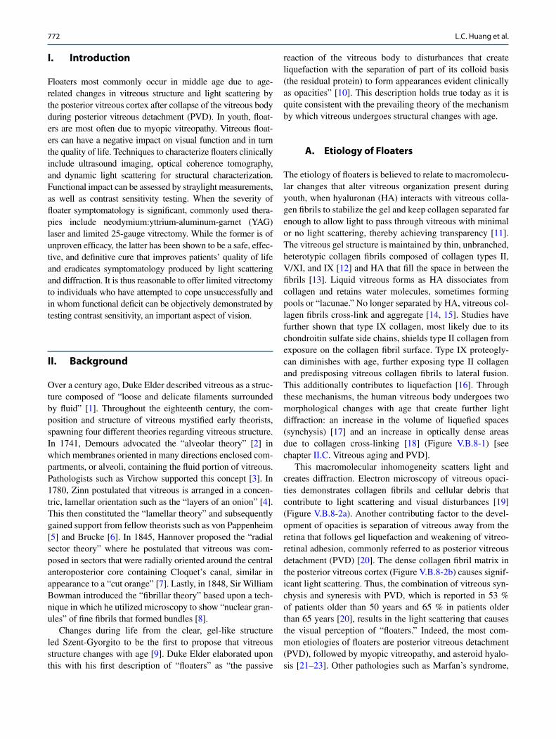
The etiology of floaters is believed to relate to macromolecu-lar changes that alter vitreous organization present during youth, when hyaluronan (HA) interacts with vitreous colla-gen fibrils to stabilize the gel and keep collagen separated far enough to allow light to pass through vitreous with minimal or no light scattering, thereby achieving transparency [11]. The vitreous gel structure is maintained by thin, unbranched, heterotypic collagen fibrils composed of collagen types II, V/XI, and IX [12] and HA that fill the space in between the fibrils [13]. Liquid vitreous forms as HA dissociates from collagen and retains water molecules, sometimes forming pools or “lacunae.” No longer separated by HA, vitreous col-lagen fibrils cross-link and aggregate [14, 15]. Studies have further shown that type IX collagen, most likely due to its chondroitin sulfate side chains, shields type II collagen from exposure on the collagen fibril surface. Type IX proteogly-can diminishes with age, further exposing type II collagen and predisposing vitreous collagen fibrils to lateral fusion. This additionally contributes to liquefaction [16]. Through these mechanisms, the human vitreous body undergoes two morphological changes with age that create further light diffraction: an increase in the volume of liquefied spaces (synchysis) [17] and an increase in optically dense areas due to collagen cross-linking [18] (Figure V.B.8-1) [see chapter II.C. Vitreous aging and PVD].
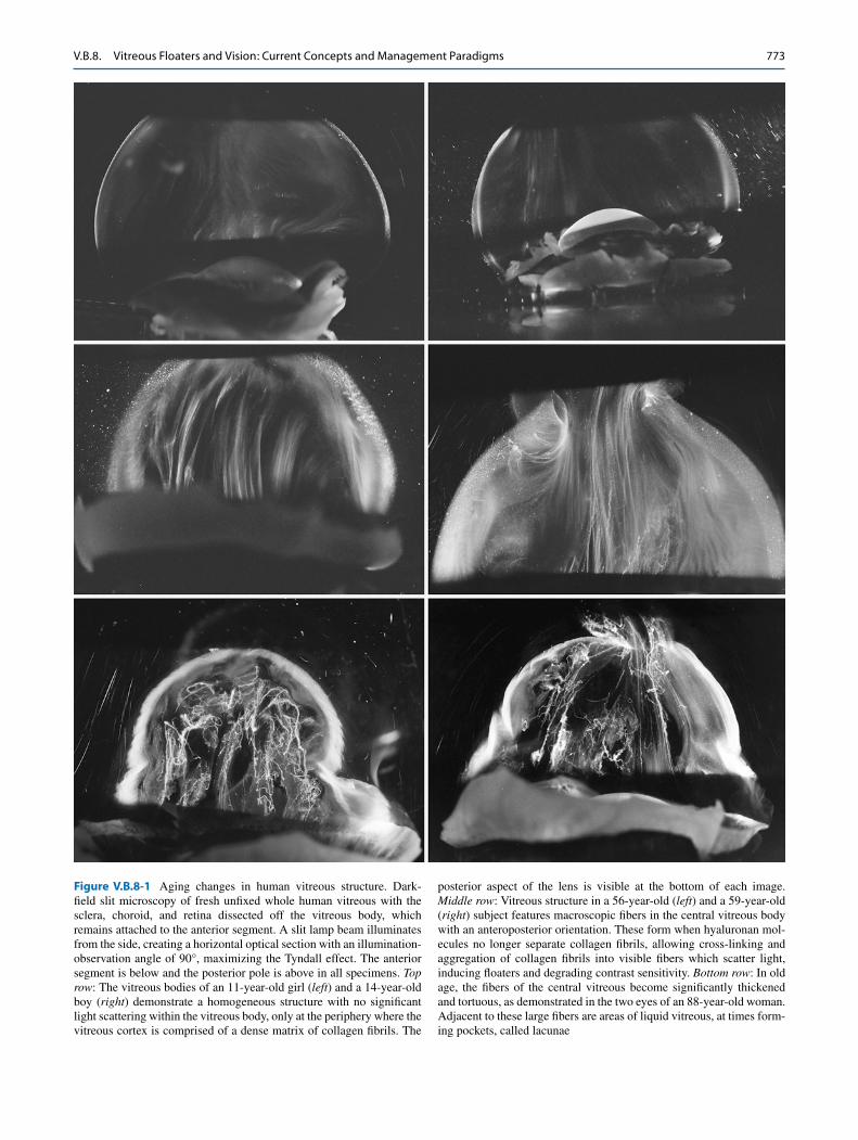
This macromolecular inhomogeneity scatters light and creates diffraction. Electron microscopy of vitreous opaci-ties demonstrates collagen fibrils and cellular debris that contribute to light scattering and visual disturbances [19] (Figure V.B.8-2a). Another contributing factor to the devel-opment of opacities is separation of vitreous away from the retina that follows gel liquefaction and weakening of vitreo-retinal adhesion, commonly referred to as posterior vitreous detachment (PVD) [20]. The dense collagen fibril matrix in the posterior vitreous cortex (Figure V.B.8-2b) causes signif-icant light scattering. Thus, the combination of vitreous syn-chysis and syneresis with PVD, which is reported in 53 % of patients older than 50 years and 65 % in patients older than 65 years [20], results in the light scattering that causes the visual perception of “floaters.” Indeed, the most com-mon etiologies of floaters are posterior vitreous detachment (PVD), followed by myopic vitreopathy, and asteroid hyalo-sis [21–23]. Other pathologies such as Marfan’s syndrome,
L.C. Huang et al.
773
Figure V.B.8-1 Aging changes in human vitreous structure. Dark-field slit microscopy of fresh unfixed whole human vitreous with the sclera, choroid, and retina dissected off the vitreous body, which remains attached to the anterior segment. A slit lamp beam illuminates from the side, creating a horizontal optical section with an illumination-observation angle of 90°, maximizing the Tyndall effect. The anterior segment is below and the posterior pole is above in all specimens. Top row: The vitreous bodies of an 11-year-old girl (left) and a 14-year-old boy (right) demonstrate a homogeneous structure with no significant light scattering within the vitreous body, only at the periphery where the vitreous cortex is comprised of a dense matrix of collagen fibrils. The
posterior aspect of the lens is visible at the bottom of each image. Middle row: Vitreous structure in a 56-year-old (left) and a 59-year-old (right) subject features macroscopic fibers in the central vitreous body with an anteroposterior orientation. These form when hyaluronan mol-ecules no longer separate collagen fibrils, allowing cross-linking and aggregation of collagen fibrils into visible fibers which scatter light, inducing floaters and degrading contrast sensitivity. Bottom row: In old age, the fibers of the central vitreous become significantly thickened and tortuous, as demonstrated in the two eyes of an 88-year-old woman. Adjacent to these large fibers are areas of liquid vitreous, at times form-ing pockets, called lacunae
V.B.8. Vitreous Floaters and Vision: Current Concepts and Management Paradigms
774
Ehlers-Danlos syndrome, and diabetic vitreopathy [see chap-ter I.E. Diabetic vitreopathy] also feature aggregation of vit-reous collagen fibers resulting in glare due to light scattering.
III. Diagnostic Considerations
Recent studies have found that floaters are more prevalent than previously appreciated. An electronic survey recently administered to 603 people via a smartphone application has demonstrated that 76 % of individuals report seeing floaters and 33 % report impairment in vision due to floaters [24]. Additionally, myopes were 3.5 times more likely to report moderate or severe floaters compared to those with normal vision. While this study suggests a significant prevalence of floaters in younger individuals (< 5 % of participants were
above age 50), this may simply be the result of selection bias since younger individuals are more likely to respond to sur-veys on smartphones than older individuals.
A. Clinical Presentation
The clinical presentation of a patient with floaters includes visual symptoms often described as gray, linear, hair-like structures with round points that appear more prominent against bright backgrounds (a white wall or clear sky), translucent strings, or a “spider web-like” image. The per-ception of these objects floating occurs during head or eye movements with an overdamping effect. To date, floaters have been viewed by the medical profession as an innocuous and benign process that will improve over time. Recent studies
a b
Figure V.B.8-2 (a) Ultrastructure of human vitreous fibers. Transmission electron microscopy of human vitreous- detected bundles of collagen fibrils shown longitudinally in the upper image and in cross section in the lower image. The inset in the upper image is a high magnification view of the bundle of fibrils demonstrating their collagenous nature. While these fibers form universally with aging, their formation is accelerated in myopia, constituting the second most common cause of floaters (From Sebag and Balazs [125]). (b) Ultrastructure of posterior vitreous cortex.
Scanning electron microscopy of the outer surface of the human posterior vitreous cortex after dissection and peeling away of the retina. The dense matrix of collagen fibrils is apparent, albeit somewhat exaggerated by the dehydration prep for electron microscopy. Nonetheless, it is apparent that when detached away from the retina, this structure will interfere with photon passage to the retina. The consequent light scattering will induce floaters and degrade contrast sensitivity (Courtesy of the Eye Research Institute of Retina Foundation, Boston, MA)
L.C. Huang et al.
775
have begun to dismiss this notion of floaters as a benign and innocuous process [25, 26]. These studies employed utility value analysis to determine that floaters have a significantly negative impact upon quality of life and symptoms can be much more disturbing than previously appreciated [27]. In one such study [25], researchers demonstrated that the degree to which floaters lower the visual quality of life is equivalent to age-related macular degeneration and greater than diabetic retinopathy and glaucoma, as well as mild angina, mild stroke, and colon cancer. Additionally, the per-ceived negative impact of floaters is underscored by the fact that patients would be willing to take an 11 % risk of death and a 7 % risk of blindness and would be willing to trade 1.1 years out of every 10 years remaining in their lives to eradicate floaters.
B. Clinical Characterization of Floaters
One of the contributing factors to clinicians’ perception of floaters as a benign condition and not a disease is the
lack of a clinical approach to characterize floaters both in terms of structure as well as the impact on visual function. Beyond visual acuity, the evaluation of the impact of floaters on vision is lacking, since rarely do such vitreous opacities affect visual acuity, the most common method for assessing visual function. Thus, both structural [see chapter II.F. To See the Invisible – the Quest of Imaging Vitreous] and func-tional assessments of floaters are needed.
1. Structural Assessment of Floatersa. Ultrasonography
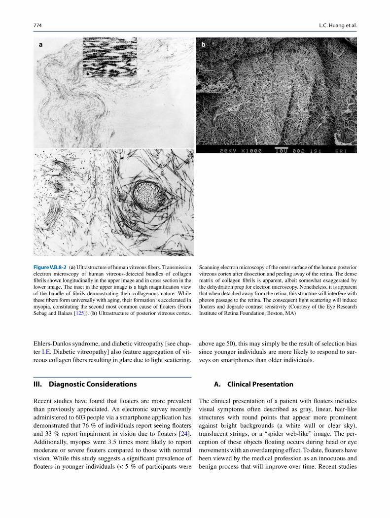
Ultrasonography may be used to image opacities within the vitreous body. Ultrasound measures differences in acoustic impedance generated by echoes produced at tissue interfaces between structures of different densities. Ultrasonography imaging within the frequencies of 8–10 MHz can produce wavelengths as small as 0.2 mm to determine pathologies such as PVD, asteroid hyalosis, vitreous hemorrhage, and large foreign bodies [28, 29] (see Figure V.B.8-3). As the vitreous ages, inhomogeneities in density develop via the aforementioned biochemical changes and echoes become
Figure V.B.8-3 B-scan ophthalmic ultrasound image of a patient demonstrating prominent echodensities in the vitreous body
V.B.8. Vitreous Floaters and Vision: Current Concepts and Management Paradigms
776
more prominent. Acoustic changes or echoes in the vitre-ous also occur when there is persistence of primary vitreous (vascular) structures, changes induced by myopic vitreopa-thy, or the presence of intraocular foreign bodies [30]. An advantage of vitreous imaging by ultrasound is the ability to visualize posterior structures regardless of media opacifi-cation anteriorly (primarily lens) and the ability to estimate lacunar size and collagen fibril density [31].
Many studies have utilized ultrasonography to image the topography of the vitreous body. In patients being considered for vitrectomy surgery, rapid B-scan imaging is the most useful technique to investigate vitreoretinal pathology [30]. Static A-scan ultrasound demonstrated an 80 % incidence of acoustic interfaces in patients 60 years old and greater, which differed drastically from the 5 % incidence in patients 21–40 years old [32]. This demon-strates the decrease in acoustic homogeneity as the vit-reous ages. Additionally, kinetic B-scan ultrasonography was utilized to quantify the vitreous of aged eyes with PVD compared to younger eyes [33]. The speckle density, referring to areas of acoustic hyper-reflectivity in areas of greater acoustic impedance, is increased in older subjects due to aggregation of collagen fibrils. Furthermore, chirp pulse encoding and synthetic focusing with an annular array was utilized to provide imaging of the vitreous with higher resolution and sensitivity through a 20-MHz ultra-sound probe [34].
Recently, quantitative ultrasonography was used to ana-lyze increased vitreous echodensity and correlate the find-ings with contrast sensitivity in patients with floaters of varying severity [35]. Investigators found that quantitative ultrasonography taken at nasal longitudinal, inferotemporal longitudinal, and inferotemporal transverse positions corre-lated strongly with contrast sensitivity (R = 0.82, p < 0.001) [35]. Quantitative ultrasonography should therefore have great utility to assess the severity of vitreous degeneration and aid in clinical decision-making regarding therapy of floaters.
b. Combined OCT/SLOTime-domain OCT was previously used to extensively analyze the vitreoretinal interface [36], but utility was lim-ited and fine details of the posterior vitreous were lacking. SD-OCT possesses inherent image resolution enhancement over time domain but also provides advantages in compari-son to ultrasound with a greater horizontal resolution as well as better imaging of the vitreoretinal interface [37]. Combined SD-OCT and scanning laser ophthalmoscopy (SLO) provides further insight on vitreous structure and evolving physiologic and pathologic vitreoretinal changes. In one study [31], SD-OCT/SLO imaging examined 202 eyes and demonstrated a high correlation between diagnosis of complete PVD found by both clinical examination and OCT.
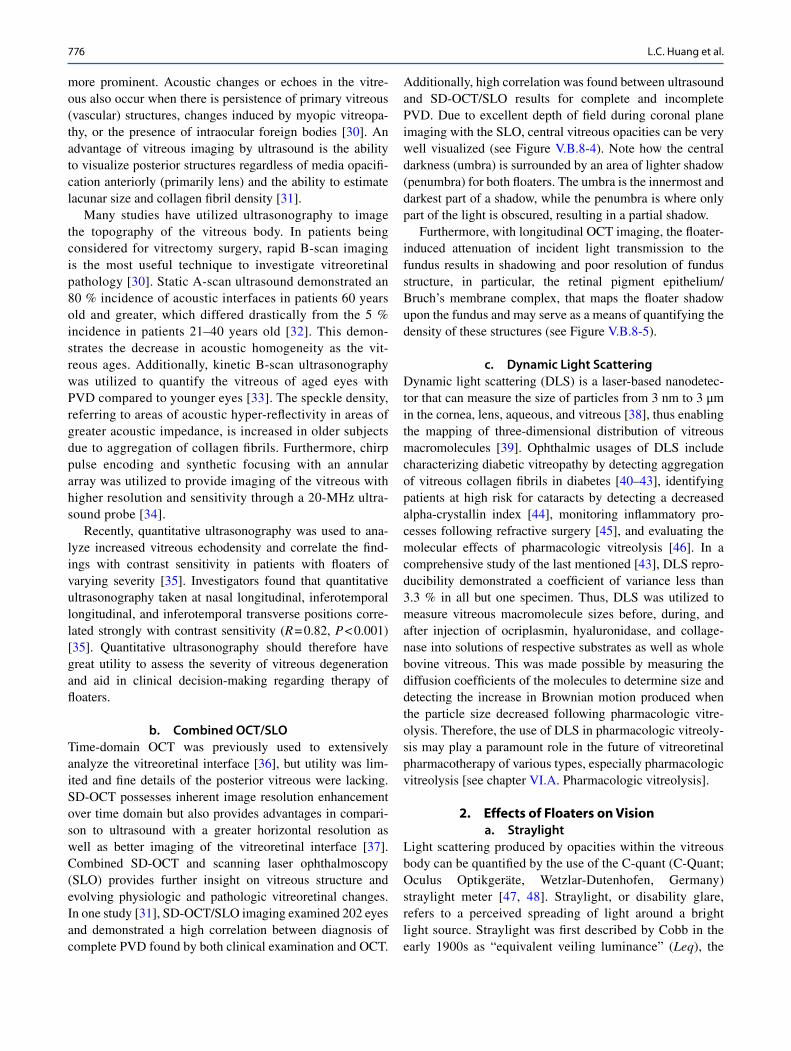
Additionally, high correlation was found between ultrasound and SD-OCT/SLO results for complete and incomplete PVD. Due to excellent depth of field during coronal plane imaging with the SLO, central vitreous opacities can be very well visualized (see Figure V.B.8-4). Note how the central darkness (umbra) is surrounded by an area of lighter shadow (penumbra) for both floaters. The umbra is the innermost and darkest part of a shadow, while the penumbra is where only part of the light is obscured, resulting in a partial shadow.
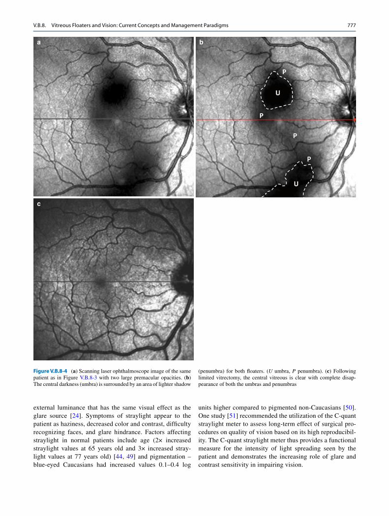
Furthermore, with longitudinal OCT imaging, the floater- induced attenuation of incident light transmission to the fundus results in shadowing and poor resolution of fundus structure, in particular, the retinal pigment epithelium/Bruch’s membrane complex, that maps the floater shadow upon the fundus and may serve as a means of quantifying the density of these structures (see Figure V.B.8-5).
c. Dynamic Light ScatteringDynamic light scattering (DLS) is a laser-based nanodetec-tor that can measure the size of particles from 3 nm to 3 μm in the cornea, lens, aqueous, and vitreous [38], thus enabling the mapping of three-dimensional distribution of vitreous macromolecules [39]. Ophthalmic usages of DLS include characterizing diabetic vitreopathy by detecting aggregation of vitreous collagen fibrils in diabetes [40–43], identifying patients at high risk for cataracts by detecting a decreased alpha-crystallin index [44], monitoring inflammatory pro-cesses following refractive surgery [45], and evaluating the molecular effects of pharmacologic vitreolysis [46]. In a comprehensive study of the last mentioned [43], DLS repro-ducibility demonstrated a coefficient of variance less than 3.3 % in all but one specimen. Thus, DLS was utilized to measure vitreous macromolecule sizes before, during, and after injection of ocriplasmin, hyaluronidase, and collage-nase into solutions of respective substrates as well as whole bovine vitreous. This was made possible by measuring the diffusion coefficients of the molecules to determine size and detecting the increase in Brownian motion produced when the particle size decreased following pharmacologic vitre-olysis. Therefore, the use of DLS in pharmacologic vitreoly-sis may play a paramount role in the future of vitreoretinal pharmacotherapy of various types, especially pharmacologic vitreolysis [see chapter VI.A. Pharmacologic vitreolysis].
2. Effects of Floaters on Visiona. Straylight
Light scattering produced by opacities within the vitreous body can be quantified by the use of the C-quant (C-Quant; Oculus Optikgeräte, Wetzlar-Dutenhofen, Germany) straylight meter [47, 48]. Straylight, or disability glare, refers to a perceived spreading of light around a bright light source. Straylight was first described by Cobb in the early 1900s as “equivalent veiling luminance” (Leq), the
L.C. Huang et al.
777
external luminance that has the same visual effect as the glare source [24]. Symptoms of straylight appear to the patient as haziness, decreased color and contrast, difficulty recognizing faces, and glare hindrance. Factors affecting straylight in normal patients include age (2× increased straylight values at 65 years old and 3× increased stray-light values at 77 years old) [44, 49] and pigmentation – blue-eyed Caucasians had increased values 0.1–0.4 log
units higher compared to pigmented non-Caucasians [50]. One study [51] recommended the utilization of the C-quant straylight meter to assess long- term effect of surgical pro-cedures on quality of vision based on its high reproducibil-ity. The C-quant straylight meter thus provides a functional measure for the intensity of light spreading seen by the patient and demonstrates the increasing role of glare and contrast sensitivity in impairing vision.
a b
c
Figure V.B.8-4 (a) Scanning laser ophthalmoscope image of the same patient as in Figure V.B.8-3 with two large premacular opacities. (b) The central darkness (umbra) is surrounded by an area of lighter shadow
(penumbra) for both floaters. (U umbra, p penumbra). (c) Following limited vitrectomy, the central vitreous is clear with complete disap-pearance of both the umbras and penumbras
V.B.8. Vitreous Floaters and Vision: Current Concepts and Management Paradigms
778
b. Contrast Sensitivity FunctionMeasuring contrast sensitivity function (CSF) provides a use-ful method to quantify floaters and the impact on vision. The measure of CSF is often used to supplement visual acuity measurements in evaluating posterior capsular opacification after cataract surgery [52] as well as demonstrate reduced visual function in patients with cataracts whose acuity is only slightly impaired [53, 54]. In one study, multiple logistic regression analysis demonstrated that reduced contrast sen-sitivity was independently associated with a vision disability score affecting tasks such as judging distance, night driving, and mobility issues [55]. In addition to anterior ocular media opacities, vitreous opacities may also degrade CSF.
This hypothesis was tested in a study [56] at the VMR Institute for Vitreous Macula Retina in California, that
utilized computer- based Freiburg Acuity Contrast test (FrACT) to evaluate CSF [57–59]. This CSF testing method had a reproducibility of 92.1 % when employing the Weber index (%W = (Luminancemax − Luminancemin)/Luminancemax) [52, 60] as the outcome measure. Studies have previously demonstrated that FrACT yields results similar to those of Pelli-Robson and is not significantly affected by group differences in visual acuity [61]. Patients with bothersome floaters have been reported to have CSF that is diminished by 67.4 % (4.0 ± 2.3 %W; N = 16) compared to age-matched controls (2.4 ± 0.9 %W; N = 16; p < 0.01). Since publication of these preliminary findings, this study has continued with increased enrollment (n = 38) obtaining similar results: in floater patients, CSF is degraded by 72.3 % (4.14 ± 1.95 %W) compared to controls (p < 0.01).
Figure V.B.8-5 OCT of the same patient as in Figure V.B.8-3 demon-strating floater-induced attenuation of imaging, especially at the retinal pigment epithelium/Bruch’s membrane complex. Solid arrows repre-
sent incident light rays, while dashed arrows represent scattered light. (Fl floater, U umbra, p penumbra)
L.C. Huang et al.
779
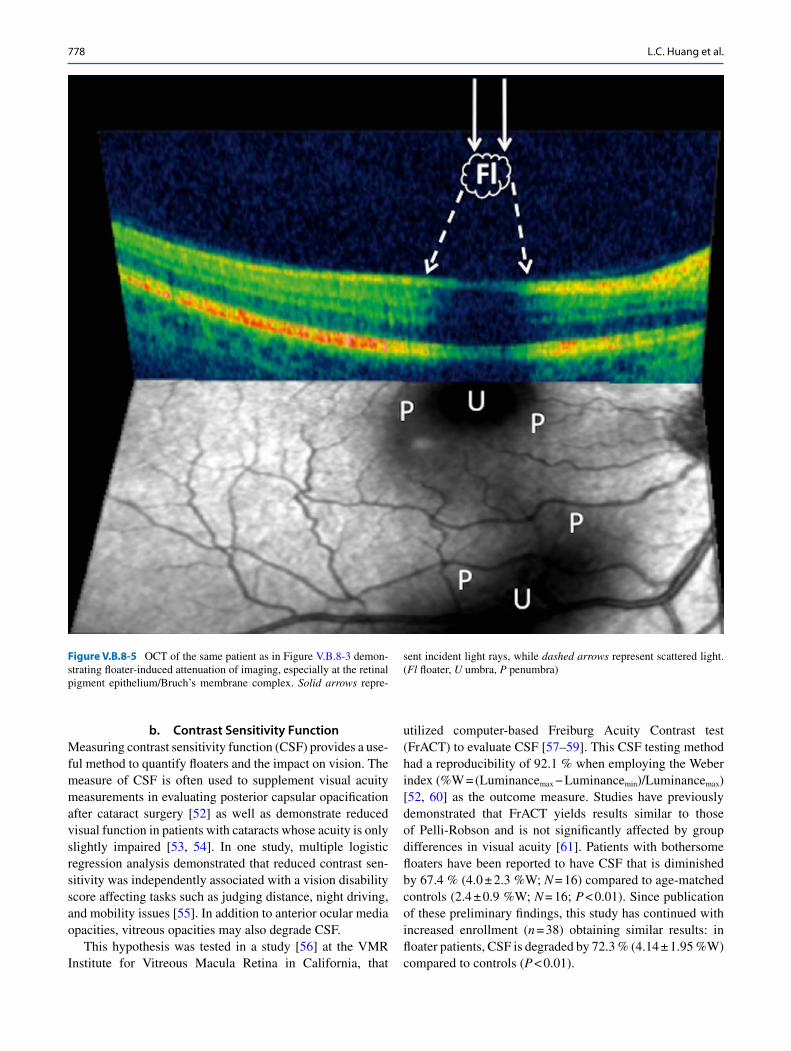
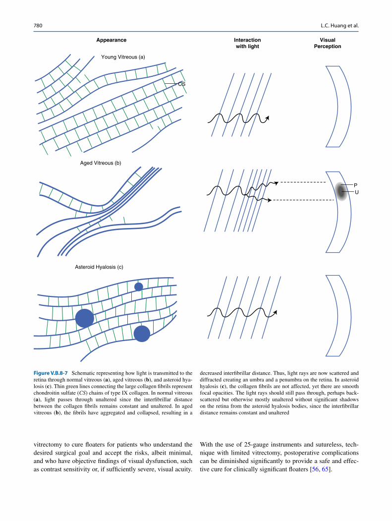
As demonstrated in Figure V.B.8-6, diminished contrast sensitivity is often a clinically significant consequence of light scattering by ocular media opacities. An opaque object with a smooth surface (e.g., an asteroid hyalosis body) will block light rays that create an umbra and a penumbra (see “a” in Figure V.B.8-6). A floater of the same size but with uneven surfaces and edges will scatter more light rays, creat-ing a smaller umbra and a larger penumbra than a smooth body (see “b” in Figure V.B.8-6). Although the objects are of the same size, the uneven surface of a floater will scatter more light, and the larger penumbra will degrade contrast sensitivity. As light interacts with collagen fibers in the vitre-ous (Figure V.B.8-7), the spacing of the collagen fibrils, or interfibrillar distance, can influence the transmission of light to the retina. When the interfibrillar distance is decreased as in aged vitreous with syneresis (b in Figure V.B.8-7), increased scattered light will create a larger penumbra and degrade contrast sensitivity.
IV. Therapeutic Considerations
To date, treatment options for patients with bothersome float-ers have been limited. Because floaters are generally viewed by doctors as benign and innocuous, patients are often told
to cope with symptoms and hope for either improvement via settling inferiorly or acceptance over time. For patients unwilling to accept this advice, eyedrops and Nd:YAG (neodymium:yttrium-aluminum-garnet (YAG) laser) are sometimes offered as therapies, though efficacy has never been demonstrated and reports are conflicting [62–64]. In one study, Nd:YAG vitreolysis eradicated floater symptoms in only 1/3 of patients with moderate clinical improvement and worsened symptoms in 7.7 % of patients [62]. As a result, the authors concluded that Nd:YAG vitreolysis provided a safe, but only mildly if at all effective, treatment for the eradication of floater symptoms. Additionally, posteriorly located vitre-ous opacities, which often cause the most disturbing symp-toms, cannot be safely treated by YAG laser [64].
A. Vitrectomy
Pars plana vitrectomy (PPV) provides the most effec-tive treatment for floaters as it offers a definitive cure with complete resolution of symptoms. However, this procedure is invasive with the possibility of complications such as endophthalmitis, retinal tears and detachments, cataract formation, glaucoma, vitreoretinal hemorrhage, and macu-lar edema [65]. Therefore, it is important to only advocate
Figure V.B.8-6 Schematic diagram of the umbra and penumbra effect of floaters. (a) Shows how the convergence of light on a smooth body results in a small umbra and a penumbra. (b) Shows how the diffraction
and scatter of light on a floater of the same size but with uneven edges results in a smaller umbra and a larger penumbra
V.B.8. Vitreous Floaters and Vision: Current Concepts and Management Paradigms
780
vitrectomy to cure floaters for patients who understand the desired surgical goal and accept the risks, albeit minimal, and who have objective findings of visual dysfunction, such as contrast sensitivity or, if sufficiently severe, visual acuity.
With the use of 25-gauge instruments and sutureless, tech-nique with limited vitrectomy, postoperative complications can be diminished significantly to provide a safe and effec-tive cure for clinically significant floaters [56, 65].
CS
Young Vitreous (a)
Appearance Interactionwith light
VisualPerception
Aged Vitreous (b)
Asteroid Hyalosis (c)
PU
Figure V.B.8-7 Schematic representing how light is transmitted to the retina through normal vitreous (a), aged vitreous (b), and asteroid hya-losis (c). Thin green lines connecting the large collagen fibrils represent chondroitin sulfate (CS) chains of type IX collagen. In normal vitreous (a), light passes through unaltered since the interfibrillar distance between the collagen fibrils remains constant and unaltered. In aged vitreous (b), the fibrils have aggregated and collapsed, resulting in a
decreased interfibrillar distance. Thus, light rays are now scattered and diffracted creating an umbra and a penumbra on the retina. In asteroid hyalosis (c), the collagen fibrils are not affected, yet there are smooth focal opacities. The light rays should still pass through, perhaps back-scattered but otherwise mostly unaltered without significant shadows on the retina from the asteroid hyalosis bodies, since the interfibrillar distance remains constant and unaltered
L.C. Huang et al.
781
1. Efficacy of Vitrectomy for FloatersVitrectomy can cure floaters by decreasing light scatter-ing and thus decrease straylight, increase contrast sensitiv-ity function (CSF), and improve the quality of life. It has been demonstrated that straylight measurements improve following vitrectomy [66]. Improvement in recognizing faces and glare hindrance occurred in 38 of 39 cases (97 %) with a statistically significant decrease in straylight glare measurements post vitrectomy, with 69 % of cases having straylight values within the normal range compared to 21 % preoperatively. The overall improvement, however, was only 18.2 % in the 39 floater cases of that study, perhaps related to the fact that 21 % of the cases had preoperative straylight measurements that were within the normal range.
Light scattering also degrades contrast sensitivity [47] underscoring that floaters negatively impact patients’ vision in more than one way. In the aforementioned prospective study of 16 cases [56], investigators at the VMR Institute in Huntington Beach, California, found that patients with float-ers had a 67 % diminution in CSF when compared to age- matched controls (p < 0.013). Following limited vitrectomy, CSF normalized at 1 week (p < 0.01) and remained normal at 1 month (p < 0.003) and at greater than 3 months (p < 0.018).
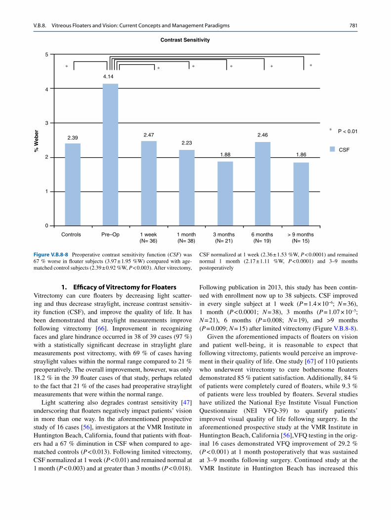
Following publication in 2013, this study has been contin-ued with enrollment now up to 38 subjects. CSF improved in every single subject at 1 week (p = 1.4 × 10⁻⁶; N = 36), 1 month (p < 0.0001; N = 38), 3 months (p = 1.07 × 10⁻⁵; N = 21), 6 months (p = 0.008; N = 19), and >9 months (p = 0.009; N = 15) after limited vitrectomy (Figure V.B.8-8).
Given the aforementioned impacts of floaters on vision and patient well-being, it is reasonable to expect that following vitrectomy, patients would perceive an improve-ment in their quality of life. One study [67] of 110 patients who underwent vitrectomy to cure bothersome floaters demonstrated 85 % patient satisfaction. Additionally, 84 % of patients were completely cured of floaters, while 9.3 % of patients were less troubled by floaters. Several studies have utilized the National Eye Institute Visual Function Questionnaire (NEI VFQ-39) to quantify patients’ improved visual quality of life following surgery. In the aforementioned prospective study at the VMR Institute in Huntington Beach, California [56],VFQ testing in the orig-inal 16 cases demonstrated VFQ improvement of 29.2 % (p < 0.001) at 1 month postoperatively that was sustained at 3–9 months following surgery. Continued study at the VMR Institute in Huntington Beach has increased this
4.14
2.472.39
Controls
5
∗ ∗ ∗ ∗ ∗ ∗
∗
4
3
2
% W
eber
1
0
Pre–Op 1 week(N= 36)
1 month(N= 38)
3 months(N= 21)
6 months(N= 19)
> 9 months(N= 15)
2.23
1.88
2.46P < 0.01
CSF
Contrast Sensitivity
1.86
Figure V.B.8-8 Preoperative contrast sensitivity function (CSF) was 67 % worse in floater subjects (3.97 ± 1.95 %W) compared with age- matched control subjects (2.39 ± 0.92 %W, p < 0.003). After vitrectomy,
CSF normalized at 1 week (2.36 ± 1.53 %W, p < 0.0001) and remained normal 1 month (2.17 ± 1.11 %W, p < 0.0001) and 3–9 months postoperatively
V.B.8. Vitreous Floaters and Vision: Current Concepts and Management Paradigms
782
series and found an average postoperative improvement in the composite index of NEI VFQ-36 of 32 % (p < 0.001). Similar improvements have been demonstrated in other studies utilizing the NEI VFQ [68–70] and other methods [71, 72] to assess patient visual quality of life before and after vitrectomy.
2. Safety of Vitrectomy for FloatersAs described above, vitrectomy offers an effective cure of floaters that improves vision and quality of life for patients who cannot cope with the symptoms of floaters, presum-ably because of profound diminution in contrast sensitivity. As an invasive procedure, however, vitrectomy has known potential complications [73–76]. Fortunately, a study by Delaney et al. [62] demonstrated a lower postoperative complication rate associated with vitrectomy for floaters as opposed to other vitreoretinal diseases. In a study of 31 patients (42 eyes) who underwent either Nd:YAG vitreoly-sis or pars plana vitrectomy for floaters, the authors com-pared the efficacy of the two treatments to eradicate floater symptomatology. Of these, 15 eyes underwent vitrectomy for floaters: 4 eyes underwent vitrectomy as a primary treatment for floaters, while 11 patients had vitrectomy subsequent to Nd:YAG laser treatment for floaters. In this admittedly small and limited study, 93.3 % of patients demonstrated complete resolution of floaters following vitrectomy with an average follow-up post vitrectomy of 31.5 months (range 6–108 months). Reported complica-tions included postoperative cataract development that underwent successful phacoemulsification surgery and postoperative retinal detachment occurring 7 weeks after a combined vitrectomy with phacoemulsification. Another study found a relatively low rate of immediate post-floater vitrectomy retinal detachment with a slightly increased risk during long-term follow-up [77]. Out of 73 eyes that underwent vitrectomy for severe and persistent floaters ≥6 months’ duration, a total of five (6.8 %) developed retinal detachments, four occurring 24–44 months after vitrectomy.
The current approach to curing floaters with vitrectomy must take into consideration the following specific risks and incorporate measures to mitigate these risks.
a. EndophthalmitisEndophthalmitis is a rare but severe complication of vit-rectomy [78]. However, instrument size (microincisional 23- or 25-gauge vs. conventional 20-gauge), beveled entry technique, and cannula removal with a blunt probe have all been demonstrated to lower the risk of this postsurgi-cal complication. Recent literature has reported that there is inconclusive evidence that 25-gauge PPV has a higher rate of endophthalmitis than 20-gauge PPV [79–82]. The one exception is the use of a straight approach to cannula
insertion, which has an increased risk for endophthalmitis as compared to a beveled approach [83, 84]. This prob-ably relates to a reduction of vitreous incarceration, which will not only mitigate endophthalmitis [85, 86] but also peripheral retinal tears [87–89] and fibrovascular prolif-eration in diabetic patients [90]. Regarding endophthalmi-tis, incisional vitreous incarceration is believed to improve postoperative sclerotomy closure, preventing the entry of bacteria into vitreous via an incisional vitreous wick [85]. Cannula removal at the end of surgery also influences the risk of postoperative endophthalmitis. In a prospective study of 118 cadaveric pig eyes that underwent vitrectomy with 23-gauge transconjunctival sclerotomies, two tech-niques of cannula removal were employed: the superior cannula was extracted with the illumination probe inserted through it and the other cannula was removed with a can-nula plug inserted [86]. Postoperative incisional vitreous entrapment was subsequently evaluated and demonstrated vitreous entrapment in 95.8 % of entry sites whose can-nulas were extracted with the plug inserted, whereas incarceration only occurred in 93.2 % of incisions whose cannulas were extracted with the light probe inside. While this was not a big difference, the authors report that this difference may be due to the peripheral vitreous being dis-placed to the inner face of sclerotomies when a plug is inserted, thus leading to a greater degree of vitreous incar-ceration. The use of non-hollow probes for cannula extrac-tion may have contributed to the safety profile observed in the ongoing study at the VMR Institute in Huntington Beach, California [56], that employs 25-gauge vitrectomy with beveled incisions and illumination probe insertion for cannula removal at the end of the case. In 98 eyes with severe floaters, there have been no cases of endophthalmi-tis. Additionally, a reduction in vitreous incarceration may also decrease post-vitrectomy retinal detachment rate [89].
b. Retinal Tears and Retinal Detachment
Retinal tears and detachments occurring intraoperatively or postoperatively are well-documented potential complica-tions [91, 92]. However, smaller instrument size (microinci-sional 23- or 25-gauge vs. conventional 20-gauge), avoiding surgical induction of PVD, and performing cannula removal as described above can decrease the incidence of retinal tears and detachment.
The use of 25-gauge vitrectomy instruments allows for an incision with an oblique scleral tunnel that can be self- sealing without any sutures, decreasing operating time and hastening postoperative recovery [93–95]. Furthermore, the use of self- retaining cannulas preserves the edge of the incisions during instrument insertion and removal and allows more effective self-sealing during sutureless surgery [96, 97]. Iatrogenic intraoperative peripheral retinal break
L.C. Huang et al.
783
incidence during 23-gauge vitrectomy was compared to con-ventional 20-gauge vitrectomy, and retinal breaks occurred significantly less often during 23-gauge (16 of 973 cases, 1.6 %) compared to conventional vitrectomy (25 of 402 cases, 6.2 %) [98]. This is consistent with another study that found a 4.9 % retinal detachment rate 14 months after vitreo-macular surgery with 20-gauge instruments versus a 1.1 % retinal detachment rate with 23-gauge instruments [99].
Additionally, the use of 360° indirect ophthalmoscopy with scleral depression to identify retinal breaks or tears for prophylactic treatment prior to or during surgery has also been shown to decrease the incidence of postoperative retinal detachments [100]. In a study of 415 eyes of 381 patients that underwent primary, standard, three-port vitrec-tomy, 65 retinal breaks were found in 48/415 eyes (11.6 %) that were treated perioperatively. Of 366 eyes in which no breaks were identified during vitrectomy, postoperative reti-nal detachment occurred in 8 (2.2 %) eyes; all occurring at least 3 months following vitrectomy. This demonstrates the importance of avoiding, detecting, and treating breaks dur-ing surgery to decrease the incidence of postoperative RD. In a study performed by Sebag et al. [56], 24/98 (24.5 %) cases received localized prophylactic laser or cryopexy to peripheral retinal breaks a minimum of 3 months prior to vitrectomy for floaters. This further contributed to the safety profile of the procedure, with no cases of retinal detachment occurring at a mean follow-up of 16.2 months (range = 3–51 months).
Lastly, avoiding the induction of PVD during vitrec-tomy will also help decrease the incidence of intraopera-tive or postoperative retinal breaks. A study of 311 patients undergoing vitrectomy for treatment of premacular mem-brane with macular pucker or macular hole demonstrates a decreased incidence of retinal tears when surgical PVD was avoided in both groups [101]. For patients undergoing vit-rectomy for macular pucker, 32.1 % had retinal breaks with induction of PVD compared to 2.1 % that had retinal breaks without induction of PVD (p = 0.006). For patients undergo-ing vitrectomy for macular hole, 12.7 % had retinal breaks with induction of PVD while only 3.1 % had breaks without induction of PVD (p = 0.008). Additionally, in a previous study that induced PVD during vitrectomy for floaters, reti-nal breaks occurred in 9/30 (30.5 %) cases [102], compared to 0/60 (0 %) that developed retinal breaks or detachments without induction of PVD (p < 0.007) [56]. Since that publi-cation, a total of 98 cases have been performed at the VMR Institute in Huntington Beach without PVD induction. There have been no cases of retinal breaks or detachment.
c. CataractsIncreased risk of cataract formation due to increased intra-vitreal levels of oxygen following vitrectomy [103–108] may be mitigated by leaving the anterior vitreous and
posterior vitreous intact to protect the lens against oxy-gen free radicals. Previous studies [103, 106] have dem-onstrated that vitreous maintains an intraocular oxygen concentration gradient from the retina to the vitreous gel [see chapter IV.B. Oxygen in vitreo-retinal physiology and pathology]. Following vitrectomy, the oxygen gradient no longer exists due to the removal of the vitreous. One study demonstrated that the difference in oxygen tension pre- and post vitrectomy is statistically significant with oxygen tension levels in the mid-vitreous and near the lens increasing greatly, remaining elevated, and becoming more uniform in distribution in patients who had previous removal of vitreous gel [103]. This led to the conclusion that vitrectomy significantly increases intraocular oxygen tension after surgery causing exposure of the crystalline lens to reactive oxygen species that contribute to cataract formation. Similar reports of vitrectomy in animal models have further confirmed that oxygen measurements taken in the nucleus of the lens post vitrectomy had increased oxy-gen levels due to diffusion from elevated oxygen tension in the vitreous [106].
Beebe et al. [104] further elucidated the concept of oxida-tive damage and its role in cataract formation. They dem-onstrated that patients with ischemic diabetic retinopathy had lower oxygen levels in the vitreous and thus had par-tial protection from nuclear cataract development within the first year after vitrectomy. Other studies have also suggested that the rate of cataract extraction following vitrectomy in diabetic patients is lower than in nondiabetic patients under-going vitrectomy [108]. Additionally, PVD has been impli-cated in possible acceleration of nuclear cataract formation due to increased oxygen tension levels following PVD. Pharmacologic vitreolysis induction of posterior vitreous detachment with ocriplasmin has also been demonstrated to increase vitreous oxygen concentration in comparison to control eyes and hyaluronidase-injected (without PVD) eyes [109]. In another study, intravitreal ocriplasmin injection that produced liquefaction and PVD was associated with a 68 % increase in lens nuclear oxygen tension compared to intravit-real hyaluronidase that only produced liquefaction and not PVD [110].
Thus, based on the strong association between increased oxygen levels and nuclear cataract formation [103, 111, 112], age-related PVD may be related to an acceleration of nuclear cataract development. Due to the increased oxygen tension levels that occur with induction of PVD and removal of the vitreous in vitrectomy, this suggests that leaving the anterior and posterior vitreous intact during surgery may result in decreased cataract formation postoperatively. In the VMR Institute study of 98 eyes with limited vitrectomy for clinically significant floaters, only 14/60 phakic eyes (23 %) needed cataract surgery, all in patients with a mean age 64.1 years and average time between vitrectomy and cataract
V.B.8. Vitreous Floaters and Vision: Current Concepts and Management Paradigms
784
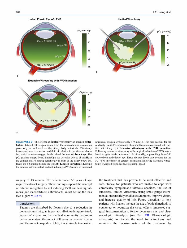
surgery of 13 months. No patients under 53 years of age required cataract surgery. These findings support the concept of cataract mitigation by not inducing PVD and leaving vit-reous (and its constituent antioxidants) intact behind the lens (see Figure V.B.8-9).
Conclusions
Patients are disturbed by floaters due to a reduction in contrast sensitivity, an important, albeit underappreciated, aspect of vision. As the medical community begins to better understand the impact of floaters on patients’ vision and the impact on quality of life, it is advisable to consider
the treatment that has proven to be most effective and safe. Today, for patients who are unable to cope with chronically symptomatic vitreous opacities, the use of sutureless, limited vitrectomy using small-gauge instru-mentation can safely eradicate symptoms, improve vision, and increase quality of life. Future directions to help patients with floaters include the use of optical methods to counteract or neutralize the visual effects, improved sur-gical instrumentation to further decrease risks, and phar-macologic vitreolysis (see Part VII. Pharmacologic vitreolysis) to obviate the need for vitrectomy and minimize the invasive nature of the treatment by
pO2 (mm Hg)
25
20
15
10
5
Intact Phakic Eye w/o PVD
pO2 = 4–6 mm Hg
pO2 = 10
pO2 = 18
pO2 = 22 mm Hg
a
pO2 (mm Hg)
25
20
15
10
5
Limited Vitrectomy
pO2= 6–9 mm Hg
pO2= 22 mm Hg
b
25
20
15
10
5
Extensive Vitrectomy with PVD Induction
c
pO2 = 22 mm Hg
pO2 = 12–14 mm Hg
pO2 (mm Hg)
Figure V.B.8-9 The effects of limited vitrectomy on oxygen distri-bution. Intravitreal oxygen arises from the retina/choroid circulation posteriorly as well as from the ciliary body anteriorly. Vitrectomy increases convective motion and fluid circulation in the vitreous cham-ber, which increases oxygen levels behind the lens. (a) Intact eye. The pO2 gradient ranges from 22 mmHg at the posterior pole to 18 mmHg at the equator and 10 mmHg peripherally in front of the ciliary body. pO2 levels are 4–6 mmHg behind the lens. (b) Limited vitrectomy. Leaving the anterior vitreous intact and not inducing a PVD results in increased
retrolental oxygen levels of only 6–9 mmHg. This may account for the relatively low (23 %) incidence of cataract formation observed with lim-ited vitrectomy. (c) Extensive vitrectomy with PVD induction. Following extensive vitrectomy with surgical induction of PVD, retro-lental oxygen levels increase to 12–14 mmHg, approaching three-fold above those in the intact eye. These elevated levels may account for the 50–76 % incidence of cataract formation following extensive vitrec-tomy. (Adapted from Beebe, Holekamp, et al.)
L.C. Huang et al.
785
pharmacologically breaking down vitreous macromole-cules, especially the aggregates of collagen that underlie the formation of floaters [46, 113–123].
It is important that the medical profession heed the cry of reasonable patients afflicted with clinically sig-nificant floaters, defined by quantitative ultrasound and contrast sensitivity testing, to achieve the goal of modern medicine [124]:
To help people die young, as late in life as possibleErnst L. Wynder MDFounding President of the American Health Foundation
References
1. Duke-Elder WS. The nature of the vitreous body. Br J Ophthalmol. 1930;14(Suppl):6.
2. Demours F. Observations anatomiques sur la structure cellulaire du corps vitre. Memoires de Paris. 1741.
3. Virchow H. Die morphologische Natur des Glask-rpergeebes. Ophthalmol Gesellsch Heidelberg. 1885; Klin Monatsbl Augenheilkd.
4. Zinn. Descriptio anatomica oculi humani. Goettingen. 1780. 5. Von Pappenheim A: Die spezielle gewebelehie des auges. 1842:
179–184; as cited by Redslob E: Le Corps Vitre. Paris: Masson et Cie, 1932:160–217
6. Brucke E. Ueber den innern Bau des Glaskörpers. Arch Anal Physiol Wissensch Med (Mueller) 1843.
7. Hannover A. Endeckung des baues des glaskorpers. Müller Archiv 1845;467–77.
8. Bowman W. Observations on the structure of the vitreous humor. Dublin Quart J Med Sci. 1848;6:102–18.
9. Szent-Györgyi A. Untersuchungen über die Struktur des Glaskörpers des Menschen. Arch f Mikroscop Anat. 1917;89.
10. Duke-Elder WS. The physico-chemical properties of the vitreous body. J Physiol (Lond). 1929;68(2):155–65.
11. Sebag J, Yee K. Vitreous – from biochemistry to clinical relevance. In: Tasman W, Jaeger EA, editors. Duane’s foundation of clini-cal ophthalmology, vol. 1. Philadelphia: Lippincott Williams & Wilkins; 1998. p. 1–34.
12. Bishop PN. Structural macromolecules and supramolecu-lar organisation of the vitreous gel. Prog Retin Eye Res. 2000; 19(3):323–44.
13. Snowden JM, Eyre DR, Swann DA. Vitreous structure. VI. Age- related changes in the thermal stability and crosslinks of vitreous, articular cartilage and tendon collagens. Biochim Biophys Acta. 1982;706(2):153–7.
14. Los LI, Van der Worp RJ, Van Luyn MJ, Hooymans JM. Age- related liquefaction of the human vitreous body: LM and TEM evaluation of the role of proteoglycans and collagen. Invest Ophthalmol Vis Sci. 2003;44(7):2828–33.
15. Van Deemter M, Kuijer R, Pas HH, van der Worp RJ, Hooymans JM, Los LI. Trypsin-mediated enzymatic degradation of type II col-lagen in the human vitreous. Mol Vis. 2013;19:1591–9.
16. Bishop PN, Holmes DF, Kadler KE, Mcleod D, Bos KJ. Age- related changes on the surface of vitreous collagen fibrils. Invest Ophthalmol Vis Sci. 2004;45(4):1041–6.
17. Foos RY, Wheeler NC. Vitreoretinal juncture. Synchysis seni-lis and posterior vitreous detachment. Ophthalmology. 1982; 89(12):1502–12.
18. Sebag J. Age-related changes in human vitreous structure. Graefes Arch Clin Exp Ophthalmol. 1987;225(2):89–93.
19. Hong PH, Han DP, Burke JM, Wirostko WJ. Vitrectomy for large vitreous opacity in retinitis pigmentosa. Am J Ophthalmol. 2001;131(1):133–4.
20. Sebag J. The vitreous—structure, function and pathobiology. New York: Springer; 1989.
21. Murakami K, Jalkh AE, Avila MP, Trempe CL, Schepens CL. Vitreous floaters. Ophthalmology. 1983;90(11):1271–6.
22. Nguyen N, Sebag J. Myopic vitreopathy – significance in anoma-lous PVD and vitreo-retinal disorders. In: Midena E, editor. Myopia and related diseases. New York: Ophthalmic Communications Society; 2005. p. 137–45.
23. Balazs EA, Denlinger JL. Aging changes in the vitreous. In: Sekuler R, Kline D, Dismukes K, editors. Aging and human visual function, vol. 2. New York: Alan R. Liss; 1982. p. 45–57.
24. Webb BF, Webb JR, Schroeder MC, North CS. Prevalence of vit-reous floaters in a community sample of smartphone users. Int J Ophthalmol. 2013;6(3):402–5.
25. Wagle AM, Lim W-Y, Yap T-P, Neelam K, Au Eong K-G. Utility values associated with vitreous floaters. Am J Ophthalmol. 2011; 152(1):60–5.
26. Sebag J. Floaters and the quality of life [editorial]. Am J Ophthalmol. 2011;152(1):3–4.
27. Zou H, Liu H, Xu X, Zhang X. The impact of persistent visually disabling vitreous floaters on health status utility values. Qual Life Res. 2013;22:1507–14.
28. Sebag J. Seeing the invisible: the challenge of imaging vitreous. J Biomed Opt. 2004;9(1):38–46.
29. Coleman DJ, Daly SW, Atencio A, Lloyd HO, Silverman RH. Ultrasonic evaluation of the vitreous and retina. Semin Ophthalmol. 1998;13(4):210–8.
30. Mcleod D, Restori M, Wright JE. Rapid B-scanning of the vitreous. Br J Ophthalmol. 1977;61(7):437–45.
31. Mojana F, Kozak I, Oster SF, Cheng L, Bartsch DU, Brar M, et al. Observations by spectral-domain optical coherence tomography combined with simultaneous scanning laser oph-thalmoscopy: imaging of the vitreous. Am J Ophthalmol. 2010; 149(4):641–50.
32. Oksala A. Ultrasonic findings in the vitreous body at vari-ous ages. Albrecht Von Graefes Arch Klin Exp Ophthalmol. 1978;207:275–80.
Abbreviations
CSF Contrast sensitivity functionDLS Dynamic light scatteringFrACT Freiburg acuity contrast testHA HyaluronanLeq Equivalent veiling luminancemmHg Millimeters of mercuryNd:YAG Neodymium:yttrium-aluminum-garnetNEI VFQ-39 National Eye Institute visual function
questionnairepO2 Partial pressure of oxygenPPV Pars plana vitrectomyPVD Posterior vitreous detachmentRD Retinal detachmentSD-OCT Spectral-domain optical coherence
tomographySLO Scanning laser ophthalmoscopy
V.B.8. Vitreous Floaters and Vision: Current Concepts and Management Paradigms
786
33. Walton KA, Meyer CH, Harkrider CJ, Cox TA, Toth CA. Age- related changes in vitreous mobility as measured by video B scan ultrasound. Exp Eye Res. 2002;74(2):173–80.
34. Silverman RH, Ketterling JA, Mamou J, Lloyd HO, Filoux E, Coleman DJ. Pulse-encoded ultrasound imaging of the vitre-ous with an annular array. Ophthalmic Surg Lasers Imaging. 2012;43(1):82–6.
35. Wa CA, Yee KM, Mamou J, Silverman R, Gale J, Ganti AK, et al. Quantitative ultrasonography of vitreous correlates with con-trast sensitivity in patients with floaters. Paper presented at: the Association for Research in Vision and Ophthalmology (ARVO), Orlando, 4–8 May 2014.
36. Mirza RG, Johnson MW, Jampol LM. Optical coherence tomogra-phy use in evaluation of the vitreoretinal interface: a review. Surv Ophthalmol. 2007;52(4):397–421.
37. Coleman DJ, Silverman RH, Chabi A, Rondeau MJ, Shung KK, Cannata J, et al. High-resolution ultrasonic imaging of the posterior segment. Ophthalmology. 2004;111(7):1344–51.
38. Sebag J. Vitreous anatomy, aging, and anomalous posterior vitreous detachment. In: Dartt DA, editor. Encyclopedia of the eye. Oxford: Academic; 2010. p. 307–15.
39. Ansari RR, Suh KI, Dunker S, Kitaya N, Sebag J. Quantitative molecular characterization of bovine vitreous and lens with non- invasive dynamic light scattering. Exp Eye Res. 2001;73(6):859–66.
40. Rovati L, Fankhauser F, Docchio F, Van Best J. Diabetic retinopa-thy assessed by dynamic light scattering and corneal autofluores-cence. J Biomed Opt. 1998;3(3):357–63.
41. Fankhauser F. Dynamic light scattering in ophthalmology: results of in vitro and in vivo experiments. Technol Health Care. 2006;14(6):521–35.
42. Fankhauser F. Analysis of diabetic vitreopathy with dynamic light scattering spectroscopy–problems and solutions related to photon correlation. Acta Ophthalmol. 2012;90(3):173–8.
43. Sebag J. To see the invisible: the quest of imaging vitreous. Dev Ophthalmol. 2008;42:5–28.
44. Datiles MB, Ansari RR, Suh KI, Vitale S, Reed GF, Zigler JS, et al. Clinical detection of precataractous lens protein changes using dynamic light scattering. Arch Ophthalmol. 2008;2008(126):12.
45. Fankhauser F. Postoperative follow up by dynamic light scattering: detection of inflammatory molecular changes in the cornea, the vit-reous and the lens. Technol Health Care. 2007;15(4):247–58.
46. Sebag J. Molecular biology of pharmacologic vitreolysis. Trans Am Ophthalmol Soc. 2005;103:473–94.
47. Van den Berg T, Franssen L, Coppens JE. Ocular media clarity and straylight. In: Dartt D, Besharse J, Dana R, editors. Encyclopedia of the eye. Oxford: Academic; 2010. p. 173–83.
48. Van den Berg TJ, Franssen L, Kruijt B, Coppens JE. History of ocular straylight measurement: a review. Z Med Phys. 2012;23(1):6–20.
49. Rozema JJ, Van den Berg T, Tassignon MJ. Retinal straylight as a function of age and ocular biometry in healthy eyes. Invest Ophthalmol Vis Sci. 2010;51(5):2795–9.
50. IJspeert JK, de Waard PW, van den Berg TJ, de Jong PT. The intra-ocular straylight function in 129 healthy volunteers; dependence on angle, age and pigmentation. Vision Res. 1990;30:699–707.
51. Guber I, Bachmann LM, Guber J, Bochmann F, Lange AP, Thiel MA. Reproducibility of straylight measurement by C-Quant for assessment of retinal straylight using the compensation comparison method. Graefes Arch Clin Exp Ophthalmol. 2011;249(9):1367–71.
52. Tan JC, Spalton DJ, Arden GB. The effect of neodymium:YAG capsulotomy on contrast sensitivity and the evaluation of methods for it assessment. Ophthalmology. 1999;4:703–9.
53. Adamsons I, Rubin GS, Vitale S, Taylor HR, Stark WJ. The effect of early cataracts on glare and contrast sensitivity. A pilot study. Arch Ophthalmol. 1992;110(8):1081–6.
54. Mäntyjärvi M, Tuppurainen K. Cataract in traffic. Graefes Arch Clin Exp Ophthalmol. 1999;237(4):278–82.
55. Rubin GS, Roche KB, Prasada-rao P, Fried LP. Visual impairment and disability in older adults. Optom Vis Sci. 1994;71(12):750–60.
56. Sebag J, Yee KMP, Wa CA, Huang LC, Sadun AA. Vitrectomy for floaters – prospective efficacy analysis and retrospective safety pro-file. Retina. 2014;34:1062–8. PMID: 24296397.
57. Bach M. The Freiburg Visual Acuity Test – variability unchanged by post-hoc re-analysis. Graefes Arch Clin Exp Ophthalmol. 2007;245(7):965–71.
58. Nielsen E, Hjortdal J. Visual acuity and contrast sensitivity after posterior lamellar keratoplasty. Acta Ophthalmol. 2012;90(8): 756–60.
59. Dennis RJ, Beer JM, Baldwin JB, Ivan DJ, Lorusso FJ, Thompson WT. Using the Freiburg Acuity and Contrast Test to measure visual performance in USAF personnel after PRK. Optom Vis Sci. 2004;81(7):516–24.
60. Woods RL, Tregear SJ, Mitchell RA. Screening for ophthalmic dis-ease in older subjects using visual acuity and contrast sensitivity. Ophthalmology. 1998;12:2318–26.
61. Neargarder SA, Stone ER, Cronin-golomb A, Oross S. The impact of acuity on performance of four clinical measures of contrast sen-sitivity in Alzheimer’s disease. J Gerontol B Psychol Sci Soc Sci. 2003;58(1):54–62.
62. Delaney YM, Oyinloye A, Benjamin L. Nd:YAG vitreolysis and pars plana vitrectomy: surgical treatment for vitreous floaters. Eye (Lond). 2002;16(1):21–6.
63. Tsai WF, Chen YC, Su CY. Treatment of vitreous floaters with neo-dymium YAG laser. Br J Ophthalmol. 1992;77(8):485–8.
64. Toczolowski J, Katski W. Use of Nd:YAG laser in treatment of vit-reous floaters. Klin Oczna. 1998;100(3):155–7.
65. Wilkinson CP. Safety of vitrectomy for floaters – how safe is safe? [editorial]. Am J Ophthalmol. 2011;151(6):919–20.
66. Mura M, Engelbrecht LA, de Smet MD, Papadaki TG, van den Berg TJ, Tan HS. Surgery for floaters. Ophthalmology. 2011;118:1894.
67. De Nie KF, Crama N, Tilanus MA, Klevering BJ, Boon CJ. Pars plana vitrectomy for disturbing primary vitreous floaters: clini-cal outcome and patient satisfaction. Graefes Arch Clin Exp Ophthalmol. 2013;251(5):1373–82.
68. Martínez-Sanz F, Velarde JI, Casuso P, Fernández-Cotero JN. Surgical solution to vitreous floaters visual problem. Arch Soc Esp Oftalmol. 2009;84(5):259–62.
69. Schiff WM, Chang S, Mandava N, Barile GR. Pars plana vitrec-tomy for persistent, visually significant vitreous opacities. Retina. 2000;20(6):591–6, forthcoming.
70. Okamoto F, Okamoto Y, Fukuda S, Hiraoka T, Oshika T. Vision- related quality of life and visual function after vitrectomy for various vitreoretinal disorders. Invest Ophthalmol Vis Sci. 2010;51(2):744–51.
71. Stoffelns BM, Vetter J, Keicher A, Mirshahi A. Pars plana vitrec-tomy for visually disturbing vitreous floaters in pseudophacic eyes. Klin Monbl Augenheilkd. 2011;228(4):293–7.
72. Hoerauf H, Müller M, Laqua H. Vitreous body floaters and vitrec-tomy with full visual acuity. Ophthalmologe. 2003;100(8):639–43.
73. Bansal AS, Hsu J, Garg SJ, Sivalingam A, Vander JF, Moster M, et al. Optic neuropathy after vitrectomy for retinal detachment: clinical features and analysis of risk factors. Ophthalmology. 2012;119(11):2364–70.
74. Gonzalez MA, Flynn HW, Smiddy WE, Albini TA, Berrocal AM, Tenzel P. Giant retinal tears after prior pars plana vitrec-tomy: management strategies and outcomes. Clin Ophthalmol. 2013;7:1687–91.
75. Hasegawa Y, Okamoto F, Sugiura Y, Okamoto Y, Hiraoka T, Oshika T. Intraocular pressure elevation after vitrectomy for various vitreo-retinal disorders. Eur J Ophthalmol. 2014;24:235–41.
76. Shousha MA, Yoo SH. Cataract surgery after pars plana vitrectomy. Curr Opin Ophthalmol. 2010;21(1):45–9.
L.C. Huang et al.
787
77. Schulz-Key S, Carlsson JO, Crafoord S. Longterm follow-up of pars plana vitrectomy for vitreous floaters: complications, outcomes and patient satisfaction. Acta Ophthalmol. 2011;89(2):159–65.
78. Chiang A, Kaiser RS, Avery RL, Dugel PU, Eliott D, Shah SP, et al. Endophthalmitis in microincision vitrectomy: outcomes of gas-filled eyes. Retina. 2011;31(8):1513–7.
79. Bahrani HM, Fazelat AA, Thomas M, et al. Endophthalmitis in the era of small gauge transconjunctival sutureless vitrectomy–meta analysis and review of literature. Semin Ophthalmol. 2010; 25(5–6):275–82.
80. Scott IU, Flynn HW, Acar N, et al. Incidence of endophthalmitis after 20-gauge vs 23-gauge vs 25-gauge pars plana vitrectomy. Graefes Arch Clin Exp Ophthalmol. 2011;249(3):377–80.
81. Hu AY, Bourges JL, Shah SP, et al. Endophthalmitis after pars plana vitrectomy a 20- and 25-gauge comparison. Ophthalmology. 2009;116(7):1360–5.
82. Wu L, Berrocal MH, Arévalo JF, et al. Endophthalmitis after pars plana vitrectomy: results of the Pan American Collaborative Retina Study Group. Retina. 2011;31(4):673–8.
83. Govetto A, Virgili G, Menchini F, Lanzetta P, Menchini U. A sys-tematic review of endophthalmitis after microincisional versus 20-gauge vitrectomy. Ophthalmology. 2013;120:2286–91.
84. Kaiser RS, Prenner J, Scott IU, et al. The Microsurgical Safety Task Force: evolving guidelines for minimizing the risk of endophthal-mitis associated with microincisional vitrectomy surgery. Retina. 2010;30(4):692–9.
85. Chen SD, Mohammed Q, Bowling B, Patel CK. Vitreous wick syn-drome—a potential cause of endophthalmitis after intravitreal injec-tion of triamcinolone through the pars plana. Am J Ophthalmol. 2004;137:1159–60.
86. Benitez-Herreros J, Lopez-Guajardo L, Camara-Gonzalez C, Silva-Mato A. Influence of the interposition of a non-hollow probe during cannula extraction on sclerotomy vitreous incarcera-tion in sutureless vitrectomy. Invest Ophthalmol Vis Sci. 2012; 53(11):7322–6.
87. Buettner H, Machemer R. Histopathologic findings in human eyes after pars plana vitrectomy and lensectomy. Arch Ophthalmol. 1977;95(11):2029–33.
88. Gosse E, Newsom R, Lochhead J. The incidence and distribution of iatrogenic retinal tears in 20-gauge and 23-gauge vitrectomy. Eye (Lond). 2012;26(1):140–3.
89. Tan HS, Mura M, De Smet MD. Iatrogenic retinal breaks in 25-gauge macular surgery. Am J Ophthalmol. 2009;148(3): 427–30.
90. Hotta K, Hirakata A, Ohi Y, et al. Ultrasound biomicroscopy for examination of the sclerotomy site in eyes with proliferative dia-betic retinopathy after vitrectomy. Retina. 2000;20(1):52–8.
91. Rahman R, Murray CD, Stephenson J. Risk factors for iatro-genic retinal breaks induced by separation of posterior hya-loid face during 23-gauge pars plana vitrectomy. Eye (Lond). 2013;27(5):652–6.
92. Al-harthi E, Abboud EB, Al-dhibi H, Dhindsa H. Incidence of scle-rotomy-related retinal breaks. Retina. 2005;25(3):281–4.
93. López-guajardo L, Pareja-esteban J, Teus-guezala MA. Oblique sclerotomy technique for prevention of incompetent wound clo-sure in transconjunctival 25-gauge vitrectomy. Am J Ophthalmol. 2006;141(6):1154–6.
94. Shimada H, Nakashizuka H, Mori R, Mizutani Y, Hattori T. 25-gauge scleral tunnel transconjunctival vitrectomy. Am J Ophthalmol. 2006;142(5):871–3.
95. Keshavamurthy R, Venkatesh P, Garg S. Ultrasound biomicros-copy findings of 25 G Transconjuctival Sutureless (TSV) and con-ventional (20G) pars plana sclerotomy in the same patient. BMC Ophthalmol. 2006;6:7.
96. Eckardt C. Transconjunctival sutureless 23-gauge vitrectomy. Retina. 2005;25(2):208–11.
97. Schweitzer C, Delyfer MN, Colin J, Korobelnik JF. 23-Gauge transconjunctival sutureless pars plana vitrectomy: results of a prospective study. Eye (Lond). 2009;23(12):2206–14.
98. Cha DM, Woo SJ, Park KH, Chung H. Intraoperative iatrogenic peripheral retinal break in 23-gauge transconjunctival sutureless vitrectomy versus 20-gauge conventional vitrectomy. Graefes Arch Clin Exp Ophthalmol. 2013;251(6):1469–74.
99. Le Rouic JF, Becquet F, Ducournau D. Does 23-gauge sutureless vitrectomy modify the risk of postoperative retinal detachment after macular surgery? A comparison with 20-gauge vitrectomy. Retina. 2011;31(5):902–8.
100. Moore JK, Kitchens JW, Smiddy WE, Mavrofrides EC, Gregorio G. Retinal breaks observed during pars plana vitrectomy. Am J Ophthalmol. 2007;144(1):32–6.
101. Chung SE, Kim KH, Kang SW. Retinal breaks associated with the induction of posterior vitreous detachment. Am J Ophthalmol. 2009;147(6):1012–6.
102. Tan HS, Mura M, Lesnik Oberstein SY, Bijl HM. Safety of vitrec-tomy for floaters. Am J Ophthalmol. 2011;151(6):995–8.
103. Holekamp NM, Shui YB, Beebe DC. Vitrectomy surgery increases oxygen exposure to the lens: a possible mecha-nism for nuclear cataract formation. Am J Ophthalmol. 2005;139(2):302–10.
104. Beebe DC, Holekamp NM, Siegfried C, Shui YB. Vitreoretinal influences on lens function and cataract. Philos Trans R Soc B Biol Sci. 2011;366(1568):1293–300.
105. Pardo-Muñoz A, Muriel-Herrero A, Abraira V, Muriel A, Munoz- Negrete FJ, Murube J, et al. Phacoemulsification in previously vit-rectomized patients: an analysis of the surgical results in 100 eyes as well as the factors contributing to the cataract formation. Eur J Ophthalmol. 2006;16(1):52–9.
106. Barbazetto IA, Liang J, Chang S, Zheng L, Spector A, Dillon JP. Oxygen tension in the rabbit lens and vitreous before and after vitrectomy. Exp Eye Res. 2004;78:917–24.
107. Van Effenterre G, Ameline B, Campinchi F, et al. Is vitrectomy cataractogenic? Study of changes of the crystalline lens after sur-gery of retinal detachment. J Fr Ophtalmol. 1992;15(8–9):449–54.
108. Smiddy WE, Feuer W. Incidence of cataract extraction after dia-betic vitrectomy. Retina. 2004;24(4):574–81.
109. Quiram PA, Leverenz VR, Baker RM, Dang L, Giblin FJ, Trese MT. Microplasmin-induced posterior vitreous detachment affects vitreous oxygen levels. Retina. 2007;27(8):1090–6.
110. Giblin FJ, Quiram PA, Leverenz VR, Baker RM, et al. Enzyme- induced posterior vitreous detachment in the rat produces increased lens nuclear pO2 levels. Exp Eye Res. 2009;88(2):286–92.
111. Palmquist BM, Philipson B, Barr PO. Nuclear cataract and myopia during hyperbaric oxygen therapy. Br J Ophthalmol. 1984;68(2):113–7.
112. Simpanya MF, Ansari RR, Suh KI, Leverenz VR, Giblin FJ. Aggregation of lens crystallins in an in vivo hyperbaric oxy-gen guinea pig model of nuclear cataract: dynamic light-scat-tering and HPLC analysis. Invest Ophthalmol Vis Sci. 2005; 46(12):4641–51.
113. Sebag J. Pharmacologic vitreolysis. Retina. 1998;18(1):1–3. 114. Sebag J, Kitaya N, Ansari RR, Karagoezian H, Yoshida A.
Preliminary findings using dynamic light scattering to study vitreous effects of Pharmacologic Vitreolysis. Proc SPIE. 2000;3908:69–77.
115. Sebag J. Is pharmacologic vitreolysis brewing? Retina. 2002;22(1):1–3.
116. Sebag J. Vitreous pathobiology and pharmacologic vitreolysis. In: Binder S, editor. The macula – diagnosis, treatment, and future trends. Wein/New York: Springer; 2004. p. 171–80.
117. Sebag J, Ansari RR, Suh KI. Pharmacologic vitreolysis with microplasmin increases vitreous diffusion coefficients. Graefes Arch Clin Exp Ophthalmol. 2007;245(4):576–80.
V.B.8. Vitreous Floaters and Vision: Current Concepts and Management Paradigms

788
118. Sebag J. Pharmacologic vitreolysis–premise and promise of the first decade. Retina. 2009;29(7):871–4.
119. Sebag J. The emerging role of pharmacologic vitreolysis. Retinal Physician. 2010;7(2):52–6.
120. Costa Ede P, Rodrigues EB, Farah ME, Sebag J, Meyer CH. Novel vitreous modulators for pharmacologic vitreolysis in the treatment of diabetic retinopathy. Curr Pharm Biotechnol. 2011;12(3):410–22.
121. Tozer K, Fink W, Sadun AA, Sebag J. Prospective three- dimensional analysis of structure and function in macular hole treated by pharmacologic vitreolysis. Retinal Cases & Brief Reports. 2013;7:57–61.
122. Sebag J, Tozer K. Vitreous biochemistry and pharmacologic vitreolysis. In: Hartnett ME, editor. Pediatric retina. 2nd ed. Philadelphia: Lippincott Williams & Wilkins; 2013.
123. Stalmans P, Duker JS, Kaiser PK, Heier JS, Dugel PU, Gandorfer A, et al. Oct-based interpretation of the vitreomac-ular interface and indications for pharmacologic vitreolysis. Retina. 2013;33:2003–11.
124. Sebag J. The diagnosis of health. First prize, national essay com-petition. Prev Med. 1979;8:76–8.
125. Sebag J, Balazs EA. Morphology and ultrastructure of human vit-reous fibers. Invest Ophthalmol Vis Sci. 1989;30:1867–71.
L.C. Huang et al.