Embed Size (px)
Citation preview
VVVVirulence mechanisms of irulence mechanisms of irulence mechanisms of irulence mechanisms of Clostridium Clostridium Clostridium Clostridium
perfringensperfringensperfringensperfringens in broiler necrotic enteritis in broiler necrotic enteritis in broiler necrotic enteritis in broiler necrotic enteritis
Leen TimbermontLeen TimbermontLeen TimbermontLeen Timbermont
Thesis submitted in fulfillment of the requirements for the degree of Doctor in Veterinary Sciences (PhD), Faculty of Veterinary Medicine, Ghent University, September 2009
Promotors:
Prof. Dr. F. Van Immerseel Prof. Dr. R. Ducatelle
Faculty of Veterinary Medicine Departement of Pathology, Bacteriology and Poultry Diseases
FACULTEIT DIERGENEESKUNDE

Table of contents
Table of contents
LIST OF ABBREVIATIONS
CHAPTER 1 GENERAL INTRODUCTION 1
1. Necrotic enteritis in broilers 3 1.1. Etiology 3 1.2. Clinical signs and gross lesions 5
1.2.1. Clinical necrotic enteritis 6 1.2.2. Sub-clinical form of Clostridium perfringens infection 6
1.3. Histopathology 7
2. Role of Clostridium perfringens in the pathogenesis of necrotic enteritis 8 2.1. Basic characteristics 8 2.2. Virulence factors 9
2.2.1. Toxins 9 2.2.1.1. Major toxins 9 2.2.1.2. Enterotoxin 10 2.2.1.3. Minor toxins 11 2.2.1.4. The role of alpha toxin in necrotic enteritis 12
2.2.2. Proteolytic enzymes 14 2.3. Genetic diversity 14 2.4. Single strain dominance in broiler necrotic enteritis 15
3. Antimicrobial proteins from bacteria 16 3.1. Bacteriocins 17 3.2. Bacteriocins of Gram-positive bacteria 18
3.2.1. General characteristics 18 3.2.2. Classification 19
3.3. Bacteriocins of Clostridium perfringens 21 3.3.1. BCN5 22 3.3.2. Bacteriocin 28 23 3.3.3. Other Clostridium perfringens bacteriocins 24 3.3.4. Bacteriocin genes in genome sequences 24
CHAPTER 2 SCIENTIFIC AIMS 43
Table of contents
CHAPTER 3 EXPERIMENTAL STUDIES 47
CHAPTER 3.1: ORIGIN OF CLOSTRIDIUM PERFRINGENS ISOLATES DETERMINES THE ABILITY
TO INDUCE NECROTIC ENTERITIS IN BROILERS 49
CHAPTER 3.2: INTRA-SPECIES GROWTH-INHIBITION BY CLOSTRIDIUM PERFRINGENS IS A
POSSIBLE VIRULENCE TRAIT IN NECROTIC ENTERITIS IN BROILERS 65
CHAPTER 3.3: PURIFICATION AND PARTIAL CHARACTERIZATION OF A NOVEL
ANTIMICROBIAL PEPTIDE FROM CLOSTRIDIUM PERFRINGENS STRAIN 56 77
CHAPTER 4 GENERAL DISCUSSION 93
SUMMARY 111
SAMENVATTING 117
CURRICULUM VITAE 123
BIBLIOGRAPHY 127
DANKWOORD 133
List of abbreviations
List of abbreviations
ADP adenosine diphosphate
AFLP amplified fragment length polymorphism
ATP adenosine triphosphate
BHI brain heart infusion
BLAST basic local alignment search tool
BLIS bacteriocin-like inhibitory substance
CFU colony forming units
DNA deoxyribonucleic acid
EF-G elongation factor G
ELISA enzyme linked immunosorbent assay
FBA fructose 1,6-biphosphate aldolase
GAPDA glyceraldehyde-3-phosphate dehydrogenase
HP hypothetical protein
LMH leghorn male hepatoma cell line
LYM lyophilisation medium
MLST multilocus sequence typing analysis
MLVA multiple-locus variable-number tandem repeat analysis
NCBI national center for biotechnology information
PBS phosphate buffered saline
PCR polymerase chain reaction
PFGE pulsed-field gel electrophoresis
PFOR pyruvate:ferredoxin oxidoreductase
RNA ribonucleic acid
TMHMM transmembrane hidden markov model
TSB tryptic soy broth

Chapter 1
General Introduction
Necrotic enteritis in broilers
Etiology
Clinical signs and gross lesions
Histopathology
Role of Clostridium perfringens in the pathogenesis
of necrotic enteritis
Basic characteristics
Virulence factors
Genetic diversity
Single strain dominance in necrotic enteritis
Antimicrobial proteins from bacteria
Bacteriocins
Bacteriocins of Gram-positive bacteria
Bacteriocins of Clostridium perfringens

Chapter 1: General Introduction
3
Chapter 1:
General Introduction
1. Necrotic enteritis in broilers
Enteric diseases are an important concern to the poultry industry because of production losses,
increased mortality, reduced welfare of birds and increased risk of contamination of poultry
products for human consumption. Necrotic enteritis was first described by Parish (1961) and
is a common enteric disease, caused by C. perfringens. The disease usually occurs in broiler
chickens at about 4 weeks after hatching and is found in all poultry-growing areas of the
world (Long, 1973; Dahiya et al., 2006).
1.1. Etiology
Today it is commonly accepted that the bacterium C. perfringens plays an important role in
the development of necrotic enteritis (Truscott and Al-Sheikhly, 1977; Gholamiandehkordi et
al., 2007). However, C. perfringens is ubiquitous in the environment and is also a member of
the normal gut microbiota of vertebrates (Songer, 1996; Porter, 1998). Moreover, chickens
without C. perfringens among the normal flora are uncommon (Shane et al., 1984; Miwa et
al., 1997a, b; Craven et al., 2001; Van Immerseel et al., 2004). The intestine of birds suffering
from necrotic enteritis contains large numbers of C. perfringens, up to 106-108 cfu/g of the
intestinal contents, whereas in healthy broilers, counts from 0-105 cfu/g of the intestinal
contents are normal (Long et al., 1974; Baba et al., 1997; Si et al., 2007). However, the
presence of C. perfringens in the intestinal tract of broiler chickens, even at high numbers, is
not sufficient to produce necrotic enteritis (Long and Truscott, 1976; Cowen et al.; 1987,
Kaldhusdal et al., 1999; Craven, 2000; Pedersen et al., 2003; Nauerby et al., 2003). Therefore,
it is generally accepted that predisposing factors or risk factors are required for these bacteria
to cause disease.
Chapter 1: General Introduction
4
The key risk factor for the development of necrotic enteritis is an intestinal environment that
favors growth of the organism. The best-known predisposing factor is mucosal damage
caused by coccidial pathogens (Williams, 2005). Coccidiosis is often seen to proceed or occur
concurrent with field outbreaks of necrotic enteritis (Long, 1973; Broussard et al., 1986;
Gazdzinski and Julian, 1992; Porter, 1998). Moreover, it is shown in experimental infection
studies that C. perfringens and Eimeria act synergistically in inducing necrotic enteritis
lesions. Coinfection with C. perfringens and Eimeria oocysts or commercial coccidiosis
vaccines containing attenuated Eimeria strains result in more animals with lesions or in higher
mortality rates compared with birds receiving only Eimeria or only C. perfringens (Al-
Sheikhly and Al-Saieg, 1980; Shane et al., 1985; Baba et al., 1997; Gholamiandehkordi et al.,
2007; Park et al., 2008; Pedersen et al., 2008). Eimeria parasites colonize the small intestine
and kill epithelial cells as a consequence of the intracellular stages of their life cycle. Through
the resulting gaps in the epithelial lining of the intestinal lumen, plasma proteins are leaking
into the gut lumen and these can be used as growth-substrate by C. perfringens strains (Van
Immerseel et al., 2004). Moreover, coccidial infection induces a T-cell mediated
inflammatory response that enhances intestinal mucogenesis. This enhanced mucus
production provides a growth advantage to C. perfringens due to its ability to use mucus as a
substrate (Collier et al., 2008). It is pertinent to note that C. perfringens is auxotrophic for
thirteen amino acids (Shimizu et al., 2002; Myers et al., 2006), an increase in available
nutrients would thus allow C. pefringens to proliferate extensively. Furthermore, Park et al.
(2008) suggested that the exacerbated pathological findings after co-infection with Eimeria
and C. perfringens are caused by an altered cytokine response.
The nature of the diet is an important non-bacterial factor that influences the incidence of
necrotic enteritis. Diets with high levels of indigestible, water-soluble non-starch
polysaccharides predispose to necrotic enteritis. So, wheat, rye, oat, and barley are risk factors
for necrotic enteritis, whereas maize is not (Branton et al., 1987; Hofshagen and Kaldhusdal,
1992; Kaldhusdal and Hofshagen, 1992; Kaldhusdal and Skjerve, 1996; Riddell and Kong,
1992; Craven, 2000; Jia et al., 2009). Some of these effects may be related to differences in
digesta viscosity, decreased nutrient digestibility and prolonged intestinal transit time (Choct
et al., 1996). High dietary concentrations of animal protein, such as fishmeal, have also been
reported to increase the incidence of necrotic enteritis (Truscott and Al-Sheikhly, 1977; Drew
Chapter 1: General Introduction
5
et al., 2004; Gholamiandehkordi et al., 2007). In general, protein-rich diets containing
relatively high concentrations of poorly digestible proteins lead to high concentrations of
protein in the gastrointestinal tract and thus act as substrates for the bacteria (Williams et al.,
2001). The dietary fat source also inflicts on the C. perfringens population. Animal fat
increases C. perfringens counts compared to vegetable oil (Knarreborg et al., 2002). Even the
physical form of the feed may influence the incidence of necrotic enteritis. Mashed feeds are
associated with higher numbers of C. perfringens in the digestive tract compared to pelleted
feed (Engberg et al., 2002). Mortality due to necrotic enteritis was higher in groups fed a
hammer-mill diet compared to groups fed a roller-mill diet (Branton et al., 1987). The feed
particle size in mashed feed and hammer-mill feed varies widely around the geometric mean,
it thus contains some large-sized and many small-sized particles. In contrast, feed particles are
uniform in size in pelleted feed and roller-mill feed.
Apart from Eimeria infections and the feed, any factor that causes stress in broiler chickens
could predispose them to necrotic enteritis because it could alter the intestinal environment in
such a way that the risk of necrotic enteritis occurring is elevated. Programmed alterations in
the feeding regime (moving from starter diets to grower diets), are frequently associated with
necrotic enteritis. Furthermore, immunosuppressive agents such as chick anaemia virus,
Gumboro disease or Marek’s disease, reduces resistance to gut infections and may increase
the severity of disease. Also physical rupture of the gastrointestinal tract by rough litter and
increases in stocking density predispose to necrotic enteritis (McDevitt et al., 2006).
It is thus generally accepted that any factor that may increase the C. perfringens counts in the
intestine is a risk factor for the development of necrotic enteritis. Nevertheless, as stated
above, presence of C. perfringens in high numbers is not sufficient to produce necrotic
enteritis. Therefore, additional, unknown factors determine if necrotic enteritis develops.
1.2. Clinical signs and gross lesions
Many signs of necrotic enteritis are aspecific. Birds are depressed, reluctant to move, and
have ruffled feathers and drooping wings and head. They may be somnolent, diarrheic,
anorexic, and dehydrated and they can emit a foul smell (Helmboldt and Bryant, 1971; Long
1973; Van Immerseel et al., 2004). The disease can occur in two forms, it may present as
acute clinical disease or sub-clinical disease.
Chapter 1: General Introduction
6
1.2.1. Clinical necrotic enteritis
The acute clinical form of the disease is characterized by a sudden increase in flock mortality,
often without premonitory signs, although wet litter is sometimes an early indicator of
disease. The course is often peracute, with death in 1-2 h. Mortality may sometimes exceed
1% daily and the duration of the outbreak in a flock is normally one week (Helmboldt and
Bryant, 1971).
Gross lesions are usually restricted to the small intestine, but lesions can also occur in other
organs, such as caeca, liver and kidney. Upon necropsy, the duodenum, jejunum and ileum are
usually thin walled and filled with gas. Confluent mucosal necrosis of large parts of the small
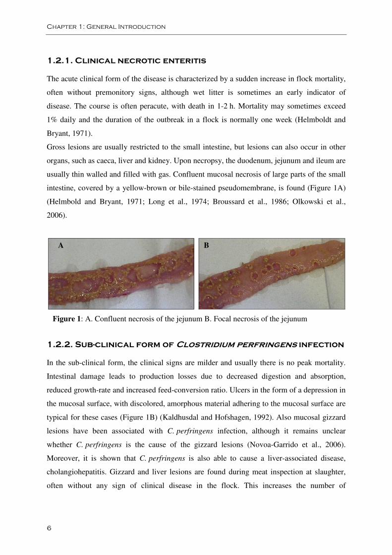
intestine, covered by a yellow-brown or bile-stained pseudomembrane, is found (Figure 1A)
(Helmbold and Bryant, 1971; Long et al., 1974; Broussard et al., 1986; Olkowski et al.,
2006).
Figure 1: A. Confluent necrosis of the jejunum B. Focal necrosis of the jejunum
1.2.2. Sub-clinical form of Clostridium perfringens infection
In the sub-clinical form, the clinical signs are milder and usually there is no peak mortality.
Intestinal damage leads to production losses due to decreased digestion and absorption,
reduced growth-rate and increased feed-conversion ratio. Ulcers in the form of a depression in
the mucosal surface, with discolored, amorphous material adhering to the mucosal surface are
typical for these cases (Figure 1B) (Kaldhusdal and Hofshagen, 1992). Also mucosal gizzard
lesions have been associated with C. perfringens infection, although it remains unclear
whether C. perfringens is the cause of the gizzard lesions (Novoa-Garrido et al., 2006).
Moreover, it is shown that C. perfringens is also able to cause a liver-associated disease,
cholangiohepatitis. Gizzard and liver lesions are found during meat inspection at slaughter,
often without any sign of clinical disease in the flock. This increases the number of
B A
Chapter 1: General Introduction
7
condemnations at processing (Onderka et al., 1990; Løvland and Kaldhusdal, 1999; Sasaki et
al., 2000). There is a general consensus that, although clinical outbreaks of necrotic enteritis
may cause high levels of mortality, the sub-clinical form of the disease is more important than
the clinical form because it may persist in broiler flocks without overt clinical manifestations.
Since the disease is undetected and birds remain untreated, sub-clinical necrosis causes the
greatest economic losses in the poultry production industry (Kaldhusdal and Hofshagen,
1992; Dahiya et al., 2006).
1.3. Histopathology
Microscopic examination of early stages of necrotic enteritis show strong inflammatory
reactions to C. perfringens. The lamina propria is hyperemic and infiltrated with numerous
inflammatory cells. Most significant changes are seen at the interface of the basal domain of
enterocytes and lamina propria. These areas are extensively edematous, allowing for the
substantial disturbance of the structural integrity between the lamina propria and the
enterocytes (Olkowski et al., 2006). Microscopic examination of later stages of necrotic
enteritis lesions shows diffuse and severe coagulative necrosis of the mucosa, involving the
luminal third to half of the mucosa. Necrosis of enterocytes in these areas is apparent on the
villi. A clear line of demarcation between necrotic and viable tissue and an accumulation of
heterophilic granulocytes at the junction is seen. If present, the pseudomembrane consists of
masses of tissue fragments, necrotic cells, cell debris, and numerous bacterial colonies
suspended in mucus. Congestion of blood vessels is seen in the lamina propria and
submucosa. Large Gram-positive rods are associated with areas of necrosis but do not invade
the epithelium or are never found to be attached to the viable mucosal epithelial cells, despite
their massive presence in the lumen tissue debris (Helmboldt and Bryant, 1971; Long et al.,
1974; Al-Sheikhly and Al-Saieg, 1980; Broussard et al., 1986; Olkowski et al., 2006).
Chapter 1: General Introduction
8
2. Role of Clostridium perfringens in the pathogenesis
of necrotic enteritis
C. perfringens was first described as Bacillus aerogenes capsulatus in 1892 and has also been
commonly known as C. welchii (Hatheway, 1990). C. perfringens may be the most widely
occurring bacterial pathogen in nature (Songer, 1996). The bacterium is commonly found in
soil and sewage and is a normal microbiota component of the intestinal tract of warm-blooded
animals and men. C. perfringens has been shown to be a cause of human diseases, including
gas gangrene, necrotic enteritis and food poisoning. It is also the most important cause of
clostridial enteric disease in domestic animals (Rood and Cole, 1991; Songer, 1996; Rood,
1998).
2.1. Basic characteristics
Clostridium perfringens is a Gram-positive, rod-shaped bacterium. The rods are relatively
large: 0.6-2.4 x 1.3-19.0 µm (Cato et al., 1986; Hatheway, 1990). C. perfringens has no
flagella but it is motile by way of type IV pili (Varga et al., 2006). It is classified as an
anaerobe, although C. perfringens is less strictly anaerobic than other Clostridia (Cato et al.,
1986; Novak and Juneja, 2002). C. perfringens can survive under extreme conditions, due to
its differentiation from vegetative cells to highly resistant dormant spores (Novak and Juneja,
2002). Growth has been shown at temperatures as high as 50 °C, while slowly arresting near
6 °C and below. Under optimal conditions (43-45 °C), C. perfringens is known as the most
rapidly multiplying organism with generation times often less than 10 min and growth is
accompanied by abundant gas production. Growth is limited at pH-values lower than 5.0 or
higher than 8.0 and optimal growth is between pH 6 and 7 (Cato et al., 1986; Novak and
Juneja, 2002). Genome analysis has revealed that C. perfringens lacks the genetic machinery
to produce 13 essential amino acids (Shimizu et al., 2002; Myers et al., 2006). As a
consequence, C. perfringens is not able to grow in an environment where amino acids are
limiting and it can obtain these via the action of exotoxins, some of which are enzymes.

Chapter 1: General Introduction
9
2.2. Virulence factors
C. perfringens does not invade healthy cells but produces an intimidating arsenal of toxins
and enzymes that are responsible for the associated lesions and symptoms (Petit et al., 1999).
2.2.1. Toxins
The C. perfringens toxins are classified in major toxins, an enterotoxin and minor toxins
(Hatheway et al., 1990). Individual strains produce only portions of this toxin repertoire and
the ability of C. perfringens to cause disease is ascribed mainly to the differential production
of four major toxins, an enterotoxin, and 9 minor protein toxins (Hatheway, 1990; Songer,
1996; Rood, 1998).
2.2.1.1. M2.2.1.1. M2.2.1.1. M2.2.1.1. Major toxinsajor toxinsajor toxinsajor toxins
The major toxins are alpha, beta, epsilon, and iota toxin, all potentially lethal depending on
the host. The bacterium is classified into 5 types (A through E) according to different
combinations of production of the four major toxins, as shown in Table I (Sterne and
Warrack, 1964; Songer, 1996).
Table I: Toxins used for typing C. perfringens (Petit et al., 1999).
Toxin(s) produced
Type
alpha toxin
beta toxin
epsilon toxin
iota toxin
A
+
-
-
-
B + + + -
C + + - -
D + - + -
E + - - +
gene
plc
cpb1
etx
iap, ibp
location chromosome plasmid plasmid plasmid
Chapter 1: General Introduction
10
C. perfringens toxin genes are located on the chromosome or on plasmids. The alpha toxin
gene is located in a very stable region on the C. perfringens chromosome (close to the origin
of replication) and this is why all C. perfringens strains carry this gene and why it is produced
in varying amounts by all isolates (Canard and Cole, 1989). Clostridium perfringens
phospholipase C (alpha toxin) is a Zn2+ metalloenzyme that degrades both lecithin and
sphingomyelin. It promotes membrane disorganization resulting in lysis or other forms of
cytotoxicity. Alpha toxin displays platelet aggregating, haemolytic, necrotic, and vascular
permeabilization activities (Songer, 1997; Rood, 1998; Titball, 1999; Sakurai et al., 2004;
Flores-Díaz et al., 2004). It is the main virulence determinant in gas gangrene, which is a life-
threatening infection with fever, pain, edema, myonecrosis and gas production. It is shown
that mutated strains that are unable to produce alpha toxin failed to cause this disease in mice
(Awad et al., 1995; Flores-Díaz and Alape-Girón, 2003). Beta toxin is a protease-sensitive
pore-forming toxin. It forms pores by the formation of toxin multimers in the cell membrane,
resulting in Ca2+, Na+, and Cl- influx and K+ efflux from the cells (Steinthorsdottir et al.,
2000; Shatursky et al., 2000; Nagahama et al., 2003). Epsilon toxin acts by forming large
membrane pores by oligomerization into a heptamer resulting in potassium and fluid leakage
of cells, which leads to the loss of cell viability (Petit et al., 2001; Miyata et al., 2001; Petit et
al., 2003). The beta and epsilon toxins seem to have key roles in enterotoxaemia in calves,
lambs, piglets and goats, and most of the domesticated livestock in developed countries are
immunized against disease with toxoid vaccines. Iota toxin is a binary toxin, it consists of two
independent components, the enzymatic component (Ia) and the binding component (Ib). Ia is
an ADP-ribosyltransferase that modifies actin. The iota toxin is the only C. perfringens toxin
that acts intracellularly. All other toxins interact with the cell membrane leading to membrane
disruption or pore formation (Rood and Cole, 1991; Songer, 1996; Petit et al., 1999; Gibert et
al., 2000; Marvaud et al., 2001).
2.2.1.2. 2.2.1.2. 2.2.1.2. 2.2.1.2. EEEEnterotoxinnterotoxinnterotoxinnterotoxin
Enterotoxin is the cause of human food poisoning. Unlike the other toxins, enterotoxin is not
secreted but is produced during sporulation (Adak et al., 2002; Brynestad and Granum, 2002;
Lukinmaa et al., 2002). It interacts with epithelial tight junction proteins and induces leakage
of water and ions by forming pores or channels in plasma membranes of host cells (McClane,
2001, Smedley et al., 2004).
Chapter 1: General Introduction
11
2.2.1.2.2.1.2.2.1.2.2.1.3333. M. M. M. Minorinorinorinor toxinstoxinstoxinstoxins
All other toxins belong to the group of minor toxins. Theta toxin, also known as theta-
hemolysin, perfringolysin O, or the thiol-activated cytolysin is located on the chromosome
and produced by all five toxin types of C. perfringens (Rood and Cole, 1991). Theta toxin is a
member of the cholesterol-binding toxin family and causes complete hemolysis of red blood
cells by forming oligomers, which subsequently form pores through the cell membrane (Petit
et al., 1999, Awad et al., 2001). A more recently discovered toxin is Beta2 toxin, a pore-
forming toxin that is associated with enteritis in neonatal pigs (Gibert et al., 1997; Jost et al.,
2005). Other known toxins produced by C. perfringens are: delta toxin, a hemolysin; kappa
toxin, a collagenase; lambda toxin, a caseinase; mu toxin, a hyaluronidase; nu toxin, a
nuclease; neuraminidase or sialidase, a N-acetylneuraminic acid glycohydrolase; and the
gamma and eta toxins, whose function is unclear (Hatheway, 1990; Rood and Cole, 1991).
The relevance in disease of most of these minor toxins is not fully understood.
The different toxin types of C. perfringens are associated with particular human or veterinary
diseases (Table II), indicating that variations in toxin production profoundly influence the
virulence properties of C. perfringens isolates. These isolate-to-isolate differences in toxin
production also help explain the pathogenic versatility of C. perfringens, which causes both
enteric and histotoxic infections and has a disease spectrum ranging from low incidence/high
mortality to high incidence/low mortality. Strains of toxin type B-E are always associated
with disease processes, indicating that they are frank pathogens. Type A strains are also
associated with disease but can equally well be part of the normal flora in the intestinal tract
of man and animal (Hatheway, 1990; Songer, 1996). Rather than being a function of a single
toxin, the virulence of different C. perfringens isolates is considered as a multifactorial trait,
with different determinants contributing to adaptation of the organism to its niche and to
production of the pathology (Canard et al., 1992; Miyamoto et al., 2006; Sawires and Songer,
2006). The pathogenesis of C. perfringens in different diseases has not yet been fully
elucidated and probably many potential toxins are yet unidentified.
Chapter 1: General Introduction
12
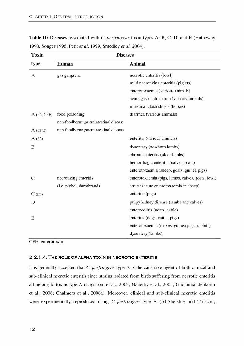
Table II: Diseases associated with C. perfringens toxin types A, B, C, D, and E (Hatheway
1990, Songer 1996, Petit et al. 1999, Smedley et al. 2004).
Diseases Toxin
type Human Animal
A
gas gangrene
necrotic enteritis (fowl)
mild necrotizing enteritis (piglets)
enterotoxaemia (various animals)
acute gastric dilatation (various animals)
intestinal clostridiosis (horses)
A (β2, CPE) food poisoning
non-foodborne gastrointestinal disease
diarrhea (various animals)
A (CPE) non-foodborne gastrointestinal disease
A (β2) enteritis (various animals)
B dysentery (newborn lambs)
chronic enteritis (older lambs)
hemorrhagic enteritis (calves, foals)
enterotoxaemia (sheep, goats, guinea pigs)
C necrotizing enteritis
(i.e. pigbel, darmbrand)
enterotoxaemia (pigs, lambs, calves, goats, fowl)
struck (acute enterotoxaemia in sheep)
C (β2) enteritis (pigs)
D pulpy kidney disease (lambs and calves)
enterocolitis (goats, cattle)
E enteritis (dogs, cattle, pigs)
enterotoxaemia (calves, guinea pigs, rabbits)
dysentery (lambs)
CPE: enterotoxin
2.2.1.4. T2.2.1.4. T2.2.1.4. T2.2.1.4. The role of alpha toxin in necrotic enteritishe role of alpha toxin in necrotic enteritishe role of alpha toxin in necrotic enteritishe role of alpha toxin in necrotic enteritis
It is generally accepted that C. perfringens type A is the causative agent of both clinical and
sub-clinical necrotic enteritis since strains isolated from birds suffering from necrotic enteritis
all belong to toxinotype A (Engström et al., 2003; Nauerby et al., 2003; Gholamiandehkordi
et al., 2006; Chalmers et al., 2008a). Moreover, clinical and sub-clinical necrotic enteritis
were experimentally reproduced using C. perfringens type A (Al-Sheikhly and Truscott,
Chapter 1: General Introduction
13
1977a; Al-Sheikhly and Truscott, 1977c; Gholamiandehkordi et al., 2007). Of the major
typing toxins, type A strains produce only alpha toxin. Therefore, for a long time it was
thought that alpha toxin was the major virulence factor in the pathogenesis of necrotic
enteritis in poultry. Several studies presented evidence for this hypothesis. Bacteria-free crude
supernatant from C. perfringens type A cultures produced necrotic lesions in broilers (Al-
Sheikly and Truscott, 1977b) or caused mortality in germ-free chickens (Fukata et al., 1988).
After addition of antibodies to C. perfringens alpha toxin to the supernatant no mortality was
seen (Fukata et al., 1988). Lovland et al. (2004) showed that maternal vaccination with a
crude C. perfringens type A and C toxoid induces antibodies against alpha toxin in chicks,
which are partially protective against necrotic enteritis. However, care must be taken when
interpreting these studies. Hence, crude supernatant was used and the assumption that the
observed effects were caused by the dominant protein present in the supernatant (i.e. alpha
toxin) did not take into account other secreted toxins that the bacteria may have produced.
Epidemiological and experimental evidence has supported the proposal that alpha toxin is an
important protective antigen. High titers of antibodies to alpha toxin are found in poultry
immune to necrotic enteritis (Heier et al., 2001; Kulkarni et al., 2006). Moreover,
immunization of broilers with purified alpha toxoid induced protection against experimentally
induced necrotic enteritis (Kulkarni et al., 2007). Thompson et al. (2006) showed that
spontaneously derived alpha toxin mutants of a virulent strain have an impaired ability to
cause NE lesions. However, since it were spontaneously derived mutants, the reduced
virulence could be due to the impairment of the production of other toxins than alpha toxin.
Against this background, the role of alpha toxin in the pathogenesis of necrotic enteritis was
called somewhat into doubt at the start of this thesis. C. perfringens outbreak strains as well as
normal broiler microbiota isolates are type A (Nauerby et al., 2003; Gholamiandehkordi et al.,
2006). Moreover, no apparent difference in the levels of alpha toxin was found when the
alpha toxin production in vitro was compared between strains associated with necrotic
enteritis and isolates derived from the microbiota of normal broilers (Gholamiandehkordi et
al., 2006). Yet another study found that the intestinal level of alpha toxin was not correlated
with disease lesion scores (Wilkie et al., 2006). More convincing evidence was produced by
Keyburn et al. (2006). They showed that an alpha toxin mutant, constructed from a virulent
chicken isolate, was equally able to cause necrotic lesion in broiler chickens compared to the
wild-type strain (Keyburn et al., 2006). Another observation that argues strongly against the
Chapter 1: General Introduction
14
role of alpha toxin in necrotic enteritis is the massive heterophil, lymphocyte, and plasma cell
infiltration in infected tissues (Al-Sheikhly and Truscott, 1977b; Gazdzinsky and Julian,
1992; Shane et al., 1985). In gas gangrene, a disease proved to be mediated by alpha toxin,
marked leukostasis and lack of inflammatory infiltrate is common in tissues infected by
C. perfringens cells (Flores-Díaz and Alape-Girón, 2003). Alpha toxin-negative mutants of
C. perfringens are not able to cause gas gangrene in mice but do promote profound
inflammatory responses (Awad et al., 1995). Thus, the massive immune-cell influx in necrotic
enteritis lesions seems to be inconsistent with the known effects of alpha toxin on the innate
immune system.
2.2.2. Proteolytic enzymes
Recent studies of initial stages of the disease process have provided new etiological details on
the development of necrotic enteritis in broilers. Olkowski et al. (2006, 2008) showed that
damage to the villi initially occurs at the level of the basement membrane and lateral domain
of the enterocytes, spreading throughout the lamina propria, while epithelial damage occurs
later in the process. The nature of the morphological changes indicates that the initiation of
the pathological process leading to necrotic enteritis involves proteolytic factors affecting the
extracellular matrix and cellular junctions. Indeed, in broilers undergoing necrotic enteritis,
the extracellular matrix is disorganized and can even be completely absent. It was shown that
C. perfringens strains isolated from field cases of necrotic enteritis secrete several potent
collagenolytic enzymes and that broilers challenged with C. perfringens show elevated levels
of several collagenolytic enzymes in the intestinal tissue in comparison to controls. It was
thus suggested that the pathology may be the result of bacterial collagenases, whose action is
enhanced when mucosal damage (e.g. induced by coccidia) is present, or of host matrix
metalloproteinases that are activated by the host-pathogen interaction (Olkowski et al., 2008).
From this point of view, it is interesting to note that one of the recently identified, potentially
protective vaccine antigens from C. perfringens might be a zinc metallopeptidase (Kulkarni et
al, 2007).
2.3. Genetic diversity
The genetic diversity among C. perfringens isolates originating from humans, calves, pigs,
sheep, rabbits, goats, poultry, horses, roe deer and food has been investigated by pulsed-field
Chapter 1: General Introduction
15
gel electrophoresis (PFGE). In all cases a high degree of genetic diversity was found (Canard
et al., 1992; Maslanka et al., 1999; Nauerby et al., 2003; Johansson et al., 2006;
Gholamiandehordi et al., 2006). Ribotyping of C. perfringens isolates obtained from food also
confirmed a great diversity (Schalch et al., 1999; Kilic et al., 2002). Using multiple-locus
variable-number tandem repeat analysis (MLVA), Sawires and Songer (2006) and Chalmers
et al. (2008b) showed also a lack of association between strain phylogeny and host species of
disease. Isolates of common host origin did not cluster, while isolates from diverse animal
origins were sometimes the same MLVA type. Moreover, some normal flora strains from
certain host species are phylogenetically close to genotypes of virulent strains from a different
host species.
The entire genome sequence of three different C. perfringens type A strains has been
published (Shimizu et al., 2002; Myers et al., 2006). The sequenced strains are a gangrene
strain (ATCC 13124) and a food poisoning strain (SM101) isolated from humans and a soil
isolate that is able to induce gas gangrene in mice (strain 13). Comparison of the three
genomes revealed considerable genomic diversity with discrete islands containing genes
likely to confer specific virulence, metabolic, or catabolic capabilities to the host strain. This
enables C. perfringens to adapt to a variety of environmental conditions and explains the
different virulence characteristics of C. perfringens (Myers et al., 2006). C. perfringens toxin
genes are located on the chromosome or on plasmids. The genomic diversity of C. perfringens
seen during typing could be the result of the presence of toxin genes on extrachromosomal
elements. It seems to be the case that, by acquisition of extrachromosomal elements
(plasmids, transposons and possibly phages) containing additional toxin genes, different
C. perfringens types have been derived from the type A strain (Petit et al., 1999). Moreover,
Sawires and Songer (2006) suggested that acquisition of the major toxin genes as well as
other plasmidborne toxin genes is a recent evolutionary event and that their maintenance is
essentially a function of the selective advantage they confer in certain niches under different
conditions
2.4. Single strain dominance in broiler necrotic
enteritis
Genetic characterization by PFGE or amplified fragment length polymorphism (AFLP) has
revealed that in healthy flocks, different genotypes of C. perfringens type A can be found,
Chapter 1: General Introduction
16
even within individual birds and within the same gut segment. In contrast, outbreak isolates
from a flock with necrotic enteritis or cholangiohepatitis are generally clonal, regardless of
the animal or the organ of isolation (Engström et al., 2003; Nauerby et al., 2003;
Gholamiandehkordi et al., 2006). These results were confirmed by multilocus sequence typing
analysis (MLST) (Chalmers et al., 2008a). After natural recovery or treatment, birds again
yield multiple genetic types (Nauerby et al., 2003). The reason for the selective presence of a
single clone in necrotic enteritis outbreaks is not known. It is possible that during an outbreak,
certain C. perfringens strains are able to secrete growth-inhibiting molecules which give them
a competitive advantage over other C. perfringens strains in the broiler gut. Indeed, inhibiting
other strains could lead to extensive and selective presence of a strain that contains the genetic
make-up to secrete toxins that cause gut lesions.
3. Antimicrobial proteins from bacteria
Antimicrobial peptides and proteins are produced by all species of life (prokaryotic and
eukaryotic): plants, insects and other invertebrates, fish, amphibians, birds, mammals -
including humans, and different microorganisms (Nissen-Meyer and Nes, 1997; Jenssen et al.,
2006). In higher organisms these compounds are produced as an innate host defense
mechanism to protect against pathogenic attack, whereas microorganisms presumably use
these compounds as weapons in the competition for space and nutrients among bacteria living
in the same ecological niche. Although they differ greatly in their primary structures, they are
nearly all fairly short molecules, cationic and very often amphiphilic (i.e. it possesses both
hydrophobic and hydrophilic properties). This is reflected in the fact that many of these
peptides kill their target cells by permeabilizing the target cell membrane, resulting in an
irreversible leakage of cellular material and consequently cell death (Nissen-Meyer and Nes,
1997). Other modes of action such as the inhibition of nucleic acid synthesis, protein
synthesis, enzyme activity and cell wall synthesis have been described (Brogden, 2005). The
antimicrobial proteins and peptides from bacteria include toxins, antibiotics, bacteriolytic
enzymes, bacteriocins, and bacteriocin-like peptides (Jack et al., 1995).
Chapter 1: General Introduction
17
3.1. Bacteriocins
Bacteriocins are defined as a heterogeneous group of ribosomally synthesised, proteinaceous
substances (with or without further modifications) produced by bacteria that kill or inhibit the
growth of other bacteria. Their mode of activity is primarily bactericidal and directed against
closely related strains and species. The bacteriocin family includes a diversity of peptides and
proteins in terms of size, amino acid sequence and composition, secretion and processing
machinery, post-translational modifications, microbial target, mode of action, and
mechanisms of resistance (Klaenhammer, 1993; Jack et al., 1995).
Bacteriocins were first identified in Gram-negative bacteria over 80 years ago when inhibition
was observed between two strains of Escherichia coli (Gratia, 1925). A heat labile product
present in cultures of Escherichia coli V was shown to be toxic to E. coli S and it was named
colicin (Gratia, 1925). Fredericq (1946) demonstrated the protein nature of colicins and their
limited range of activity due to the presence or absence of specific receptors on the surface of
sensitive cells (Fredericq, 1946). Since then, bacteriocins have been found in all major
bacterial lineages and, more recently, some members of the Archaea have also been seen to
produce similar antimicrobial proteins (O’Connor and Shand, 2002). According to
Klaenhammer (1988), 99% of all bacteria may produce at least one bacteriocin, and the only
reason why there have not been isolated more is that few researchers have looked for them.
Since bacteriocin production is detected in all surveyed lineages of prokaryotes, bacteriocins
must serve some function in microbial communities (Klaenhammer, 1988). They may play a
defensive role by inhibiting the invasion of other strains or species into an occupied niche or
they may serve as anti-competitors enabling the invasion of a strain into an established
microbial community (Riley and Wertz, 2002). Additional roles have been proposed; they
may mediate quorum sensing by acting as a signal molecule that induces the transcription of
genes involved in its biosynthesis and they may act as communication signals in bacterial
consortia e.g. biofilms (Gobbetti et al., 2007; Gillor et al., 2008).
Bacteriocins can be divided into two main groups: those produced by Gram-negative and
Gram-positive bacteria (Gillor et al., 2008). Within the scope of the thesis we will focus on
bacteriocins of Gram-positive bacteria.
Chapter 1: General Introduction
18
3.2. Bacteriocins of Gram-positive bacteria
3.2.1. General characteristics
Bacteriocins of Gram-positive bacteria are abundant and diverse. They resemble many of the
antimicrobial peptides produced by eukaryotes; they are generally cationic, amphiphilic, heat
stable, membrane-permeabilizing peptides, and smaller than 8 kDa (Maqueda et al., 2008).
However, some Gram-positive bacteria have been shown to form relatively high-molecular-
weight, heat labile bacteriocin-like substances (Jack et al., 1995). Although by definition all
bacteriocins have a protein or peptide component that is essential for their bactericidal
function, some have been reported to consist of combinations of different proteins, or are
composites of proteins together with lipid or carbohydrate moieties (Jack et al., 1995).
Bacteriocins can specifically target a particular subset of bacterial strains or species. The
conventional wisdom about the spectrum of Gram-positive bacteriocins is that they are
restricted to killing other Gram-positives (Gillor et al., 2008). However, the range of killing
can vary significantly, from relatively narrow to extraordinarily broad. Some bacteriocins are
only active against closely related strains, others against a wide range of Gram-positive
bacteria and some are even able to inhibit Gram-negative species (Morency et al., 2001).
Interpretation of spectra of inhibitory activity in terms of specific bacteriocin activities can
sometimes be difficult if the producer strains release more than one bacteriocin-like agent
(Higa et al., 1991).
Many bacteriocins appear to elicit their lethal effects by permeabilizing the cell membrane of
target organisms, in certain cases by targeting intermediates of cell wall biosynthesis or
possibly proteins of sugar phosphotransferase systems (Garneau et al., 2002). Creating pores
in the membrane of target cells has deleterious effects such as dissipation of proton motive
force, ATP depletion and leakage of nutrients and metabolites. The size, stability, and
conductivity of these pores differ considerably from one bacteriocin to another. To form a
pore, bacteriocins have to interact with the cytoplasmic membrane of target cells. This process
is at least in part governed by electrostatic interactions between the positively charged peptide
and the anionic lipids that are abundantly present in the membranes of Gram-positive bacteria
(Eijsink et al., 2002). Several factors may contribute to making a cell resistant towards
bacteriocins. The composition and structure of both cell wall and cellular membrane(s) may
be such that the bacteriocin is physically unable to reach its target. Alternatively, certain
Chapter 1: General Introduction
19
cellular components (‘receptors’) that are essential for bacteriocin action may be lacking or
may be mutated. In some cases, the presence of (aspecific) proteases in and near the target
cell may reduce bacteriocin effectiveness. Finally, the ease at which a membrane-bound
bacteriocin actually can form pores can be affected by the physiological state of the target cell
(Driessen et al., 1995; Eijsink et al., 2002).
Production of bacteriocins in Gram-positive bacteria is generally associated with the shift
from logarithmic phase to stationary phase. Gram-positive bacteriocins require several genes
and these bacteriocin-associated genes appear to be characteristically arranged in multigene
operon-like structures, the first gene typically (but not always) encodes the structural protein
(Klaenhammer, 1993). Additional genes encode for proteins that aid in the regulation,
proteins that aid in the processing to the active form, proteins that aid in the transport of the
bacteriocin across the membrane and proteins that confer immunity to the host producer
(Engelke et al., 1992; Klein et al., 1993; Engelke et al., 1994; Klein and Entian, 1994; Diep et
al., 1996; Qiao et al., 1996; Diep et al., 2007; Dufour et al., 2007). Due to these additional
genes, bacteriocins produced by Gram-positive bacteria are generally not lethal to the
producing cell.
3.2.2. Classification
There is a lack of consensus in the classification of bacteriocins from Gram-positive bacteria.
Bacteriocins are commonly divided into three or four main categories (Nes et al., 2007; Gillor
et al., 2008). This classification is based on bacteriocins produced by lactic acid bacteria since
these are the best characterized of this group. Class I is comprised of lantibiotics. These are
small, posttranslationally modified peptides that contain unusual amino acids (Guder et al.,
2000; Twomey et al., 2002). Class II includes heat stable non-lantibiotics (Eijsink et al., 2002;
Héchard and Sahl, 2002; Drider et al., 2006). Larger, heat labile bacteriocins are classified as
class III. For this category much less information is available than for the first two. Class IV
is comprised of complex bacteriocins that require lipid or carbohydrate moieties for activity.
However, presently, no such bacteriocins have been purified and it is suggested that this type
of bacteriocin is an artefact due to the cationic and hydrophobic properties of bacteriocins
which result in complexing with other macromolecules in the crude extract (Cleveland et al.,
2001; Garneau et al., 2002; Gillor et al., 2008). Class I and II are subdivided in subgroups.
Chapter 1: General Introduction
20
Lantibiotics are posttranslationally modified peptides that contain the unusual amino acids
lanthionine and β-methyl lanthionine as part of additional intramolecular rings, and often
possess other modified residues such as dehydro amino acids (Guder et al., 2000; Twomey et
al., 2002; Nagao et al., 2006). They target a broad range of Gram-positive bacteria and are
subdivided into three groups on the basis of their structure and mode of action: Type A
lantibiotics are small (2 - 5kDa), elongated molecules with a flexible structure in solution, that
contain positively charged molecules, which kill via the formation of pores in the bacterial
membrane, leading to the dissipation of membrane potential and the efflux of small
metabolites from the sensitive cells. Type B lantibiotics tend to have a more rigid and
globular structure. They kill by interfering with cellular enzymatic reactions, such as cell wall
synthesis (Pag and Sahl, 2002; Garneau et al., 2002). Another subgroup of the lantibiotics is
composed of two-component lantibiotics, consisting of two lantibiotic peptides that
synergistically display antimicrobial activity (Breukink, 2006; Wiedemann et al., 2006).
Class II bacteriocins are small non-lanthionine containing peptides (Garneau et al., 2002;
Drider et al., 2006). The majority of bacteriocins in this group kill by inducing membrane
permeabilization and the subsequent leakage of molecules from target bacteria. These
bacteriocins are organized into four subgroups. Class IIa is the largest group and its members
share activity against Listeria and a conserved amino-terminal sequence (YGNGVXaaC) that
is thought to facilitate nonspecific binding to the target surface. Class IIa bacteriocins act
through the formation of pores in the cytoplasmic membrane. Class IIb bacteriocins are two-
peptide bacteriocins and form cation- or anion-specific pores, composed of two different
proteins, in the membrane of their target cells (Héchard and Sahl, 2002; Oppegård et al.,
2007). A third subgroup (IIc) has been proposed, which consists of leaderless peptide-
bacteriocins that are secreted via the general secretory (sec) pathway and not having its own
dedicated and specific mechanism (Nes et al., 1996; Garneau et al., 2002). Circular
posttranslationally modified bacteriocins are classified in a fourth subgroup (IId) (Nes et al.,
2007).
Chapter 1: General Introduction
21
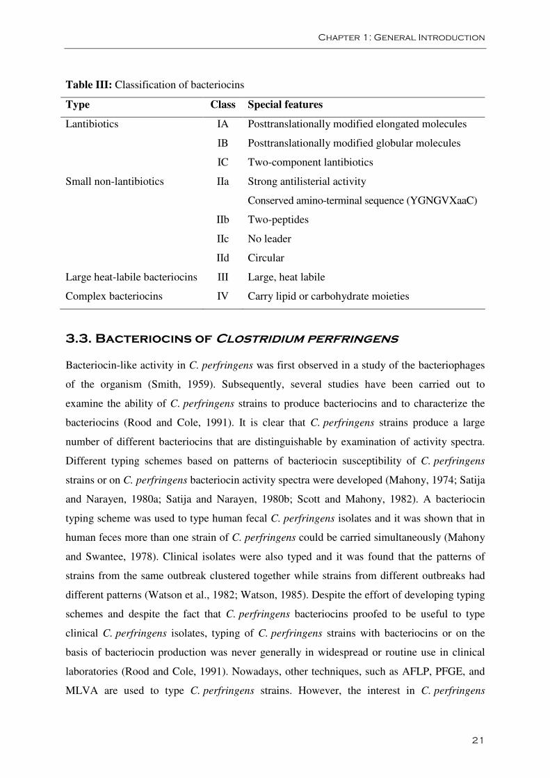
Table III: Classification of bacteriocins
Type Class Special features
Lantibiotics IA Posttranslationally modified elongated molecules
IB Posttranslationally modified globular molecules
IC Two-component lantibiotics
Small non-lantibiotics IIa Strong antilisterial activity
Conserved amino-terminal sequence (YGNGVXaaC)
IIb Two-peptides
IIc No leader
IId Circular
Large heat-labile bacteriocins III Large, heat labile
Complex bacteriocins IV Carry lipid or carbohydrate moieties
3.3. Bacteriocins of Clostridium perfringens
Bacteriocin-like activity in C. perfringens was first observed in a study of the bacteriophages
of the organism (Smith, 1959). Subsequently, several studies have been carried out to
examine the ability of C. perfringens strains to produce bacteriocins and to characterize the
bacteriocins (Rood and Cole, 1991). It is clear that C. perfringens strains produce a large
number of different bacteriocins that are distinguishable by examination of activity spectra.
Different typing schemes based on patterns of bacteriocin susceptibility of C. perfringens
strains or on C. perfringens bacteriocin activity spectra were developed (Mahony, 1974; Satija
and Narayen, 1980a; Satija and Narayen, 1980b; Scott and Mahony, 1982). A bacteriocin
typing scheme was used to type human fecal C. perfringens isolates and it was shown that in
human feces more than one strain of C. perfringens could be carried simultaneously (Mahony
and Swantee, 1978). Clinical isolates were also typed and it was found that the patterns of
strains from the same outbreak clustered together while strains from different outbreaks had
different patterns (Watson et al., 1982; Watson, 1985). Despite the effort of developing typing
schemes and despite the fact that C. perfringens bacteriocins proofed to be useful to type
clinical C. perfringens isolates, typing of C. perfringens strains with bacteriocins or on the
basis of bacteriocin production was never generally in widespread or routine use in clinical
laboratories (Rood and Cole, 1991). Nowadays, other techniques, such as AFLP, PFGE, and
MLVA are used to type C. perfringens strains. However, the interest in C. perfringens
Chapter 1: General Introduction
22
bacteriocins and the analysis of bacteriocin-encoding plasmids has played a very important
role in the development of C. perfringens genetics and different C. perfringens bacteriocins
are described in literature.
3.3.1. BCN5
BCN5 (or N5) produced by C. perfringens CPN50 (or C. perfringens BP6k-N5) is the best
characterized bacteriocin of C. perfringens. BCN5 was first purified by Wolff and Ionesco
(1975). They found a simple protein with an estimated molecular mass of 82 kDa. The
purified bacteriocin was inactivated by proteolytic enzymes and was heat labile. Since BCN5
is a large heat labile bacteriocin, it can be classified as class III bacteriocin according to the
currently used classification. BCN5 inhibits the initiation of the germination of spores of
C. perfringens and it inhibits the DNA-, RNA-, and protein synthesis simultaneously in
sensitive cells, without DNA degradation (Sebald and Ionesco, 1974; Ionesco and Wolff,
1975). C. perfringens CPN50 produces the bacteriocin upon UV induction (Ionesco and
Bouanchaud, 1973; Ionesco et al., 1974), which suggest that the production is controlled by
an SOS-like system and is induced by DNA-damaging treatment (Walker, 1984). Following
activation of the bcn gene, copious amounts of a 96 kDa protein accumulate in the cytoplasm,
to be released into the medium 90 to 180 minutes after induction by lysis of the bacteria
(Ionesco et al., 1974; Garnier and Cole, 1986). From the nucleotide sequence of the bcn gene,
the primary structure of BCN5 was deduced and revealed a protein with a molecular weight of
96.591 kDa, with high glycine content (11.5%). This is common for bacteriocins and is
believed to facilitate their transfer across cell membranes. The primary structure of the protein
reveals the presence of an extended lipophilic region near the COOH terminus. This suggests
that it may function as an ionophore (Garnier and Cole, 1986). The discrepancy between the
estimated molecular weight of 82 kDa by Wolff and Ionesco (1975) could be due to
proteolytic processing of the 95 kDa precursor molecule (Garnier and Cole, 1986).
Curing of C. perfringens CPN50 has been accomplished after acriflavine treatment (Ionesco
and Bouanchaud, 1973). It was shown that these cured derivates produced no longer
bacteriocin and that this bacteriocin loss was associated with the loss of plasmid DNA
(Ionesco and Bouanchaud, 1973). Electron-microscopic analysis showed more specifically
that the loss of both bacteriocin production and immunity to the bacteriocin was associated
with the loss of a small (5.7 MDa) plasmid, pIP404. Thus, in addition to encoding BCN5,
Chapter 1: General Introduction
23
pIP404 codes for bacteriocin immunity (Ionesco et al., 1976; Brefort et al., 1977). To locate
the bcn gene on pIP404, differential dot blot hybridization was performed with RNA samples
prepared from cultures before and after induction with UV irradiation. This approach revealed
that UV irradiation induces a high rate of transcription of a 4kb segment of pIP404 that
includes two contiguous transcription units, uviAB and bcn (Garnier and Cole, 1986; Garnier
and Cole, 1988). The bcn gene, encoding BCN5, is transcribed from three promoters (P1, P2
and P3) all of which are dependent on UviA for activation. P1, the promoter closest to the
coding sequence, appears to be the strongest of the three (Dupuy et al., 2005). Transcription
of uviAB is directed by two promoters (P4 and P5). It was hypothesized that the uviAB operon
might encode proteins needed for BCN5 synthesis or secretion or for immunity to the
bacteriocin (Garnier and Cole, 1986; Garnier and Cole, 1988). Dupuy et al. (2005) showed
that UviA is an RNA polymerase σ factor. Promotor P4 provides an UviA-independent, basal
level of gene expression while the stronger, UviA-dependent promoter (P5) was only utilized
after the cell experienced DNA damage. As a result, BCN5 synthesis is induced by treatment
with UV light or mitomycin C. The role of UviB, the second product of the uviAB operon,
has never been determined. As BCN5-producing cells are immune to the bacteriocin, UviB
might be the immunity protein (Dupuy et al., 2005).
3.3.2. Bacteriocin 28
Mahony and Butler (1971) screened 33 C. perfringens strains for bacteriocin production and
four bacteriocin-producing strains were detected. One of the bacteriocins, bacteriocin 28
produced by C. perfringens strain 28, was chosen by Mahony and associates to study in more
detail. Bacteriocin production by C. perfringens strain 28 is associated with a 5.6 MDa
plasmid since a cured variant lost its immunity and its ability to produce bacteriocin (Li et al.,
1980). High titres of bacteriocin 28 are produced during late logarithmic growth although
higher yields are obtainable by induction with mitomycin C but not with UV light (Mahony
and Butler, 1971; Mahony, 1977). The bacteriocin was heat labile, only stable at pH between
5 and 7 and sensitive to the action of trypsin and pronase (Mahony and Butler, 1971; Mahony
and Li, 1978). Bacteriocin 28 is a glycoprotein with hydrophobic properties and an estimated
molecular mass of 100 kDa (Li et al., 1982). Like BCN5, bacteriocin 28 is a large heat labile
bacteriocin and can be classified as class III bacteriocin. Investigation of its mode of action
indicated that it seemed bacteriostatic and that it does not inhibit the synthesis of DNA, RNA
Chapter 1: General Introduction
24
or proteins but acts on the cell wall of viable indicator cultures, causing conversion of
indicator strains to spheroplasts, either by removing the existing wall or inhibiting cell wall
synthesis. These spheroplasts are capable of growing as L-form colonies on sucrose
containing media. Such colonies were composed of spherical and amorphous structures of
considerable size and demonstrated a dense center containing many granular structures
(Mahony et al., 1971).
3.3.3. Other Clostridium perfringens bacteriocins
Mahony and Li (1978) compared nine other C. perfringens bacteriocins with bacteriocin 28.
Two bacteriocins were stable over a wide range of pH values and resisted boiling and three
other bacteriocins were resistant to trypsin. Essentially, the bacteriocins could be divided in
two major groups, those that inhibit DNA, RNA, and protein synthesis, such as BCN5, and
those that interfere with the cell wall of sensitive C. perfringens strains, like bacteriocin 28
(Mahony and Li, 1987; Mahony, 1982). Clarke et al. (1975) described perfringocin 11105.
This bacteriocin is produced by C. perfringens NCIB11105 and is heat stable, trypsin
susceptible and stable over a wide range of pH. It has a molecular weight of 76 kDa and the
properties of an amphiphilic protein. Perfringocin 11105 is produced at the onset of the
stationary phase of the C. perfringens culture, and its subsequent production coincides with
some lysis of the producer organism. Yield of perfringocin 11105 can be enhanced by
mitomycin C treatment (Clarke et al., 1975). Mihelc et al. (1978) reported that a 5.6 MDa
plasmid (pCW4) is associated with bacteriocin production and immunity in C. perfringens
strain CW55. Higa et al. (1991) showed that C. perfringens SN-17 produced two types of
bacteriocin successively one after the other during late exponential phase, without induction
by UV radiation or mitomycin C. The molecular weight of SN-a and SN-b was determined to
be about 70 kDa and 100 kDa respectively. Both bacteriocins were heat labile, sensitive to
pronase and more stable in alkaline pH than in acidic pH. Both bacteriocins adsorbed to
resting cells or even dead cells, indicating that they bind to receptors on the surface of
sensitive cells (Higa et al., 1991).
3.3.4. Bacteriocin genes in genome sequences
Analysis of the genome sequences of the three sequenced C. perfringens strains revealed a
single bacteriocin gene (bcn5) on the chromosome of strain 13 in association with the
Chapter 1: General Introduction
25
prophage remnant (Shimizu et al., 2002). No bacteriocin or accessory genes are found in the
ATCC 13124 genome. By contrast, several bacteriocin loci are found in the SM101 genome
and plasmid sequences. The two SM101 plasmids each bear a complete UV-inducible
bacteriocin operon (uviA-uviB-bcn5), similar to the archetypal clostridial bacteriocin bearing
plasmid pIP404 of CPN50 (Myer et al., 2006).
Chapter 1: General Introduction
26
REFERENCES
Adak G.K., Long S.M., O’Brien S.J. (2002). Trends in indigenous foodborne disease and
deaths, England and Wales: 1992 to 2000. Gut. 51, 832-841.
Al-Sheikhly F., Al-Saieg A. (1980). Role of Coccidia in the occurrence of necrotic enteritis of
chickens. Avian Dis. 24, 324-333.
Al-Sheikhly F., Truscott R.B. (1977a). The pathology of necrotic enteritis of chickens
following infusion of broth cultures of Clostridium perfringens into the duodenum. Avian
Dis. 21, 230-240.
Al-Sheikhly F., Truscott R.B. (1977b). The pathology of necrotic enteritis of chickens
following infusion of crude toxins of Clostridium perfringens into the duodenum. Avian Dis.
21, 230-240.
Al-Sheikhly F., Truscott R.B. (1977c). The interaction of Clostridium perfringens and its
toxins in the production of necrotic enteritis of chickens. Avian Dis. 21, 256-263.
Awad M.M., Bryant A.E., Stevens D.L., Rood J.I. (1995). Virulence studies on chromosomal
alpha-toxin and theta-toxin mutants constructed by allelic exchange provide genetic evidence
for the essential role of alpha-toxin in Clostridium perfringens-mediated gas gangrene. Mol.
Microbiol. 15, 191-202.
Awad M.M., Ellemor D.M., Boyd R.L., Emmins J.J., Rood J.I. (2001). Synergistic effects of
alpha-toxin and perfringolysin O in Clostridium perfringens-mediated gas gangrene. Infect.
Immun. 69, 7904-7910.
Baba E., Ikemoto T., Fukata T., Sasai K., Arakawa A., McDougald L.R. (1997). Clostridial
population and the intestinal lesions in chickens infected with Clostridium perfringens and
Eimeria necatrix. Vet. Microbiol. 54, 301-308.
Branton S.L., Reece F.N., Hagler W.M. Jr. (1987). Influence of a wheat diet on mortality of
broiler chickens associated with necrotic enteritis. Poult. Sci. 66, 1326-1330.
Chapter 1: General Introduction
27
Brefort G., Magot M., Ionesco H., Sebald M. (1977). Characterization and transferability of
Clostridium perfringens plasmids. Plasmid. 1, 52-66.
Breukink E. (2006). A lesson in efficient killing from two-component lantibiotics. Mol.
Microbiol. 61, 271-273.
Brogden K.A. (2005). Antimicrobial peptides: pore formers or metabolic inhibitors in
bacteria? Nat. Rev. Microbiol. 3, 238-250.
Broussard C.T., Hofacre C.L., Page R.K., Fletcher O.J. (1986). Necrotic enteritis in cage-
reared commercial layer pullets. Avian Dis. 30, 617-619.
Brynestad S., Granum P.E. (2002). Clostridium perfringens and foodborne infections. Int. J.
Food Mircrobiol. 74, 195-202.
Canard B., Cole S.T. (1989). Genome organization of the anaerobic pathogen Clostridium
perfringens. Proc. Natl. Acad. Sci. USA. 86, 6676-6680.
Canard B., Saint-Joanis B., Cole S.T. (1992). Genomic diversity and organization of virulence
genes in the pathogenic anaerobe Clostridium perfringens. Mol. Microbiol. 6, 1421-1429.
Cato E.P., George W.L., Finegold S.M. (1986). Genus Clostridium. In Bergey’s Manual of
Systematic Bacteriology Volume 2. Edited by P.H.A. Sneath, N.S. Mair, M.E. Sharpe, J.G.
Holt. Williams and Wilkins, Baltimore, USA, 1141-1200.
Chalmers G., Bruce H.L., Hunter D.B., Parreira V.R., Kulkarni R.R., Jiang Y.-F., Prescott J.F.
Boerlin P. (2008a). Multilocus sequence typing analysis of Clostridium perfringens isolates
from necrotic enteritis outbreaks in broiler chicken populations. J. Clin. Microbiol. 46, 3957-
3964.
Chalmers G., Martin S.W., Prescott J.F., Boerlin P. (2008b). Typing of Clostridium
perfringens by multiple-locus variable number of tandem repeats analysis. Vet. Microbiol.
128, 126-135.
Chapter 1: General Introduction
28
Choct M., Hughes R.J., Wang J., Bedford M.R., Morgan A.J., Annison G. (1996). Increased
small intestinal fermentation is partly responsible for the anti-nutritive activity of non-starch
polysaccharides in chickens. Br. Poult. Sci. 37, 609-621.
Clarke D.J., Robson R.M., Morris J.G. (1975). Purification of two Clostridium perfringens
bacteriocins by procedures appropriate to hydrophobic proteins. Antimicrob Agents
Chemother. 7, 256-264.
Cleveland J., Montville T.J., Nes I.F., Chikindas M.L. (2001). Bacteriocins: safe, natural
antimicrobials for food preservation. Int. J. Food Microbiol. 71, 1-20.
Collier C.T., Hofacre C.L., Payne A.M., Anderson D.B., Kaiser P., Mackie R.I., Gaskins H.R.
(2008). Coccidia-induced mucogenesis promotes the onset of necrotic enteritis by supporting
Clostridium perfringens growth. Vet. Immun. Immunopath. 122, 104-115.
Cowen B.S., Schwartz L.D., Wilson R.A., Ambrus S.I. (1987). Experimentally induced
necrotic enteritis in chickens. Avian Dis. 31, 904-906.
Craven S.E. (2000). Colonization of the intestinal tract by Clostridium perfringens and fecal
shedding in diet-stressed and unstressed broiler chickens. Poult. Sci. 79, 843-849.
Craven S.E., Stern N.J., Bailey J.S., Cox N.A. (2001). Incidence of Clostridium perfringens in
broiler chickens and their environment during production and processing. Avian Dis. 45, 887-
896.
Dahiya J.P., Wilkie D.C., Van Kessel A.G., Drew M.D. (2006). Potential strategies for
controlling necrotic enteritis in broiler chickens in post-antibiotic era. Anim. Feed Sci.
Technol. 129, 60-88.
Diep D.B., Håvarstein L.S., Nes I.F. (1996). Characterization of the locus responsible for the
bacteriocin production in Lactobacillus plantarum C11. J. Bacteriol. 178, 4472-4483.
Diep D.B., Skaugen M., Salehian Z., Holo H., Nes I.F. (2007). Common mechanisms of
target cell recognition and immunity for class II bacteriocins. Proc. Natl. Acad. Sci. U.S.A.
104, 2384-2389.
Chapter 1: General Introduction
29
Drew M.D., Syed N.A., Goldade B.G., Laarveld B., Van Kessel A.G. (2004). Effects of
dietary protein source and level on intestinal populations of Clostridium perfringens in broiler
chickens. Poult. Sci. 83, 414-420.
Drider D., Fimland G., Héchard Y., McMullen L.M., Prévost H. (2006). The continuing story
of class IIa bacteriocins. Microbiol. Mol. Biol. Rev. 70, 564-582.
Driessen A.J., van den Hooven H.W., Kuiper W., van de Kamp M., Sahi H.G., Konings R.N.,
Konings W.N. (1995). Mechanistic studies of lantibiotic-induced permeabilization of
phospholipid vesicles. Biocheminstry. 34, 1606-1614.
Dufour A., Hindré T., Haras D., Le Pennec J.P. (2007). The biology of lantibiotics from the
lacticin 481 group is coming of age. FEMS Microbiol. Rev. 31, 134-167.
Dupuy B., Mani N., Katayama S., Sonenshein A.L. (2005). Transcription activation of a UV-
inducible Clostridium perfringens bacteriocin gene by a novel sigma factor. Mol. Microbiol.
55, 1196-1206.
Eijsink V.G., Axelsson L., Diep D.B., Håvarstein L.S., Holo H., Nes I.F. (2002). Production
of class II bacteriocins by lactic acid bacteria: an example of biological warfare and
communication. Antonie Van Leeuwenhoek. 81, 639-654.
Engberg R.M., Hedemann M.S., Jensen B.B. (2002). The influence of grinding and pelleting
of feed on the microbial composition and activity in the digestive tract of broiler chickens. Br.
Poult. Sci. 43, 569-579.
Engelke G., Gutowski-Eckel Z., Hammelmann M., Entian K.D. (1992). Biosynthesis of the
lantibiotic nisin: genomic organization and membrane localization of the NisB protein. Appl.
Environ. Microbiol. 58, 3730-3743.
Engelke G., Gutowski-Eckel Z., Kiesau P., Siegers K., Hammelmann M., Entian K.D.
(1994). Regulation of nisin biosynthesis and immunity in Lactococcus lactis 6F3. Appl.
Environ. Microbiol. 60, 814-825.
Chapter 1: General Introduction
30
Engström B.E., Fermér C., Lindberg A., Saarinen E., Båverud V., Gunnarson A. (2003).
Molecular typing of isolates of Clostridium perfringens from healthy and diseased poultry.
Vet. Microbiol. 94, 225-235.
Flores-Díaz M., Alape-Girón A. (2003). Role of Clostridium perfringens phospholipase C in
the pathogenesis of gas gangrene. Toxicon. 15, 979-986.
Flores-Díaz M., Thelestam M., Clark G.C., Titball R.W., Alape-Girón A. (2004). Effects of
Clostridium perfringens phospholipase C in mammalian cells. Anaerobe. 10, 115-123.
Fredericq P. (1946). Sur la pluralité des récepteurs d’antibiose de E. coli. C. R. Soc. Biol.
(Paris). 140, 1189-1194.
Fukata T., Hadate Y., Baba E., Uemura T., Arakawa A. (1988). Influence of Clostridium
perfringens and its toxins in germ-free chickens. Res. Vet. Sci. 44, 68-70.
Garneau S., Martin N.I., Vederas J.C. (2002). Two-peptide bacteriocins produced by lactic
acid bacteria. Biochimie. 84, 577-592.
Garnier T., Cole S.T. (1986). Characterization of a bacteriocinogenic plasmid from
Clostridium perfringens and molecular genetic analysis of the bacteriocin-encoding gene. J.
Bacteriol. 168, 1189-1196.
Garnier T., Cole S.T. (1988). Studies of UV-inducible promoters from Clostridium
perfringens in vivo and in vitro. Mol. Microbiol. 2, 607-614.
Gazdzinski P., Julian R.J. (1992). Necrotic enteritis in turkeys. Avian Dis. 36, 792-798.
Gholamiandehkordi A.R., Ducatelle R., Heyndrickx M., Haesebrouck F., Van Immerseel F.
(2006). Molecular and phenotypical characterization of Clostridium perfringens isolates from
poultry flocks with different disease status. Vet. Microbiol. 113, 143-152.
Gholamiandehkordi A.R., Timbermont L., Lanckriet A., Van den Broeck W., Pedersen K.,
Dewulf J., Pasmans F., Haesebrouck F., Ducatelle R., Van Immerseel F. (2007).
Quantification of gut lesions in a subclinical necrotic enteritis model. Avian Pathol. 36, 375-
382.
Chapter 1: General Introduction
31
Gibert M., Jolivet-Reynaud C., Popoff M.R. (1997). Beta2 toxin, a novel toxin produced by
Clostridium perfringens. Gene. 203, 65-73.
Gibert M., Petit L., Raffestin S., Okabe A., Popoff M.R. (2000). Clostridium perfringens iota-
toxin requires activation of both binding and enzymatic components for cytopathic activity.
Infect. Immun. 68, 3848-3853.
Gillor O., Etzion A., Riley M.A. (2008). The dual role of bacteriocins as anti- and probiotics.
Appl. Microbiol. Biotechnol. 81, 591-606.
Gobbetti M., De Angelis M., Di Cagno R., Minervini F., Limitone A. (2007). Cell-cell
communication in food related bacteria. Int. J. Food Microbiol. 120, 34-45.
Gratia A. (1925). Sur un remarquable example d’antagonisme entre deux souches de
colibacille. C. R. Soc. Biol. (Paris). 93, 1040-1042.
Guder A., Wiedemann I., Sahl H.G. (2000). Posttranslationally modified bacteriocins-the
lantibiotics. Biopolymers. 55, 62-73.
Hatheway C.L. (1990). Toxigenic Clostridia. Clin. Microbiol. Rev. 3, 66-98.
Héchard Y., Sahl H.G. (2002). Mode of action of modified and unmodified bacteriocins from
Gram-positive bacteria. Biochimie. 84, 545-557.
Heier B.T., Lovland A., Soleim K.B., Kaldhusdal M., Jarp J. (2001). A field study of
naturally occurring specific antibodies against Clostridium perfringens alpha toxin in
Norwegian broiler flocks. Avian Dis. 45, 724-732.
Helmboldt C.F., Bryant E.S. (1971). The pathology of necrotic enteritis in domestic fowl.
Avian Dis. 15, 775-780.
Higa A., Yoshida E., Miyoshi Y. (1991). Characterization of two bacteriocins produced by
Clostridium perfringens. Microbiol. Immunol. 35, 411-421.
Hofshagen M., Kaldhusdal M. (1992). Barley inclusion and avoparcin supplementation in
broiler diets. 1. Effect on small intestinal bacterial flora and performance. Poult. Sci. 71, 959-
969.
Chapter 1: General Introduction
32
Ionesco H., Bieth G., Dauguet C., Bouanchaud D. (1976). Identification of two plasmids
isolated from a bacteriocinogenic strain of Clostridium perfringens. Ann. Microbiol. (Paris).
127B, 283-294.
Ionesco H., Bouanchaud D. (1973). Bacteriocin production linked to the presence of a
plasmid, in Clostridium perfringens, type A. C. R. Acad. Sci. Hebd. Seances Acad. Sci. D.
276, 2855-2857.
Ionesco H., Wolff A. (1975). The mode of action of bacteriocin N5 purified from Clostridium
perfringens. C. R. Acad. Sci. Hebd. Seances Acad. Sci. D. 281, 2033-2036.
Ionesco H., Wolff A., Sebald M. (1974). The induced production of bacteriocin and
bacteriophage by the BP6K-N-5 strain of “Clostridium perfringens”. Ann. Microbiol. (Paris).
125B, 335-346.
Jack R.W., Tagg J.R., Ray B. (1995). Bacteriocins of Gram-positive bacteria. Microbiol. Rev.
59, 171-200.
Jenssen H., Hamill P., Hancock R.E. (2006). Peptide antimicrobial agents. Clin. Microbiol.
Rev. 19, 491-511.
Jia W., Slominski B.A., Bruce H.L., Blank G., Crow G., Jones O. (2009). Effect of diet type
and enzyme addition on growth-performance and gut health of broiler chickens during
subclinical Clostridium perfringens challenge. Poult. Sci. 88, 132-140.
Johansson A., Aspan A., Bagge E., Båverud V., Engström B.E., Johansson K.E. (2006).
Genetic diversity of Clostridium perfringens type A isolates from animals, food poisoning
outbreaks and sludge. BMC Microbiol. 6, 47-58.
Jost B.H., Billington S.J., Trinh H.T., Beuschel D.M., Songer J.G. (2005). Atypical cpb2
genes, encoding beta2-toxin in Clostridium perfringens isolates of nonporcine origin. Infect.
Immun. 73, 652-656.
Kaldhusdal M., Hofshagen M. (1992). Barley inclusion and avoparcin supplementation in
broiler diets. 2. Clinical, pathological, and bacteriological findings in a mild form of necrotic
enteritis. Poult. Sci. 71, 1145-1153.
Chapter 1: General Introduction
33
Kaldhusdal M., Hofshagen M. Løvland A., Langstrand H., Redhead K. (1999). Necrotic
enteritis challenge models with broiler chickens raised on litter: evaluation of preconditions,
Clostridium perfringens strains and outcome variables. FEMS Immunol. Med. Microbiol. 24,
337-343.
Kaldhusdal M., Skjerve E. (1996). Association between cereal contents in the diet and
incidence of necrotic enteritis in broiler chickens in Norway. Prev. Vet. Med. 28, 1-16.
Keyburn A.L., Sheedy S.A., Ford M.E., Williamson M.M., Awad M.M., Rood J.I., Moore
R.J. (2006). Alpha-toxin of Clostridium perfringens is not an essential virulence factor in
necrotic enteritis in chickens. Infect. Immun. 74, 6496-6500.
Kilic U., Schalch B., Stolle A. (2002). Ribotyping of Clostridium perfringens from
industrially produced ground meat. Lett. Appl. Microbiol. 34, 238-243.
Klaenhammer T.R. (1988). Bacteriocins of lactic acid bacteria. Biochimie. 70, 337-349.
Klaenhammer T.R. (1993). Genetics of bacteriocins produced by lactic acid bacteria. FEMS
Microbiol. Rev. 12, 39-85.
Klein C., Entian K.D. (1994). Genes involved in self-protection against the lantibiotic
subtilin, produced by Bacillus subtilis ATCC 6633. Appl. Environ. Microbiol. 60, 2793-2801.
Klein C., Kaletta C., Entian K.D. (1993). Biosynthesis of the lantibiotic subtilin is regulated
by a histidine kinase/response regulator system. Appl. Environ. Microbiol. 59, 296-303.
Knarreborg A., Simon M.A., Engberg R.M., Jensen B.B., Tannock G.W. (2002). Effects of
dietary fat source and subtherapeutic levels of antibiotic on the bacterial community in the
ileum of broiler chickens at various ages. Appl. Environ. Microbiol. 68, 5918-5924.
Kulkarni R.R., Parreira V.R., Sharif S., Prescott J.F. (2006). Clostridium perfringens antigens
recognized by broiler chickens immune to necrotic enteritis. Clon. Vaccine Immunol. 13,
1358-1362.
Chapter 1: General Introduction
34
Kulkarni R.R., Parreira V.R., Sharif S., Prescott J.F. (2007). Immunization of broiler chickens
against Clostridium perfringens induced necrotic enteritis. Clin. Vaccine Immunol. 14, 1070-
1077.
Li A.W., Krell P.J., Mahony D.E. (1980). Plasmid detection in a bacteriocinogenic strain of
Clostridium perfringens. Can. J. Microbiol. 26, 1018-1022.
Li A.W., Verpoorte J.A., Lewis R.G., Mahony D.E. (1982). Characterization of bacteriocin 28
produced by Clostridium perfringens. Can. J. Microbiol. 28, 860-873.
Long J.R. (1973). Necrotic enteritis in broiler chickens. I. A review of the literature and the
prevalence of the disease in Ontario. Can. J. Comp. Med. 37, 302-308.
Long J.R., Pettit J.R., Barnum D.A. (1974). Necrotic enteritis in broiler chickens. II.
Pathology and proposed pathogenesis. Can. J. Comp. Med. 38, 467-474.
Long J.R., Truscott R.B. (1976). Necrotic enteritis in broiler chickens. III. Reproduction of
the disease. Can. J. Comp. Med. 40, 53-59.
Løvland A., Kaldhusdal M. (1999). Liver lesions seen at slaughter as an indicator of necrotic
enteritis in broiler flocks. FEMS Immunol. Med. Microbiol. 24, 345-351.
Lovland A., Kaldhusdal M., Redhead K., Skjerve E., Lillehaug A. (2004). Maternal
vaccination against subclinical necrotic enteritis in broilers. Avian Pathol. 33, 83-92.
Lukinmaa S., Takkunen E., Siitonen A. (2002). Molecular epidemiology of Clostridium
perfringens related to food-borne outbreaks of disease in Finland from 1984 to 1999. Appl.
Environ. Microbiol. 68, 3744-3749.
Mahony D.E. (1974). Bacteriocin susceptibility of Clostridium perfringens: a provisional
typing schema. Appl. Microbiol. 28, 172-176.
Mahony D.E. (1977). Induction of bacteriocins from Clostridium perfringens by treatment
with mitomycin C. Antimicrob. Agents Chemother. 11, 1067-1078.
Mahony D.E. (1982). A simple device for growing Clostridium perfringens and its
application in bacteriocin studies. Can. J. Microbiol. 28, 709-713.
Chapter 1: General Introduction
35
Mahony D.E., Butler M.E. (1971). Bacteriocins of Clostridium perfringens. 1. Isolation and
preliminary studies. Can. J. Microbiol., 17, 1-6.
Mahony D.E., Butler M.E. Lewis R.G. (1971). Bacteriocins of Clostridium perfringens. 2.
Studies on mode of action. Can. J. Microbiol., 17, 1435-1442.
Mahony D.E., Li A. (1978). Comparative study of ten bacteriocins of Clostridium
perfringens. Antimicrob. Agents Chemother. 14, 886-892.
Mahony D.E., Swantee C.A. (1978). Bacteriocin typing of Clostridium perfringens in human
feces. J. Clin. Microbiol. 7, 307-309.
Maqueda M., Sánchez-Hidalgo M., Fernández M., Montalbán-López M., Valdivia E.,
Martinez-Bueno M. (2008). Genetic features of circular bacteriocins produced by Gram-
positive bacteria. FEMS Microbiol. Rev.32, 2-22.
Marvaud J.C., Smith T., Hale M.L., Popoff M.R., Smith L.A., Stiles B.G. (2001). Clostridium
perfringens iota-toxin: mapping of receptor binding and Ia docking domains on Ib. Infect.
Immun. 69, 2435-2441.
Maslanka S.E., Kerr J.G., Williams G., Barbaree J.M., Carson L.A., Miller J.M.,
Swaminathan B. (1999). Molecular subtyping of Clostridium perfringens by pulsed-field gel
electrophoresis to facilitate food-borne-disease outbreak investigation. J. Clin. Microbiol. 37,
2209-2214.
McClane B.A. (2001). The complex interactions between Clostridium perfringens enterotoxin
and epithelial tight junctions. Toxicon. 39, 1781-1791.
McDevitt R.M., Brooker J.D., Acamovic T., Sparks N.H.C. (2006). Necrotic enteritis; a
continuing challenge for the poultry industry. Worlds Poult. Sci. J. 62, 221-247.
Mihelc V.A., Duncan C.L., Chambliss G.H. (1978). Characterization of a bacteriocinogenic
plasmid in Clostridium perfringens CW55. Antimicrob. Agents Chemother. 14, 771-119.
Chapter 1: General Introduction
36
Miwa N., Nishina T., Kubo S., Atsumi M. (1997a). Most probable number method combined
with nested polymerase chain reaction for detection and enumeration of enterotoxigenic
Clostridium perfringens in intestinal contents of cattle, pig and chicken. J. Vet. Med. Sci. 59,
89-92.
Miwa N., Nishina T., Kubo S., Honda H. (1997b). Most probable numbers of enterotoxigenic
Clostridium perfringens in intestinal contents of domestic livestock detected by nested PCR.
J. Vet. Med. Sci. 59, 557-560.
Miyamoto K., Fisher D.J., Sayeed S., Akimoto S., McClane B.A. (2006). Complete
sequencing and diversity analysis of the enterotoxin-encoding plasmids in Clostridium
perfringens type A notn-food-borne human gastrointestinal disease isolates. J. Bacteriol. 188,
1585-1598.
Miyata S., Matsushita O., Minami J., Katayama S., Shimamoto S., Okabe A. (2001).
Cleavage of a C-terminal peptide is essential for heptamerization of Clostridium perfringens
epsilon-toxin in the synaptosomal membrane. J. Biol. Chem. 276, 13778-13783.
Morency H., Mota-Meira M., LaPointe G., Lacroix C., Lavoie M.C. (2001). Comparison of
the activity spectra against pathogens of bacterial strains producing a mutacin or a lantibiotic.
Can. J. Microbiol. 47, 322-331.
Myers G.S.A., Rasko D.A., Cheung J.K., Ravel J., Seshadri R., DeBoy R.T., Ren Q., Varga J.,
Awad M.M., Brinkac L.M., Daugherty S.C., Haft D.H., Dodson R.J., Madupu R., Nelson W.C.,
Rosovitz M.J., Sullivan S.A., Khouri H., Dimitrov G.I., Watkins K.L., Mulligan S., Benton J.,
Radune D., Fisher D.J., Atkins H.S., Hiscox T., Jost B.H., Billington S.J., Songer J.G., McClane
B.A., Titball R.W., Rood J.I., Melville S.B., Paulsen I.T. (2006). Skewed genomic variability in
strains of the toxigenic bacterial pathogen, Clostridium perfringens. Genome Res. 16, 1031-
1040.
Nagahama M., Hayashi S., Morimitsu S., Sakurai J. (2003). Biological activities and pore
formation of Clostridium perfringens beta toxin in HL 60 cells. J. Biol. Chem. 278, 36934-
36941.
Chapter 1: General Introduction
37
Nagao J., Asaduzzaman S.M., Aso Y., Okuda K., Nakayama J., Sonomoto K. (2006).
Lantibiotics: insight and foresight for new paradigm. J. Biosci. Bioeng. 102, 139-149.
Nauerby B., Pedersen K., Madsen M. (2003). Analysis by pulsed-field gel electrophoresis of
the genetic diversity among Clostridium perfringens isolates from chickens. Vet. Microbiol.
94, 257-266.
Nes I.F., Diep D.B., Håvarstein L.S., Brurberg M.B., Eijsink V., Holo H. (1996). Biosynthesis
of bacteriocins in lactic acid bacteria. Antonie Van Leeuwenhoek. 70, 113-128.
Nes I.F., Diep D.B., Holo H. (2007). Bacteriocin diversity in Streptococcus and
Enterococcus. J. Bacteriol. 189, 1189-1198.
Nissen-Meyer J., Nes I.F. (1997). Ribosomally synthesised antimicrobial peptides: their
function, structure, biogenesis, and mechanism of action. Arch. Microbiol. 167, 67-77.
Novoa-Garrido M., Larsen S., Kaldhusdal M. (2006). Association between gizzard lesions
and increased caecal Clostridium perfringens counts in broiler chickens. Avian Pathol. 35,
367-372.
Novak J.S., Juneja V.K. (2002). Clostridium perfringens: hazards in new generation foods.
Innov. Food Sci. Emerg. Techn. 3, 127-132
O’Connor E.M., Shand R.F. (2002). Halocins and sulfolobicins: the emerging story of
archaeal protein and peptide antibiotics. J. Ind. Micriobol. Biotechnol. 28, 23-31.
Olkowski A.A., Wojnarowicz C., Chinio-Trejo M., Drew M.D. (2006). Responses of broiler
chickens orally challenged with Clostridium perfringens isolated from field cases of necrotic
enteritis. Res. Vet. Sci. 81, 99-108.
Olkowski A.A., Wojnarowicz C., Chirino-Trejo M., Laarveld B., Sawicki G. (2008). Sub-
clinical necrotic enteritis in broiler chickens: Novel etiological consideration based on ultra-
structural and molecular changes in the intestinal tissue. Res. Vet. Sci. 85, 543-553.
Chapter 1: General Introduction
38
Onderka D.K., Langevin C.C., Hanson J.A. (1990). Fibrosing cholehepatisis in broiler
chickens induced by bile duct ligations or inoculation of Clostridium perfringens. Can. J. Vet.
Res. 54, 285-290.
Oppegård C., Rogne P., Emanuelsen L., Kristiansen P.E., Fimland G., Nissen-Meyer J.
(2007). The two-peptide class II bacteriocins: structure, production, and mode of action. J.
Mol. Microbiol. Biotechnol. 13, 210-219.
Pag U., Sahl H.G. (2002). Multiple activities in lantibiotics - models for the design of novel
antibiotics? Curr. Pharm. Des. 8, 815-833.
Parish W.E. (1961). Necrotic enteritis in fowl (Gallus Gallus domesticus). 1. Histopathology
of the disease and isolation of a strain of Clostridium welchii. J. Com. Pathol. 71, 337-393.
Park S.S., Lillehoj H.S., Allen P.C., Park D.W., FitzCoy S., Baurista D.A. Lillehoj E.P.
(2008). Immunopathology and cytokine responses in broiler chickens coinfected with Eimeria
maxima and Clostridium perfringens with the use of an animal model of necrotic enteritis.
Avian Dis. 52, 14-22.
Pedersen K., Bjerrum L. Heuer O.E., Lo Fo Wong D.M., Nauerby B. (2008). Reproducible
infection model for Clostridium perfringens in broiler chickens. Avian Dis. 52, 34-39.
Pedersen K., Bjerrum L., Nauerby B., Madsen M. (2003). Experimental infections with
rifampicin-resistant Clostridium perfringens strains in broiler chickens using isolator
facilities. Avian Pathol. 32, 403-411.
Petit L., Gibert M., Gourch A., Bens M., Vandewalle A., Popoff M.R. (2003). Clostridium
perfringens epsilon toxin rapidly decreases membrane barrier permeability of polarized
MDCK cells. Cell Microbiol. 5, 155-164.
Petit L., Gibert M., Popoff M.R. (1999). Clostridium perfringens: toxinotype and genotype.
Trends Microbiol. 7, 104-110.
Petit L., Maier E., Gibert M., Popoff M.R., Benz R. (2001). Clostridium perfringens epsilon
toxin induces a rapid change of cell membrane permeability to ions and forms channels in
artificial lipid bilayers. J. Biol. Chem. 276, 15736-15740.
Chapter 1: General Introduction
39
Porter R.E. Jr. (1998). Bacterial enteritides of poultry. Poult. Sci. 77, 1159-1165.
Qiao M., Yes S., Koponen O., Ra R., Usabiaga M., Immonen T., Saris P.E. (1996).
Regulation of the nisin operons in Lactococcus lactis N8. J. Appl. Bacteriol. 80, 626-634.
Riddell C., Kong X.M. (1992). The influence of diet on necrotic enteritis in broiler chickens.
Avian Dis. 36, 499-503.
Riley M.A., Wertz J.E. (2002). Bacteriocin diversity: ecological and evolutionary perspective.
Biochimie. 84, 357-364.
Rood J.I. (1998). Virulence genes of Clostridium perfringens. Annu. Rev. Microbiol. 52, 333-
360.
Rood J.I., Cole S.T. (1991). Molecular genetics and pathogenesis of Clostridium perfringens.
Microbiol. Rev. 55, 621-648.
Sakurai J., Nagahama M., Oda M. (2004). Clostridium perfringens alpha-toxin:
characterization and mode of action. J. Biochem. 136, 569-574.
Sasaki J., Goryo M., Okoshi N., Furukawa H., Honda J., Okada K. (2000). Cholangiohepatitis
in broiler chickens in Japan: histopathological, immunohistochemical and microbiological
studies of spontaneous disease. Acta. Vet. Hung. 48, 59-67.
Satija K.C., Narayan K.G. (1980a). Active perfringocin typing of food poisoning strains of
Clostridium perfringens type A – a new tool for epidemiological investigations. Int. J.
Zoonoses. 7, 78-84.
Satija K.C., Narayan K.G. (1980b). Passive perfringocin typing of Clostridium perfringens
type A causing food poisoning for epidemiologic studies. J. Infect. Dis. 142, 899-902.
Sawires Y.S., Songer J.G. (2006). Clostridium perfringens: insight into virulence evolution
and population structure. Anaerobe. 12, 23-43.
Schalch B., Sperner B., Eisgruber H., Stolle A. (1999). Molecular methods for the analysis of
Clostridium perfringens relevant to food hygiene. FEMS Immunol. Med. Microbiol. 24, 281-
286.
Chapter 1: General Introduction
40
Scott H.G., Mahony D.E. (1982). Further development of a bacteriocin typing system for
Clostridium perfringens. J. Appl. Bacteriol. 53, 363-369.
Sebald M., Ionesco H. (1974). Inhibition of the initiation of the germination of A type
Clostridium perfringens spores by the bacteriocin from Clostridium perfringens BP6K-N5. C.
R. Acad. Sci. Hebd. Seances Acad. Sci. D. 279, 1503-1506.
Shane S.M., Gyimah J.E., Harrington K.S., Snider T.G. 3rd (1985). Etiology and pathogenesis
of necrotic enteritis. Vet. Res. Commun. 9, 269-287.
Shane S.M., Koetting D.G., Harrington K.S. (1984). The occurrence of Clostridium
perfringens in the intestine of chicks. Avian Dis. 28, 1120-1124.
Shatursky O., Bayles R., Rogers M., Jost B.H., Songer J.G., Tweten R.K. (2000). Clostridium
perfringens beta-toxin forms potential-dependent, cation-selective channels in lipid bilayers.
Infect. Immun. 68, 5546-5551.
Shimizu T., Ohtani K., Hirakawa H.,Ohshima K.,Yamashita A., Shiba T., Ogasawara N.,
Hattori M., Kuhara S., Hayashi H. (2002). Complete genome sequence of Clostridium
perfringens, an anaerobic flesh-eater. Proc. Natl. Acad. Sci. USA. 99, 996-1001.
Si W., Gong J., Han Y., Yu H., Brennan J., Zhou H., Chen S. (2007). Quantification of cell
proliferation and alpha-toxin gene expression of Clostridium perfringens in the development
of necrotic enteritis in broiler chickens. Appl. Environ. Microbiol. 73, 7110-7113.
Smedley J.G. 3rd, Fisher D.J., Sayeed S., Chakrabarti G., McClane B.A. (2004). The enteric
toxins of Clostridium perfringens. Rev. Physiol. Biochem. Pharmacol. 152, 183-204.
Smith H.W. (1959). The bacteriophages of Clostridium perfringens. J. Gen. Microbiol. 21,
622-630.
Songer J.G. (1996). Clostridial enteric diseases of domestic animals. Clin. Microbiol. Rev. 9,
216-234.
Songer J.G. (1997). Bacterial phospholipases and their role in virulence. Trends Microbiol. 5,
156-161.
Chapter 1: General Introduction
41
Steinthorsdottir V., Halldórsson H., Andrésson O.S. (2000). Clostridium perfringens beta-
toxin forms multimeric transmembrane spores in human endothelial cells. Microb. Pathog. 28,
45-50.
Sterne M., Warrack G.H. (1964). The types of Clostridium perfringens. J. Pathol. Bacteriol.
88, 279-283.
Thompson D.R., Parreira V.R., Kulkarni R.R., Prescott J.F. (2006). Live attenuated vaccine-
based control of necrotic enteritis of broiler chickens. Vet Microbiol. 113, 25-34.
Titball R.W., Naylor C.E., Basak A.K. (1999). The Clostridium perfringens alpha toxin.
Anaerobe 5, 51-64.
Truscott R.B., Al-Sheikhly F. (1977). Reproduction and treatment of necrotic enteritis in
broilers. Am. J. Vet. Res. 38, 857-861.
Twomey D., Ross R.P., Ryan M., Meaney B., Hill C. (2002). Lantibiotics produced by lactic
acid bacteria: structure, function and applications. Antonie Van Leeuwenhoek. 82, 165-185.
Van Immerseel F., De Buck J., Pasmans F., Huyghebaert G., Haesebrouck F., Ducatelle R.
(2004). Clostridium perfringens in poultry: an emerging threat of animal and public health.
Avian Pathol. 33, 537-549.
Varga J.J., Nguyen V., O’Brien D.K., Rodgers K., Walker R.A., Melville S.B. (2006). Type
IV pili-dependent gliding motility in the Gram-positive pathogen Clostridium perfringens and
other Clostridia. Mol. Microbiol. 62, 680-694.
Walker G.C. (1984). Mutagenesis and inducible responses to deoxyribonucleic acid damage
in Escherichia coli. Microbiol. Rev. 48, 60-93.
Watson G.C. (1985). The assessment and application of a bacteriocin typing scheme for
Clostridium perfringens. J. Hyg. (Lond). 94, 69-79.
Watson G.C., Stringer M.F., Gilbert R.J., Mahony D.E. (1982). The potential of bacteriocin
typing in the study of Clostridium perfringens food poisoning. J. Clin. Pathol. 35, 1361-1365.
Chapter 1: General Introduction
42
Wiedemann I., Böttiger T., Bonelli R.R., Wiese A., Hagge S.O., Gutsmann T., Seydel U.,
Deegan L., Hill C., Ross P., Sahl H.G. (2006). The mode of action of the lantibiotic lacticin
3147-a complex mechanism involving specific interactions of two peptides and the cell wall
precursor lipid II. Mol. Microbiol. 61, 285-296.
Wilkie D.C., Van Kessel A.G., Dumonceaux T.J., Drew M.D. (2006). The effect of hen-egg
antibodies on Clostridium perfringens colonization of the gastrointestinal tract of broiler
chickens. Prev. Vet. Med. 74, 279-292.
Williams B.A., Verstegen M.W., Tamminga S. (2001). Fermentation in the large intestine of
single-stomached animals and its relationship to animal health. Nutr. Res. Rev. 14, 207-228.
Williams R.B. (2005) Intercurrent coccidiosis and necrotic enteritis of chickens: rational,
integrated disease management by maintenance of gut integrity. Avian Pathol. 34, 159-180.
Wolff A., Ionesco H. (1975). Purification and characterization of “Clostridium perfringens”
BPK6K-N5. Ann. Microbiol. (Paris). 126, 343-356.

Chapter 2
Scientific Aims

Chapter 2: Scientific Aims
45
Chapter 2:
Scientific Aims
Clostridium perfringens type A is a member of the normal intestinal microbiota but can also
induce necrotic enteritis in broiler chickens. Necrotic enteritis is characterized clinically by a
sudden increase in flock mortality, often without premonitory signs. Also a sub-clinical form
of necrotic enteritis is described. At necropsy, necrotic foci are found in the small intestinal
mucosa. Despite the clinical and economic importance of necrotic enteritis, the pathogenesis
of C. perfringens associated disease in poultry is still not fully understood. For that reason, the
general aim of this work was to get more insight into the pathogenesis of necrotic enteritis.
C. perfringens is associated with necrotic enteritis but can also be detected in high numbers in
the intestinal tract of apparently normal birds. Consequently, the first aim of this works
(Chapter 3.1) was to compare C. perfringens isolates from both clinically healthy broilers
and broilers suffering from necrotic enteritis for their ability to induce necrotic enteritis using
an experimental necrotic enteritis model in broiler chickens.
Clinically healthy chickens can carry several different C. perfringens clones in their intestine.
However, in flocks suffering from necrotic enteritis mostly only one single clone is isolated
from the gut of all the diseased animals. The reason for the presence of a single clone in
necrotic enteritis outbreaks is not known.
Therefore the second aim of this study (Chapter 3.2) was to compare virulent and avirulent
C. perfringens isolates for their ability to inhibit the growth of other C. perfringens strains
using a lawn-spotting test. The identification of the factor(s) responsible for growth-inhibition
was the third aim of the study (Chapter 3.3).

Chapter 3
Experimental Studies
Chapter 3.1: Origin of Clostridium perfringens isolates
determines the ability to induce necrotic enteritis in
broilers
Chapter 3.2: Intra-species growth-inhibition by Clostridium
perfringens is a possible virulence trait in necrotic
enteritis in broilers
Chapter 3.3: Purification and partial characterization of
a novel antimicrobial peptide from Clostridium
perfringens strain 56

Chapter 3.1:
Origin of Clostridium perfringens
isolates determines the ability to
induce necrotic enteritis in broilers
Leen Timbermont, Anouk Lanckriet, Ahmad R. Gholamiandehkordi, Frank
Pasmans, An Martel, Freddy Haesebrouck, Richard Ducatelle and Filip Van
Immerseel
Research Group Veterinary Public Health and Zoonoses, Department of Pathology,
Bacteriology and Poultry Diseases, , Faculty of Veterinary Medicine, Ghent University,
Salisburylaan 133, 9820 Merelbeke, Belgium.
Comparative Immunology, Microbiology and Infectious Diseases, 2009, 32: 503-512

Chapter 3.1: Virulence
51
Abstract
Since the ban on growth-promoting antibiotics in animal feed in the European Union, necrotic
enteritis has become a major cause of mortality in broiler chickens. Despite the importance of
the disease, the pathogenesis is still not completely understood. In the current study,
Clostridium perfringens strains isolated from healthy flocks and isolates from outbreaks of
necrotic enteritis were evaluated for the ability to cause gut necrosis in an intestinal loop
model in laying hens and in an experimental infection model in broilers. High, intermediate
and low alpha toxin producing strains were chosen from each isolation source. Only the
isolates from field outbreaks induced necrotic gut lesions, independent of the amount of alpha
toxin produced in vitro. It was also shown that alpha toxin producing isolates from calf
hemorrhagic enteritis cases were not able to induce necrotic enteritis in poultry. These results
suggest the presence of host specific virulence factors in Clostridium perfringens strains,
isolated from chickens with intestinal necrotic enteritis lesions.
Key words: Clostridium perfringens; Broiler; In vivo; Alpha toxin; Host specificity
Chapter 3.1: Virulence
52
1. Introduction
Clostridium perfringens is a Gram-positive spore-forming anaerobic bacterium that is
commonly found in soil and sewage and in the gastro-intestinal tract of animals and humans
as a member of the normal gut microbiota (Hatheway, 1990; Songer, 1996). C. perfringens
strains are classified into five types (A-E) on the basis of the production of four major toxins
(known as the alpha, beta, epsilon and iota toxins) (McDonel, 1980; Petit et al., 1999).
Necrotic enteritis in poultry is an enteric disease which is caused by C. perfringens type A
(Engström et al., 2003; Nauerby et al., 2003; Gholamiandehkordi et al., 2006). This toxin type
produces of the major toxins only alpha toxin. Since the ban on supplementing growth-
promoting antibiotics in animal feed in the European Union, C. perfringens induced necrotic
enteritis and related sub-clinical disease have become economically significant problems in
the broiler industry (Grave et al., 2004; Van Immerseel et al., 2004; Williams, 2005). Despite
the clinical and economic importance of necrotic enteritis, the pathogenesis of C. perfringens
associated disease in poultry is still not fully understood. Development of necrotic enteritis
depends on the presence of predisposing factors, two of the most important being mucosal
damage caused by coccidial pathogens and feed containing high protein levels (Kaldhusdal
and Skjerve, 1996; Williams, 2005; Dahiya et al., 2006). These predisposing factors are
thought to trigger the proliferation of C. perfringens to high numbers in the small intestine,
leading to intestinal necrosis due to the production of toxins.
The alpha toxin, the major toxin involved in the pathogenesis of human gas gangrene, has for
years been considered as the major virulence factor involved in necrotic enteritis in chickens
(Al-sheikhly and Truscott, 1977; Fukata et al., 1988; Hofshagen, and Stenwig, 1992).
However, recent studies indicate that alpha toxin is not an essential virulence factor in the
induction of necrotic enteritis. An alpha toxin knock-out mutant of C. perfringens could cause
necrotic enteritis in broilers, and the number and severity of lesions were similar compared to
the wild-type strain (Keyburn et al., 2006). Additionally, it was shown that strains isolated
from necrotic enteritis outbreaks did not produce more alpha toxin compared to isolates from
the gut of clinically healthy broilers (Gholamiandehkordi et al., 2006). To our knowledge, it
has not been reported in literature whether any C. perfringens strain isolated from any normal,
clinically healthy chicken gut can induce necrotic enteritis. If not, this is a specific
Chapter 3.1: Virulence
53
characteristic of those strains that are isolated from necrotic enteritis cases. Moreover, it is not
described in the literature whether alpha toxin producing isolates from other animal species
presenting C. perfringens associated enteric disease, such as hemorrhagic enteritis in cattle,
are able to cause disease in poultry.
Therefore, in the work described below, C. perfringens isolates from both healthy and
diseased poultry, and from calf hemorrhagic enteritis cases, producing different
concentrations of alpha toxin in vitro, were tested for their ability to induce necrotic enteritis
in broilers.
2. Materials and methods
2.1. Strains and vaccines
C. perfringens strains 7, 8 and 17 were isolated from the normal gut microbiota of healthy
broiler chickens, while strains 48, 56 and 61 were isolated from the intestine of broiler
chickens with severe necrotic gut lesions (Gholamiandehkordi et al., 2006). Strain 56 has
been used to optimize the in vivo necrotic enteritis model (Gholamiandehkordi et al., 2007),
and this strain was used in the present study as a positive control. A chicken C. perfringens
strain isolated from a field case of necrotic enteritis and its alpha toxin knock-out mutant
(EHE-NE18 and EHE-N18-M1) were kindly provided by A. Keyburn, Department of
Microbiology, Monash University, Australia (Keyburn et al., 2006). Strains 669 and 670 were
isolates from calf hemorrhagic enteritis cases and were kindly provided by Prof. P. Deprez,
Department of Internal Medicine and Clinical Biology of Large Animals, Ghent University,
Belgium.
Before inoculation of chickens, the bacteria were cultured in Brain Heart Infusion (BHI,
Oxoid, Basingstoke, England) broth for 24 h at 37 °C.
The commercial Gumboro vaccine, Nobilis Gumboro D78 (Intervet, Mechelen, Belgium) and
the anticoccidial vaccine Paracox-5TM (Schering-Plough Animal Health, Brussels, Belgium),
containing live, attenuated oocysts of Eimeria acervulina (E. acervulina), E. maxima (two
lines), E. mitis and E. tenella were used in this study.
Chapter 3.1: Virulence
54
2.2. Toxinotyping by PCR
The toxin type of the C. perfringens isolates was determined by a multiplex PCR, as
described by Yoo et al. (1997), while the presence of the enterotoxin and the beta2 toxin
genes were detected with single PCR reactions (Meer and Songer, 1997; Herholz et al., 1999).
2.3. Alpha toxin quantification by ELISA
For detection of alpha toxin in C. perfringens culture supernatant, the Bio-X Alpha Toxin
Elisa Kit (Bio-X Diagnostics, Jemelle, Belgium) was used according to the instructions of the
manufacturer. A standard curve was constructed based on two-fold serial dilutions of the
alpha toxin standard and their corresponding net absorbance values. Based on this standard
curve, the amount of alpha toxin produced by the isolates was calculated according to Zhang
et al. (2006).
2.4. Birds and housing
Intestinal loop assays were performed in 12 commercial laying hens (Isa Brown Warren) of
twenty weeks old.
In vivo necrotic enteritis trials using experimental inoculations were performed with Ross 308
broiler chickens that were obtained as one-day-old chicks from a commercial hatchery. The
animals were kept in isolation. All treatment groups were housed in the same room, in cages
of 1 m², on litter floor. All cages were separated by solid walls to prevent contact between
birds from different treatment groups. Before each trial, the stables were decontaminated with
peracetic acid and hydrogen peroxide (Hygiasept® vaporizer climasept, SARL Hygiasept,
Sevrey, France) and a commercial anticoccidial desinfectant (OO-CIDE, DuPont Animal
Health Solutions, Wilmington, US). For the first trial two hundred and seventy (270) chickens
and for the second trial eighty-one (81) chickens were reared, 27 per treatment group. They
were given ad libitum drinking water and feed. A 23 h/1 h light/darkness program was
applied.
The animal experiments were carried out according to the recommendations and following
approval of the Ethical Committee of the Faculty of Veterinary Medicine, Ghent University.
Chapter 3.1: Virulence
55
2.5. Intestinal loop assay
Hens were premedicated with 0.05mg/kg buprenorphine hydrochloride (Temgesic, Schering-
Plough, New Jersey, US) and 0.05 mg/kg atropine intramuscularly. The chickens were
anaesthetized using mask induction with isoflurane (Schering-Plough) in 100% oxygen.
Following intubation with a 3.0 uncuffed tracheal tube (Hudson RCI, Temecula, California,
USA), a continuous oxygen flow of 1.5-2.0 l/min was administered carrying 1.5-3% of
isoflurane, depending on the depth of the anaesthesia required. The birds were covered with a
sterile surgical blanket and the abdominal surface was plucked. After disinfection of the
incision area with a polyvidone iodine solution (Braunol, B. Braunol Medical, Prague, Czech
Republic), the abdomen was opened through a midline incision avoiding the perforation of the
ventral air sacs, and the jejunum was carefully exposed. Jejunal loops of 1.5-2 cm long were
ligated using surgical suture (Vicryl 3.0, Johnson-Johnson, New Brunswick, New Jersey, US).
Sufficient blood supply was ensured to all separate loops.
In each chicken, 8 loops were created. Six C. perfringens strains (7, 8, 17, 48, 56 and 61)
isolated from poultry were inoculated in six separate loops and two loops were used as
controls. The loops were inoculated with 1 ml of an overnight culture of C. perfringens strains
in BHI (about 4 × 10⁸ cfu/ml) or with BHI (control loops) using a 27-gauge needle. The loops
were repositioned into the abdomen, and the abdominal wall was sutured. After six, ten or
24 h the hens were euthanized by intravenous injection of T61 (Intervet, Mechelen, Belgium).
At necropsy, the mucosa of the intestinal loops was macroscopically examined for lesions and
samples were taken for histopathology as described below. These experiments were
conducted in two chickens per time point and repeated twice to verify results. A total of 12
chickens were used.
2.6. In vivo necrotic enteritis model
These trials were performed as described previously (Gholamiandehkordi et al., 2007). In
short: broilers were fed a wheat/rye (43%/7.5%) based diet, with soybean meal as protein
source. Gumboro vaccine was given in the drinking water at day 18 in all groups. From day
19 onwards, the same diet was used with the exception that fishmeal (30%) was used as
protein source. All groups were challenged orally (three times a day) with approximately
4 × 10⁸ cfu C. perfringens bacteria at days 19, 20, 21 and 22. At day 20 all birds, except the
Chapter 3.1: Virulence
56
ones in the control group, were orally inoculated with a 10-fold dose of Paracox-5TM. At days
23, 24 and 25, nine animals of each group were euthanized by intravenous T61 (Intervet,
Mechelen, Belgium) injection. Necrotic enteritis lesion scoring and sample collection was
performed, as described below.
In a first trial the three isolates from the gut of healthy broiler chickens (7, 8 and 17) were
compared to the C. perfringens strains isolated from necrotic enteritis cases (48, 56, 61) for
their ability to induce necrotic lesions in the gut. Two control groups were included: a
negative control group (not inoculated with C. perfringens, not vaccinated with Paracox) and
a group only inoculated with a 10-fold dose of Paracox vaccine. A chicken C. perfringens
strain isolated from a field case of necrotic enteritis and its alpha toxin mutant were also
included (EHE-NE18 and EHE-NE18-M1).
In a second trial, the necrotic enteritis inducing ability of C. perfringens strains 669 and 670,
isolated from bovine enteritis cases, was compared to that of strain 56.
2.7. Macroscopical lesion scoring
Intestinal lesions in the small intestine (duodenum to ileum) were scored as previously
described (Keyburn et al., 2006). Briefly, the scoring was as follows: 0 = no gross lesions; 1 =
congested intestinal mucosa; 2 = small focal necrosis or ulceration (1-5 foci); 3 = focal
necrosis or ulceration (6-15 foci); 4 = focal necrosis or ulceration (16 or more foci); 5 =
patches of necrosis 2-3 cm long; 6 = diffuse necrosis typical of field cases. Lesion scores of 2
or more were classified as necrotic enteritis positive.
2.8. Histopathology
Samples from duodenum, jejunum and ileum were fixed in phosphate buffered formalin for at
least 24 h, after which they were embedded in paraffin. Sections of 5 µm were cut and stained
with haematoxylin and eosin.
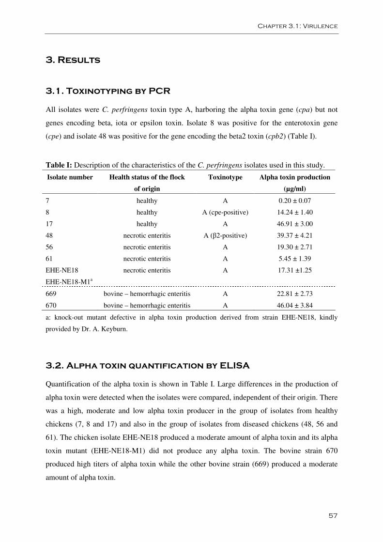
Chapter 3.1: Virulence
57
3. Results
3.1. Toxinotyping by PCR
All isolates were C. perfringens toxin type A, harboring the alpha toxin gene (cpa) but not
genes encoding beta, iota or epsilon toxin. Isolate 8 was positive for the enterotoxin gene
(cpe) and isolate 48 was positive for the gene encoding the beta2 toxin (cpb2) (Table I).
Table I: Description of the characteristics of the C. perfringens isolates used in this study.
Isolate number Health status of the flock
of origin
Toxinotype Alpha toxin production
(µg/ml)
7 healthy A 0.20 ± 0.07
8 healthy A (cpe-positive) 14.24 ± 1.40
17 healthy A 46.91 ± 3.00
48 necrotic enteritis A (β2-positive) 39.37 ± 4.21
56 necrotic enteritis A 19.30 ± 2.71
61 necrotic enteritis A 5.45 ± 1.39
EHE-NE18 necrotic enteritis A 17.31 ±1.25
EHE-NE18-M1a
669 bovine – hemorrhagic enteritis A 22.81 ± 2.73
670 bovine – hemorrhagic enteritis A 46.04 ± 3.84
a: knock-out mutant defective in alpha toxin production derived from strain EHE-NE18, kindly
provided by Dr. A. Keyburn.
3.2. Alpha toxin quantification by ELISA
Quantification of the alpha toxin is shown in Table I. Large differences in the production of
alpha toxin were detected when the isolates were compared, independent of their origin. There
was a high, moderate and low alpha toxin producer in the group of isolates from healthy
chickens (7, 8 and 17) and also in the group of isolates from diseased chickens (48, 56 and
61). The chicken isolate EHE-NE18 produced a moderate amount of alpha toxin and its alpha
toxin mutant (EHE-NE18-M1) did not produce any alpha toxin. The bovine strain 670
produced high titers of alpha toxin while the other bovine strain (669) produced a moderate
amount of alpha toxin.
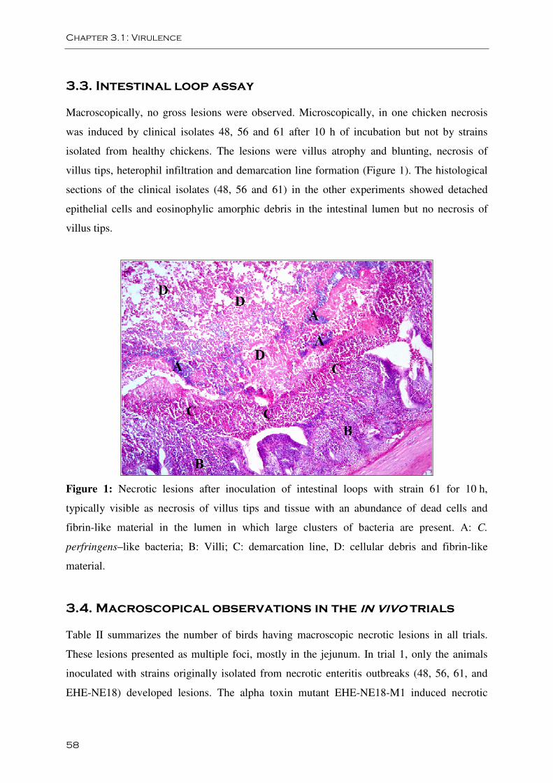
Chapter 3.1: Virulence
58
3.3. Intestinal loop assay
Macroscopically, no gross lesions were observed. Microscopically, in one chicken necrosis
was induced by clinical isolates 48, 56 and 61 after 10 h of incubation but not by strains
isolated from healthy chickens. The lesions were villus atrophy and blunting, necrosis of
villus tips, heterophil infiltration and demarcation line formation (Figure 1). The histological
sections of the clinical isolates (48, 56 and 61) in the other experiments showed detached
epithelial cells and eosinophylic amorphic debris in the intestinal lumen but no necrosis of
villus tips.
Figure 1: Necrotic lesions after inoculation of intestinal loops with strain 61 for 10 h,
typically visible as necrosis of villus tips and tissue with an abundance of dead cells and
fibrin-like material in the lumen in which large clusters of bacteria are present. A: C.
perfringens–like bacteria; B: Villi; C: demarcation line, D: cellular debris and fibrin-like
material.
3.4. Macroscopical observations in the in vivo trials
Table II summarizes the number of birds having macroscopic necrotic lesions in all trials.
These lesions presented as multiple foci, mostly in the jejunum. In trial 1, only the animals
inoculated with strains originally isolated from necrotic enteritis outbreaks (48, 56, 61, and
EHE-NE18) developed lesions. The alpha toxin mutant EHE-NE18-M1 induced necrotic
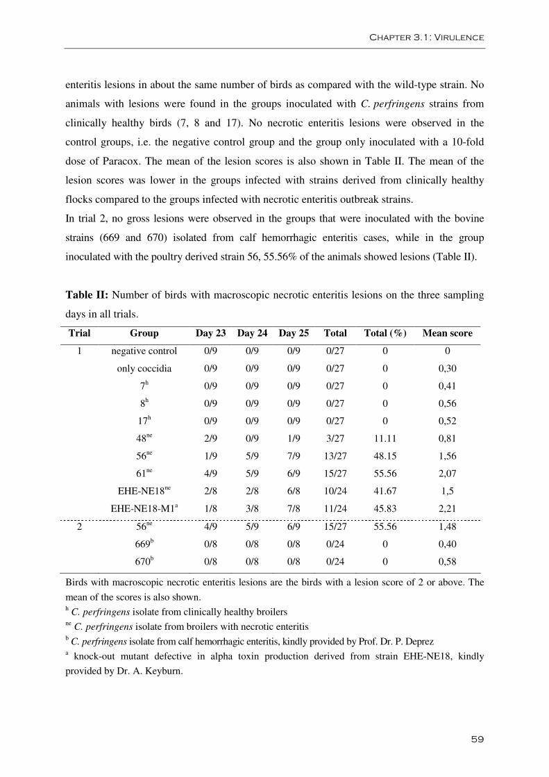
Chapter 3.1: Virulence
59
enteritis lesions in about the same number of birds as compared with the wild-type strain. No
animals with lesions were found in the groups inoculated with C. perfringens strains from
clinically healthy birds (7, 8 and 17). No necrotic enteritis lesions were observed in the
control groups, i.e. the negative control group and the group only inoculated with a 10-fold
dose of Paracox. The mean of the lesion scores is also shown in Table II. The mean of the
lesion scores was lower in the groups infected with strains derived from clinically healthy
flocks compared to the groups infected with necrotic enteritis outbreak strains.
In trial 2, no gross lesions were observed in the groups that were inoculated with the bovine
strains (669 and 670) isolated from calf hemorrhagic enteritis cases, while in the group
inoculated with the poultry derived strain 56, 55.56% of the animals showed lesions (Table II).
Table II: Number of birds with macroscopic necrotic enteritis lesions on the three sampling
days in all trials.
Trial Group Day 23 Day 24 Day 25 Total Total (%) Mean score
1 negative control 0/9 0/9 0/9 0/27 0 0
only coccidia 0/9 0/9 0/9 0/27 0 0,30
7h 0/9 0/9 0/9 0/27 0 0,41
8h 0/9 0/9 0/9 0/27 0 0,56
17h 0/9 0/9 0/9 0/27 0 0,52
48ne 2/9 0/9 1/9 3/27 11.11 0,81
56ne 1/9 5/9 7/9 13/27 48.15 1,56
61ne 4/9 5/9 6/9 15/27 55.56 2,07
EHE-NE18ne 2/8 2/8 6/8 10/24 41.67 1,5
EHE-NE18-M1a 1/8 3/8 7/8 11/24 45.83 2,21
2 56ne 4/9 5/9 6/9 15/27 55.56 1,48
669b 0/8 0/8 0/8 0/24 0 0,40
670b 0/8 0/8 0/8 0/24 0 0,58
Birds with macroscopic necrotic enteritis lesions are the birds with a lesion score of 2 or above. The
mean of the scores is also shown. h C. perfringens isolate from clinically healthy broilers ne C. perfringens isolate from broilers with necrotic enteritis b C. perfringens isolate from calf hemorrhagic enteritis, kindly provided by Prof. Dr. P. Deprez a knock-out mutant defective in alpha toxin production derived from strain EHE-NE18, kindly
provided by Dr. A. Keyburn.
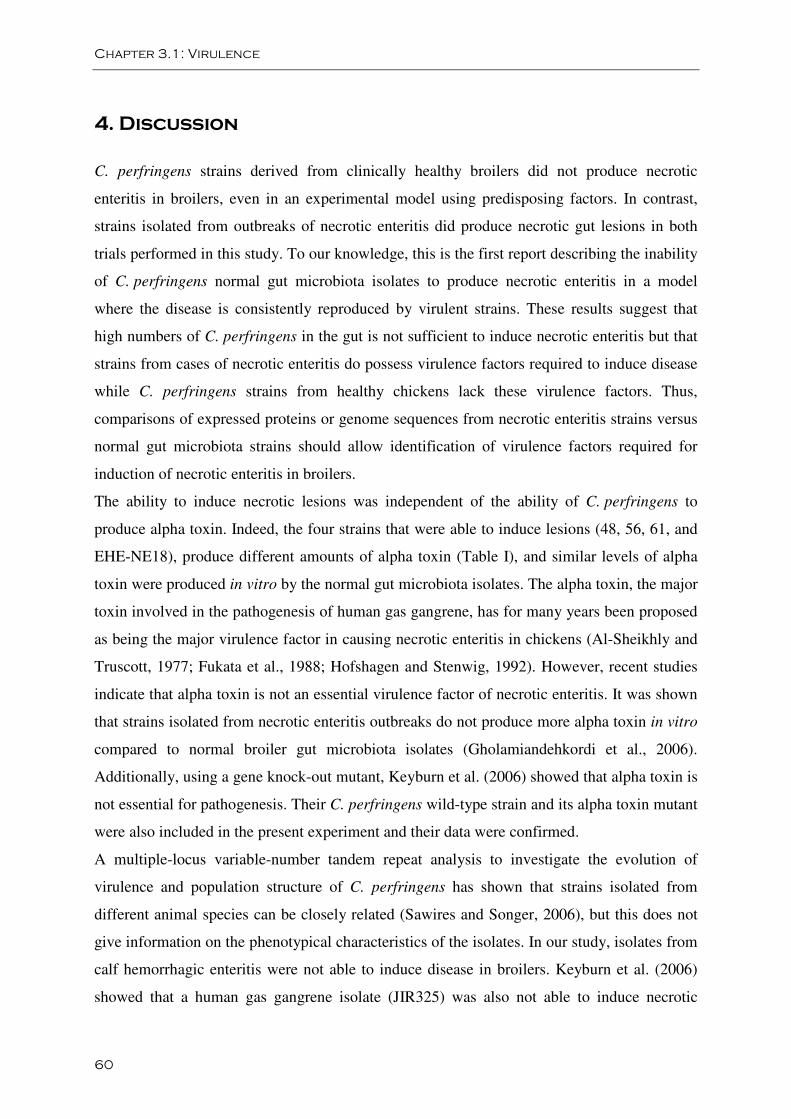
Chapter 3.1: Virulence
60
4. Discussion
C. perfringens strains derived from clinically healthy broilers did not produce necrotic
enteritis in broilers, even in an experimental model using predisposing factors. In contrast,
strains isolated from outbreaks of necrotic enteritis did produce necrotic gut lesions in both
trials performed in this study. To our knowledge, this is the first report describing the inability
of C. perfringens normal gut microbiota isolates to produce necrotic enteritis in a model
where the disease is consistently reproduced by virulent strains. These results suggest that
high numbers of C. perfringens in the gut is not sufficient to induce necrotic enteritis but that
strains from cases of necrotic enteritis do possess virulence factors required to induce disease
while C. perfringens strains from healthy chickens lack these virulence factors. Thus,
comparisons of expressed proteins or genome sequences from necrotic enteritis strains versus
normal gut microbiota strains should allow identification of virulence factors required for
induction of necrotic enteritis in broilers.
The ability to induce necrotic lesions was independent of the ability of C. perfringens to
produce alpha toxin. Indeed, the four strains that were able to induce lesions (48, 56, 61, and
EHE-NE18), produce different amounts of alpha toxin (Table I), and similar levels of alpha
toxin were produced in vitro by the normal gut microbiota isolates. The alpha toxin, the major
toxin involved in the pathogenesis of human gas gangrene, has for many years been proposed
as being the major virulence factor in causing necrotic enteritis in chickens (Al-Sheikhly and
Truscott, 1977; Fukata et al., 1988; Hofshagen and Stenwig, 1992). However, recent studies
indicate that alpha toxin is not an essential virulence factor of necrotic enteritis. It was shown
that strains isolated from necrotic enteritis outbreaks do not produce more alpha toxin in vitro
compared to normal broiler gut microbiota isolates (Gholamiandehkordi et al., 2006).
Additionally, using a gene knock-out mutant, Keyburn et al. (2006) showed that alpha toxin is
not essential for pathogenesis. Their C. perfringens wild-type strain and its alpha toxin mutant
were also included in the present experiment and their data were confirmed.
A multiple-locus variable-number tandem repeat analysis to investigate the evolution of
virulence and population structure of C. perfringens has shown that strains isolated from
different animal species can be closely related (Sawires and Songer, 2006), but this does not
give information on the phenotypical characteristics of the isolates. In our study, isolates from
calf hemorrhagic enteritis were not able to induce disease in broilers. Keyburn et al. (2006)
showed that a human gas gangrene isolate (JIR325) was also not able to induce necrotic
Chapter 3.1: Virulence
61
enteritis in broilers. It is therefore likely that certain C. perfringens strains engage in host-
specific interactions with the chicken host. Especially cytotoxic effects and induction of
lesions, but maybe not colonization of the gut itself, could be host-specific. Although more
studies are needed to definitely prove this hypothesis, our data suggest that C. perfringens
type A isolates from hemorrhagic enteritis in calves do not cause disease in broilers. It is
unclear whether these strains could be part of the normal gut microbiota of broilers.
In conclusion, data were produced in agreement with the hypothesis that isolates from broilers
with necrotic enteritis are virulent while isolates from healthy chickens are not. In addition it
was demonstrated that induction of necrotic lesions in the broiler gut is not associated with
the ability to produce alpha toxin in vitro. Moreover, the results also suggest that the virulence
of C. perfringens strains is to some extent host specific since two C. perfringens strains
isolated from calf hemorrhagic enteritis were not able to produce necrotic lesions in chickens.
Acknowledgements
We would like to thank Veerle Flama, Renzo Vercammen, Christian Puttevils and Delphine
Ameye for their skilful technical assistance. Dr. Gerard Huyghebaert (Institute for
Agricultural and Fisheries Research, Melle, Belgium) is thanked for formulation of the feed.
Prof. Dr. P. Deprez and Dr. A. Keyburn are acknowledged for providing strains. This work
was supported by the Institute for Science and Technology, Flanders (IWT). Dr. F. Van
Immerseel is Postdoctoral Research Fellow of the Research Foundation - Flanders (FWO).
Chapter 3.1: Virulence
62
References
Al-Sheikhly F., Truscott R.B. (1977). The interaction of Clostridium perfringens and its
toxins in the production of necrotic enteritis of chickens. Avian Dis. 21, 256-263.
Dahiya J.P., Wilkie D.C., Van Kessel A.G., Drew M.D. (2006). Potential strategies for
controlling necrotic enteritis in broiler chickens in post-antibiotic era. Anim. Feed Sci.
Technol. 129, 60-88.
Engström B.E., Fermér C., Lindberg A., Saarinen E., Båverud V., Gunnarsson A. (2003).
Molecular typing of isolates of Clostridium perfringens from healthy and diseased poultry.
Vet. Microbiol. 94, 225-235.
Fukata T., Hadate Y., Baba E., Uemara T., Arakawa A. (1988). Influence of Clostridium
perfringens and its toxin in germ-free chickens. Res. Vet. Sci. 44, 68-70.
Gholamiandehkordi A.R., Ducatelle R., Heyndrickx M., Haesebrouck F., Van Immerseel F.
(2006). Molecular and phenotypical characterization of Clostridium perfringens isolates from
poultry flocks with different disease status. Vet. Microbiol. 113, 143-152.
Gholamiandehkordi A.R., Timbermont L., Lanckriet A., Van Den Broeck W., Pedersen K.,
Dewulf J., Pasmans F., Haesebrouck F., Ducatelle R., Van Immerseel F. (2007).
Quantification of gut lesions in a subclinical necrotic enteritis model. Avian Pathol. 36, 375-
382.
Grave K., Kaldhusdal M.C., Kruse H., Harr L.M., Flatlandsmo K. (2004). What has happened
in Norway after the ban of avoparcin? Consumption of antimicrobials by poultry. Prev. Vet.
Med. 62, 59-72.
Hatheway C.L. (1999). Toxigenic Clostridia. Clin. Microbiol. Rev. 3, 66-98.
Herholz C., Miserez R., Nicolet J., Frey J., Popoff M., Gibert M., Gerber H., Straub R.
(1999). Prevalence of β2-toxigenic Clostridium perfringens in horses with intestinal disorders.
J. Clin. Microbiol. 37, 358-361.
Chapter 3.1: Virulence
63
Hofshagen M., Stenwig H. (1992). Toxin production by Clostridium perfringens isolated
from broiler chickens and capercaillies (Tetrao urogallus) with and without necrotizing
enteritis. Avian Dis. 36, 837-843.
Kaldhusdal M., Skjerve E. (1996). Association between cereal content in the diet and
incidence of necrotic enteritis in broiler chickens in Norway. Prev. Vet. Med. 28, 1-16.
Keyburn A.L., Sheedy S.A., Ford M.E., Williamson M.M., Awad M.M., Rood J.I., Moore
R.J. (2006). Alpha toxin of Clostridium perfringens is not an essential virulence factor in
necrotic enteritis in chickens. Infect. Immun. 74, 6496-6500.
McDonel J.L. (1980). Clostridium perfringens toxins (type A, B, C, D, E). Pharmacol. Ther.
10, 617-655.
Meer R.R., Songer J.G. (1997). Multiplex polymerase chain reaction assay for genotyping
Clostridium perfringens. Am. J. Vet. Res. 58, 702-705.
Nauerby B., Pedersen K., Madsen M. (2003). Analysis by pulsed-field gel electrophoresis of
the genetic diversity among Clostridium perfringens isolates from chickens. Vet. Microbiol.
94, 257-266.
Petit L., Gibert M., Popoff M.R. (1999). Clostridium perfringens: toxinotype and genotype.
Trends Microbiol. 7, 104-110.
Sawires Y.S., Songer J.G. (2006). Clostridium perfringens: Insight into virulence evolution
and population structure. Anaerobe 12, 23-43.
Songer J.G. (1996). Clostridial enteric diseases of domestic animals. Clin. Microbiol. Rev. 9,
216-234.
Van Immerseel F., De Buck J., Pasmans F., Huyghebaert G., Haesebrouck F., Ducatelle R.
(2004). Clostridium perfringens in poultry: an emerging threat for animal and public health.
Avian Pathol. 33, 537-549.
Williams R.B. (2005). Intercurrent coccidiosis and necrotic enteritis of chickens: rational,
integrated disease management by maintenance of gut integrity. Avian Pathol. 34, 159-180.
Chapter 3.1: Virulence
64
Yoo H.S., Lee S.U., Park K.Y., Park Y.H. (1997). Molecular typing and epidemiological
survey of prevalence of Clostridium perfringens types by multiplex PCR. J. Clin. Microbiol.
35, 228-232.
Zhang G., Darius S., Smith S.R., Ritchie S.J. (2006). In vitro inhibitory effect of hen egg
white lysozyme on Clostridium perfringens type A associated with broiler necrotic enteritis
and its α-toxin production. Lett. Appl. Microbiol. 42, 138-143.
Chapter 3.2:
Intra-species growth-inhibition by
Clostridium perfringens is a possible
virulence trait in necrotic enteritis in
broilers
Leen Timbermont, Anouk Lanckriet, Frank Pasmans, Freddy Haesebrouck,
Richard Ducatelle and Filip Van Immerseel
Department of Pathology, Bacteriology and Poultry Diseases, Research Group Veterinary
Public Health and Zoonoses, Faculty of Veterinary Medicine, Ghent University, Salisburylaan
133, 9820 Merelbeke, Belgium.
Veterinary Microbiology, 2009, 137: 388-391

Chapter 3.2: Growth-Inhibition
67
Abstract
Necrotic enteritis in broiler chickens is associated with Clostridium perfringens type A,
carrying the NetB toxin. Clostridium perfringens type A is also a member of the normal
intestinal microbiota of broilers. Clinically healthy chickens carry several different
Clostridium perfringens clones in their intestine. In flocks suffering from necrotic enteritis,
however, mostly only one single clone is isolated from the gut of all the diseased animals.
Selective proliferation of these clinical outbreak strains in the gut and spread within the flock
seems likely, but an explanation has not yet been given. The hypothesis that necrotic enteritis
associated Clostridium perfringens strains might suppress the growth of normal microbiota
Clostridium perfringens strains, was therefore tested. Twenty-six Clostridium perfringens
strains isolated from healthy broilers and 24 clinical outbreak isolates were evaluated for their
ability to induce intra-species growth-inhibition in an in vitro setup. A significantly higher
proportion of the Clostridium perfringens clinical outbreak strains inhibited the growth of
other Clostridium perfringens strains compared to Clostridium perfringens strains isolated
from the gut of healthy chickens. It is proposed that, in addition to toxin production, intra-
species growth-inhibition may be a virulence trait that contributes to the ability of certain
Clostridium perfringens strains to cause necrotic enteritis in broilers.
Keywords: Clostridium perfringens; Broiler; Necrotic enteritis; Growth-inhibition
Chapter 3.2: Growth-Inhibition
68
1. Introduction
Since the ban on growth-promoting antibiotics in animal feed in the European Union,
Clostridium perfringens associated necrotic enteritis is re-emerging in broilers (Grave et al.,
2004; Van Immerseel et al., 2004; Williams, 2005). Necrotic enteritis in poultry is associated
with C. perfringens type A, carrying the NetB toxin (Keyburn et al., 2008; Van Immerseel et
al., 2009). C. perfringens type A is also a member of the normal intestinal microbiota of
broilers. Strains isolated from healthy broilers, however, do not induce necrotic enteritis in an
experimental model using predisposing factors, in contrast to strains isolated from outbreaks
of necrotic enteritis (Timbermont et al., 2009).
In C. perfringens isolates from healthy birds, a high degree of genetic diversity is found, even
between isolates from the same animal. In contrast, different isolates from a flock suffering
from a clinical outbreak are generally of the same pulsed-field gel electrophoresis (PFGE)
type, regardless of the animal or the part of the intestine from which the strain was isolated
(Nauerby et al., 2003; Gholamiandehkordi et al., 2006). The reason for the presence of a
single clone in necrotic enteritis outbreaks is not known. It is speculated that during an
outbreak, certain C. perfringens strains have a competitive advantage over other
C. perfringens strains in the broiler gut.
In the present study, strains isolated from healthy broilers and strains isolated from broilers
suffering from necrotic enteritis were compared with respect to their capacity of intra-species
growth-inhibition in an in vitro inhibition assay.
2. Materals and methods
2.1. Bacteria
Fifty C. perfringens type A strains belonging to different genotypes, as analyzed by PFGE,
were included. Thirty-five strains were isolated from broiler chickens in Belgium: 26 strains
from clinically healthy broiler chickens and 9 strains from broilers suffering from necrotic
enteritis (Gholamiandehkordi et al., 2006). Fifteen Danish C. perfringens isolates from
necrotic enteritis cases were kindly provided by Dr. L. Bjerrum (Nauerby et al., 2003).
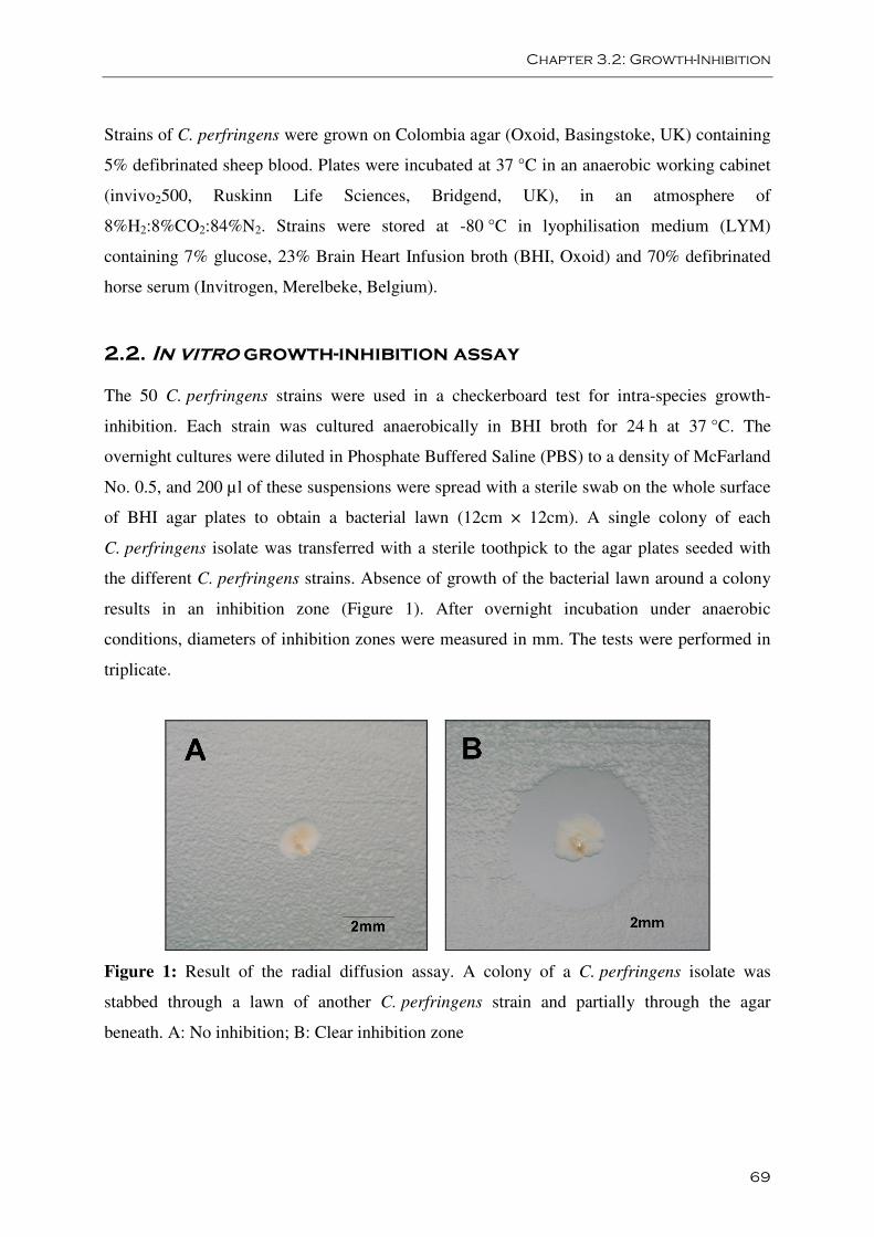
Chapter 3.2: Growth-Inhibition
69
Strains of C. perfringens were grown on Colombia agar (Oxoid, Basingstoke, UK) containing
5% defibrinated sheep blood. Plates were incubated at 37 °C in an anaerobic working cabinet
(invivo2500, Ruskinn Life Sciences, Bridgend, UK), in an atmosphere of
8%H2:8%CO2:84%N2. Strains were stored at -80 °C in lyophilisation medium (LYM)
containing 7% glucose, 23% Brain Heart Infusion broth (BHI, Oxoid) and 70% defibrinated
horse serum (Invitrogen, Merelbeke, Belgium).
2.2. In vitro growth-inhibition assay
The 50 C. perfringens strains were used in a checkerboard test for intra-species growth-
inhibition. Each strain was cultured anaerobically in BHI broth for 24 h at 37 °C. The
overnight cultures were diluted in Phosphate Buffered Saline (PBS) to a density of McFarland
No. 0.5, and 200 µl of these suspensions were spread with a sterile swab on the whole surface
of BHI agar plates to obtain a bacterial lawn (12cm × 12cm). A single colony of each
C. perfringens isolate was transferred with a sterile toothpick to the agar plates seeded with
the different C. perfringens strains. Absence of growth of the bacterial lawn around a colony
results in an inhibition zone (Figure 1). After overnight incubation under anaerobic
conditions, diameters of inhibition zones were measured in mm. The tests were performed in
triplicate.
Figure 1: Result of the radial diffusion assay. A colony of a C. perfringens isolate was
stabbed through a lawn of another C. perfringens strain and partially through the agar
beneath. A: No inhibition; B: Clear inhibition zone
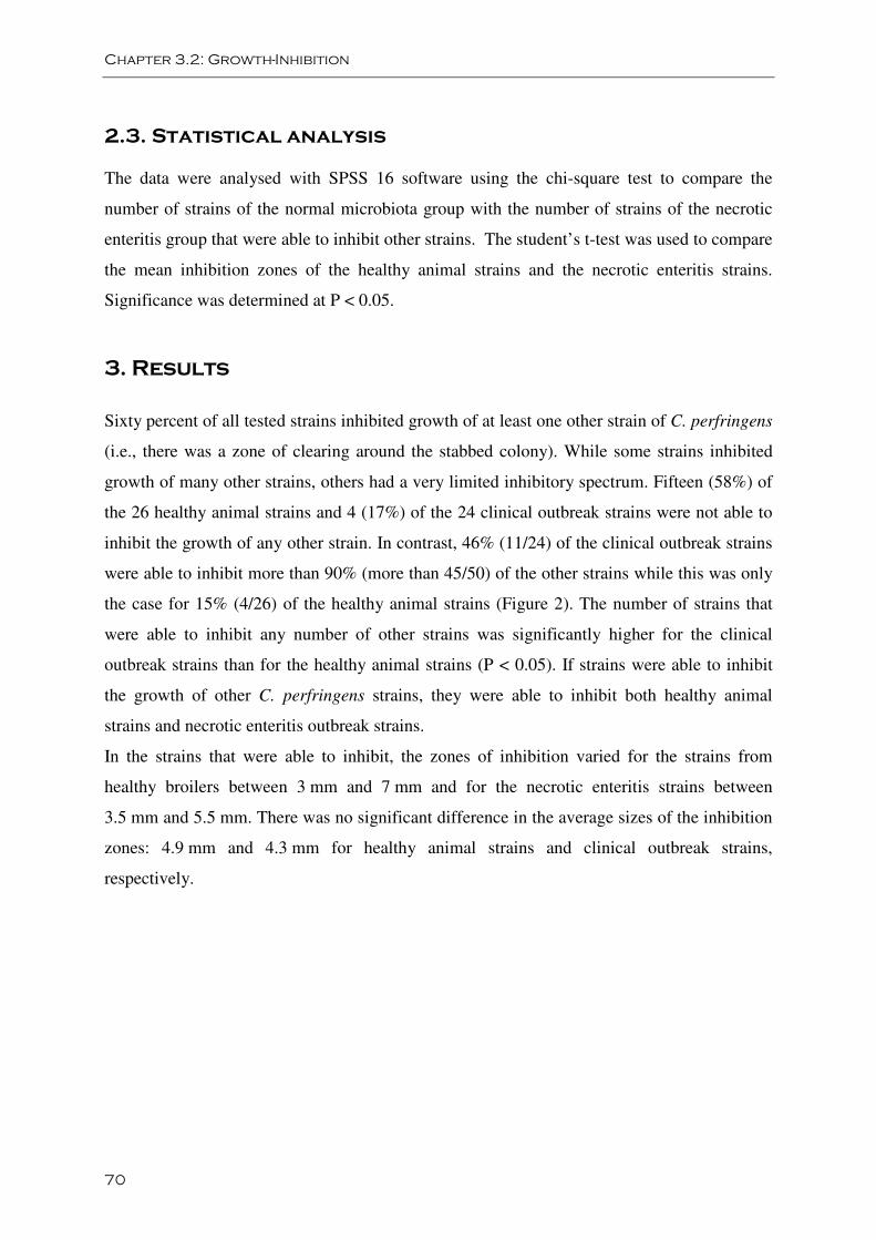
Chapter 3.2: Growth-Inhibition
70
2.3. Statistical analysis
The data were analysed with SPSS 16 software using the chi-square test to compare the
number of strains of the normal microbiota group with the number of strains of the necrotic
enteritis group that were able to inhibit other strains. The student’s t-test was used to compare
the mean inhibition zones of the healthy animal strains and the necrotic enteritis strains.
Significance was determined at P < 0.05.
3. Results
Sixty percent of all tested strains inhibited growth of at least one other strain of C. perfringens
(i.e., there was a zone of clearing around the stabbed colony). While some strains inhibited
growth of many other strains, others had a very limited inhibitory spectrum. Fifteen (58%) of
the 26 healthy animal strains and 4 (17%) of the 24 clinical outbreak strains were not able to
inhibit the growth of any other strain. In contrast, 46% (11/24) of the clinical outbreak strains
were able to inhibit more than 90% (more than 45/50) of the other strains while this was only
the case for 15% (4/26) of the healthy animal strains (Figure 2). The number of strains that
were able to inhibit any number of other strains was significantly higher for the clinical
outbreak strains than for the healthy animal strains (P < 0.05). If strains were able to inhibit
the growth of other C. perfringens strains, they were able to inhibit both healthy animal
strains and necrotic enteritis outbreak strains.
In the strains that were able to inhibit, the zones of inhibition varied for the strains from
healthy broilers between 3 mm and 7 mm and for the necrotic enteritis strains between
3.5 mm and 5.5 mm. There was no significant difference in the average sizes of the inhibition
zones: 4.9 mm and 4.3 mm for healthy animal strains and clinical outbreak strains,
respectively.
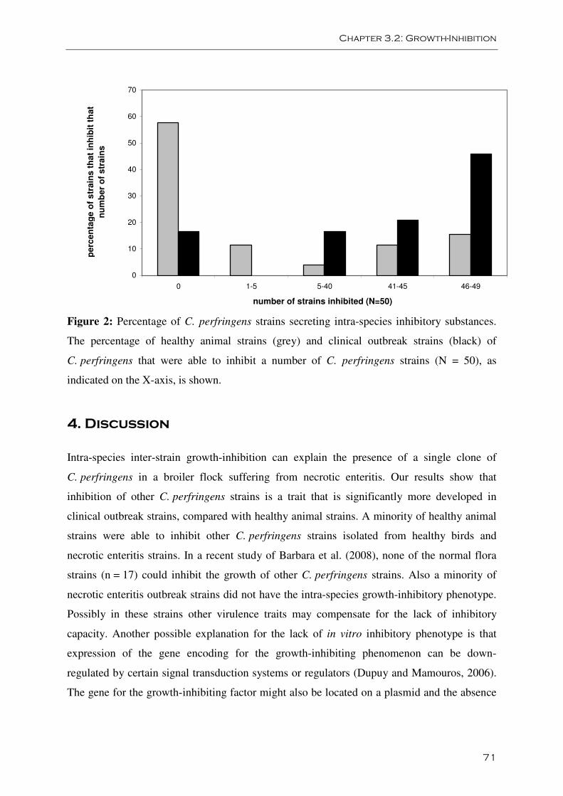
Chapter 3.2: Growth-Inhibition
71
0
10
20
30
40
50
60
70
0 1-5 5-40 41-45 46-49
number of strains inhibited (N=50)
pe
rce
nta
ge
of
str
ain
s t
ha
t in
hib
it t
ha
t
nu
mb
er
of
str
ain
s
Figure 2: Percentage of C. perfringens strains secreting intra-species inhibitory substances.
The percentage of healthy animal strains (grey) and clinical outbreak strains (black) of
C. perfringens that were able to inhibit a number of C. perfringens strains (N = 50), as
indicated on the X-axis, is shown.
4. Discussion
Intra-species inter-strain growth-inhibition can explain the presence of a single clone of
C. perfringens in a broiler flock suffering from necrotic enteritis. Our results show that
inhibition of other C. perfringens strains is a trait that is significantly more developed in
clinical outbreak strains, compared with healthy animal strains. A minority of healthy animal
strains were able to inhibit other C. perfringens strains isolated from healthy birds and
necrotic enteritis strains. In a recent study of Barbara et al. (2008), none of the normal flora
strains (n = 17) could inhibit the growth of other C. perfringens strains. Also a minority of
necrotic enteritis outbreak strains did not have the intra-species growth-inhibitory phenotype.
Possibly in these strains other virulence traits may compensate for the lack of inhibitory
capacity. Another possible explanation for the lack of in vitro inhibitory phenotype is that
expression of the gene encoding for the growth-inhibiting phenomenon can be down-
regulated by certain signal transduction systems or regulators (Dupuy and Mamouros, 2006).
The gene for the growth-inhibiting factor might also be located on a plasmid and the absence
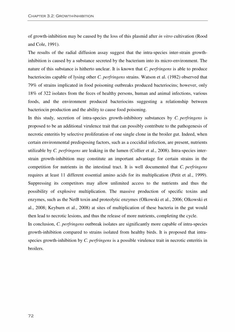
Chapter 3.2: Growth-Inhibition
72
of growth-inhibition may be caused by the loss of this plasmid after in vitro cultivation (Rood
and Cole, 1991).
The results of the radial diffusion assay suggest that the intra-species inter-strain growth-
inhibition is caused by a substance secreted by the bacterium into its micro-environment. The
nature of this substance is hitherto unclear. It is known that C. perfringens is able to produce
bacteriocins capable of lysing other C. perfringens strains. Watson et al. (1982) observed that
79% of strains implicated in food poisoning outbreaks produced bacteriocins; however, only
18% of 322 isolates from the feces of healthy persons, human and animal infections, various
foods, and the environment produced bacteriocins suggesting a relationship between
bacteriocin production and the ability to cause food poisoning.
In this study, secretion of intra-species growth-inhibitory substances by C. perfringens is
proposed to be an additional virulence trait that can possibly contribute to the pathogenesis of
necrotic enteritis by selective proliferation of one single clone in the broiler gut. Indeed, when
certain environmental predisposing factors, such as a coccidial infection, are present, nutrients
utilizable by C. perfringens are leaking in the lumen (Collier et al., 2008). Intra-species inter-
strain growth-inhibition may constitute an important advantage for certain strains in the
competition for nutrients in the intestinal tract. It is well documented that C. perfringens
requires at least 11 different essential amino acids for its multiplication (Petit et al., 1999).
Suppressing its competitors may allow unlimited access to the nutrients and thus the
possibility of explosive multiplication. The massive production of specific toxins and
enzymes, such as the NetB toxin and proteolytic enzymes (Olkowski et al., 2006; Olkowski et
al., 2008; Keyburn et al., 2008) at sites of multiplication of these bacteria in the gut would
then lead to necrotic lesions, and thus the release of more nutrients, completing the cycle.
In conclusion, C. perfringens outbreak isolates are significantly more capable of intra-species
growth-inhibition compared to strains isolated from healthy birds. It is proposed that intra-
species growth-inhibition by C. perfringens is a possible virulence trait in necrotic enteritis in
broilers.
Chapter 3.2: Growth-Inhibition
73
Acknowledgements
We would like to thank Veerle Flama for her skilful technical assistance. Dr. L. Bjerrum is
acknowledged for providing strains. This work was supported by the Institute for Science and
Technology, Flanders (IWT). F. Van Immerseel is supported by a Postdoctoral Research
Grant of the Research Foundation - Flanders (FWO) and by the Research Fund of Ghent
University (BOF).
Chapter 3.2: Growth-Inhibition
74
References
Barbara A.J., Trinh H.T., Glock R.D., Songer J.G. (2008). Necrotic enteritis-producing strains
of Clostridium perfringens displace non-necrotic enteritis strains from the gut of chicks. Vet.
Microbiol. 126, 377-382.
Dupuy B., Matamouros S. (2006). Regulation of toxin and bacteriocin synthesis in
Clostridium species by a new subgroup of RNA polymerase σ-factors. Res. Microbiol. 157,
201-205.
Collier C.T., Hofacre C.L., Payne A.M., Anderson D.B., Kaiser P., Mackie R.I., Gaskins H.R.
(2008). Coccidia-inuced mucogenesis promotes the onset of necrotic enteritis by supporting
Clostridium perfringens growth. Vet. Immunol. Immunopathol. 122, 105-114.
Gholamiandehkordi A.R., Ducatelle R., Heyndrickx M., Haesebrouck F., Van Immerseel F.
(2006). Molecular and phenotypical characterization of Clostridium perfringens isolates from
poultry flocks with different disease status. Vet. Microbiol. 113, 143-152.
Grave K., Kaldhusdal M.C., Kruse H., Harr L.M., Flatlandsmo K. (2004). What has happened
in Norway after the ban of avoparcin? Consumption of antimicrobials by poultry. Prev. Vet.
Med. 62, 59-72.
Keyburn A.L., Boyce J.D., Vaz P., Bannam T.L., Ford M.E., Parker D., Di Rubbo A., Rood
J.I., Moore R.J. (2008). NetB, a new toxin that is associated with avian necrotic enteritis
caused by Clostridium perfringens. PLoS Pathog. 4(2): e26. doi:10.1371/journal.ppat.
0040026.
Nauerby B., Pedersen K., Madsen M. (2003). Analysis by pulsed-field gel electrophoresis of
the genetic diversity among Clostridium perfringens isolates from chickens. Vet. Microbiol.
94, 257-266.
Olkowski A.A., Wojnarowicz C., Chirino-Trejo M., Drew M.D. (2006). Responses of broiler
chickens orally challenged with Clostridium perfringens isolated from field cases of necrotic
enteritis. Res. Vet. Sci. 8, 99-108.
Chapter 3.2: Growth-Inhibition
75
Olkowski A.A., Wojnarowicz C., Chirino-Trejo M., Laarveld B., Sawicki G. (2008). Sub-
clinical necrotic enteritis in broiler chickens: Novel etiological consideration based on ultra-
structural and molecular changes in the intestinal tissue. Res. Vet. Sci. 85, 543-553.
Petit L., Gibert M., Popoff M.R. (1999). Clostridium perfringens: toxinotypes and genotype.
Trends in Microbiol. 7, 104-110.
Rood J.I., Cole S.T. (1991). Molecular genetics and pathogenesis of Clostridium perfringens.
Microbiol. rev. 55, 621-648.
Timbermont L., Lanckriet A., Gholamiandehkordi A.R., Pasmans F., Martel A., Haesebrouck
F., Ducatelle R., Van Immerseel F. (2009). Origin of Clostridium perfringens isolates
determines the ability to induce necrotic enteritis in broilers. Comp. Immunol. Microbiol.
Infect. Dis. 32, 503-512.
Van Immerseel F., De Buck J., Pasmans F., Huyghebaert G., Haesebrouck F., Ducatelle R.
(2004). Clostridium perfringens in poultry: an emerging threat for animal and public health.
Avian Pathol. 33, 537-549.
Van Immerseel F., Rood J.I., Moore R.J., Titball R.W. (2009). Rethinking our understanding
of the pathogenesis of necrotic enteritis in chickens. Trends Microbiol. 17, 32-36.
Watson G.N., Stringer M.F., Gilbert R.J., Mahony D.E. (1982). The potential of bacteriocin
typing in the study of Clostridium perfringens food poisoning. J. Clin. Pathol. 35, 1361-1365.
Williams R.B. (2005). Intercurrent coccidiosis and necrotic enteritis of chickens: rational,
integrated disease management by maintenance of gut integrity. Avian Pathol. 34, 159-180.

Chapter 3.3:
Purification and partial
characterization of a novel
antimicrobial peptide from Clostridium
perfringens strain 56
Leen Timbermont§, Lina De Smet‡, Anouk Lanckriet§, Gonzalez Van
Driessche‡, Freddy Haesebrouck§, Richard Ducatelle§, Bart Devreese‡, Filip
Van Immerseel§
§ Department of Pathology, Bacteriology and Poultry Diseases, Research Group Veterinary
Public Health and Zoonoses, Faculty of Veterinary Medicine, Ghent University, Salisburylaan
133, B-9820 Merelbeke, Belgium.
‡ Department of Biochemistry, Physiology and Microbiology, Faculty of Sciences, Ghent
University, K. L. Ledeganckstraat 35, 9000 Gent, Belgium.

Chapter 3.3: Purification and characterization
79
Abstract
Necrotic enteritis in broiler chickens is associated with Clostridium perfringens type A.
Single strain dominance is found in the gut of broiler chickens suffering from necrotic
enteritis: within an outbreak, all affected animals carry the same clonal
Clostridium perfringens strain in the affected tissue. It is known that Clostridium perfringens
is capable of secreting factors inhibiting growth of other Clostridium perfringens strains and
this characteristic is more prevalent in outbreak strains compared to normal microbiota
strains. This characteristic could lead to extensive and selective presence of a strain that
contains the genetic make-up enabling to secrete toxins that cause gut lesions. This report
describes the purification and the characterization of a bacteriocin-like inhibitory substance
(BLIS) from a necrotic enteritis associated Clostridium perfringens strain exhibiting
antibacterial activity against other Clostridium perfringens strains. Amino acid sequence
analysis indicated that the BLIS is an 11.5 kDa fragment of a 22 kDa protein and that it
constitutes the C-terminal part of this protein. The 22 kDa protein is a cytoplasmic membrane
protein without signal peptide. The antibacterial activity of the purified BLIS was abolished
by proteolytic enzymes trypsin and proteinase K and by heat treatment (10 min at 80 °C). The
purified BLIS showed inhibitory activity over a wide pH-range (4.0 to 10.0). Since the
antibacterial activity against other Clostridium perfringens strains of the purified BLIS had a
narrower spectrum than the crude supernatant of the secreting strain, most probably different
of these bacteriocins are produced.
Key words: Clostridium perfringens; Necrotic enteritis; Broiler; Bacteriocin
Chapter 3.3: Purification and characterization
80
1. Introduction
Clostridium perfringens type A is widely prevalent in the environment and in the intestinal
tract of animals and men. Necrotic enteritis in poultry is associated with a specific
subpopulation of C. perfringens type A, i.e. those carrying the NetB toxin (Keyburn et al.,
2008; Van Immerseel et al., 2009). As a consequence, strains isolated from outbreaks of
necrotic enteritis are able to induce necrotic enteritis in an experimental model using
predisposing factors, in contrast to strains isolated from the gut of healthy broilers
(Timbermont et al., 2009a). In C. perfringens isolates from healthy birds, a high degree of
genetic diversity is found, even between isolates within the gut of a single animal. In contrast,
in a flock suffering from a clinical outbreak, different isolates are generally of the same
pulsed-field gel electrophoresis (PFGE) type, regardless of which animal in the flock or which
part of the intestine the strain was isolated from (Nauerby et al., 2003; Gholamiandehkordi et
al., 2006). Recent data show that C. perfringens is capable of secreting factors inhibiting
growth of other C. perfringens strains. The intra-species inhibitory phenotype was shown to
be more prevalent in outbreak strains compared to normal microbiota strains (Barbara et al.,
2008; Timbermont et al., 2009b). This characteristic could lead to extensive and selective
presence of a single strain in the gut, and if it contains the genetic make-up enabling to secrete
toxins, it could consequently cause gut lesions.
It is known that many C. perfringens strains are able to produce antibacterial proteins, called
bacteriocins (Tagg et al., 1976). Bacteriocins are proteinaceous toxic compounds produced by
bacteria that generally inhibit the growth of closely related strains (Jack et al., 1995), and are
thus potential candidates involved in the intra-species inhibitory phenotype of C. perfringens.
The nature of the inhibitory component that causes intra-species growth-inhibition in broiler
outbreak strains was hitherto unknown. Therefore, in the present study, the purification and
partial characterization of a novel antimicrobial peptide from a C. perfringens outbreak strain
is described.
Chapter 3.3: Purification and characterization
81
2. Materials and methods
2.1. Strains
Fifty C. perfringens type A strains belonging to different genotypes, as analyzed by PFGE,
were included. Thirty-five strains were isolated from broiler chickens in Belgium: 26 strains
from clinically healthy broiler chickens and 9 strains from broilers suffering from necrotic
enteritis. Fifteen Danish C. perfringens isolates from necrotic enteritis cases were kindly
provided by Dr. L. Bjerrum (Nauerby et al., 2003; Gholamiandehkordi et al., 2006;
Timbermont et al., 2009b). Strain 56 was isolated from the intestine of a broiler chicken with
severe necrotic gut lesions. It was selected from the strains because it is shown to be a virulent
strain (Timbermont et al., 2009a) and inhibits the growth of 41 of the 50 strains included
(Timbermont et al., 2009b). C. perfringens strain 6 was isolated from the normal gut
microbiota of a healthy broiler chicken and was used as indicator strain because it is not able
to inhibit other C. perfringens strains and its growth is inhibited by strain 56 (Timbermont et
al., 2009b).
2.2. Agar spot test
For the agar spot test, 20 µl drops of the fractions to be tested were spotted on the lawns of
bacteria. Lawns were prepared by resuspension of colonies in PBS to a density of McFarland
1, and 100 µl of these suspensions were spread with a sterile swab on the surface of agar
plates.
2.3. Antimicrobial protein purification and identification
C. perfringens strain 56 was grown in Tryptic Soy Broth (TSB, Oxoid, Basingstoke, England)
for 24 h at 42 °C (500ml). Culture supernatant was obtained by centrifugation at 17000 g for
15 min at 4 °C. The supernatant was filter sterilized (0.22 µm) and supernatant proteins
precipitated by overnight incubation in 50% (w/v) (NH4)2SO4 at 4 °C followed by
centrifugation at 17000 g for 2 h at 4 °C. The precipitate was resuspended in 5 ml of
phosphate buffered saline (PBS) and dialysed against 10 mM Tris-HCl buffer, pH 8.5 for 24 h
at 4 °C. This concentrated supernatant was loaded onto a SP-sepharose cation exchange
Chapter 3.3: Purification and characterization
82
column in 10 mM NaOAc buffer, pH 4. Proteins were eluted stepwise from the column, with
increasing concentrations of NaCl (up to 1 M). To determine the antimicrobial activity of the
fractions, two-fold dilutions of the fractions were spotted on a lawn of strain 6 in the agar spot
test. The most active fractions in the antimicrobial assay were pooled and loaded onto a butyl-
sepharose hydrophobic interaction column in 50 mM Sodium phosphate buffer with 1 M
(NH4)2SO4, pH 7.4. Proteins were stepwise eluted from the column, with decreasing
concentrations of (NH4)2SO4. Again, the activity of the fractions was determined and the most
active fraction was concentrated and analyzed by SDS-PAGE. Precision Plus Protein
Standard, All Blue (Bio-Rad Laboratories, Hercules, USA) was used as protein marker. The
protein band was cut from the gel and subjected to in-gel protein digestion with trypsin
(Devreese et al., 2002) followed by mass spectrometric characterization. After mixing of 1 µl
of the digestion mixture with 10 µl a-cyano siniapinic acid (5 mg/ml), one microliter was
spotted onto the target plate and analyzed with the 4800 plus MALDI TOF/TOF Analyzer
(Applied Biosystems, Foster City, CA). A NCBI BLAST-search was done with the obtained
amino acid sequences (http://blast.ncbi.nlm.nih.gov/Blast/). The protein fragment sequences
were also compared with the incomplete EHE-NE18 sequence, kindly provided by Prof. J.
Rood (Monash University). Prediction of transmembrane helices was done with TMHMM
(Krogh et al, 2001).
2.4. Characterization of the antimicrobial peptide
Thermal, pH and protease stability of the purified bacteriocin-like inhibitory substance (BLIS)
was tested. The remaining antibacterial activity after treatment was determined by the agar
spot test and compared with the activity of a corresponding control. Dilutions of treated and
untreated (control) BLIS were spotted on lawns of strain 6. First, thermal stability of the
growth-inhibiting factor was investigated by determination of the residual antibacterial
activity after incubation at 4, 24, 37, 42, 60, 80 and 100 °C for 10, 30 and 60 min. To evaluate
the influence of pH on activity, the BLIS sample was adjusted to a pH of 2.0, 4.0, 6.0, 8.0,
10.0 and 12.0 with HCl or NaOH, mixed, incubated at room temperature for 1 h, neutralized
to pH 7, and tested for activity. Effect of trypsin and proteinase K on bacteriocin activity was
tested. Each enzyme was prepared at a concentration of 10 mg/ml and added to the purified
BLIS at a final concentration of 1 and 0.1 mg/ml. After incubation for 1 h at 37 °C, inhibitory
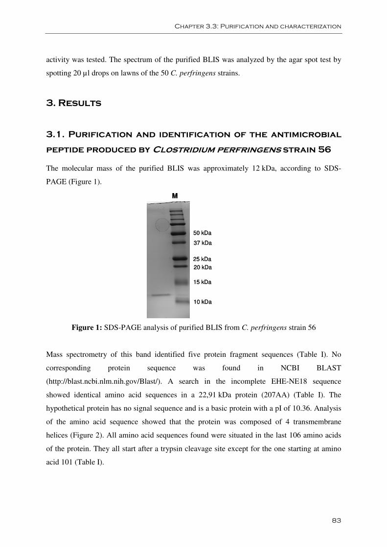
Chapter 3.3: Purification and characterization
83
activity was tested. The spectrum of the purified BLIS was analyzed by the agar spot test by
spotting 20 µl drops on lawns of the 50 C. perfringens strains.
3. Results
3.1. Purification and identification of the antimicrobial
peptide produced by Clostridium perfringens strain 56
The molecular mass of the purified BLIS was approximately 12 kDa, according to SDS-
PAGE (Figure 1).
50 kDa
37 kDa
25 kDa
20 kDa
15 kDa
10 kDa
M
50 kDa
37 kDa
25 kDa
20 kDa
15 kDa
10 kDa
M
Figure 1: SDS-PAGE analysis of purified BLIS from C. perfringens strain 56
Mass spectrometry of this band identified five protein fragment sequences (Table I). No
corresponding protein sequence was found in NCBI BLAST
(http://blast.ncbi.nlm.nih.gov/Blast/). A search in the incomplete EHE-NE18 sequence
showed identical amino acid sequences in a 22,91 kDa protein (207AA) (Table I). The
hypothetical protein has no signal sequence and is a basic protein with a pI of 10.36. Analysis
of the amino acid sequence showed that the protein was composed of 4 transmembrane
helices (Figure 2). All amino acid sequences found were situated in the last 106 amino acids
of the protein. They all start after a trypsin cleavage site except for the one starting at amino
acid 101 (Table I).
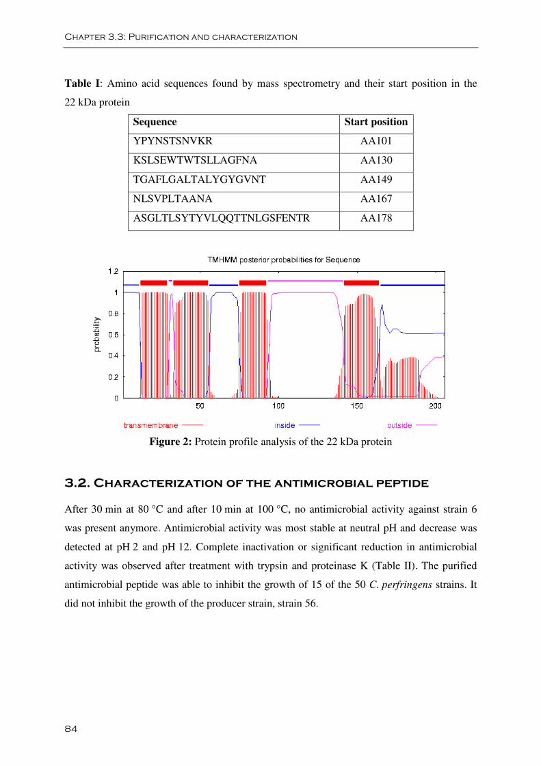
Chapter 3.3: Purification and characterization
84
Table I: Amino acid sequences found by mass spectrometry and their start position in the
22 kDa protein
Sequence Start position
YPYNSTSNVKR AA101
KSLSEWTWTSLLAGFNA AA130
TGAFLGALTALYGYGVNT AA149
NLSVPLTAANA AA167
ASGLTLSYTYVLQQTTNLGSFENTR AA178
Figure 2: Protein profile analysis of the 22 kDa protein
3.2. Characterization of the antimicrobial peptide
After 30 min at 80 °C and after 10 min at 100 °C, no antimicrobial activity against strain 6
was present anymore. Antimicrobial activity was most stable at neutral pH and decrease was
detected at pH 2 and pH 12. Complete inactivation or significant reduction in antimicrobial
activity was observed after treatment with trypsin and proteinase K (Table II). The purified
antimicrobial peptide was able to inhibit the growth of 15 of the 50 C. perfringens strains. It
did not inhibit the growth of the producer strain, strain 56.
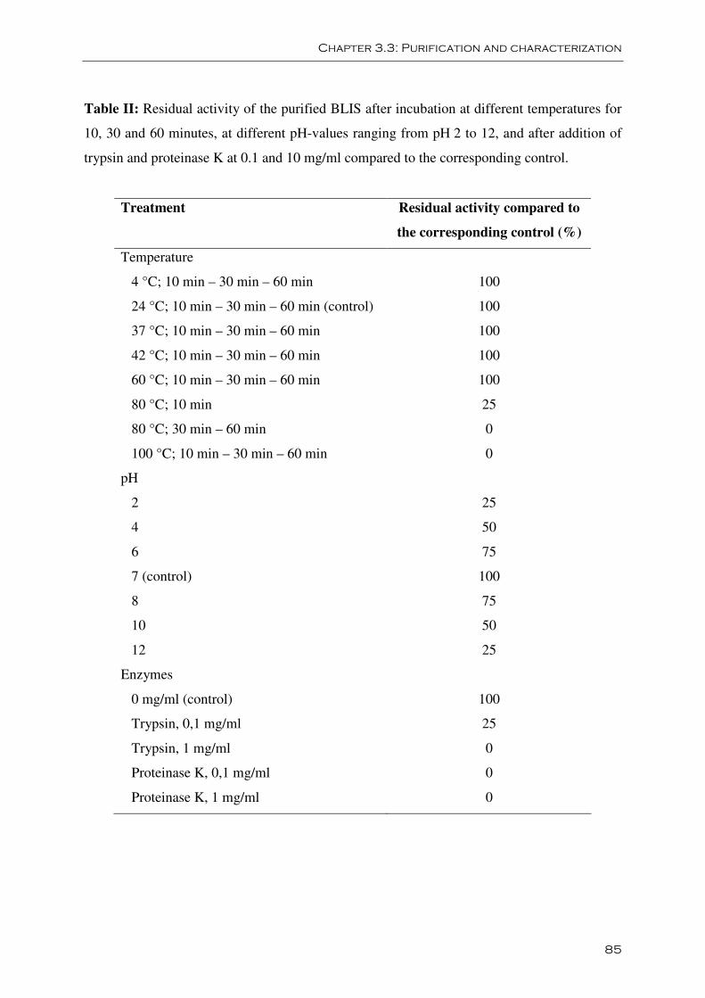
Chapter 3.3: Purification and characterization
85
Table II: Residual activity of the purified BLIS after incubation at different temperatures for
10, 30 and 60 minutes, at different pH-values ranging from pH 2 to 12, and after addition of
trypsin and proteinase K at 0.1 and 10 mg/ml compared to the corresponding control.
Treatment Residual activity compared to
the corresponding control (%)
Temperature
4 °C; 10 min – 30 min – 60 min 100
24 °C; 10 min – 30 min – 60 min (control) 100
37 °C; 10 min – 30 min – 60 min 100
42 °C; 10 min – 30 min – 60 min 100
60 °C; 10 min – 30 min – 60 min 100
80 °C; 10 min 25
80 °C; 30 min – 60 min 0
100 °C; 10 min – 30 min – 60 min 0
pH
2 25
4 50
6 75
7 (control) 100
8 75
10 50
12 25
Enzymes
0 mg/ml (control) 100
Trypsin, 0,1 mg/ml 25
Trypsin, 1 mg/ml 0
Proteinase K, 0,1 mg/ml 0
Proteinase K, 1 mg/ml 0
Chapter 3.3: Purification and characterization
86
4. Discussion
Bacteriocin production might be involved in the pathogenesis of necrotic enteritis, since in
outbreaks of necrotic enteritis single clone dominance is found (Nauerby et al., 2003;
Gholamiandehkordi et al., 2006) and intra-species inter-strain growth-inhibition was found to
be more prevalent in outbreak strains from broilers compared to normal microbiota strains
(Barbara et al., 2008; Timbermont et al., 2009b).
The antibacterial peptide identified in this study is a novel peptide without any homology with
currently known bacteriocins or antimicrobial proteins. No sequence similarity was found
with any genes or proteins in database searches. Even more, no similarity was found with
gene or protein sequences of any of the 3 genome sequenced C. perfringens type A strains
(Shimizu et al., 2002; Myers et al., 2006). The sequenced strains are a gangrene strain and a
food poisoning strain isolated from humans and a gangrene strain isolated from soil. The
antimicrobial peptide may be specific for animal and even for broiler necrotic enteritis strains,
since a match was found in the sequence of EHE-NE18, a strain isolated from a chicken
suffering from necrotic enteritis (Keyburn et al., 2008).
The newly identified bacteriocin-like peptide is hypothesized to be released by proteolytic
activity from a 22 kDa protein without signal peptide that is located in the cytoplasmic
membrane. The amino acid sequence of the protein indicates the presence of four
transmembrane helices. An extracellular loop is located between the third and the fourth
transmembrane helix (Figure 2). Protease activity may cleave the protein at this position,
releasing a 12 kDa protein fragment extracellullarly. All amino acid sequences found by mass
spectrometry, were situated in the last 106 amino acids of the 22 kDa protein. Remarkably, all
amino acid sequences started after a trypsin cleavage site except the one starting at amino acid
101 (Y101; Table I). This result suggests that the purified fragment starts at Y101 since an in-
gel trypsin digestion was performed before mass spectrometry. A fragment starting at Y101
would have a molecular mass of 11,538 kDa which is in accordance with the estimated
molecular mass of the purified antimicrobial peptide (Figure 1). The extracellular loop starts
around amino acid at position 100 and a cleavage site of proteinase K is located before Y101.
Bacteriocins from Gram-positive bacteria are usually produced as precursors with N-terminal
signal peptides (Jack et al., 1995). The signal peptides serve as recognition signals for the
export of the bacteriocins out of the cells, and they also protect the producers against their
own bacteriocin by making them biologically inactive inside the cell. Concomitant with the
Chapter 3.3: Purification and characterization
87
export from the cell, bacteriocins and antimicrobial peptides are processed and activated.
However, it has been shown that antimicrobial peptides can be produced from the degradation
of larger proteins. For example, Faye et al. (2002) showed that Propionibacterium jensenii
secretes an inactive proprotein which is proteolytically activated in the environment. To our
knowledge, this is the first description of a bacteriocin-like peptide that may be formed by an
external protease from a bacterial putative cytoplasm membrane associated protein.
The transmembrane helix at the C-terminal part of the 12 kDa part of the protein, might
suggest that the antimicrobial peptide acts by pore formation. This is a feature similar to those
of many characterized antimicrobial peptides from Gram-positive bacteria (Jack et al., 1995).
It has indeed been shown that the C-terminal region of class IIa bacteriocins forms one or two
helices and they penetrate the cell membrane, thereby inducing leakage and cell death (Moll
et al., 2000; Morisset et al., 2004).
The C. perfringens strain 56 most likely secretes or produces more bacteriocins in addition to
the protein identified in this work. Indeed, the inhibitory spectrum of C. perfringens strain 56
and that of the purified BLIS are not identical. It is not unusual that a C. perfringens strain
produces more than 1 bacteriocin simultaneously. As an example, Higa et al. (1991) showed
that C. perfringens SN-17 produced two bacteriocins in succession. Moreover, it was also
shown that these bacteriocins have different inhibitory spectra. To detect the other
antimicrobial factor(s), screening of the protein fractions derived from the supernatant must
be done using other indicator strains. C. perfringens strain 56 is not sensitive to its own
bacteriocin. Genes encoding membrane-associated molecules that confer a degree of specific
protection upon the producer strain have been found. For example, Diep et al. (2007) showed
that an immunity protein prevents Lactococcus lactis from being killed by its permeabilizing
peptide-bacteriocin, lactococcin A, by formation of a strong complex with the receptor
proteins and the bacteriocin. Moreover, it was shown that the immunity protein is only
expressed if mature bacteriocin is produced or if it is present in the environment (Diep et al.,
2007).
In conclusion, a new antimicrobial peptide derived from a necrotic enteritis inducing
C. perfringens strain was purified and partly characterized. Further studies are necessary to
determine the mode of action and the target of the bacteriocin-like peptide and to elucidate its
role in the pathogenesis of necrotic enteritis.
Chapter 3.3: Purification and characterization
88
Acknowledgements
We would like to thank J. Rood for searching the EHE-NE18 sequence and translating the
protein sequence into a DNA sequence. This work was supported by the Institute for Science
and Technology, Flanders (IWT). F. Van Immerseel is supported by a Postdoctoral Research
Grant of the Research Foundation - Flanders (FWO) and the Research Council of Ghent
University (BOF).
Chapter 3.3: Purification and characterization
89
References
Barbara A.J., Trinh H.T., Glock R.D., Songer J.G. (2008). Necrotic enteritis-producing strains
of Clostridium perfringens displace non-necrotic enteritis strains from the gut of chicks. Vet.
Microbiol. 126, 377-382.
Devreese B., Janssen K.P.C., Vanrobaeys F., Van Herp F., Martens G.J.M. and Van Beeumen
J. (2002). Automated nanoflow liquid chromatography – tandem mass spectrometry for a
differential display proteomic study on Xenopus laevis neuroendocrine cells. J. chromatogr. A
976, 113-121.
Diep D.B., Skaugen M., Salehian Z., Holo H., Nes I.F. (2007). Common mechanisms of
target cell recognition and immunity for class II bacteriocins. PNAS 104, 2384-2389.
Faye T., Brede D.A., Langsrud T., Nes I.F., Holo H. (2002). An antimicrobial peptide is
produced by extracellular processing of a protein from Propionibacterium jensenii. J. Bact.
184, 3649-3656.
Gholamiandehkordi A.R., Ducatelle R., Heyndrickx M., Haesebrouck F., Van Immerseel F.
(2006). Molecular and phenotypical characterization of Clostridium perfringens isolates from
poultry flocks with different disease status. Vet. Microbiol. 113, 143-152.
Higa A., Yoshida E., Mryoshi Y. (1991). Characterization of two bacteriocins produced by
C. perfringens. Microbiol. Immunol. 35, 411-421.
Jack R.W., Tagg J.R., Ray B. (1995). Bacteriocins of Gram-positive bacteria. Microbiol. Rev.
59, 171-200.
Keyburn A.L., Boyce J.D., Vaz P., Bannam T.L., Ford M.E., Parker D., Di Rubbo A., Rood J.I.,
Moore R.J. (2008). NetB, a new toxin that is associated with avian necrotic enteritis caused by
Clostridium perfringens. PLoS Pathog. 4(2): e26. doi:10.1371/journal.ppat.0040026.
Krogh A., Larsson B., von Heijne G., Sonnhammer E. L. L. (2001). Predicting
transmembrane protein topology with a hidden Markov model: Application to complete
genomes. J. Molec. Biol. 305, 567-580.
Chapter 3.3: Purification and characterization
90
Moll G.N., Brul S., Konings W.N., Driessen A.J. (2000). Comparison of the membrane
interaction and permeabilization by the designed peptide Ac-MB21-NH2 and truncated
dermaseptin S3. Biochem. 39, 11907-11912.
Morisset D., Berjeaud J.M., Marion D., Lacombe C., Frère J. (2004). Mutational analysis of
mesentericin Y105, an anti-Listeria bacteriocin, for determination of impact on bactericidal
activity, in vitro secondary structure, and membrane interaction. Appl. Environ. Microbiol.
70, 4672-4680.
Myers G.S.A., Rasko D.A., Cheung J.K., Ravel J., Seshadri R., DeBoy R.T., Ren Q., Varga J.,
Awad M.M., Brinkac L.M., Daugherty S.C., Haft D.H., Dodson R.J., Madupu R., Nelson W.C.,
Rosovitz M.J., Sullivan S.A., Khouri H., Dimitrov G.I., Watkins K.L., Mulligan S., Benton J.,
Radune D., Fisher D.J., Atkins H.S., Hiscox T., Jost B.H., Billington S.J., Songer J.G., McClane
B.A., Titball R.W., Rood J.I., Melville S.B., Paulsen I.T. (2006). Skewed genomic variability in
strains of the toxigenic bacterial pathogen, Clostridium perfringens. Genome Res. 16, 1031-
1040.
Nauerby B., Pedersen K., Madsen M. (2003). Analysis by pulsed-field gel electrophoresis of
the genetic diversity among Clostridium perfringens isolates from chickens. Vet. Microbiol.
94, 257-266.
Shimizu T. Ohtani K., Hirakawa H., Ohshima K., Yamashita A., Shiba T., Ogasawara N.,
Hattori M., Kuhara S., Hayashi H. (2002). Complete genome sequence of Clostridium
perfringens, an anaerobic flesh-eater. Proc. Natl. Acad. Sci. 99, 996-1001.
Tagg J.R., Dajani A.S., Wannamaker L.W. (1976). Bacteriocins of Gram-positive bacteria.
Bacteriol. Rev. 40, 722-756.
Timbermont L., Lanckriet A., Gholamiandehkordi A.R., Pasmans F., Martel A., Haesebrouck
F., Ducatelle R., Van Immerseel F. (2009a). Origin of Clostridium perfringens isolates
determines the ability to induce necrotic enteritis in broilers. Comp. Immunol. Microbiol.
Infect. Dis. 32, 503-512.
Timbermont L., Lanckriet A., Pasmans F., Haesebrouck F., Ducatelle R., Van Immerseel F.
(2009b). Intra-species growth-inhibition by Clostridium perfringens is a possible virulence
trait in necrotic enteritis in broilers. Vet. Microbiol. 137, 388-391.
Chapter 3.3: Purification and characterization
91
Van Immerseel F., Rood J.I., Moore R.J., Titball R.W. (2009). Rethinking our understanding
of the pathogenesis of necrotic enteritis in chickens. Trends Microbiol.. 17, 32-36.

Chapter 4
General Discussion
Virulent and avirulent Clostridium perfringens
strains
Not alpha toxin but NetB is important for virulence
Growth-inhibition
New insights in pathogenesis, a model
Implications: improvement of treatments
Alpha toxin as vaccine
Other antigens as vaccine
GMO probiotics expressing bacteriocins
Conclusion

Chapter 4: General Discussion
95
Chapter 4:
General Discussion
1. Virulent and avirulent Clostridium perfringens
strains
For over 30 years, it was believed that alpha toxin was the key virulence factor in necrotic
enteritis in broiler chickens caused by C. perfringens. As a consequence, it seemed logical
that, given appropriate conditions in the chicken intestinal tract, all strains of C. perfringens
were able to produce necrotic enteritis lesions. However, using a gene knock-out mutant it
was shown that alpha toxin is not essential for virulence (Keyburn et al., 2006). Moreover, it
is now known that only specific strains are able to produce necrotic enteritis in broilers, i.e.
those strains that are isolated from outbreaks of the disease (Chapter 3.1). Chicken normal
microbiota strains or strains from enteric disease in other species are not virulent for broiler
chickens in an experimental infection model. Thus not only predisposing factors are necessary
to induce necrotic enteritis, but the genetic make-up of the strain is also a crucial factor.
Therefore, key virulence determinants specific for necrotic enteritis outbreak isolates were
searched by researchers in the last few years.
1.1. Not alpha toxin but NetB is important for virulence
Recently, a novel toxin has been described that is associated with necrotic enteritis in broilers
(Keyburn et al., 2008). The toxin has amino acid sequence similarity to beta toxin from
C. perfringens (38% amino acid sequence identity) and therefore, it is called NetB (Necrotic
Enteritis Toxin B-like). The toxin was identified using two complementary approaches. The
supernatant of C. perfringens cultures was screened in vitro for proteins that were cytotoxic
for a chicken leghorn male hepatoma cell line (LMH) and the netB structural gene was
identified by sequencing the genome of a necrotic enteritis outbreak isolate and by comparing
Chapter 4: General Discussion
96
the genome of the necrotic enteritis isolate with the known genomes of other Clostridia. Both
native and recombinant NetB caused cell rounding and lysis of LMH cells, and it was shown
that small hydrophilic 1.6 - 1.8nm pores were formed. Moreover, a netB mutant of
C. perfringens failed to cause necrotic lesions in the gut of experimentally infected broilers,
but a mutant complemented with the wild-type netB gene was as virulent as the wild-type
strain (Keyburn et al., 2008). Confirmation for the role of NetB in disease came from the
finding that most necrotic enteritis outbreak strains carry the netB gene, whereas non-necrotic
enteritis derived C. perfringens isolates lack this gene (Keyburn et al., 2008). Several groups
screened C. perfringens isolates derived from different countries for the occurrence of netB.
In a Canadian study, 95% (39/41) C. perfringens isolates from broilers with necrotic enteritis
were positive for netB while only 35% (7/20) isolates from healthy boilers were netB positive
(Chalmers et al., 2008). A screening of isolates from the US reported a carriage rate of 58%
(7/12) for the necrotic enteritis isolates while only 8.75% (7/80) normal microbiota strains
carried the netB gene (Martin and Smyth, 2009). The isolates used in Chapter 3.2 were also
screened. Of the 26 isolates from healthy broilers from Belgium, only 1 (3.8%) was netB
positive while 20 of the 24 (83%) Belgian and Danish necrotic enteritis isolates were netB
positive (unpublished data). This is in accordance with NetB being important for necrotic
enteritis. However, all these studies have also found some isolates from diseased birds that do
not carry the netB gene. A plausible explanation is that these isolates may possess a sequence
divergent of the published toxin gene sequence. Another possibility is that the toxin gene is
lost during culture. Such possible instability of the virulent phenotype might occur if the netB
gene is on a plasmid that is readily lost during the colony purification process. Loss of
virulence following in vitro culture is not an uncommon occurrence in pathogenic bacteria.
Also the isolation protocol is an important issue here. Clostridium perfringens strains are
always present in the gut of animals and isolation of strains should be performed with care,
preferentially within the lesions, to ensure picking the causal strains.
To determine if netB is essential for virulence, it is important to determine the disease
producing capability of both netB positive strains recovered from healthy chickens, and netB
negative strains recovered from chickens suffering from necrotic enteritis. In Chapter 3.1,
strain 56 and strain 61 were able to induce necrotic enteritis lesions in 48.15% and 55.56% of
infected chickens, respectively. They both carry the netB gene. Strain 48 was able to induce
disease in 11.11% of the animals while it is a netB negative strain. However, the genotype of
Chapter 4: General Discussion
97
the isolates recovered from lesions was not compared with the genotype of the test strain.
Therefore, it can not be ruled out that the small amount of lesions is caused by an
environmentally derived organism, especially since the chickens in this experimental model
are exposed to a lot of predisposing factors (Gholamiandehkordi et al., 2007) and are thus
more prone to developing necrotic enteritis. Strain 23, the only netB positive strain isolated
from healthy broilers used in Chapter 3.2 was tested in our in vivo model and it was able to
induce lesions in 36% infected broiler chickens. Moreover, a necrotic enteritis isolate without
the netB gene and a C. perfringens isolate from a healthy broiler with the netB gene but with a
premature stop codon were also tested in our in vivo model. Both strains were unable to
induce necrotic enteritis lesions in the infected animals (own unpublished data). These results
support the hypothesis that NetB plays a central role in necrotic enteritis. A recent
unpublished study, using a large number of NetB positive and negative C. perfringens strains
to inoculate broiler chickens, showed a perfect match between necrotic enteritis induction and
the ability to produce NetB (Keyburn et al., personal communication).
1.2. Growth-inhibition
1.2.1. Bacteriocin or antibiotic?
The peptide identified in Chapter 3.3 is a bacteriocin and not an antibiotic. Indeed,
bacteriocins are often confused in the literature with antibiotics. All classes of bacteriocins are
ribosomally synthesized and are encoded by one structural gene. Only class I bacteriocins, the
lantibiotics, are post-translationally modified to produce the active form. In contrast,
antibiotics are generally considered secondary metabolites and they are not ribosomally
synthesized. Antibiotics may be composed of amino acids but they are enzymatically
synthesized. Peptide antimicrobials are nonribosomally synthesized by the multiple-carrier
thiotemplate mechanism. In this template-driven assembly, peptide synthetases assemble
amino acids to form the antibiotic molecule (Hancock and Chapple, 1999). Generally,
antibiotics are considered to have a wide spectrum while most bacteriocins have a narrow
spectrum by killing closely related bacteria. Also the mode of action distinguishes
bacteriocins from antibiotics. Bacteriocins act mostly by pore formation while most
antibiotics inhibit RNA, DNA or protein synthesis and cell wall synthesis. Antibiotic
resistance is usually associated with a genetic determinant, facilitating the transfer of
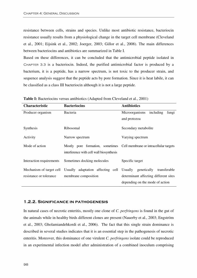
Chapter 4: General Discussion
98
resistance between cells, strains and species. Unlike most antibiotic resistance, bacteriocin
resistance usually results from a physiological change in the target cell membrane (Cleveland
et al., 2001; Eijsink et al., 2002; Joerger, 2003; Gillor et al., 2008). The main differences
between bacteriocins and antibiotics are summarized in Table I.
Based on these differences, it can be concluded that the antimicrobial peptide isolated in
Chapter 3.3 is a bacteriocin. Indeed, the purified antimicrobial factor is produced by a
bacterium, it is a peptide, has a narrow spectrum, is not toxic to the producer strain, and
sequence analysis suggest that the peptide acts by pore formation. Since it is heat labile, it can
be classified as a class III bacteriocin although it is not a large peptide.
Table I: Bacteriocins versus antibiotics (Adapted from Cleveland et al., 2001)
Characteristic Bacteriocins Antibiotics
Producer organism Bacteria Microorganisms including fungi
and protozoa
Synthesis Ribosomal Secondary metabolite
Activity Narrow spectrum Varying spectrum
Mode of action Mostly pore formation, sometimes
interference with cell wall biosynthesis
Cell membrane or intracellular targets
Interaction requirements Sometimes docking molecules Specific target
Mechanism of target cell
resistance or tolerance
Usually adaptation affecting cell
membrane composition
Usually genetically transferable
determinant affecting different sites
depending on the mode of action
1.2.2. Significance in pathogenesis
In natural cases of necrotic enteritis, mostly one clone of C. perfringens is found in the gut of
the animals while in healthy birds different clones are present (Nauerby et al., 2003; Engström
et al., 2003; Gholamiandehkordi et al., 2006). The fact that this single strain dominance is
described in several studies indicates that it is an essential step in the pathogenesis of necrotic
enteritis. Moreover, this dominance of one virulent C. perfringens isolate could be reproduced
in an experimental infection model after administration of a combined inoculum comprising
Chapter 4: General Discussion
99
different necrotic enteritis strains or a combination of virulent and avirulent C. perfringens
strains (Barbara et al., 2008). Finally, the secretion of factors inhibiting growth of other
C. perfringens strains is shown to be more prevalent in outbreak strains than in normal
microbiota strains in vitro (Chapter 3.2.; Barbara et al., 2008). Taken together, these results
suggest that this is a trait associated with virulence. Although there is no information on in
vivo bacteriocin production by C. perfringens, production of bacteriocins by a virulent strain
in the gut would allow this strain to displace the C. perfringens strains present. Inhibiting the
growth of other C. perfringens strains in the gut is also important as part of the competition
for nutrients. Indeed, C. perfringens is not able to produce 13 amino acids and is thus
dependent on environmental presence of these molecules.
It is pertinent to note that there are also examples of other antimicrobial factors of Gram-
positive bacteria that are associated with virulence. In persistent endodontic infections where
Enterococcus faecalis is dominant, a low number of other species is found. It is therefore
suggested that AS-48 and other bacteriocincs of E. faecalis, although they are not strictly
acting as virulence factors, contribute to its dominance in persistent endodontic infections and
may serve as a means to obtain ecological advantages which can result in disease (Kayaoglu
and Ørstavik, 2004).
2. New insights in pathogenesis, a presumed model
After Keyburn et al. (2006) showed that alpha toxin is not an essential virulence factor in
necrotic enteritis in broiler chickens, new C. perfringens virulence factors have been proposed
and new insight was gained into the mechanisms by which Eimeria predispose to Clostridium
perfringens induced necrotic enteritis. The current knowledge is summarized in Figure 1.
In short, Eimeria parasites induce leakage of plasma proteins by killing epithelial cells as a
consequence of the intracellular stages of their life cycle. Moreover, according to Collier et al.
(2008), a coccidial infection enhances mucus production in the intestine. Both effects provide
an increase in available nutrients and create an environment favorable for proliferation of
C. perfringens. Indeed, C. perfringens lacks the machinery to produce 13 amino acids but is
able to utilize mucus as a substrate. Finally, Park et al. (2008) suggested that the exacerbated
pathological findings after co-infection with Eimeria and C. perfringens are caused by an
altered cytokine response. Taken together, these data show that an Eimeria infection aids in
Chapter 4: General Discussion
100
the first stage of pathogenesis: massive proliferation of C. perfringens in the gut. The factors
involved in the second stage of pathogenesis, possibly are bacteriocins. A significantly higher
proportion of virulent C. perfringens strains produce bacteriocins compared to normal
microbiota C. perfringens strains (Chapter 3.2). By inhibiting other C. perfringens strains,
the virulent strain is able to obtain the maximum benefit of the increased nutrient availability
due to the Eimeria infection. The virulent strain is thus able to proliferate massively and as a
consequence it secretes virulence factors playing a role in the final step in pathogenesis: the
induction of necrotic lesions. Olkowski et al. (2006; 2008) suggested that the initial
pathological changes are due to the activity of collagenolytic enzymes. The nature of the
morphological changes seen in the early stages of necrotic enteritis is consistent with the
action of collagenolytic enzymes. Indeed, the pathological changes start at the basal and
lateral domains of enterocytes and then invade progressively the entire lamina propria. Both
host collagenases and collagenolytic enzymes secreted by proliferating pathogens may play a
role. It was shown that virulent C. perfringens strains are able to secrete collagenolytic
enzymes and high levels of different collagenolytic enzymes were found in intestinal tissue of
broilers challenged with C. perfringens, in comparison to controls (Olkowski et al., 2008).
Furthermore, it is shown that the NetB toxin is crucial for the induction of necrotic enteritis
(Keyburn et al., 2008). NetB forms pores in a chicken epithelial cell line in vitro. Its activity
in vivo is not shown yet, but it may induce necrotic lesions by forming pores in enterocytes
leading to cell death. By inducing necrotic enteritis lesions, more nutrients are leaking into the
gut lumen which again can be used by C. perfringens strains.
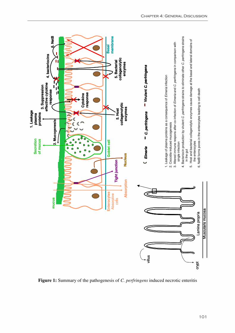
Chapter 4: General Discussion
101
Figure 1: Summary of the pathogenesis of C. perfringens induced necrotic enteritis
La
min
apro
pri
a
Muscu
laris
mu
cosa
vill
us
cry
pt
La
min
apro
pri
a
Muscu
laris
mu
cosa
vill
us
cry
pt
Ab
so
rpti
onTig
ht
jun
cti
on
En
tero
cyte
sA
bsorp
tive
cells
Go
ble
tcell
Se
cre
tio
no
f m
ucu
s
2. M
uc
og
en
esis
1.
Leakag
e
pla
sm
ap
rote
ins
Cyto
kin
ere
sp
on
se
3. S
up
pre
ss
ion
eff
ecti
ve
cyto
kin
ere
sp
on
se
4. b
acte
rio
cin
s
6. N
etB
C.
perf
rin
gen
sE
ime
ria
Vir
ule
nt
C. p
erf
rin
gen
s
5. H
ost
co
llag
en
oly
tic
en
zym
es
5. B
acte
rial
co
llag
en
oly
tic
en
zym
es
Ba
sal
mem
bra
ne
Nu
cle
us
mu
cu
s
Ab
so
rpti
onTig
ht
jun
cti
on
En
tero
cyte
sA
bsorp
tive
cells
Go
ble
tcell
Se
cre
tio
no
f m
ucu
s
2. M
uc
og
en
esis
1.
Leakag
e
pla
sm
ap
rote
ins
Cyto
kin
ere
sp
on
se
3. S
up
pre
ss
ion
eff
ecti
ve
cyto
kin
ere
sp
on
se
4. b
acte
rio
cin
s
6. N
etB
C.
perf
rin
gen
sE
ime
ria
Vir
ule
nt
C. p
erf
rin
gen
sC
. p
erf
rin
gen
sE
ime
ria
Vir
ule
nt
C. p
erf
rin
gen
s
5. H
ost
co
llag
en
oly
tic
en
zym
es
5. B
acte
rial
co
llag
en
oly
tic
en
zym
es
Ba
sal
mem
bra
ne
Nu
cle
us
mu
cu
s
1.
Leakage
of
pla
sm
a p
rote
ins
as a
conse
que
nce
of
Eim
eri
ain
fectio
n
2.
Coccid
ia-in
du
ced
mucogen
esis
3.
Alte
red
imm
une r
espo
ns a
fte
rco-infe
ction
of
Eim
eri
aa
nd C
. p
erf
ringe
ns
in c
om
pariso
nw
ith
sin
gle
infe
ction
4.
Bacte
riocin
pro
duction
by
viru
lent
C.
perf
rin
gen
sstr
ain
sto
elim
ina
teoth
er
C. p
erf
ringen
sstr
ain
sin
the
gut
5. H
ost
an
d b
acte
ria
lco
llag
en
oly
tic
enzym
es
cau
se
dam
age
at th
e b
asa
la
nd la
tera
ldom
ain
sof
the e
nte
rocyte
s
6.
NetB
form
sp
ore
sin
the e
nte
rocyte
sle
ad
ing
toce
lldeath
Chapter 4: General Discussion
102
3. Implications: possible improvement of control measures
Control of necrotic enteritis has commonly been accomplished by administration of
antimicrobials in feed or water. However, concerns are rising about effects on human health
of such routine use of antimicrobials because of the emergence of antibiotic resistance.
Therefore, the European Union banned the use of antibiotics as antimicrobial growth
promoters in animal production (Van Immerseel et al., 2009). Alternative non-antibiotic
approaches to control necrotic enteritis are thus searched for. Vaccination is a widely used
preventive measure against many infectious diseases in mammals and poultry (Dahiya et al.,
2006). Because bacteriocins are commonly found in food and thus have been consumed for
centuries, they are considered natural and nontoxic to eukaryotic cells, in contrast to
traditional antibiotics (Cleveland et al., 2001; Lin, 2009).
3.1. Alpha toxin as vaccine
Despite the fact that it has been shown that alpha toxin is not essential for the development of
necrotic enteritis (Keyburn et al., 2006), specific antibodies against alpha toxin protect birds
from disease. Vaccines using recombinant alpha toxin in toxoid and active form and live
delivery of the C-terminal (non-toxic) domain of alpha toxin by an attenuated Salmonella
enterica serovar Typhimurium strain have been tested and shown to give partial protection
against necrotic enteritis in experimental challenge models (Kulkarni et al., 2007; Zekarias et
al., 2008; Cooper et al., 2009). Initially, it was not clear how antibodies against alpha toxin
could offer any level of protection if alpha toxin did not play a part in the pathogenesis.
However, Zekerias et al. (2008) found that serum antibodies, from broilers that had been
vaccinated, bound directly to the cell surface of C. perfringens and suppressed its growth in
vitro. Antibodies binding to the membrane-bound preprotein might block protein transport
channels and as a consequence, inhibit proliferation of the bacteria (Zekarias et al., 2008).
Alpha toxin can thus be used as a protective antigen to vaccinate broilers, but this does not
necessarily mean that alpha toxin plays a primary role in the pathogenesis of necrotic enteritis.
Chapter 4: General Discussion
103
3.2. Other antigens as vaccine
Thompson et al. (2006) have used alpha toxin deficient mutants to immunize broilers orally
and found that these strains protected birds from experimental necrotic enteritis. These results
clearly demonstrate that antigens other that alpha toxin can provide immunity against necrotic
enteritis. Subsequently, Kulkarni et al. (2006) identified six proteins that reacted to serum
antibodies from birds immune to necrotic enteritis in the supernatant of virulent
C. perfringens strains and not in the supernatant of avirulent C. perfringens strains. These
proteins are a hypothetical protein (HP, possibly a zinc-binding protease); pyruvate:ferredoxin
oxidoreductase (PFOR); Elongation factor G (EF-G); Perfringolysin O; glyceraldehyde-3-
phosphate dehydrogenase (GAPDA) and fructose 1,6-biphosphate aldolase (FBA).
Recombinant HP, PFOR, GAPDA and FBA were tested as vaccine and all proteins protected
broiler chickens against a mild challenge while immunization with HP and PFOR also offered
protection against a severe challenge (Kulkarni et al., 2007). Broilers orally immunized with
an avirulent Salmonella enterica serovar Typhimurium vaccine vector expressing FBA or HP
proteins were also significantly protected against challenge (Kulkarni et al., 2008). Also other
proteins that have been shown to play a role in the pathogenesis of necrotic enteritis might be
good candidates as vaccine antigens. These include the metalloproteinases that are suggested
to have a role in early lesion development and the NetB protein (Olkowski et al., 2008;
Keyburn et al., 2008). Candidate protective antigens have thus been identified. This does not
mean that a solution to the necrotic enteritis problem is found, but the results are promising
for an answer in the near future.
3.3. Gmo probiotics expressing bacteriocins
The use of bacteriocins active against C. perfringens could also be a good alternative for
antibiotic control of necrotic enteritis. Bacteriocins of C. perfringens are naturally present in
the intestine, and can be considered as safe. There are two possible ways to deliver the
bacteriocins in the intestinal tract: by the administration of bacteriocin-producing bacteria or
by the administration of purified bacteriocins (Kirkup, 2006). The purified bacteriocins can be
included in the feed. However, sensitivity of bacteriocins to gastric acidity and the presence of
proteases in the gastrointestinal tract can potentially result in loss of activity when the
Chapter 4: General Discussion
104
bacteriocins are orally administered. An additional difficulty is the challenge of bulk
production of purified bacteriocins.
To circumvent the loss of activity, the genes for bacteriocin production can be cloned and
expressed in an appropriate bacterial carrier. This approach not only prevents in vivo
degradation of the bacteriocin during gastric transit, but it also facilitates continued
bacteriocin production at the site of infection. It is important that the bacterial carrier is able to
colonize the gut and is not pathogenic to the host. Probiotic strains can be used as bacterial
carrier. Probiotics are ‘live microorganisms, which, when consumed in adequate amounts,
confer a health benefit on the host’. The potential of genetically modified bacteriocin-
producing bacteria has already been shown. An avian E. coli strain genetically engineered to
produce the bacteriocin microcin 24 lowered intestinal Salmonella Typhimurium counts in
chickens when administered continuously in the water supply (Wooley et al., 1999).
As with any antimicrobial compound, also for bacteriocins it is important to consider the issue
of resistance. Although the mechanism of action is not known for all bacteriocins, most of the
bacteriocins appear to interact with the bacterial membrane. Therefore, resistance is usually
the result of changes in the membrane of bacteria targeted by a bacteriocin (Joerger, 2003).
However, in the producer strains, bacteriocins are accompanied by specific immunity proteins
that can theoretically be transferred horizontally to pathogens (Kirkup, 2006).
If bacteriocins are going to be used as a strategy for the prevention of necrotic enteritis or
other infectious diseases, there are some important requirements, such as stability, lack of
toxicity, a specific activity spectrum, lack of development of resistance, and a thorough
understanding of their biochemical and genetic properties. A detailed knowledge of the
isolated bacteriocin structures and properties is therefore needed. The same goes for the
carrier strain.
4. Conclusion
The past decade, great progress has been made in understanding broiler necrotic enteritis. It is
shown that the single strain dominance found in natural cases of necrotic enteritis is likely to
be due to bacteriocin production by C. perfringens strains. The assumption that alpha toxin
was the major virulence factor in necrotic enteritis is refuted and a novel toxin, NetB, is now
associated with necrotic enteritis. Proteolytic enzymes are suggested to have a role in the early
Chapter 4: General Discussion
105
stages of pathogenesis. This insight in the etiology and the pathogenesis of C. perfringens
infection in the gut provides important information that will be valuable to develop effective
disease control strategies.
Chapter 4: General Discussion
106
References
Barbara A.J., Trinh H.T., Glock R.D., Songer J.G. (2008). Necrotic enteritis-producing strains
of Clostridium perfringens displace non-necrotic enteritis strains from the gut of chicks. Vet.
Microbiol. 126, 377-382.
Chalmers G., Bruce H.L., Hunter D.B., Parreira V.R., Kulkarni R.R., Jiang Y.-F., Prescott
J.F., Boerlin P. (2008). Multilocus sequence typing analysis of Clostridium perfringens
isolates from necrotic enteritis outbreaks in broiler chicken populations. J. Clin. Microbiol.
46, 3957-3964.
Cleveland J., Montville T.J., Nes I.F., Chikindas M.L. (2001). Bacteriocins: safe, natural
antimicrobials for food preservation. Int. J. Food Microbiol. 71, 1-20.
Collier C.T., Hofacre C.L., Payne A.M., Anderson D.B., Kaiser P., Mackie R.I., Gaskins H.R.
(2008). Coccidia-induced mucogenesis promotes the onset of necrotic enteritis by supporting
Clostridium perfringens growth. Vet. Immun. Immunopath. 122, 104-115.
Cooper K.K., Trinh H.T., Songer J.G. (2009). Immunization with recombinant alpha toxin
partially protects broiler chickens against experimental challenge with Clostridium
perfringens. Vet. Microbiol. 133, 92-97.
Dahiya J.P., Wilkie D.C., Van Kessel A.G., Drew M.D. (2006). Potential strategies for
controlling necrotic enteritis in broiler chickens in post-antibiotic era. Anim. Feed Sci.
Technol. 129, 60-88.
Eijsink V.G., Axelsson L., Diep D.B., Håvarstein L.S., Holo H., Nes I.F. (2002). Production
of class II bacteriocins by lactic acid bacteria: an example of biological warfare and
communication. Antonie Van Leeuwenhoek. 81, 639-654.
Engström B.E., Fermér C., Lindberg A., Saarinen E., Båverud V., Gunnarson A. (2003).
Molecular typing of isolates of Clostridium perfringens from healthy and diseased poultry.
Vet. Microbiol. 94, 225-235.
Chapter 4: General Discussion
107
Gholamiandehkordi A.R., Ducatelle R., Heyndrickx M., Haesebrouck F., Van Immerseel F.
(2006). Molecular and phenotypical characterization of Clostridium perfringens isolates from
poultry flocks with different disease status. Vet. Microbiol. 113, 143-152.
Gholamiandehkordi A.R., Timbermont L., Lanckriet A., Van den Broeck W., Pedersen K.,
Dewulf J., Pasmans F., Haesebrouck F., Ducatelle R., Van Immerseel F. (2007).
Quantification of gut lesions in a subclinical necrotic enteritis model. Avian Pathol. 36, 375-
382.
Gillor O., Etzion A., Riley M.A. (2008). The dual role of bacteriocins as anti- and probiotics.
Appl. Microbiol. Biotechnol. 81, 591-606.
Hancock R.E., Chapple D.S. (1999). Peptide antibiotics. Animicrob. Agents Chemother. 43,
1317-1323.
Joerger R.D. (2003). Alternatives to antibiotics: bacteriocins, antimicrobial peptides and
bacteriophages. Poultry Sci. 82, 640-647.
Kayaoglu G., Ørstavik D. (2004). Virulence factors of Enterococcus faecalis: relationship to
endodontic disease. Crit. Rev. Oral Biol. Med. 15, 308-320.
Keyburn A.L., Boyce J.D., Vaz P., Bannam T.L., Ford M.E., Parker D., Di Rubbo A., Rood
J.I., Moore R.J. (2008). NetB, a new toxin that is associated with avian necrotic enteritis
caused by Clostridium perfringens. PLoS Pathog. 4(2): e26. doi:10.1371/journal.ppat.
0040026.
Keyburn A.L., Sheedy S.A., Ford M.E., Williamson M.M., Awad M.M., Rood J.I., Moore
R.J. (2006). Alpha-toxin of Clostridium perfringens is not an essential virulence factor in
necrotic enteritis in chickens. Infect. Immun. 74, 6496-6500.
Kirkup Jr. B.C. (2006). Bacteriocins as oral and gastrointestinal antibiotics: theoretical
considerations, applied research, and practical applications. Curr. Med. Chem. 13, 3335-3350.
Kulkarni R.R., Parreira V.R., Sharif S., Prescott J.F. (2006). Clostridium perfringens antigens
recognized by broiler chickens immune to necrotic enteritis. Clin. Vaccine Immunol. 13,
1358-1362.
Chapter 4: General Discussion
108
Kulkarni R.R., Parreira V.R., Sharif S., Prescott J.F. (2007). Immunization of broiler chickens
against Clostridium perfringens induced necrotic enteritis. Clin. Vaccine Immunol. 14, 1070-
1077.
Kulkarni R.R., Parreira V.R., Sharif S., Prescott J.F. (2008). Oral immunization of broiler
chickens against necrotic enteritis with an attenuated Salmonella vaccine vector expressing
Clostridium perfringens antigens. Vaccine. 26, 4194-4203.
Lin J. (2009). Novel approaches for Campylobacter control in poultry. Foodborne Pathog Dis.
Doi: 10.1089/fpd.2008.0247.
Martin T.G., Smyth J.A. (2009). Prevalence of netB among some clinical isolates of
Clostridium perfringens from animals in the United States. Vet. Microbiol. 136, 202-205.
Nauerby B., Pedersen K., Madsen M. (2003). Analysis by pulsed-field gel electrophoresis of
the genetic diversity among Clostridium perfringens isolates from chickens. Vet. Microbiol.
94, 257-266.
Olkowski A.A., Wojnarowicz C., Chinio-Trejo M., Drew M.D. (2006). Responses of broiler
chickens orally challenged with Clostridium perfringens isolated from field cases of necrotic
enteritis. Res. Vet. Sci. 81, 99-108.
Olkowski A.A., Wojnarowicz C., Chirino-Trejo M., Laarveld B., Sawicki G. (2008). Sub-
clinical necrotic enteritis in broiler chickens: Novel etiological consideration based on ultra-
structural and molecular changes in the intestinal tissue. Res. Vet. Sci. 85, 543-553.
Park S.S., Lillehoj H.S., Allen P.C., Park D.W., FitzCoy S., Baurista D.A. Lillehoj E.P.
(2008). Immunopathology and cytokine responses in broiler chickens coinfected with Eimeria
maxima and Clostridium perfringens with the use of an animal model of necrotic enteritis.
Avian Dis. 52, 14-22.
Thompson D.R., Parreira V.R., Kulkarni R.R., Prescott J.F. (2006). Live attenuated vaccine-
based control of necrotic enteritis of broiler chickens. Vet. Microbiol. 113, 25-34.
Van Immerseel F., Rood J.I., Moore R.J., Titball R.W. (2009). Rethinking our understanding
of the pathogenesis of necrotic enteritis in chickens. Trends Microbiol.. 17, 32-36.
Chapter 4: General Discussion
109
Wooley R.E., Gibbs P.S., Shotts E.B. Jr. (1999). Inhibition of Salmonella Typhimurium in the
chicken intestinal tract by a transformed avirulent avian Escherichia coli. Avian Dis. 43, 245-
250.
Zekerias B., Mo H., Curtiss III R. (2008). Recombinant attenuated Salmonella enterica
Serovar Typhimurium expressing the carboxy-terminal domain of alpha toxin from
Clostridium perfringens induces protective responses against necrotic enteritis in chickens.
Clin. Vaccine Immunol. 15, 805-816.


Summary

Summary
113
Summary
Necrotic enteritis was first described in 1961. It is a common enteric disease, caused by
Clostridium perfringens type A. However, C. perfringens type A can also be present in the
intestine of healthy broilers. Necrotic enteritis is characterized clinically by a sudden increase
in flock mortality, often without premonitory signs. Also a sub-clinical form of necrotic
enteritis is described. At necropsy, necrotic foci are found in the small intestinal mucosa. The
disease usually occurs in broiler chickens at about 4 weeks after hatching and is found in all
poultry-growing areas of the world. Despite the clinical and economic importance of necrotic
enteritis, the pathogenesis of C. perfringens associated disease in poultry is still not fully
understood. For a long time, it was believed that alpha toxin was the key virulence factor in
necrotic enteritis in broiler chickens caused by C. perfringens but, at the start of this thesis, its
role in the pathogenesis of necrotic enteritis was called into doubt.
C. perfringens type A is associated with necrotic enteritis but can also be detected in
relatively high numbers in the intestinal tract of apparently normal chickens. Consequently, in
the first study of this thesis (Chapter 3.1), the hypothesis was tested that the necrosis
inducing capability would be a characteristic of specific strains. C. perfringens isolates from
both clinically healthy broilers and broilers suffering from necrotic enteritis were compared
for their ability to induce necrotic lesions using an experimental necrotic enteritis model in
broiler chickens. High, intermediate and low alpha toxin producing strains were chosen from
each isolation source to determine the role of alpha toxin in the pathogenesis of necrotic
enteritis. In this experimental model, only the isolates from field outbreaks induced necrotic
gut lesions, independent of the amount of alpha toxin produced in vitro. It was also shown
that alpha toxin producing isolates from calf hemorrhagic enteritis cases were not able to
induce necrotic enteritis in poultry. These results suggest that alpha toxin is not important to
induce necrotic lesions in broiler chickens. Moreover, the results are indicative of the
presence of host specific virulence factors in certain C. perfringens strains, isolated from
chickens with intestinal necrotic enteritis lesions.
Summary
114
Clinically healthy chickens carry several different C. perfringens clones in their intestine. In
flocks suffering from necrotic enteritis, however, mostly only one single clone is isolated
from the gut of all the diseased animals. Selective proliferation of such clinical outbreak strain
in the gut and spread within the flock seems likely. The hypothesis that necrotic enteritis
associated C. perfringens strains might suppress the growth of normal microbiota
C. perfringens strains, was therefore tested (Chapter 3.2). Twenty-six C. perfringens strains
isolated from healthy broilers and 24 clinical outbreak isolates were evaluated for their ability
to induce intra-species growth-inhibition in an in vitro setup. A significantly higher
proportion of the C. perfringens clinical outbreak strains inhibited the growth of other
C. perfringens strains compared to C. perfringens strains isolated from the gut of healthy
chickens. It is proposed that, in addition to toxin production, intra-species growth-inhibition
may be a virulence trait that contributes to the ability of certain C. perfringens strains to cause
necrotic enteritis in broilers. The secretion of factors inhibiting the growth of other
C. perfringens strains could lead to extensive and selective presence of a strain that contains
the genetic make-up enabling to secrete toxins that cause gut lesions.
In Chapter 3.3, the purification and the characterization of such a growth-inhibiting factor is
described. A bacteriocin-like inhibitory substance (BLIS) was purified from a necrotic
enteritis associated C. perfringens strain exhibiting antibacterial activity against other
C. perfringens strains. Amino acid sequence analysis indicated that the BLIS is an 11.5 kDa
fragment of a 22 kDa protein and that it constitutes the C-terminal part of this protein. The
22 kDa protein is a cytoplasmic membrane protein without signal peptide. The antibacterial
activity of the purified BLIS was abolished by proteolytic enzymes trypsin and proteinase K
and by heat treatment (10 min at 80 °C). The purified BLIS showed inhibitory activity over a
wide pH range (4.0 to 10.0). Since the antibacterial activity against other C. perfringens
strains of the purified BLIS had a narrower spectrum than the crude supernatant of the
secreting strain, most probably different of these bacteriocins are produced.
In conclusion, this thesis offers new insights in the pathogenesis of C. perfringens necrotic
enteritis in broiler chickens. It was shown that only a specific subpopulation of C. perfringens
type A strains are able to induce necrotic enteritis lesions. We suggest that the single clone
dominance found in necrotic enteritis is due to the secretion of growth-inhibiting factors, also
Summary
115
called bacteriocins, secreted by these virulent strains. This provides important information
that will be valuable to develop effective disease control strategies.


Samenvatting

Samenvatting
119
Samenvatting
Necrotische enteritis werd voor het eerst in 1961 beschreven. Het is een vaak voorkomende
darmziekte veroorzaakt door Clostridium perfringens type A. Hoewel C. perfringens dus in
staat is om necrotische enteritis te veroorzaken, komt deze bacterie bijna altijd voor in de
darm van gezonde vleeskippen. Necrotische enteritis wordt klinisch gekarakteriseerd door een
plotse stijging van sterfte in een toom, meestal zonder voorafgaande tekenen. Er is ook een
subklinische vorm van necrotische enteritis beschreven. Bij lijkschouwing worden necrotische
haarden gevonden in de mucosa van de dunne darm. De ziekte komt meestal voor bij
vleeskippen van ongeveer 4 weken oud en komt voor in alle landen waar pluimvee gefokt
wordt. Ondanks het klinische en economische belang van necrotische enteritis, is de
pathogenese nog niet volledig ontrafeld. Lange tijd werd er gedacht dat alfa toxine de
belangrijkste virulentiefactor was van C. perfringens die een rol speelt in necrotische enteritis.
Bij het begin van deze thesis werd de rol van alfa toxine in de pathogenese van necrotische
enteritis echter in twijfel getrokken.
C. perfringens type A wordt geassocieerd met necrotische enteritis maar kan ook in relatief
hoge aantallen teruggevonden worden in de darm van gezonde vleeskippen. Daarom werd in
het eerste hoofdstuk van deze thesis (Chapter 3.1), de hypothese getoetst dat necrotische
enteritis zou veroorzaakt worden door welbepaalde stammen van C. perfringens. Hiervoor
werden C. perfringens isolaten van zowel klinisch gezonde vleeskippen als van vleeskippen
met necrotische enteritis getest op hun capaciteit om necrotische letsels te induceren in een
experimenteel necrotisch enteritis model in vivo. Om de rol van alfa toxine in de pathogenese
na te gaan werden C. perfringens isolaten uit kippen met necrotische enteritis en isolaten uit
klinisch gezonde kippen gekozen die in vitro hoge, gemiddelde en lage concentraties alfa
toxine produceerden. Enkel de isolaten geïsoleerd uit kippen met necrotische enteritis konden
necrotische letsels induceren, ongeacht de concentratie alfa toxine die ze produceerden in
vitro. Er werd ook aangetoond dat alfa toxine producerende isolaten van gevallen van
hemorragische enteritis bij het kalf niet in staat waren om necrotische enteritis in vleeskippen
te induceren. Deze resultaten suggereren dat alfa toxine niet belangrijk is om necrotische
Samenvatting
120
letsels te induceren bij vleeskippen. Bovendien doen deze resultaten vermoeden dat de
C. perfringens stammen geïsoleerd uit kippen met necrotische letsels, over gastheer specifieke
virulentiefactoren beschikken.
Klinisch gezonde vleeskippen hebben verschillende C. perfringens klonen in hun darm terwijl
in een toom die necrotische enteritis doormaakt, meestal één en dezelfde kloon geïsoleerd
wordt uit de darm van verschillende vleeskippen. Selectieve proliferatie in de darm van één
stam en verspreiding in de toom lijkt waarschijnlijk. De hypothese dat necrotische enteritis
geassocieerde C. perfringens stammen de groei van C. perfringens stammen uit de normale
flora inhiberen, werd daarom getest (Chapter 3.2). Bij 26 C. perfringens isolaten uit
gezonde vleeskippen en 24 isolaten uit klinische uitbraken werd nagegaan in hoeverre ze over
de eigenschap beschikken om elkaars groei te inhiberen in vitro. Significant meer
C. perfringens stammen uit klinische uitbraken inhibeerden de groei van de andere
C. perfringens stammen in vergelijking met C. perfringens stammen geïsoleerd uit de darm
van gezonde dieren. Naast toxine productie zou groei-inhibitie een virulentie eigenschap
kunnen zijn die bijdraagt tot de eigenschap van bepaalde C. perfringens stammen om
necrotische enteritis te induceren in vleeskippen. De secretie van factoren die de groei
inhiberen van andere C. perfringens stammen kan leiden tot proliferatie en selectieve
aanwezigheid van een stam die de genetische eigenschappen heeft om toxines uit te scheiden
die darmletsels veroorzaken.
In het laatste hoofdstuk (Chapter 3.3) wordt de opzuivering en karakterisatie van een groei-
inhiberende factor beschreven. De bacteriocine-achtige inhibitorische factor werd
opgezuiverd uit het supernatans van een C. perfringens stam geïsoleerd uit een kip met
necrotische enteritis. Deze stam vertoonde antibacteriële activiteit tegen andere C. perfringens
stammen. Aminozuur sequentie analyse toonde aan dat de factor een 11.5 kDa groot fragment
van een proteïne van 22 kDa en dat de factor het C-terminale deel van dit proteïne is. Het
22 kDa proteïne is een cytoplasmatisch membraan proteïne zonder signaal sequentie. Na
incubatie met de proteolytische enzymen trypsine en proteinase K of na hittebehandeling
(10 min, 80 °C) werd geen antibacteriële activiteit meer waargenomen. De inhibitorische
activiteit was stabiel bij een brede pH range (4.00 tot 10.00). Aangezien de intra-species
antibacteriële activiteit van de opgezuiverde bacteriocine-achtige inhibitorische factor een
Samenvatting
121
nauwer spectrum had dan het ruwe supernatans van de producerende stam, kan verondersteld
worden dat deze stam meerdere dergelijke bacteriocine-achtige factoren aanmaakt.
De resultaten van deze thesis hebben geleid tot een beter inzicht in de pathogenese van
C. perfringens geïnduceerde necrotische enteritis bij vleeskippen. Er werd aangetoond dat
slechts een subpopulatie van C. perfringens type A in staat is necrotische letsels te induceren.
Onze resultaten tonen voorts aan dat het feit dat slechts één kloon teruggevonden wordt bij
vleeskippen met necrotische enteritis waarschijnlijk te wijten is aan de secretie van groei-
inhiberende factoren, ook bacteriocines genoemd, gesecreteerd door C. perfringens type A
stammen die pathogeen zijn voor de kip. Dit levert belangrijke informatie die waardevol zal
zijn om meer efficiënte bestrijdingsstrategieën te ontwikkelen.


Curriculum vitae

Curriculum Vitae
125
Curriculum vitae
Leen Timbermont werd geboren op 10 november 1981 te Dendermonde. Na het beëindigen
van haar studies algemeen secundair onderwijs, richting Latijn-Wiskunde, aan het
Koninklijke Atheneum Dendermonde begon ze in 1999 aan de studies Bio-ingenieur aan de
Vrije Universiteit Brussel. In 2004 behaalde zij met onderscheiding het diploma Bio-
ingenieur optie cel- en genbiotechnologie.
Daarna trad zij in dienst bij de vakgroep Pathologie, Bacteriologie en Pluimveeziekten voor
onderzoek naar het gedrag van Salmonella Enteritidis in leghennen en eieren. In 2005 startte
ze haar doctoraatsonderzoek aan dezelfde vakgroep met Prof. Dr. F. Van Immerseel en Prof.
Dr. R. Ducatelle als promotoren. Gedurende vier jaar werkte zij aan het onderzoeksproject:
“Studie van pathogenese en bestrijding van Clostridium perfringens bij pluimvee’ dat
gefinancierd werd door het IWT, afdeling landbouwkundig onderzoek.
In 2007 behaalde zij aan de Faculteit Psychologie en Pedagogische Wetenschappen (UGent)
het diploma van de Academische Initiële Lerarenopleiding (AILO) en in 2008 voltooide zij de
doctoraatsopleiding in de Diergeneeskundige Wetenschappen.
Leen Timbermont is auteur of medeauteur van meerdere wetenschappelijke publicaties in
nationale en internationale tijdschriften. Zij nam deel aan verschillende congressen en
presenteerde de resultaten van haar onderzoek in de vorm van voordrachten en posters.


Bibliography

Bibliography
129
Bibliography
Publications in national and international journals
Gholamiandehkordi A.R., Eeckhaut V., Lanckriet A., Timbermont L., Bjerrum L., Ducatelle
R., Haesebrouck F., Van Immerseel F. (2009). Animicrobial resistance in Clostridium
perfringens isolates from broilers in Belgium. Veterinary Research Communications, doi:
10.1007/s11259-009-9306-4.
Lanckriet A., Timbermont L., Happonen L.J., Pajunen M.I., Pasmans F., Haesebrouck F.,
Ducatelle R., Savilahti H., Van Immerseel F. (2009). Generation of single-copy transposon
insertions in Clostridium perfringens by electroporation of phage Mu DNA transposition
complexes. Applied Environmental Microbiology, 75, 2638-2642.
Timbermont L., Lanckriet A., Pasmans F., Haesebrouck F., Ducatelle R., Van Immerseel F.
(2009). Intra-species growth-inhibition by Clostridium perfringens is a possible virulence trait
in necrotic enteritis in broilers. Veterinary Microbiology, 137, 388-391.
Timbermont L., Lanckriet A., Gholamiandehkordi A.R., Pasmans F., Martel A.,
Haesebrouck F., Ducatelle R., Van Immerseel F. (2009). Origin of Clostridium perfringens
isolates determines the ability to induce necrotic enteritis in broilers. Comparative
Immunology, Microbiology and Infectious Diseases, 32, 503-512.
Gholamiandehkordi A.R., Timbermont L., Lanckriet A., Broeck W.V., Pedersen K., Dewulf
J., Pasmans F., Haesebrouck F., Ducatelle R., Van Immerseel F. (2007). Quantification of gut
lesions in a subclinical necrotic enteritis model. Avian Pathology, 36, 375-382.
Van Immerseel F., Timbermont L., Lanckriet A., Gholamiandehkordi A.R., Pasmans F.,
Haesebrouck F., Ducatelle R. (2006). Clostridium perfringens bij pluimvee: ubiquitaire kiem,
normale darmbewoner of pathogeen? Vlaams Diergeneeskundig Tijdschrift, 75, 353-363.
Bibiliography
130
Gantois I., Ducatelle R., Timbermont L., Boyen F., Bohez L., Haesebrouck F., Pasmans F.,
Van Immerseel F. (2006). Oral Immunisation of laying hens with the live vaccine strains of
TAD Salmonella vac(®) E and TAD Salmonella vac(®) T reduces internal egg contamination
with Salmonella Enteritidis (2006). Vaccine, 24, 6250-6255.
Van Immerseel F., Russel J.B., Flythe M.D., Gantois I., Timbermont L., Pasmans F.,
Haesebrouck F., Ducatelle R. (2006). The use of organic acids to combat Salmonella in
poultry: a mechanistic explanation of the efficacy. Avian Pathology, 35, 182-188.
Van Immerseel F., Boyen F., Gantois I., Timbermont L., Bohez L., Pasmans F.,
Haesebrouck F., Ducatelle R. (2005). Supplementation of coated butyric acid in the feed
reduces colonization and shedding of Salmonella in poultry. Poultry Science, 84, 1851-1856.
Van Immerseel F., De Buck J., Timbermont L., Gantois I., Bohez L., Boyen F., Pasmans F.,
Bertrand S., Collard J.M., Saegerman C., Hooyberghs J., Haesebrouck F., Ducatelle R.
(2005). Salmonella in pluimveevlees en eieren: een gevaar voor de consument die om
efficiënte bestrijdingsprogramma’s vraagt. Vlaams Diergeneeskundig Tijdschrift, 74, 258-
275.
Abstracts and proceedings on international meetings
Timbermont L., Lanckriet A., Pasmans F., Haesebrouck R., Ducatelle R., Van Immerseel F.
(2008). Intra-species growth-inhibition is an additional virulence trait of Clostridium
perfringens in broiler necrotic enteritis. The Conference – Clostridium perfringens, Torquay,
Devon, UK, 24.
Lanckriet A, Timbermont L, Happonen L.J., Pajunen M., Pasmans F., Haesebrouck F.,
Ducatelle R. , Savilahti S., Van Immerseel F (2008). Generation of single-copy transposon
insertions in Clostridium perfringens by electroporation of phage Mu DNA transposition
complexes. The Conference – Clostridium perfringens, Torquay, Devon, UK, 12
Timbermont L., Lanckriet A., Gholamiandehkordi A., Marien M., Vancraeynest D., De
Gussem M., Pasmans F., Ducatelle R., Haesebrouck F., Van Immerseel F. (2008). Evaluation of
the efficacy of a unique Lactobacillus-based product in a necrotic enteritis model in broilers.
XXIII World’s Poultry Congress, Brisbane, Queensland, Australia, 189-190.
Bibliography
131
Timbermont L., Gholamiandehkordi A.R., Lanckriet A., Haesebrouck F., Ducatelle R., Van
Immerseel F. (2007). A semi-quantitative scoring system for gut lesions in a subclinical necrotic
enteritis model. International Symposium on Protozoal Infections in Poultry, Vienna, Austria,
42.
Timbermont L., Lanckriet A., Van Immerseel F., Ducatelle R. (2006). Studies on the
pathogenesis of Clostridium perfringens induced necrotic enteritis in poultry. Poultry
Veterinary Study Group of the EU (PVSG), Antwerpen, België.
Van Immerseel F., Gholamiandehkordi A.R., Timbermont L., Lanckriet A., Haesebrouck F.
Ducatelle R. (2006). Clinical isolates of Clostridium perfringens in poultry are not superior
alpha toxin producers in vitro. 5th International Meeting on the Molecular Biology and
Pathogenesis of Clostridia, Nottingham, United Kingdom, 37.
Ducatelle R., Timbermont L., lanckriet A., Gholamiandehkordi A., Haesebrouck F., Van
Immerseel F. (2006). Lesions associated with Clostridium perfringens infection in a chicken
intestinal loop model. 24th Meeting of the European Society of Veterinary Pathology,
Edinburgh, United Kingdom.
Van Immerseel F., Gantois I., Bohez L., Timbermont L., Boyen F., Hautefort I., Hinton J.,
Pasmans F., Haesebrouck F., Ducatelle R. (2005). Developments and Pitfalls of Feed
Acidification in Controlling Gut Pathogens in Poultry, with Emphasis on Salmonella. Avian
gut function in health and disease, WPSA Meeting, Bristol, UK, 275-293
Oral presentations on national and international meetings
Timbermont L., Lanckriet A., Pasmans F., Haesebrouck R., Ducatelle R., Van Immerseel F.
(2008). Intra-species growth-inhibition is an additional virulence trait of Clostridium
perfringens in broiler necrotic enteritis. The Conference – Clostridium perfringens, Torquay,
Devon, UK.
Timbermont L., Gholamiandehkordi A.R., Lanckriet A., Haesebrouck F., Ducatelle R., Van
Immerseel F. (2007). A semi-quantitative scoring system for gut lesions in a subclinical necrotic
enteritis model. International Symposium on Protozoal Infections in Poultry, Vienna, Austria.
Bibiliography
132
Timbermont L., Lanckriet A., Van Immerseel F., Ducatelle R. (2006). Studies on the
pathogenesis of Clostridium perfringens induced necrotic enteritis in poultry. Poultry
Veterinary Study Group of the EU (PVSG), Antwerpen, België.
Van Immerseel F., Gantois I., Bohez L., Timbermont L., Boyen F., Hautefort I., Hinton J.,
Pasmans F., Haesebrouck F., Ducatelle R. (2005). Developments and Pitfalls of Feed
Acidification in Controlling Gut Pathogens in Poultry, with Emphasis on Salmonella Avian
gut function in health and disease, WPSA Meeting, Bristol.
Poster presentation on national and international meetings
Timbermont L., Lanckriet A., Pasmans F., Haesebrouck F., Ducatelle R., Van Immerseel F.
(2009). Intra-species growth-inhibition by Clostridium perfringens is a possible virulence trait
in necrotic enteritis in broilers. RESEARCH, Gent, Belgium.
Timbermont L., Lanckriet A., Nollet N, Schwarzer K., Haesebrouck F., Ducatelle R., Van
Immerseel F. (2008). Control of Clostridium perfringens induced necrotic enteritis in broilers
by butyric acid, medium chain fatty acids and essential oils. The Conference – Clostridium
perfringens, Torquay, Devon, UK.
Timbermont L., Lanckriet A., Gholamiandehkordi A., Pasmans F., Martel A., Haesebrouck
F., Ducatelle R., Van Immerseel F. (2008). Origin of Clostridium perfringens isolates
determines the ability to induce necrotic enteritis in broilers. The Conference – Clostridium
perfringens, Torquay, Devon, UK.

Dankwoord

Dankwoord
135
Dankwoord
En dan zijn we nu aanbeland bij het laatste en misschien wel het meest gelezen stukje van dit
doctoraat: het dankwoord. Het leuke aan het schrijven van een dankwoord is dat al het harde
werk achter de rug is, het moeilijke ervan is niemand te vergeten! Het afleggen van een
doctoraat is niet mogelijk zonder de steun, het vertrouwen en de hulp van vele mensen. Ik zou
daarom graag de mensen bedanken die een bijdrage leverden aan deze thesis.
In de eerste plaats wil ik mijn promotoren bedanken voor de steun de voorbije 4 jaar. Prof. Dr.
F. Van Immerseel, of Filip, dank zij jou heb ik de kans gekregen om mij te verdiepen in de
wondere wereld van C. perfringens. Zoals dat bij onderzoek gaat, zijn de proeven niet altijd
even vlekkeloos verlopen maar ik apprecieer enorm de tijd die je voor mij hebt vrijgemaakt.
Ook Prof. Dr. Ducatelle, wil ik bedanken voor de begeleiding. Uw enthousiasme en
verrassende ideeën zorgden ervoor dat er altijd wel iets nieuws uit de bus kwam om verder uit
te pluizen.
Ook wil ik graag de vakgroepvoorzitter, Prof. Dr. Haesebrouck bedanken voor zijn steun,
vertrouwen en interesse in mijn onderzoek.
Dit onderzoek werd financieel gesteund door IWT Vlaanderen, afdeling landbouwkundig
onderzoek, waarvoor uiteraard dank.
Mijn dank gaat eveneens uit naar de leden van de begeleidingscommissie, Prof. Deprez en
Prof. Houf, voor het kritisch nalezen van dit manuscript en het geven van constructieve
commentaar. Ook de leden van de examencommissie wil ik graag bedanken voor het grondig
nalezen van dit doctoraat: Prof. Dr. H. De Brabander, Prof. Dr. A. Martel, Prof. Dr. P. De
Herdt, Prof. Dr. B. Devreese, I would like to thank Prof. Dr. M. Kaldhusdal for reviewing this
manuscript.
Dankwoord
136
Een niet te onderschatten factor van wetenschappelijk onderzoek is de aangename werksfeer
waarin het kan uitgeoefend worden. Daarom wil ik de collega’s van de vakgroep Pathologie,
Bacteriologie en Pluimveeziekten ook in het zonnetje zetten. In de eerste plaats de
Clostridium-groep. Veerle, jij hebt een zeer belangrijke rol gespeeld in het tot stand komen
van deze thesis. Waarschijnlijk kon je een tijdje geen tandenstokers meer zien ;-) maar heel
erg bedankt voor je zeer nauwgezet werk en je snelle uitvoering van alle mogelijke
experimenten. Veel succes in je (ondertussen al niet meer zo) nieuwe job. Anouk, bedankt
voor de leuke vrouwenbabbels op de bureau, zowel wetenschappelijke als niet-
wetenschappelijke. Samen hebben we ons beestje proberen kleinkrijgen. De ene keer hadden
we al wat meer succes dan de andere keer wat soms voor veel frustratie heeft gezorgd maar
het einde is nabij! Nog veel goede moed in je laatste spurt naar het afronden van je doctoraat
en veel geluk in al je verdere plannen. Renzo, de man die voor de vrolijke noot zorgde in het
labo. Bedankt voor je hulp bij de in vivo proeven. We zullen je zangstonden missen maar ik
ben er van overtuigd dat je een enthousiaste leerkracht zal zijn. Ahmad, you were the pioneer
of the Clostridium research at our lab. Thanks to your strain collection and in vivo model, we
could immediately start our research. Marleen wil ik graag bedanken voor de uitstekende
technische hulp aan het begin van het project. Gelukkig kan ik je nog altijd lastigvallen om
pakjes te versturen. Ik kan heel erg genieten van onze etentjes op het terras van Caritas! Toen
ik hier op het labo toekwam kon ik rekenen op de technische hulp van Paul. Het was niet
altijd gemakkelijk om de dagtaak uit te stippelen van iemand die qua leeftijd en ervaring mijn
vader had kunnen zijn. Bedankt voor je nauwgezet werk en ik ben “den arrangeur” nog altijd
niet vergeten! Lieve, heel erg bedankt voor de goede zorgen voor de konijntjes. Frank en An,
heel erg bedankt voor de hulp bij het leggen van de loops.
Nog een speciaal woordje van dank voor de Salmonella collegaatjes: Ruth, Jantina, Emma,
Sophie en Xavier. Bedankt voor de hulp bij de staalnames! Inne, bedankt voor alle nuttige en
praktische tips om dit werkje tot een goed einde te brengen. Altijd handig als iemand de hele
procedure nog niet lang geleden ook heeft moeten doorlopen. Venessa, bedankt voor al je
hulp. Ik wens je veel succes met het afronden van je doctoraat. Dan is het mijn beurt om
nuttige en praktische tips te geven! Lotte, jou wens ik een fantastische tijd met Mauro.
Dankwoord
137
Marc, het was leuk om telkens samen te spurten naar het station omdat we weer eens te laat
vertrokken waren. We hebben ons record ettelijke malen verbeterd! Misschien zat de
conditietraining met Els en Katleen daar voor iets tussen. Meisjes, het was altijd heel leuk om
samen te gaan lopen. Els, veel succes in je verdere carrière. Katleen, proficiat met je
toekomstige spruit! Je gaat dat goed doen, geniet ervan.
Graag wil ik ook de secretariaatsmensen bedanken voor de hulp op vlak van administratie:
Isabel, Koen, Gunter en Jo. Met Delphine en Sarah als excellente vervangsters! Delphine en
Christian, een extra woordje van dank gaat uit naar jullie voor het kleuren van de coupes. Ook
wil ik graag volgende mensen bedanken voor het proper houden van de werkvloer: Erna
(bureaus, labo), Johan en Michiel (het olijke duo van de snijzaal) en Magda, (de grote vriend
van de dieren en de superkuisvrouw van de stallen).
Verder wil ik iedereen bedanken voor de goede sfeer in het labo en de hulp waar nodig:
Arlette, Serge, Hanne, Sofie, Nathalie, Rosalie, Roselien, An, Bregje, David, Bram, Filip,
Frank, Katleen, Dries, Jana, Lieve, Miet, Elin, Lies, Kim, Pascale, Lieven, Tom, Alexander,
Mostafa, Rebecca, Sebastiaan, Urszula, Sandrine, Annemieke, Smitha, Virginie, Paul, Koen,
Sara, Griet, Beatrice, Florence,…
Sinds de aanvang van mijn doctoraat zijn er een aantal mensen die nu niet meer op het labo
rondlopen, maar die wel een blijvende indruk op mij hebben gemaakt en die ik zeker niet zal
vergeten! Ik denk hierbij aan Audrey, Tom, Julien, Hilde, Steven, Maja, Dieter, Evelyne,
Margo, Eef, Linn, Jurgen, Kathleen, Ellen, Dieter, Katty, Karolien, Ann, Annemie, Jeroen,
Liesbeth, Manuelle,……….
Maar gelukkig is er ook nog een leven buiten de wetenschap en wat zou dat voorstellen
zonder vrienden en familie. Marijke en Filip; Joke, Tijl en Arlieke; Ann, Filip en Rune, alias
de MMFFD’kes: bedankt voor alle gezellige, grappige, leuke, ontroerende,… momenten.
Marijke en Filip, veel geluk met het aankomende boeleke! Laura, Natalie en Joost, het VUB-
clubje houdt nog altijd stand. We zien elkaar niet zoveel als we wel zouden willen maar als
het nog eens lukt om af te spreken is het weer net als vroeger. Elke, Liesbet, Julie, Iris,
Muriel, Lien en Sarah, bedankt voor de vrouwenavondjes! Karen en Katrien, bedankt voor de
gezellige babbels op de trein.
Dankwoord
138
Zoals de traditie het wil, komen de belangrijkste mensen altijd achteraan in het dankwoord.
Mama en papa, zonder jullie was dit doctoraat er nooit gekomen. Jullie hebben mij alle
kansen gegeven om te studeren en mijn eigen weg te volgen, waarvoor dank! Ook de voorbije
vier jaar hebben jullie sterk meegeleefd met mij en het verloop van mijn onderzoek. Toon,
bedankt voor het meeleven en altijd welkom voor een frisse pint tijdens één van je
fietstochten. Stijn, veel plezier gehad met het nalezen van de tekst? Joke, bedankt voor de
nodige ontspanning op tijd en stond: shoppen! Pépé Zele, jaja, uw metekind is nu doctor maar
toch geen dokter, verwarrend! Liliane, Wouter, Jan, Toon, Guido en Dany, bedankt om zo
mee te leven met mijn doctoraat (maar eigenlijk vooral met de kippen). Ook de rest van de
familie wil ik bedanken voor hun interesse en steun. Pépé en Mémé Winnik, ook jullie hebben
me tijdens het doctoraat zeer sterk gesteund. Het stemt me dan ook verdrietig dat jullie dit
moment niet kunnen meemaken. Ik troost me wel met de gedachte dat jullie vandaag zouden
glunderen van trots.
En tot slot, Stijn! Juist voor het begin van het doctoraat ben ik jou tegengekomen, je hebt dus
alle hoogte- en dieptepunten tijdens mijn doctoraat van dichtbij meegemaakt. Maar je kon me
altijd troosten en opvrolijken als het onderzoek wat minder vlotte. Bedankt voor je geduld, je
steun, je liefde, … We hebben veel gezegd: dat is voor na het doctoraat. Wel, het is gelukt,
mijn ‘boekske’ is af!