Embed Size (px)
Citation preview
WHERE AM I? Online Anatomy Module 1
APPENDICULAR SKELETON
CELL
INTRO & TERMS
EPITHELIUM
CONNECTIVE TISSUE
MUSCLE
NERVOUS SYSTEM
AXIAL SKELETON
MUSCLES
EMBRYOLOGY
MUSCLE
see Marieb pp. 82-84, 153-166
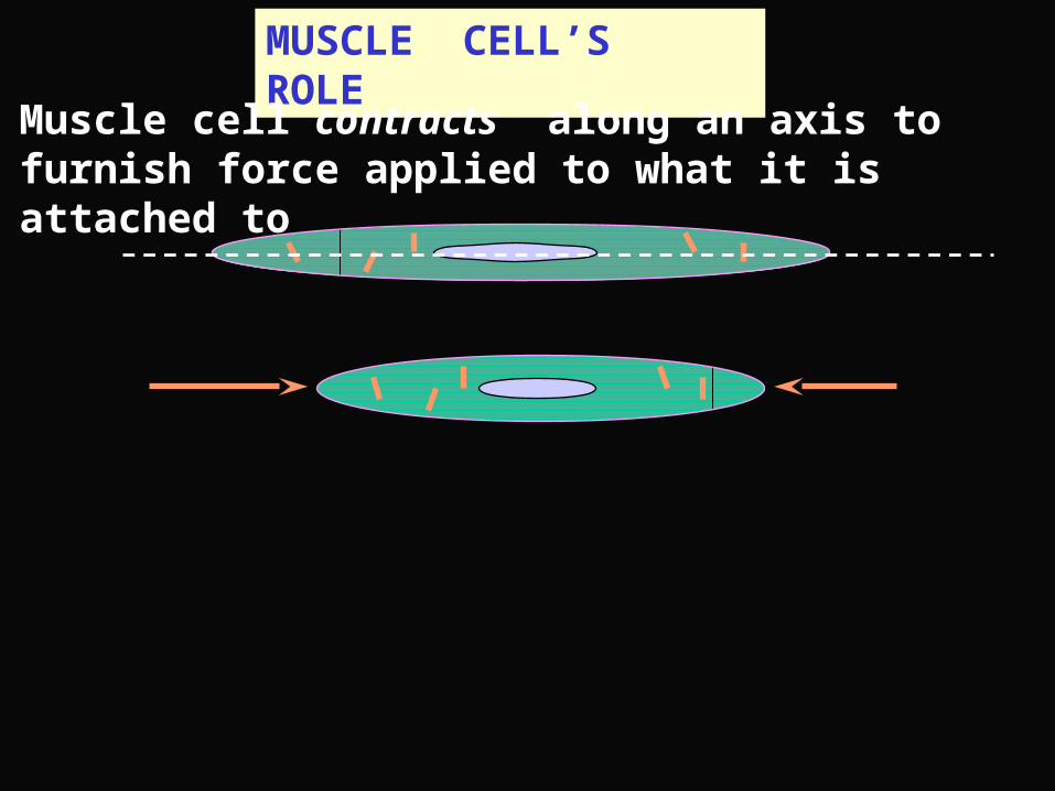
MUSCLE CELL’S ROLE
Muscle cell contracts along an axis to furnish force applied to what it is attached to
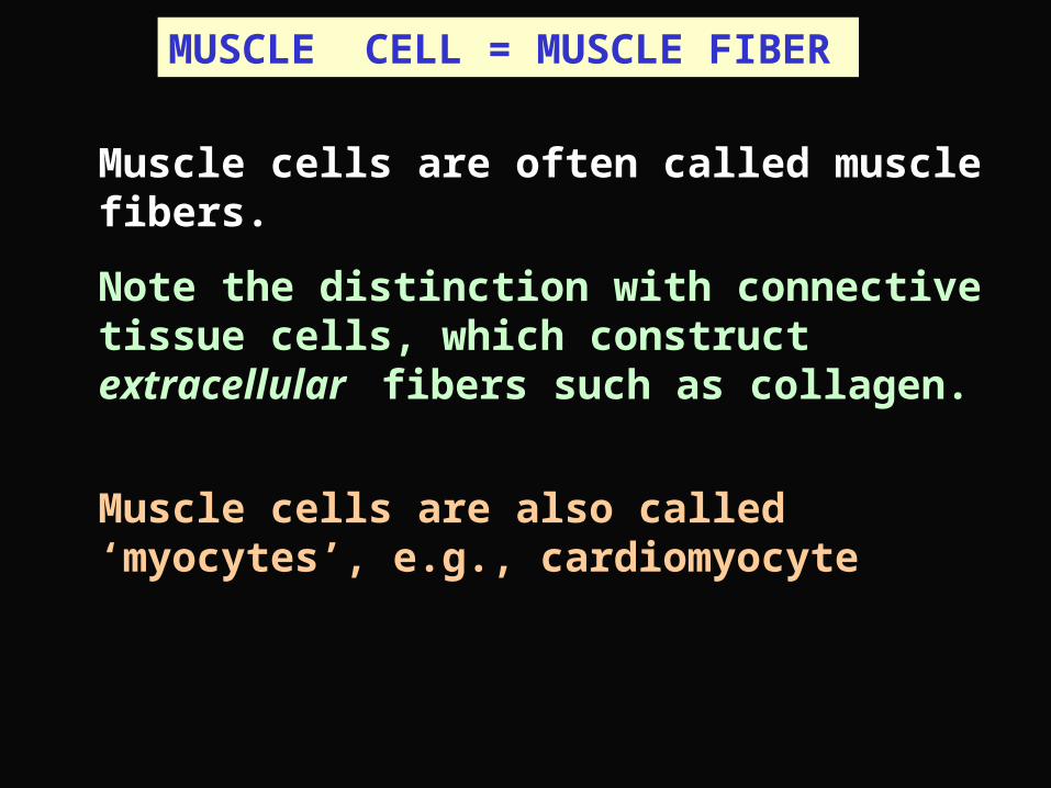
Muscle cells are often called muscle fibers.
Note the distinction with connective tissue cells, which construct extracellular fibers such as collagen.
Muscle cells are also called ‘myocytes’, e.g., cardiomyocyte
MUSCLE CELL = MUSCLE FIBER
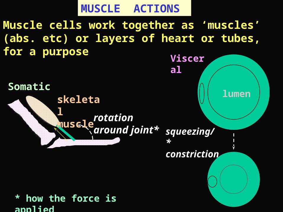
MUSCLE ACTIONS
Muscle cells work together as ‘muscles’ (abs. etc) or layers of heart or tubes, for a purpose
Somatic skeletal muscle
rotation around joint*
Visceral
squeezing/ * constriction
lumen
* how the force is applied
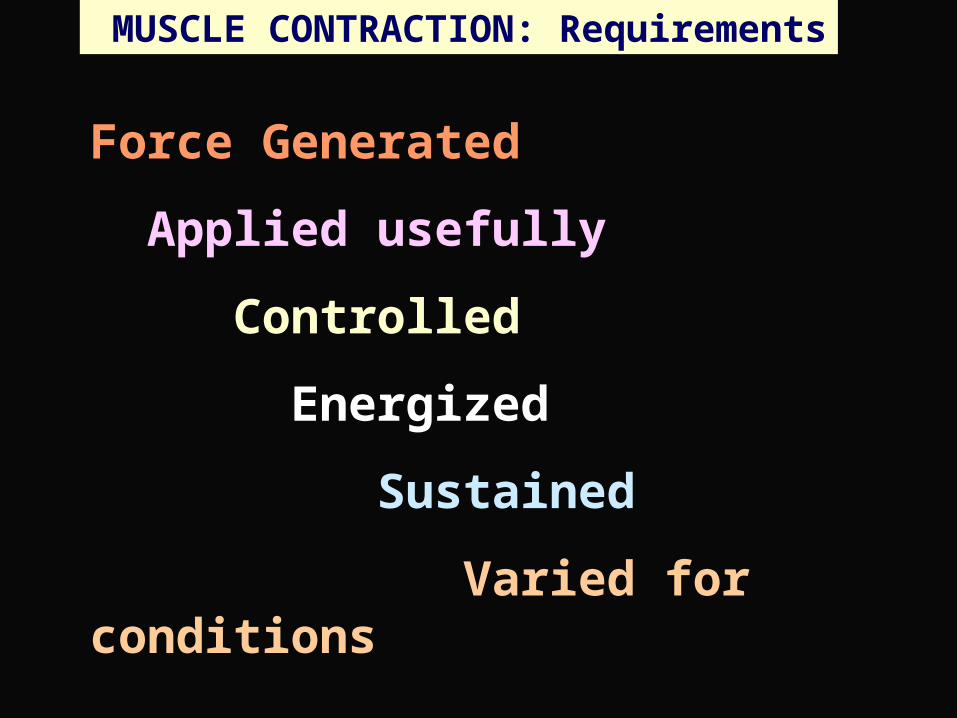
MUSCLE CONTRACTION: Requirements
Force Generated
Applied usefully
Controlled
Energized
Sustained
Varied for conditions
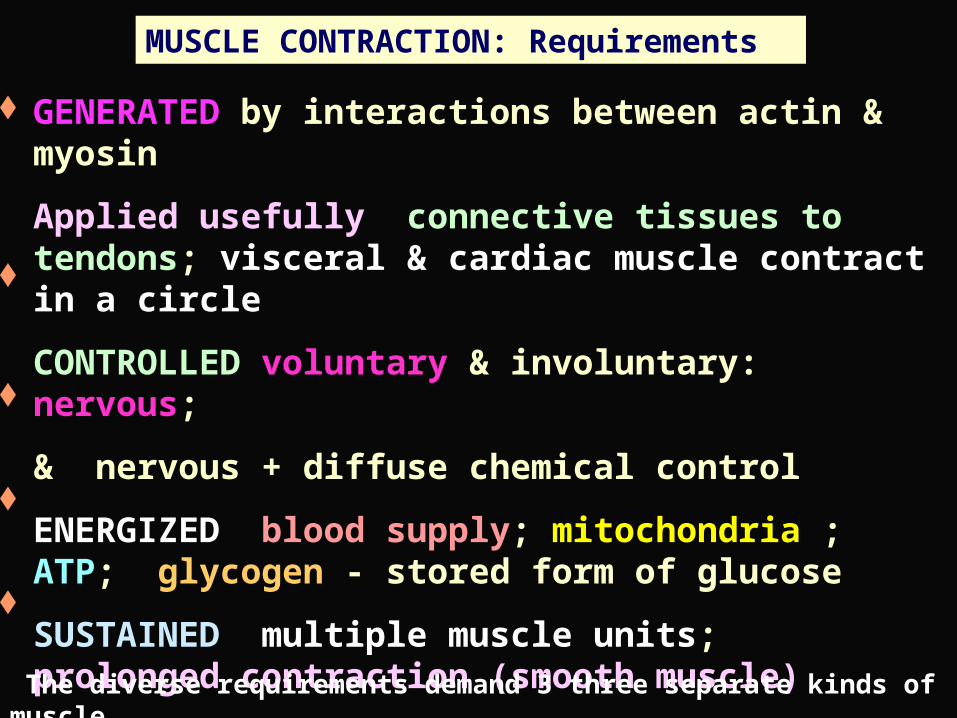
MUSCLE CONTRACTION: Requirements
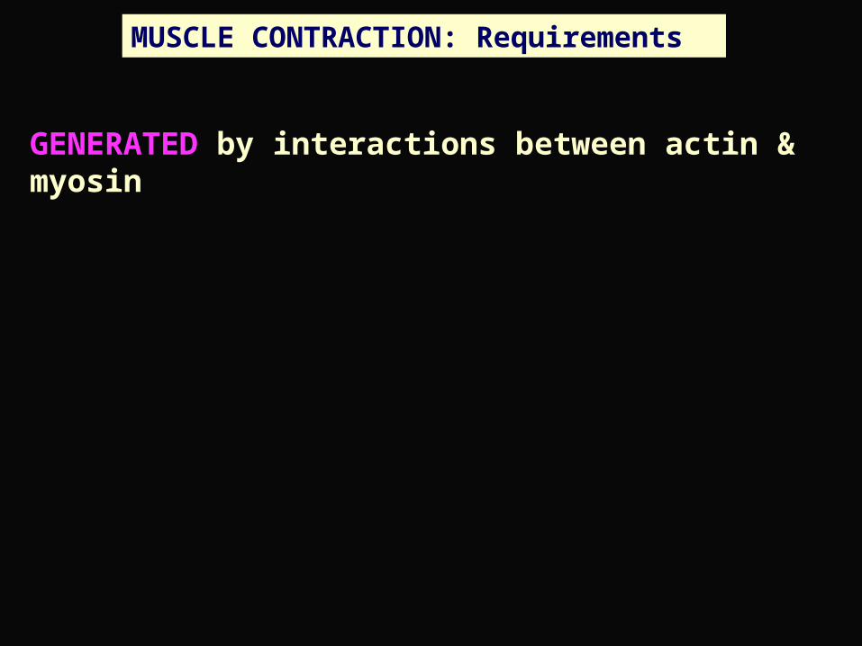
GENERATED by interactions between actin & myosin
Applied usefully connective tissues to tendons; visceral & cardiac muscle contract in a circle
CONTROLLED voluntary & involuntary: nervous;
& nervous + diffuse chemical control
ENERGIZED blood supply; mitochondria ; ATP; glycogen - stored form of glucose
SUSTAINED multiple muscle units; prolonged contraction (smooth muscle)
VARIED FOR CONDITIONS sub-types of muscle
The diverse requirements demand 3 three separate kinds of muscle
MUSCLE CONTRACTION: Requirements
GENERATED by interactions between actin & myosin
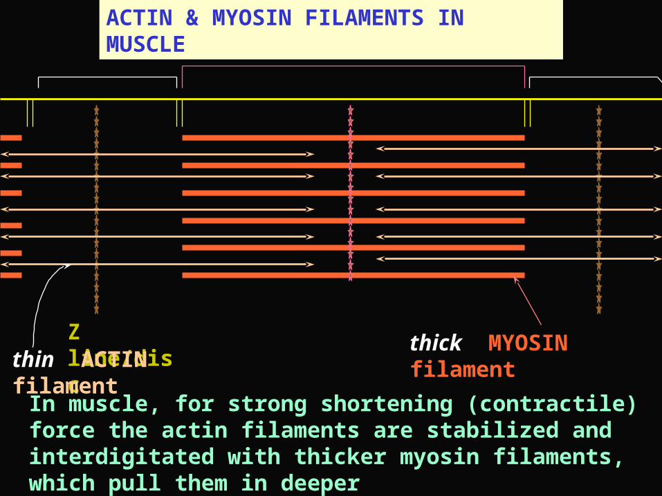
ACTIN & MYOSIN FILAMENTS IN MUSCLE
Z line/disc thick MYOSIN filament thin ACTIN filament
In muscle, for strong shortening (contractile) force the actin filaments are stabilized and interdigitated with thicker myosin filaments, which pull them in deeper
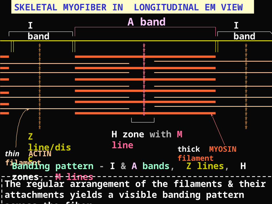
SKELETAL MYOFIBER IN LONGITUDINAL EM VIEW
Z line/disc
I band A band I band
H zone with M line
thick MYOSIN filament thin ACTIN filament
Banding pattern - I & A bands, Z lines, H zones, M lines
The regular arrangement of the filaments & their attachments yields a visible banding pattern across the fiber
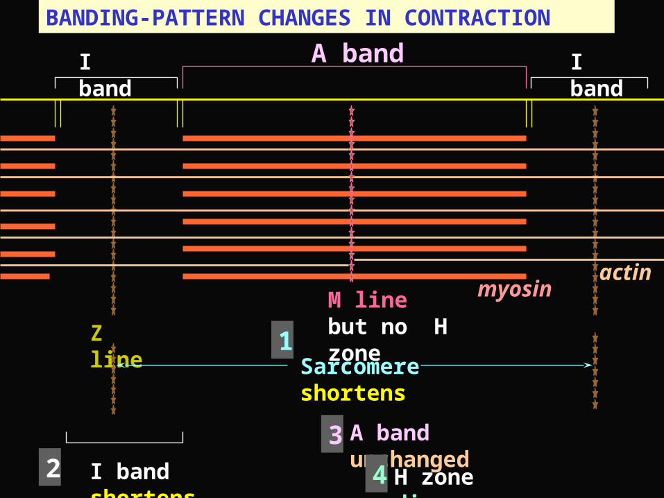
BANDING-PATTERN CHANGES IN CONTRACTION
Z line
I band A band I band
M line but no H zone
myosinactin
Sarcomere shortens
I band shortens
A band unchanged
H zone disappears
1
4
32
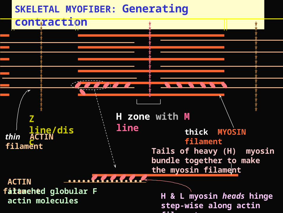
SKELETAL MYOFIBER: Generating contraction
Z line/disc H zone with M line
thick MYOSIN filament thin ACTIN filament
ACTIN filament
H & L myosin heads hinge step-wise along actin filament
Tails of heavy (H) myosin bundle together to make the myosin filament
attached globular F actin molecules
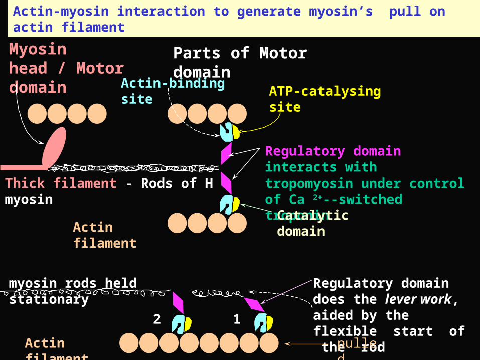
Regulatory domain interacts with tropomyosin under control of Ca 2+--switched troponinThick filament - Rods of H myosin
ATP-catalysing siteActin-binding site
Catalytic domainActin filament
Myosin head / Motor domain
Parts of Motor domain
Actin filament
Regulatory domain does the lever work, aided by the flexible start of the rod
Actin-myosin interaction to generate myosin’s pull on actin filament
12
pulled
myosin rods held stationary
MUSCLE CONTRACTION: Requirements
Applied usefully connective tissues to tendons;
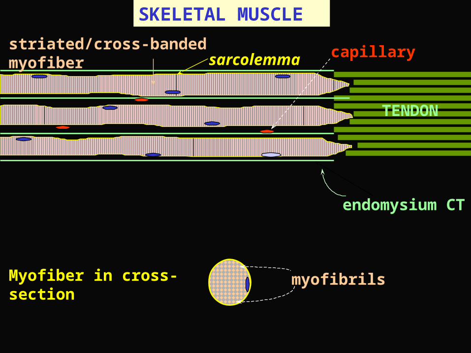
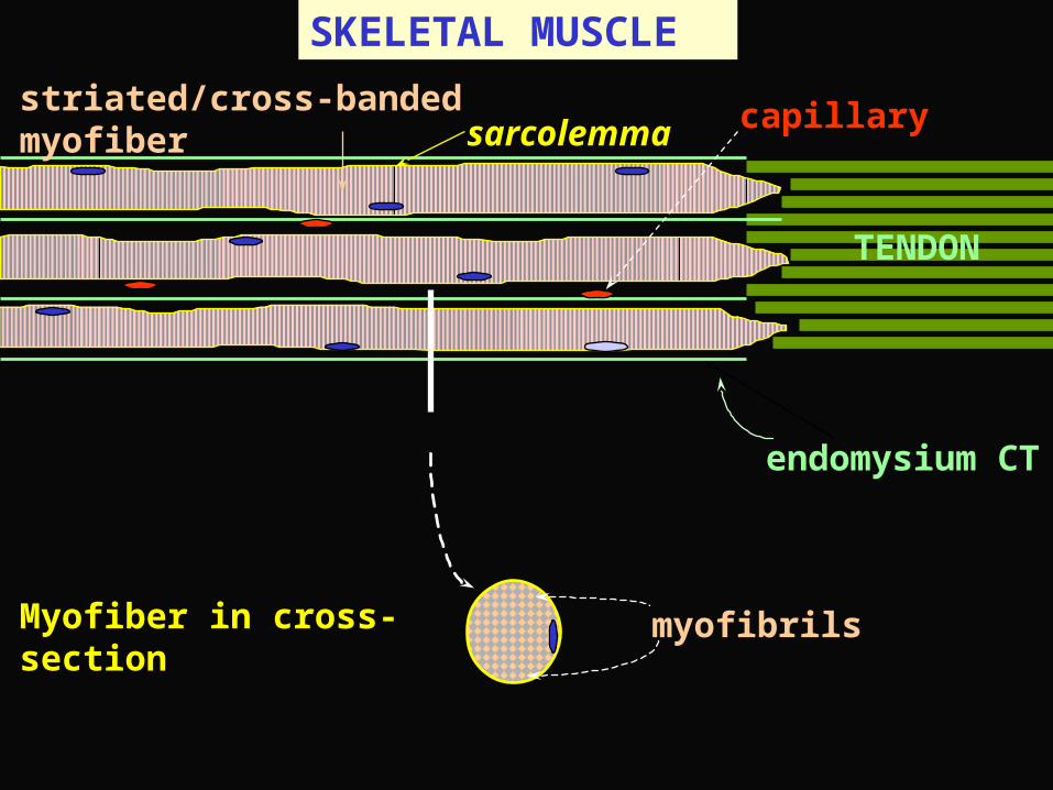
SKELETAL MUSCLE
endomysium CT
striated/cross-banded myofiber sarcolemma
TENDON
capillary
Myofiber in cross-section myofibrils
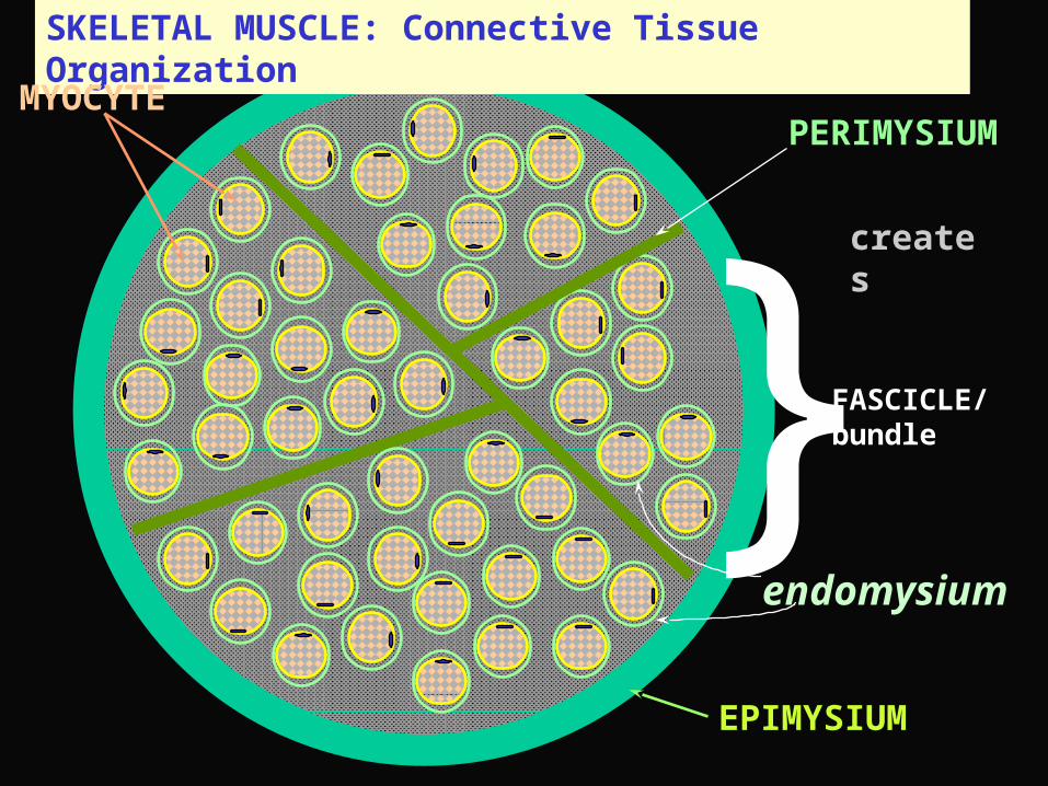
SKELETAL MUSCLE: Connective Tissue Organization
}FASCICLE/ bundle
PERIMYSIUM
EPIMYSIUM
endomysium
creates
MYOCYTE
SKELETAL MUSCLE
endomysium CT
striated/cross-banded myofiber sarcolemma
TENDON
capillary
Myofiber in cross-section myofibrils
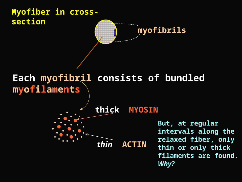
Myofiber in cross-section
myofibrils
Each myofibril consists of bundled myofilaments
thick MYOSIN
thin ACTIN
But, at regular intervals along the relaxed fiber, only thin or only thick filaments are found. Why?
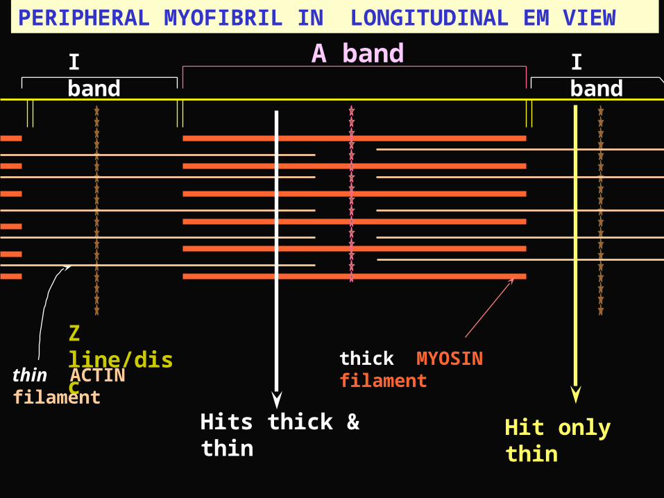
PERIPHERAL MYOFIBRIL IN LONGITUDINAL EM VIEW
Z line/disc
I band A band I band
thick MYOSIN filament thin ACTIN filament
Hit only thinHits thick & thin
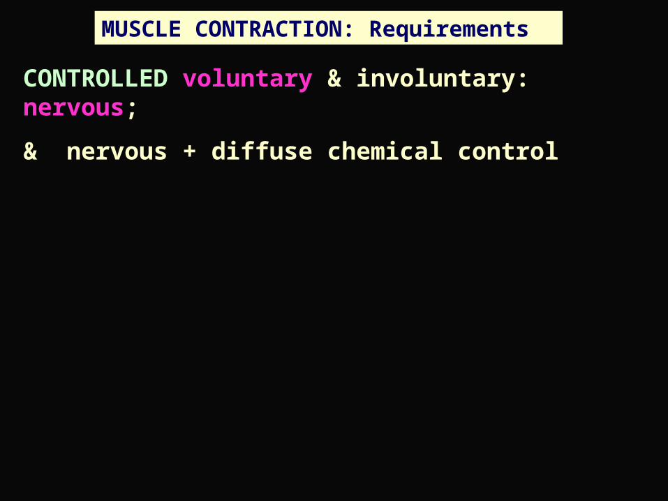
MUSCLE CONTRACTION: Requirements
CONTROLLED voluntary & involuntary: nervous;
& nervous + diffuse chemical control
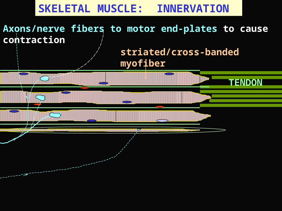
SKELETAL MUSCLE: INNERVATION
striated/cross-banded myofiber
TENDON
Axons/nerve fibers to motor end-plates to cause contraction
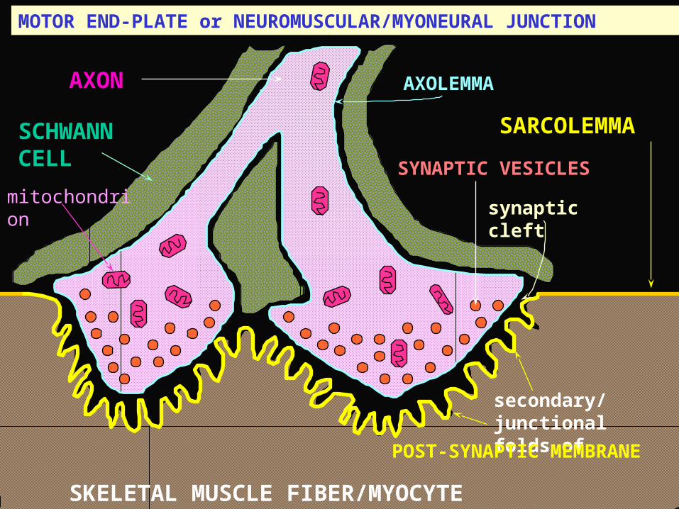
MOTOR END-PLATE or NEUROMUSCULAR/MYONEURAL JUNCTION
AXON
SCHWANN CELL
SARCOLEMMA
SKELETAL MUSCLE FIBER/MYOCYTE
synaptic cleft
secondary/ junctional folds of
POST-SYNAPTIC MEMBRANE
AXOLEMMA
SYNAPTIC VESICLES
mitochondrion

MOTOR END-PLATE: LOCATION OF ‘TRANSMISSION’ MOLECULES
SARCOLEMMA
SKELETAL MUSCLE FIBER/MYOCYTE
synaptic cleft
POST-SYNAPTIC MEMBRANE
AXOLEMMA
SYNAPTIC VESICLES
voltage-gated ion channels
voltage-gated ion channels
ACh receptors
Ligand-gated ion channels
Cholinesterase
Acetyl Choline/ACh
PRE-SYNAPTIC MEMBRANE
Ca2+ channels

SKELETAL MYOFIBER: Initiating contraction
Z line
motor end-plate
sarcolemmaT/transverse tubule
Sarcoplasmic reticulum wraps around myofibril and releases Calcium ion, when stimulated via T-tubule & feet
Feet
Terminal cisterna of SR
Triad = T-tubule + two terminal cisternae
}
Motor end-plate - Sarcolemma AP - T-tubule AP - Feet - SR - Ca 2+ release
A-I junction

MUSCLE CONTRACTION: Requirements
CONTROLLED voluntary & involuntary: nervous;
& nervous + diffuse chemical control
ENERGIZED blood supply; mitochondria ; ATP; glycogen - stored form of glucose

MYOFIBRIL
AROUND EACH MYOFIBRIL, meaning between myofibrils
Glycogen granules Sarcoplasmic reticulum
Mitochondria
energize
energize
control
Myofilaments generate force

THREE MAIN TYPES OF MUSCLE
SMOOTH small but prolongable force; diverse types, uses, & controls; controlled partly by autonomic/involuntary nervous system, partly by chemicals released from nearby cells, and by cell-to-cell connections
CARDIAC strong rhythmic contractions; controlled by own cell-to-cell connections; pace determined by autonomic innervation to a little of the cardiac muscle
SKELETAL most forceful kind, but contracts only in response to voluntary/somatic nervous system activity; applies its force via well-organized connective tissue; strength of contraction needs high internal organization within the muscle cell/fiber

THREE MAIN TYPES OF MUSCLE I
SMOOTH
small but prolongable force;
diverse types, uses, & controls;
controlled partly by autonomic/ involuntary nervous system, partly by chemicals released from nearby cells, and by cell-to-cell connections

THREE MAIN TYPES OF MUSCLE II
CARDIAC
strong rhythmic contractions;
controlled by own cell-to-cell connections;
pace determined by autonomic innervation to a little of the cardiac muscle

THREE MAIN TYPES OF MUSCLE III
SKELETAL most forceful kind;
but contracts only in response to voluntary/somatic nervous system activity;
applies its force via well-organized connective tissue;
strength of contraction needs high internal organization within the muscle cell/fiber

THREE MAIN TYPES OF MUSCLE IV
Muscle cells are often called muscle fibers. Note the distinction with connective tissue cells, which construct extracellular fibers such as collagen.
Muscle cells are also called ‘myocytes’, e.g., cardiomyocyte

THREE MAIN TYPES OF MUSCLE: Sub-types
SMOOTH skin, cardiovascular, airway, uterine, other reproductive, urinary, gastrointestinal (GI)CARDIAC atrial, ventricular, nodal, PurkinjeSKELETAL type I - slow, type IIa - fast oxidative, type IIb - fast glycolytic

SKELETAL MYOFIBER: Needs determining structure
Generation
Force generation
Stabilization
Force application
Control of contraction
Energize

CARDIAC MUSCLE
striated/cross-banded CARDIOMYOCYTES
Capillary
INTERCALATED DISK
central NUCLEUS
branching muscle fibersSarcolemma & external lamina
Reticular fiber

INTERCALATED DISC - electro-mechanical union
ID is a strong myocyte-myocyte attachment + electrical connections
Fascia adherens strength
Maculae adherens strength
Gap junction transmits contraction

PURKINJE FIBER
} Endocardium
Sub-endocardium
Large, pale cell specialized for conduction, not contraction
Myofilaments
Glycogen
ventricle

SMOOTH MUSCLE
SMOOTH MUSCLE CELL has same contractile & control *machinery as skeletal myocyte, but less organized
Reticular fiber
Gap junction/NexusAutonomic nerve axon
Myocyte plasmalemma + glycoprotein External lamina
* There is the important difference that smooth muscle uses Myosin Light-chain Kinase (MLCK) to phosphorylate the regulatory myson light chain as the main means to provoke the actomyosin ATPase to start contraction

SMOOTH MUSCLE
SMOOTH MUSCLE CELL has same contractile & control machinery* as skeletal myocyte, but less organized
Filaments attach to DENSE BODIES serving the role of Z-lines
CAVEOLAE for stimulus-contraction coupling serve role of T-tubule & SR system

SMOOTH MUSCLE
* There is the important difference that smooth muscle uses Myosin Light-chain Kinase (MLCK) to phosphorylate the regulatory myosin light chain as the main means to provoke the actomyosin ATPase to start contraction

CAVEOLA
Caveolae are plasma membrane invaginations found in most cell types of all four tissues. They are conspicuous in endothelial cells & smooth muscle.
Membrane molecules:
Caveolin - characteristic integral
membrane protein
Cholesterol (lots)
Molecules related to - Transcytosis
Endocytosis or
Signal transduction
Plasmalemma

SMOOTH MUSCLEView with H & E staining - solid pink mass (stained sarcoplasm)
cross-section
long.-section
Unseen are reticular and nerve fibers, plasmalemmas & external laminae
Trichrome stains distinguish smooth muscle cells from collagen fibers

SKELETAL MYOFIBER: Needs determining structure
Generation
Force generation
Stabilization
Force application
Control of contraction
Energize

MYOFIBER: Stabilization* & Force Application materials
Z line
M line*
a-actinin*
Sarcolemma External lamina
Nebulin*
Titin* (“elastic”)
Dystrophin
Desmin* intermediate filaments
Integrin

SKELETAL MUSCLE: SENSORY INNERVATION
striated/cross-banded myofiber
TENDON
Golgi tendon receptor
Muscle spindle
Sensory axon & spindle receptor
The fine control of contraction in individual myofibers requires abundant sensory feedback on how the muscle as a whole is performing

CARDIAC MUSCLE
striated/cross-banded CARDIOMYOCYTES
Capillary
INTERCALATED DISK
central NUCLEUS
branching muscle fibersSarcolemma & external lamina
Reticular fiber

CARDIAC PATHOLOGY
Enlarged, but altered and weakened muscle of Ventricular hypertrophy
Capillary
Reticular fiber
Blocked vessels damaged heart muscle (Cardiac infarct)
More & thicker fibers of Fibrosis
Bad gap junctions Arrythmiaaltered connexin

WHERE AM I? Online Anatomy Module 1
APPENDICULAR SKELETON
CELL
ORIENTATION
EPITHELIUM
CONNECTIVE TISSUE
MUSCLE
NERVOUS SYSTEM
AXIAL SKELETON
MUSCLES
EMBRYOLOGY
You are at the End
Caution how you exit.BACK on yourbrowser is neededUnfortunately there isno way that you candirectly reach othertopics listed here byclicking on them. Youget there by going backto the ParamedicalAnatomy menu