Embed Size (px)
Citation preview
NHS Training for Physiotherapy Support Workers
Workbook 14
The respiratory system
© NHS Education for Scotland 2012. You can copy or reproduce the information in this
document for use within NHSScotland and for non-commercial educational purposes.
Use of this document for commercial purposes is permitted only with the written
permission of NES.
Contents
Workbook 14 The respiratory system 1
14.1 Aim 3
14.2 Learning outcomes 3
14.3 The respiratory system 4
14.4 Breathing 6
14.5 Respiratory disorders 7
14.6 Recognising respiratory distress 10
14.7 Treatment of respiratory distress or failure 11
14.8 Exercise in respiratory disease 14
14.9 The respiratory system workbook completion 16
14.10 The respiratory system reflection 17
Workbook 14 Page 3
NHS Training for Physiotherapy Support Workers Workbook 14 | The respiratory system
Workbook 14
The respiratory system
14.1 Aim
The aim of this workbook is to introduce the Healthcare Support Worker
(HCSW) to the structure and function of the respiratory system.
14.2 Learning outcomes
By the end of this workbook you will be able to:
■ Describe the structure and function of the respiratory system.
■ Describe and recognise some of the common signs that you may observe when a patient has a disorder of the respiratory system.
■ Describe some common respiratory conditions.
■ Describe briefly the benefits of exercise in respiratory conditions.
Workbook 14 Page 4
NHS Training for Physiotherapy Support Workers Workbook 14 | The respiratory system
14.3 The respiratory system
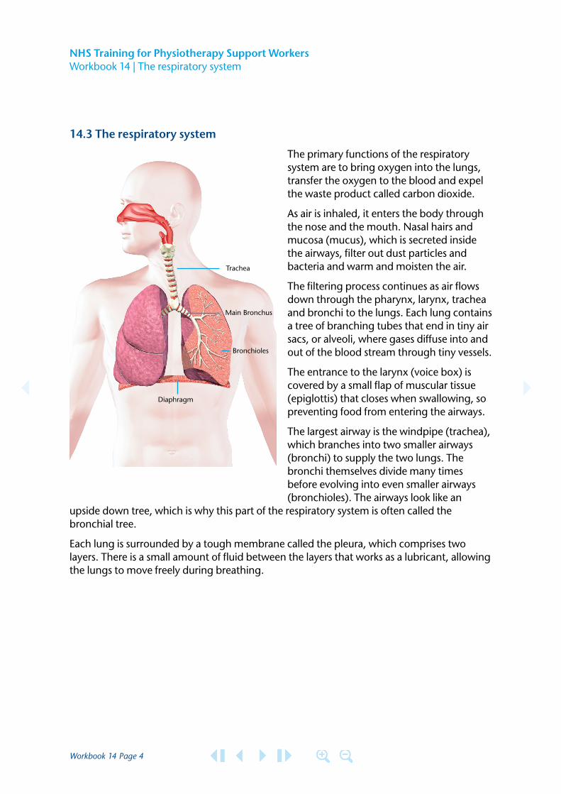
The primary functions of the respiratory system are to bring oxygen into the lungs, transfer the oxygen to the blood and expel the waste product called carbon dioxide.
As air is inhaled, it enters the body through the nose and the mouth. Nasal hairs and mucosa (mucus), which is secreted inside the airways, filter out dust particles and bacteria and warm and moisten the air.
The filtering process continues as air flows down through the pharynx, larynx, trachea and bronchi to the lungs. Each lung contains a tree of branching tubes that end in tiny air sacs, or alveoli, where gases diffuse into and out of the blood stream through tiny vessels.
The entrance to the larynx (voice box) is covered by a small flap of muscular tissue (epiglottis) that closes when swallowing, so preventing food from entering the airways.
The largest airway is the windpipe (trachea), which branches into two smaller airways (bronchi) to supply the two lungs. The bronchi themselves divide many times before evolving into even smaller airways (bronchioles). The airways look like an
upside down tree, which is why this part of the respiratory system is often called the bronchial tree.
Each lung is surrounded by a tough membrane called the pleura, which comprises two layers. There is a small amount of fluid between the layers that works as a lubricant, allowing the lungs to move freely during breathing.
Trachea
Main Bronchus
Bronchioles
Diaphragm
Workbook 14 Page 5
NHS Training for Physiotherapy Support Workers Workbook 14 | The respiratory system
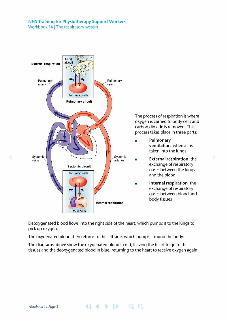
The process of respiration is where oxygen is carried to body cells and carbon dioxide is removed. This process takes place in three parts:
■ Pulmonary ventilation when air is taken into the lungs
■ External respiration the exchange of respiratory gases between the lungs and the blood
■ Internal respiration the exchange of respiratory gases between blood and body tissues
Deoxygenated blood flows into the right side of the heart, which pumps it to the lungs to pick up oxygen.
The oxygenated blood then returns to the left side, which pumps it round the body.
The diagrams above show the oxygenated blood in red, leaving the heart to go to the tissues and the deoxygenated blood in blue, returning to the heart to receive oxygen again.
Workbook 14 Page 6
NHS Training for Physiotherapy Support Workers Workbook 14 | The respiratory system
Evidence
Describe the main parts of the respiratory system. Name each part and what it does.
14.4 Breathing
Air is drawn into the lungs by the diaphragm and intercostal muscles which between them lift the ribs with the lungs attached to them, increasing the volume of the lungs so that air passes into them. When the muscles relax again, the volume of the lungs reduces and the air passes out again.
The amount of air breathed in and out varies with health and activity. The lungs of a healthy adult hold about 3 litres of air when breathing quietly and about half a litre is exchanged with each breath. When active or exercising, the volume of the lung increases considerably to as much as 6 or 7 litres because more oxygen is needed more frequently and the rate and depth of breathing increases.
Gaseous exchange
In the lungs, carbon dioxide (CO2) from the blood passes into the alveoli through the respiratory membrane, a thin barrier that has several layers. Oxygen (O2) crosses the membrane in the opposite direction, from the alveoli to the blood capillaries. Oxygen is then absorbed in the red blood cells which carry the blood to the tissues.
Workbook 14 Page 7
NHS Training for Physiotherapy Support Workers Workbook 14 | The respiratory system
Evidence
Explain how the respiratory system works and the main function of the system. For example; how and why we breathe and how gas is exchanged?
14.5 Respiratory disorders
As a support worker you will often come across patients with respiratory disorders, some of which are described below.
Cough
Coughing is a familiar but complicated reflex. It is one way in which the lungs and airways are protected. Along with other mechanisms, coughing helps protect the lungs against particles that have been inhaled (aspiration). Coughing sometimes leads to the movement or clearance of sputum - a mixture of mucus, debris and cells that is produced in the lungs in greater quantities than is normal.
Chest physiotherapy may be required to assist patients to cough to clear sputum from their lungs. If people do not keep their lungs clear, they may become short of breath because the gas exchange in the lungs is impaired. The lungs may be damaged if over time they collect large volumes of sputum.
The physiotherapist may assist patients to clear sputum from their lungs using postural drainage, percussion, vibrations, breathing exercises, or relaxation. When the patient is unable to cough and clear the secretions they may require to be suctioned.
Dyspnoea shortness of breath – the unpleasant sensation of difficulty in breathing
A healthy person breathes faster during exercise and at high altitude. Although faster breathing is rarely uncomfortable, it may limit the amount of exercise that can be performed.
With dyspnoea the sensation that the person is running out of air and can’t breathe fast enough or deeply enough accompanies the faster breathing. Dyspnoea is a sign of serious disease of the airway, lungs, or heart. The onset of dyspnoea should not be ignored but is reason to seek medical attention.
Workbook 14 Page 8
NHS Training for Physiotherapy Support Workers Workbook 14 | The respiratory system
Wheeze a whistling, musical sound during breathing that results from partially obstructed airways
It may be caused by a general narrowing of the airways, (asthma or chronic obstructive pulmonary disease), by a local narrowing (as with a tumour), or by a foreign particle lodged in an airway. The most common cause of recurring wheezing is asthma although many people who have never had asthma wheeze at some time in their lives. A wheeze is sometimes audible without the use of a stethoscope.
Stridor a crowing sound heard during breathing, mainly during inhalation, that results from a partial blockage of the throat (pharynx), voice box (larynx) or windpipe (trachea)
Stridor is usually loud enough to be heard at some distance, but it may only be audible during a deep breath. The sound is caused by turbulent airflow through a narrowed upper airway – possible causes could be a tumour, an abscess or perhaps swelling in the upper airway, or a malfunction of the vocal chords.
Stridor can be a symptom of a life-threatening emergency. In such cases, a tube may be inserted through the person’s mouth or nose (tracheal intubation) or directly into the trachea (tracheostomy) to allow air to get past the blockage and to save the persons life.
Cyanosis a bluish discolouration of the skin resulting from an inadequate amount of oxygen in the blood
Occurs when oxygen depleted blood which is bluish rather than red, circulates through the skin. Cyanosis restricted to the fingers and toes usually occurs because blood flows through the limbs very slowly. It may result when the pumping action of the heart is weak or when a person is exposed to the cold. Cyanosis that occurs throughout the body can be caused by many types of severe lung disease. Also by certain blood vessels and heart malformations that shunt blood from the venous to the arterial side of the circulation.
If you are with a patient who suddenly becomes cyanosed seek help immediately.
Workbook 14 Page 9
NHS Training for Physiotherapy Support Workers Workbook 14 | The respiratory system
Hypoxia is a lack of oxygen (O2 ) supply to the tissues
Hypoxia can occur in the following ways:
■ Hypoxic not enough oxygen to go round ■ Anaemic not enough haemoglobin to carry the oxygen ■ Stagnant not enough blood flow ■ Histotoxic chemical poisoning
Evidence
Describe what is meant by the following:
Cough
Dyspnoea
Wheeze
Stridor
Cyanosis
Hypoxia
Workbook 14 Page 10
NHS Training for Physiotherapy Support Workers Workbook 14 | The respiratory system
Evidence
Can you describe an occasion on which you observed a patient with any of these signs?
What action did you take?
14.6 Recognising respiratory distress
You may come across patients who have become unwell and who are in respiratory distress. This situation requires medical attention which should be found immediately. As a support worker you should be able to recognise when the patient is unwell.
■ General appearance Does the patient appear to be in pain, fatigued, or lethargic? Are they restless or incoherent?
■ Colour Is there a change in the patient’s colour since you last saw the patient? Are they pale or flushed? Blue at the mouth and lips - may indicate that the oxygen level is low in the blood. You should report this immediately to a nurse, doctor or qualified physiotherapist.
■ Temperature Fever is an indication of infection so the patient may feel too unwell to perform some activities with you.
■ Pulse Pulse may be raised if the patient has low oxygen levels, fever or is very anxious.
■ Breathing rate Is the patient breathing faster or more slowly than normal? Are the neck muscles working too hard? You may notice the patient pursing their lips during breathing – this is to create pressure in the airways to prevent airway collapse.
■ Oedema Does the patient have swollen ankles or hands? Is this new for them? If so, inform a member of staff.
■ Respiratory failure In some conditions, such as chronic bronchitis, cystic fibrosis or asthma the level of blood oxygen may become dangerously low, or the level of carbon dioxide may become dangerously high. This occurs when the movement of air in and out of the lungs is inadequate.
Workbook 14 Page 11
NHS Training for Physiotherapy Support Workers Workbook 14 | The respiratory system
Evidence
Describe how you might recognise that a patient may be suffering from respiratory distress.
If you have observed any of these signs in a patient that you have been treating, describe here what you did.
14.7 Treatment of respiratory distress or failure
Almost always, oxygen is given initially. Usually, the amount given is more than is needed, unless the person has a chronic chest condition. Such people tend to slow their breathing when they’re over-treated with oxygen. The underlying cause also must be treated. Antibiotics are used to fight infection, and bronchodilators are used to open the airways. Other medications may be given to decrease inflammation or prevent blood clots.
Some very ill patients need mechanical ventilation to aid breathing:
A plastic tube is inserted through the nose or mouth and into the trachea; this tube is attached to a machine that forces air into the lungs. Exhalation occurs passively because of the elastic recoil of the lungs. Many types of ventilators and modes of operation may be used, depending on the underlying disorder. If the lungs aren’t functioning well, additional oxygen may be delivered through the ventilator. Mechanical ventilation can be lifesaving whenever patients are not able to move enough air in and out of their lungs.
Workbook 14 Page 12
NHS Training for Physiotherapy Support Workers Workbook 14 | The respiratory system
Asthma
Asthma affects the small airways (bronchioles) that carry air in and out of the lungs. About 100 million people worldwide have the condition, 5.1 million in the UK alone – 1 in 13 adults and 1 in 8 children.
An ‘asthma attack’ describes the symptoms of tightness in the chest, a wheezing or whistling noise in the chest, coughing and difficulty breathing. These symptoms occur when the airway becomes narrowed, inflamed and blocked by plugs of mucus.
Patients are treated using drugs known as bronchodilators, which reduce the narrowing of the airways, and by inhaled steroids which reduce the inflammation.
Pleurisy
The pleura is the double-layered membrane that covers the lungs. One layer is attached to the surface of the lung and the other to the inside of the chest wall. The two layers are in close contact and are separated only by a thin layer of lubricating fluid produced by the membranes.
Pleurisy causes roughening of the surfaces in contact, and as these rub against one another there is a sharp pain. This usually occurs at a particular point on breathing in, often at the end of a deep breath. It may also be brought on by coughing or by twisting the body. There is likely also to be fever and general discomfort.
There are many causes of pleurisy. Pleurisy may develop if there is some disease process in the underlying lung, such as pneumonia, cancer or tuberculosis. Other causes include viral infections of the pleura. Pleurisy may also be a feature of rheumatoid arthritis.
Workbook 14 Page 13
NHS Training for Physiotherapy Support Workers Workbook 14 | The respiratory system
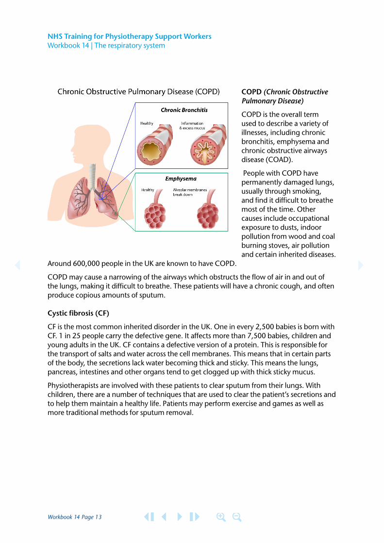
COPD (Chronic Obstructive Pulmonary Disease)
COPD is the overall term used to describe a variety of illnesses, including chronic bronchitis, emphysema and chronic obstructive airways disease (COAD).
People with COPD have permanently damaged lungs, usually through smoking, and find it difficult to breathe most of the time. Other causes include occupational exposure to dusts, indoor pollution from wood and coal burning stoves, air pollution and certain inherited diseases.
Around 600,000 people in the UK are known to have COPD.
COPD may cause a narrowing of the airways which obstructs the flow of air in and out of the lungs, making it difficult to breathe. These patients will have a chronic cough, and often produce copious amounts of sputum.
Cystic fibrosis (CF)
CF is the most common inherited disorder in the UK. One in every 2,500 babies is born with CF. 1 in 25 people carry the defective gene. It affects more than 7,500 babies, children and young adults in the UK. CF contains a defective version of a protein. This is responsible for the transport of salts and water across the cell membranes. This means that in certain parts of the body, the secretions lack water becoming thick and sticky. This means the lungs, pancreas, intestines and other organs tend to get clogged up with thick sticky mucus.
Physiotherapists are involved with these patients to clear sputum from their lungs. With children, there are a number of techniques that are used to clear the patient’s secretions and to help them maintain a healthy life. Patients may perform exercise and games as well as more traditional methods for sputum removal.
Workbook 14 Page 14
NHS Training for Physiotherapy Support Workers Workbook 14 | The respiratory system
Evidence
Describe what is meant by the following:
Asthma
COPD
Cystic fibrosis
14.8 Exercise in respiratory disease
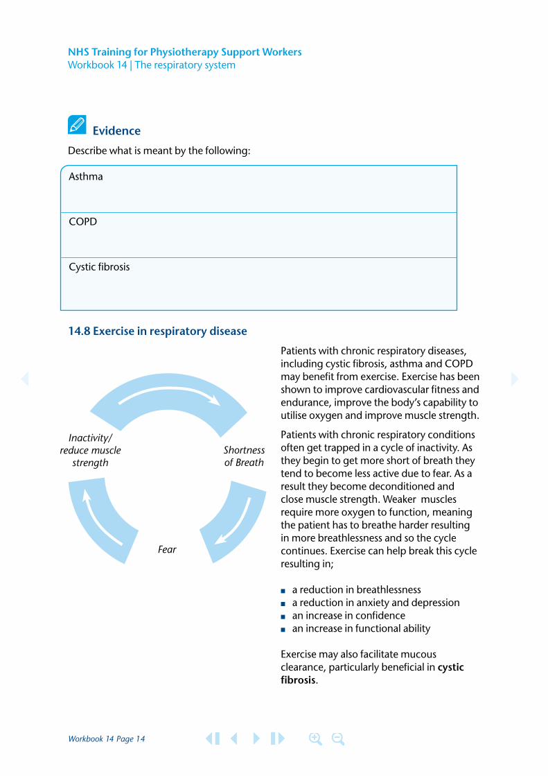
Patients with chronic respiratory diseases, including cystic fibrosis, asthma and COPD may benefit from exercise. Exercise has been shown to improve cardiovascular fitness and endurance, improve the body’s capability to utilise oxygen and improve muscle strength.
Patients with chronic respiratory conditions often get trapped in a cycle of inactivity. As they begin to get more short of breath they tend to become less active due to fear. As aresult they become deconditioned and close muscle strength. Weaker muscles require more oxygen to function, meaning the patient has to breathe harder resulting in more breathlessness and so the cycle continues. Exercise can help break this cycle resulting in;
■ a reduction in breathlessness ■ a reduction in anxiety and depression ■ an increase in confidence ■ an increase in functional ability
Exercise may also facilitate mucous clearance, particularly beneficial in cystic fibrosis.
Inactivity/ reduce muscle
strength
Fear
Shortness of Breath
Workbook 14 Page 15
NHS Training for Physiotherapy Support Workers Workbook 14 | The respiratory system
Evidence
Describe briefly the benefits of exercise in respiratory conditions.
Acknowledgements NHS Tayside
Workbook 14 Page 16
NHS Training for Physiotherapy Support Workers Workbook 14 | The respiratory system
14.9 The respiratory system workbook completion
Your supervising physiotherapist will sign your portfolio to indicate that you have completed this workbook successfully.
Objective Physiotherapist’s signature Date
Describe the main parts of the respiratory system and describe the function of the system
Describe some common signs of respiratory disorders
Discuss how you might recognise a patient that may be suffering from respiratory distress
Describe and discuss some common respiratory diseases
Explain the effect exercise can have on respiratory conditions
Support worker (name)
Support worker’s signature
Physiotherapist (name)
Physiotherapist’s signature
Date
Workbook 14 Page 17
NHS Training for Physiotherapy Support Workers Workbook 14 | The respiratory system
14.10 The respiratory system reflection
Suggested KSF Dimensions: C2, HWB2, HBW7This form should be placed in the appropriate section of your portfolio.
What did you learn from this module?
How has this influenced your work?
Date module completed
TaysideGreater Glasgow and Clyde


![Respiratory System [โหมดความเข้ากันได้] · PATHOLOGY OF RESPIRATORY SYSTEM นพ. อรรณพ นาคะป ท Respiratory system U it](https://img.pdfslide.net/doc/110x75/5fa578efd4e80f055f6b3401/respiratory-system-aaaaaaaaaaaaaaaaaa-pathology.jpg)


![Anatomy and Physiology Respiratory System [Tab 2] Respiratory System](https://img.pdfslide.net/doc/110x75/56649ebd5503460f94bc631f/anatomy-and-physiology-respiratory-system-tab-2-respiratory-system.jpg)


