Embed Size (px)
Citation preview
www.soran.edu.iq
Cell division functions in reproduction, growth, and repair
In unicellular organisms, the division of one cell to form two reproduces an entire organism (e.g., bacteria, yeast, Amoeba)
In multicellular organisms, cell division allows:
• Growth and development from the fertilized egg• Replacement of damaged or dead cells
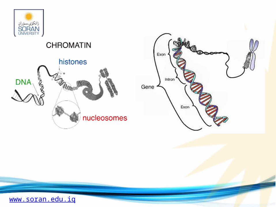
Cell division is a finely controlled process that results in the distribution of identical hereditary material—DNA—to two daughter cells.
The total hereditary endowment of a cell of a particular species is called its genome.
The genomes of some species are quite small (e.g., prokaryotes), while the genomes of other species are quite large (e.g., eukaryotes).
www.soran.edu.iq
Cell division usually proceeds in two sequential steps: nuclear division (mitosis) and division of the cytoplasm (cytokinesis). Not all cells undergo cytokinesis following mitosis.
The cell cycle alternates between the mitotic (M) phase, or dividing phase, and interphase, the nondividing phase:
• M phase, the shortest part of the cell cycle and the phase during which the cell divides, includes:
1. Mitosis - Division of the nucleus
2. Cytokinesis - Division of the cytoplasm
• Interphase, the nondividing phase, includes most of a cell's growth and metabolic activities.
• Is about 90% of the cell cycle
• Is a period of intense biochemical activity during which the cell grows and copies its chromosomes in preparation for cell division
www.soran.edu.iq
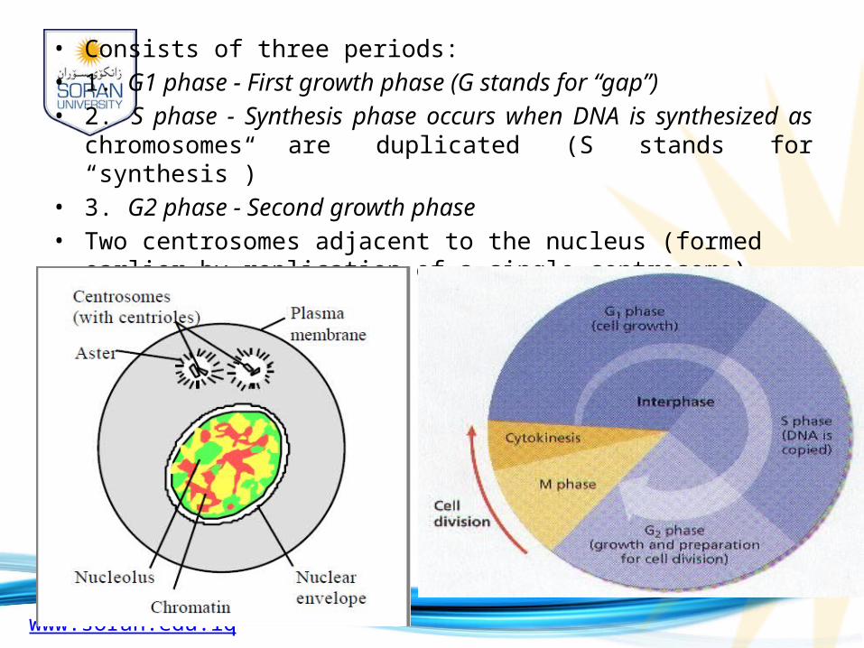
• Consists of three periods:• 1. G1 phase - First growth phase (G stands for “gap”)• 2. S phase - Synthesis phase occurs when DNA is synthesized as
chromosomes are duplicated (S stands for “synthesis”)• 3. G2 phase - Second growth phase• Two centrosomes adjacent to the nucleus (formed earlier by replication of
a single centrosome)
www.soran.edu.iq
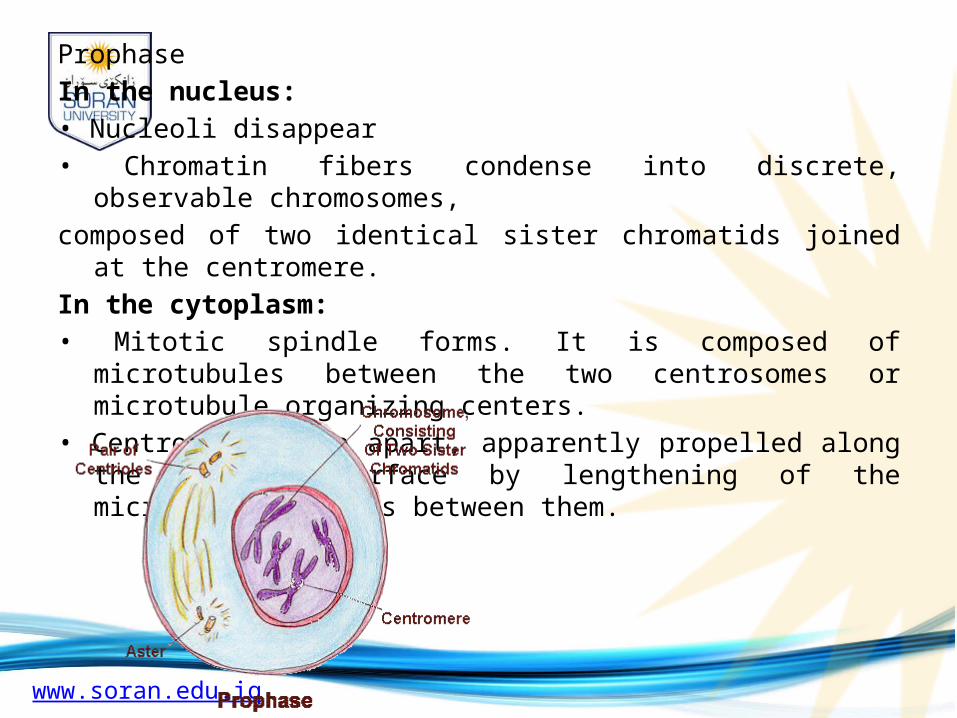
Prophase
In the nucleus:
• Nucleoli disappear
• Chromatin fibers condense into discrete, observable chromosomes,
composed of two identical sister chromatids joined at the centromere.
In the cytoplasm:
• Mitotic spindle forms. It is composed of microtubules between the two centrosomes or microtubule organizing centers.
• Centrosomes move apart, apparently propelled along the nuclear surface by lengthening of the microtubule bundles between them.
www.soran.edu.iq
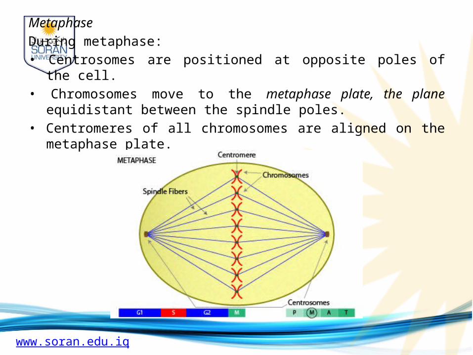
Metaphase
During metaphase:
• Centrosomes are positioned at opposite poles of the cell.
• Chromosomes move to the metaphase plate, the plane equidistant between the spindle poles.
• Centromeres of all chromosomes are aligned on the metaphase plate.
www.soran.edu.iq
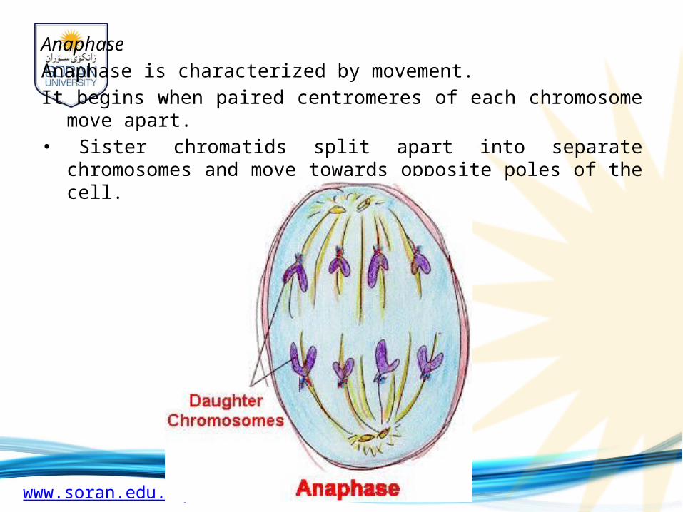
Anaphase
Anaphase is characterized by movement.
It begins when paired centromeres of each chromosome move apart.
• Sister chromatids split apart into separate chromosomes and move towards opposite poles of the cell.
www.soran.edu.iq
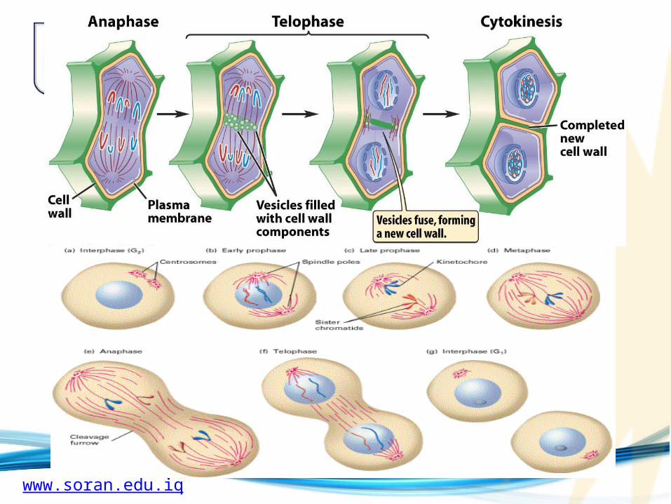
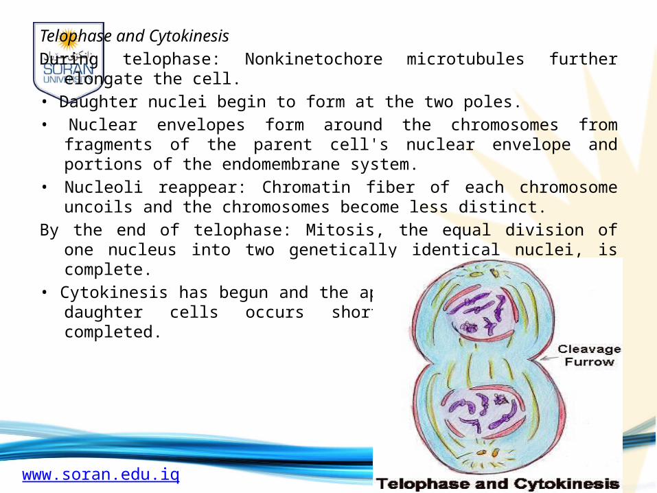
Telophase and Cytokinesis
During telophase: Nonkinetochore microtubules further elongate the cell.
• Daughter nuclei begin to form at the two poles.
• Nuclear envelopes form around the chromosomes from fragments of the parent cell's nuclear envelope and portions of the endomembrane system.
• Nucleoli reappear: Chromatin fiber of each chromosome uncoils and the chromosomes become less distinct.
By the end of telophase: Mitosis, the equal division of one nucleus into two genetically identical nuclei, is complete.
• Cytokinesis has begun and the appearance of two separate daughter cells occurs shortly after mitosis is completed.
www.soran.edu.iq
Internal and external cues help regulate the cell cycle
The cell-cycle control system integrates a variety of internal (intracellular) and external (extracellular) information.
Chemical factors
• If essential nutrients are left out of the culture medium, cells will not divide.
• Specific regulatory substances called growth factors are necessary for most cultured mammalian cells to divide, even if all other conditions are favorable.
Physical factors
• Crowding inhibits cell division in a phenomenon called density-dependent inhibition. Cultured cells stop dividing when they form a single layer on the container’s inner surface. If some cells are removed, those bordering the open space divide again until the vacancy is filled.
www.soran.edu.iq
Cancer cells have escaped from cell-cycle controls
Cancer cells do not respond normally to the body’s control mechanisms. They divide excessively, invade other tissues and, if unchecked, can kill the whole organism.
Cancer cells in culture do not stop growing in response to cell density (density dependent inhibition); they do not stop dividing when growth factors are depleted.
• Cancer cells may make growth factors themselves.
• Cancer cells may have an abnormal growth factor signaling system.
• Cancer cells in culture are immortal in that they continue to divide indefinitely, as long as nutrients are available. Normal mammalian cells in culture divide only about 20 to 50 times before they stop.