Embed Size (px)
Citation preview
NEAFS Y-mtDNA Workshop (Butler and Coble)mtDNA
November 1, 2006
http://www.cstl.nist.gov/biotech/strbase/training.htm 1
Y-Chromosome and Mitochondrial DNA Analysis
NEAFS 2006 WorkshopRye Brook, NY
November 1, 2006
Dr. John M. ButlerDr. Michael D. Coble
mitochondrial DNA
[email protected]@afip.osd.mil
Goals and Objectives
• Overview and theory behind mtDNA analysis• The science behind mtDNA sequencing.• Forensic casework applications of mtDNA.• Tools for mtDNA screening – Linear Arrays.• Emerging mtDNA technologies – mtDNA
genome sequencing for increased discrimination, mtDNA micro-chip technology.
• Summary and Questions
June 26, 2000
Associated Press
“A day for the ages”
NEAFS Y-mtDNA Workshop (Butler and Coble)mtDNA
November 1, 2006
http://www.cstl.nist.gov/biotech/strbase/training.htm 2
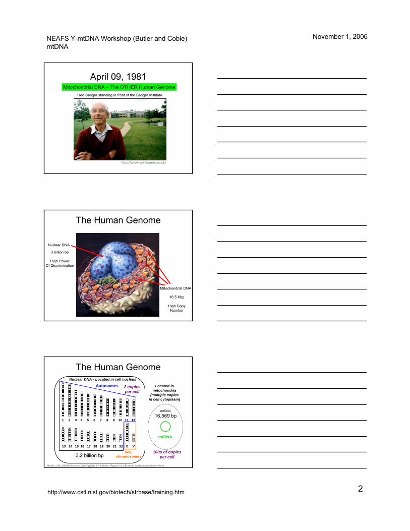
April 09, 1981
Fred Sanger standing in front of the Sanger Institute
http://www.wellcome.ac.uk/
Mitochondrial DNA – The OTHER Human Genome
The Human Genome
Nuclear DNA
3 billion bp
High PowerOf Discrimination
Mitochondrial DNA
16.5 Kbp
High CopyNumber
The Human Genome
http
://w
ww
.ncb
i.nlm
.nih
.gov
/gen
ome/
guid
e/
1 2 3 4 5 6 7 8 9 10 11 12
13 14 15 16 17 18 19 20 21 22 X YSex-
chromosomes
Autosomes
3.2 billion bp
Nuclear DNA - Located in cell nucleus
2 copies per cell
mtDNA
16,569 bp
mtDNA
Located in mitochondria
(multiple copies in cell cytoplasm)
100s of copies per cell
Butler, J.M. (2005) Forensic DNA Typing, 2nd Edition, Figure 2.3, ©Elsevier Science/Academic Press
NEAFS Y-mtDNA Workshop (Butler and Coble)mtDNA
November 1, 2006
http://www.cstl.nist.gov/biotech/strbase/training.htm 3
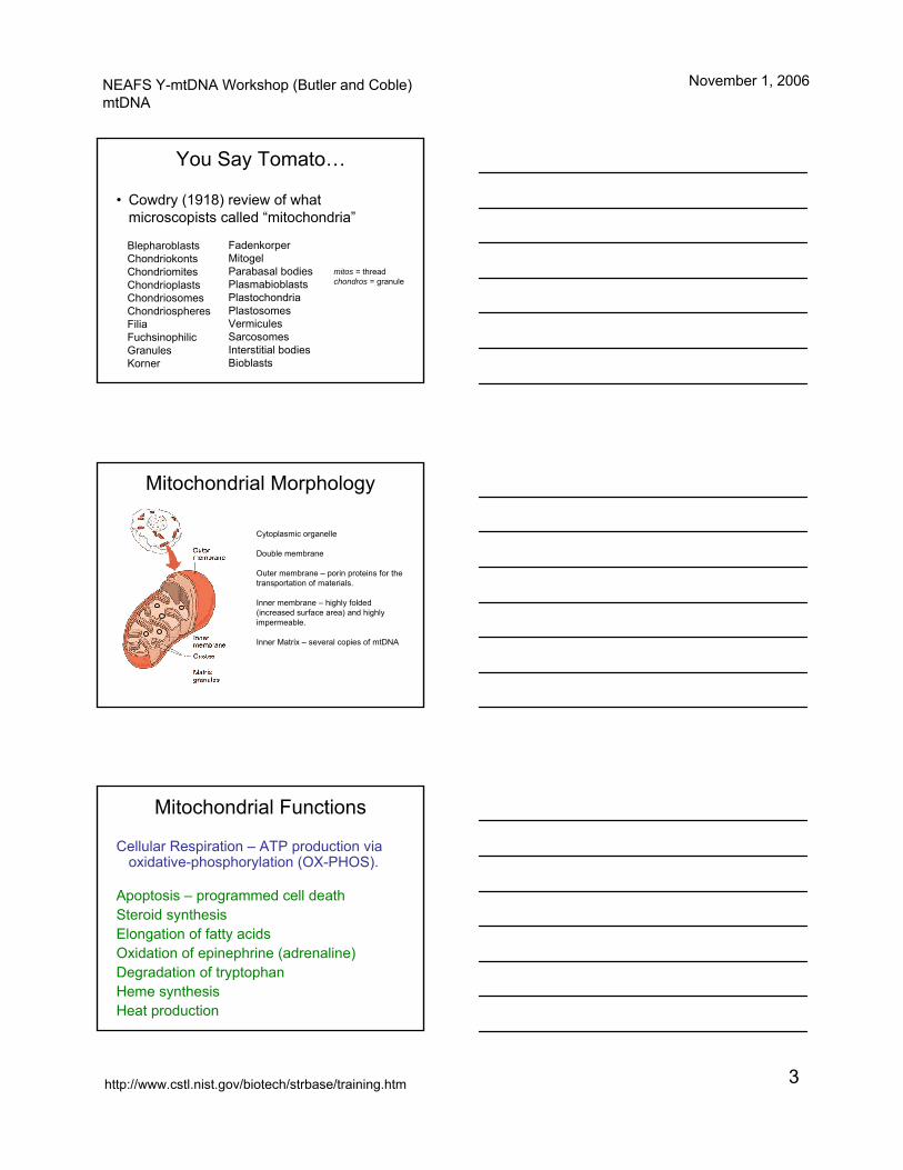
You Say Tomato…
• Cowdry (1918) review of what microscopists called “mitochondria”
BlepharoblastsChondriokontsChondriomitesChondrioplastsChondriosomesChondriospheresFiliaFuchsinophilicGranulesKorner
FadenkorperMitogelParabasal bodiesPlasmabioblastsPlastochondriaPlastosomesVermiculesSarcosomesInterstitial bodiesBioblasts
mitos = threadchondros = granule
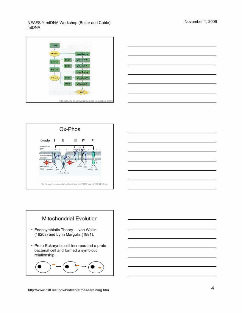
Mitochondrial Morphology
Cytoplasmic organelle
Double membrane
Outer membrane – porin proteins for the transportation of materials.
Inner membrane – highly folded (increased surface area) and highlyimpermeable.
Inner Matrix – several copies of mtDNA
Mitochondrial Functions
Cellular Respiration – ATP production via oxidative-phosphorylation (OX-PHOS).
Apoptosis – programmed cell deathSteroid synthesisElongation of fatty acidsOxidation of epinephrine (adrenaline) Degradation of tryptophanHeme synthesis Heat production
NEAFS Y-mtDNA Workshop (Butler and Coble)mtDNA
November 1, 2006
http://www.cstl.nist.gov/biotech/strbase/training.htm 4
http://sps.k12.ar.us/massengale/cell_respiration_bi.htm
Ox-Phos
http://myweb.uiowa.edu/bballard/Research%20Figures/OXPHOS.jpg
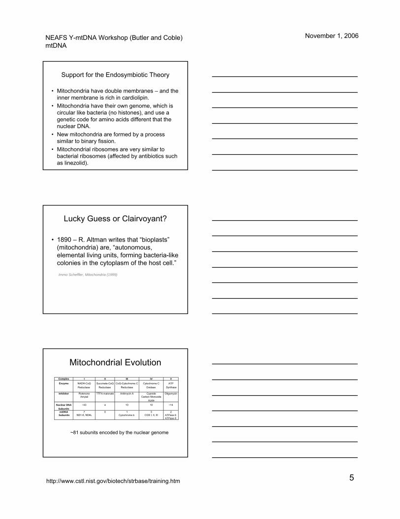
Mitochondrial Evolution
• Endosymbiotic Theory – Ivan Wallin(1920s) and Lynn Margulis (1981).
• Proto-Eukaryotic cell incorporated a proto-bacterial cell and formed a symbiotic relationship.
NEAFS Y-mtDNA Workshop (Butler and Coble)mtDNA
November 1, 2006
http://www.cstl.nist.gov/biotech/strbase/training.htm 5
Support for the Endosymbiotic Theory
• Mitochondria have double membranes – and the inner membrane is rich in cardiolipin.
• Mitochondria have their own genome, which is circular like bacteria (no histones), and use a genetic code for amino acids different that the nuclear DNA.
• New mitochondria are formed by a process similar to binary fission.
• Mitochondrial ribosomes are very similar to bacterial ribosomes (affected by antibiotics such as linezolid).
Lucky Guess or Clairvoyant?
• 1890 – R. Altman writes that “bioplasts”(mitochondria) are, “autonomous, elemental living units, forming bacteria-like colonies in the cytoplasm of the host cell.”
Immo Scheffler, Mitochondria (1999)
Mitochondrial EvolutionComplex I II III IV V
Enzyme NADH-CoQ Succinate-CoQ CoQ-Cytochrome C Cytochrome C ATPReductase Reductase Reductase Oxidase Synthase
Inhibitor Rotenone TTFA malonate Antimycin A Cyanide OligomycinAmytal Carbon Monoxide
Azide
Nuclear DNA ~43 4 10 10 ~14SubunitsmtDNA 7 0 1 3 2
Subunits ND1-6, ND4L Cytochrome b COX I, II, III ATPase 6 ATPase 8
~81 subunits encoded by the nuclear genome
NEAFS Y-mtDNA Workshop (Butler and Coble)mtDNA
November 1, 2006
http://www.cstl.nist.gov/biotech/strbase/training.htm 6
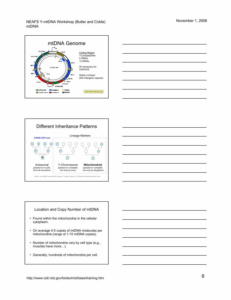
mtDNA Genome
http://www.mitomap.org/
Coding Region13 polypeptides2 rRNAs13 tRNAs
All necessary for OXPHOS
Highly compact(few intergenic spaces)
Different Inheritance Patterns
Autosomal(passed on in part, from all ancestors)
Y-Chromosome(passed on complete,
but only by sons)
Mitochondrial(passed on complete, but only by daughters)
Lineage Markers
Butler, J.M. (2005) Forensic DNA Typing, 2nd Edition, Figure 9.1, ©Elsevier Science/Academic Press
CODIS STR Loci
Location and Copy Number of mtDNA
• Found within the mitochondria in the cellular cytoplasm.
• On average 4-5 copies of mtDNA molecules per mitochondria (range of 1-15 mtDNA copies).
• Number of mitochondria vary by cell type (e.g., muscles have more…).
• Generally, hundreds of mitochondria per cell.
NEAFS Y-mtDNA Workshop (Butler and Coble)mtDNA
November 1, 2006
http://www.cstl.nist.gov/biotech/strbase/training.htm 7
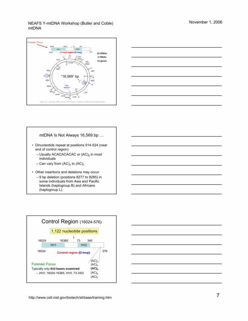
Control region (D-loop)
1/16,569
cyt b
ND5ND6
ND4
ND4L
ND3COIII
ATP6ATP8 COII
12S rRNA
16S rRNA
ND1
ND2
COI
OH
9-bp deletion
OL
FV
L1
IQM
W
ANCY
S1
DK
G
R
HS2L2
EP
T
HV1 HV216024 16365 73 340
16024 576
“16,569” bp
1
22 tRNAs2 rRNAs
13 genes
Figure 10.1, J.M. Butler (2005) Forensic DNA Typing, 2nd Edition © 2005 Elsevier Academic Press
Forensic Focus
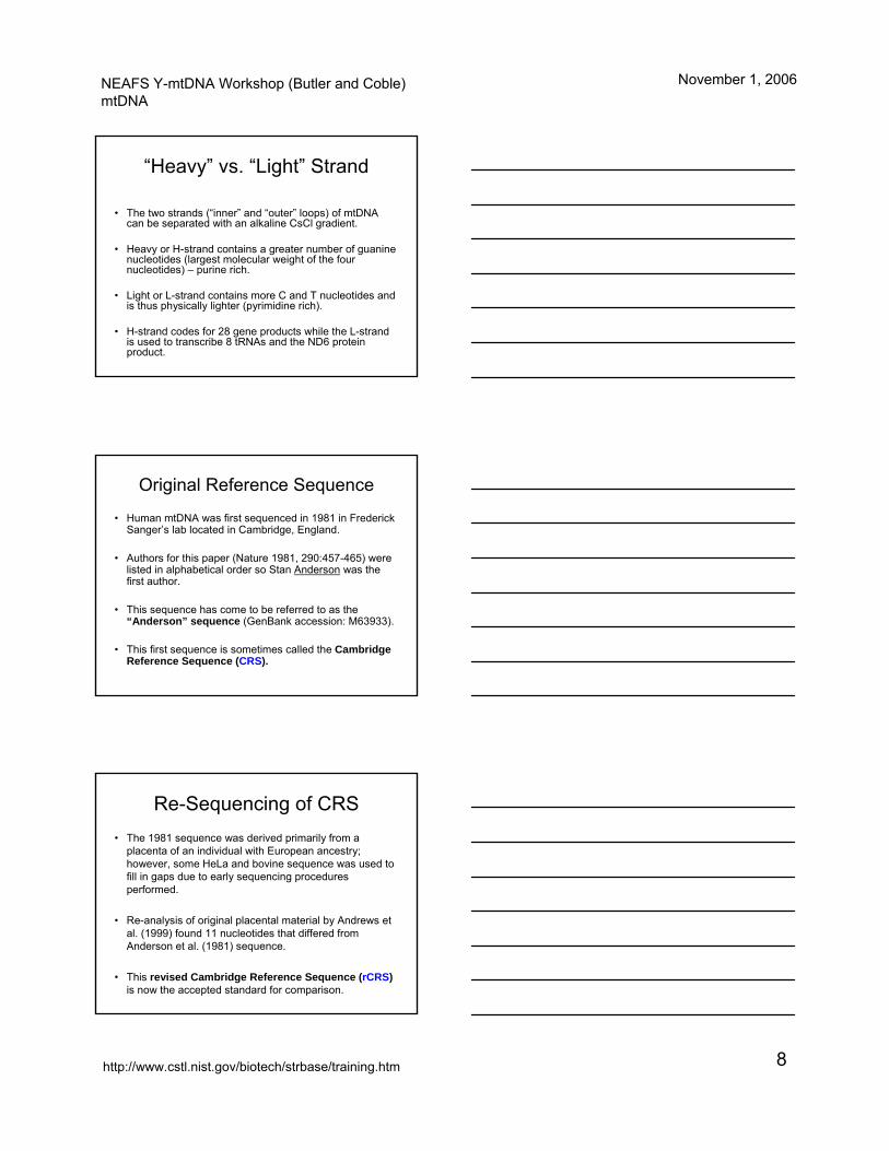
mtDNA Is Not Always 16,569 bp …
• Dinucleotide repeat at positions 514-524 (near end of control region)– Usually ACACACACAC or (AC)5 in most
individuals– Can vary from (AC)3 to (AC)7
• Other insertions and deletions may occur– 9 bp deletion (positions 8277 to 8285) in
some individuals from Asia and Pacific Islands (haplogroup B) and Africans (haplogroup L).
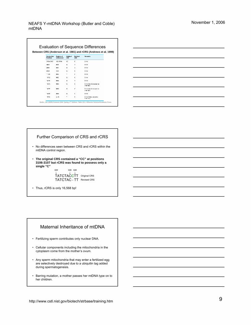
Control Region (16024-576)
Forensic FocusTypically only 610 bases examined
– (HVI: 16024-16365; HVII: 73-340)
Control region (D-loop)
HV1 HV216024 16365 73 340
16024 576
1
(AC)3(AC)4(AC)5
(AC)6(AC)7
1,122 nucleotide positions
NEAFS Y-mtDNA Workshop (Butler and Coble)mtDNA
November 1, 2006
http://www.cstl.nist.gov/biotech/strbase/training.htm 8
“Heavy” vs. “Light” Strand
• The two strands (“inner” and “outer” loops) of mtDNA can be separated with an alkaline CsCl gradient.
• Heavy or H-strand contains a greater number of guanine nucleotides (largest molecular weight of the four nucleotides) – purine rich.
• Light or L-strand contains more C and T nucleotides and is thus physically lighter (pyrimidine rich).
• H-strand codes for 28 gene products while the L-strand is used to transcribe 8 tRNAs and the ND6 protein product.
Original Reference Sequence
• Human mtDNA was first sequenced in 1981 in Frederick Sanger’s lab located in Cambridge, England.
• Authors for this paper (Nature 1981, 290:457-465) were listed in alphabetical order so Stan Anderson was the first author.
• This sequence has come to be referred to as the “Anderson” sequence (GenBank accession: M63933).
• This first sequence is sometimes called the Cambridge Reference Sequence (CRS).
Re-Sequencing of CRS• The 1981 sequence was derived primarily from a
placenta of an individual with European ancestry; however, some HeLa and bovine sequence was used to fill in gaps due to early sequencing procedures performed.
• Re-analysis of original placental material by Andrews et al. (1999) found 11 nucleotides that differed from Anderson et al. (1981) sequence.
• This revised Cambridge Reference Sequence (rCRS)is now the accepted standard for comparison.
NEAFS Y-mtDNA Workshop (Butler and Coble)mtDNA
November 1, 2006
http://www.cstl.nist.gov/biotech/strbase/training.htm 9
Evaluation of Sequence Differences Between CRS (Anderson et al. 1981) and rCRS (Andrews et al. 1999)
Butler, J.M. (2005) Forensic DNA Typing, 2nd Edition, Table 10.3, ©Elsevier Science/Academic Press
Further Comparison of CRS and rCRS
• No differences seen between CRS and rCRS within the mtDNA control region.
• The original CRS contained a “CC” at positions 3106-3107 but rCRS was found to possess only a single “C”
• Thus, rCRS is only 16,568 bp!
TATCTACCTT Original CRS
TATCTAC - TT Revised CRS
3100 31083106
Maternal Inheritance of mtDNA
• Fertilizing sperm contributes only nuclear DNA.
• Cellular components including the mitochondria in the cytoplasm come from the mother’s ovum.
• Any sperm mitochondria that may enter a fertilized egg are selectively destroyed due to a ubiquitin tag added during spermatogenesis.
• Barring mutation, a mother passes her mtDNA type on to her children.
NEAFS Y-mtDNA Workshop (Butler and Coble)mtDNA
November 1, 2006
http://www.cstl.nist.gov/biotech/strbase/training.htm 10
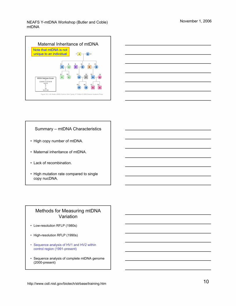
Maternal Inheritance of mtDNA
MtDNA Haplotype Groups:1
2,3,6,8,11,13,15,164,9,10
5712
14,17,18
MtDNA Haplotype Groups:1
2,3,6,8,11,13,15,164,9,10
5712
14,17,18
1 2
3 54
1211109
6 7 8
181715 16
13 14
A B
B C
C C
D B
B
B
B
B B
E
F
G
G
G
Figure 10.2, J.M. Butler (2005) Forensic DNA Typing, 2nd Edition © 2005 Elsevier Academic Press
Note that mtDNA is not unique to an individual
Summary – mtDNA Characteristics
• High copy number of mtDNA.
• Maternal inheritance of mtDNA.
• Lack of recombination.
• High mutation rate compared to single copy nucDNA.
Methods for Measuring mtDNA Variation
• Low-resolution RFLP (1980s)
• High-resolution RFLP (1990s)
• Sequence analysis of HV1 and HV2 within control region (1991-present)
• Sequence analysis of complete mtDNA genome (2000-present)
NEAFS Y-mtDNA Workshop (Butler and Coble)mtDNA
November 1, 2006
http://www.cstl.nist.gov/biotech/strbase/training.htm 11
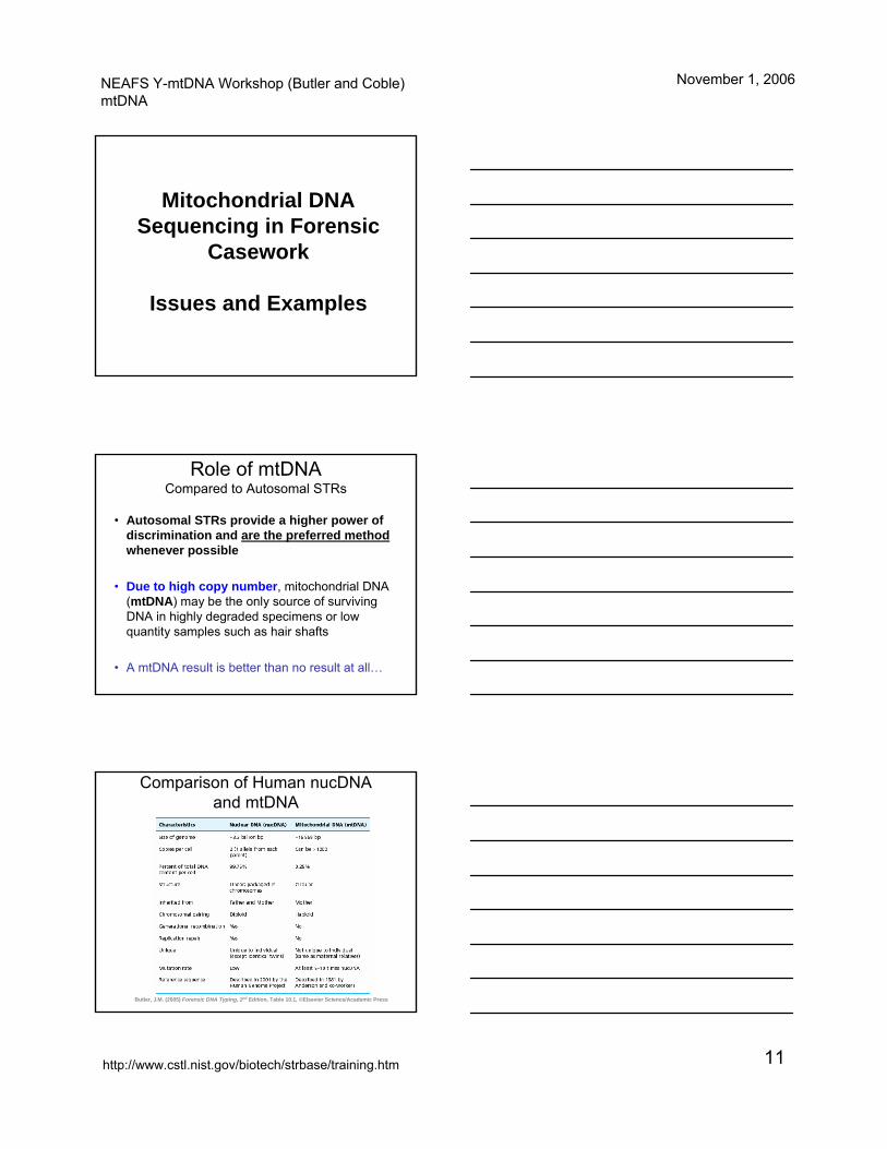
Mitochondrial DNA Sequencing in Forensic
Casework
Issues and Examples
Role of mtDNACompared to Autosomal STRs
• Autosomal STRs provide a higher power of discrimination and are the preferred methodwhenever possible
• Due to high copy number, mitochondrial DNA (mtDNA) may be the only source of surviving DNA in highly degraded specimens or low quantity samples such as hair shafts
• A mtDNA result is better than no result at all…
Comparison of Human nucDNAand mtDNA
Butler, J.M. (2005) Forensic DNA Typing, 2nd Edition, Table 10.1, ©Elsevier Science/Academic Press
NEAFS Y-mtDNA Workshop (Butler and Coble)mtDNA
November 1, 2006
http://www.cstl.nist.gov/biotech/strbase/training.htm 12
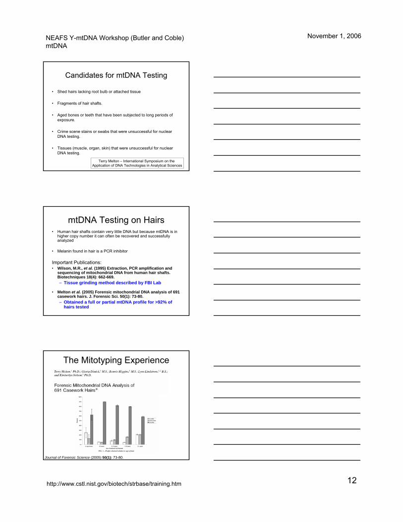
Candidates for mtDNA Testing
• Shed hairs lacking root bulb or attached tissue
• Fragments of hair shafts.
• Aged bones or teeth that have been subjected to long periods of exposure.
• Crime scene stains or swabs that were unsuccessful for nuclear DNA testing.
• Tissues (muscle, organ, skin) that were unsuccessful for nuclearDNA testing.
Terry Melton – International Symposium on the Application of DNA Technologies in Analytical Sciences
• Human hair shafts contain very little DNA but because mtDNA is in higher copy number it can often be recovered and successfully analyzed
• Melanin found in hair is a PCR inhibitor
Important Publications:• Wilson, M.R., et al. (1995) Extraction, PCR amplification and
sequencing of mitochondrial DNA from human hair shafts. Biotechniques 18(4): 662-669.– Tissue grinding method described by FBI Lab
• Melton et al. (2005) Forensic mitochondrial DNA analysis of 691 casework hairs. J. Forensic Sci. 50(1): 73-80.– Obtained a full or partial mtDNA profile for >92% of
hairs tested
mtDNA Testing on Hairs
The Mitotyping Experience
Journal of Forensic Science (2005) 50(1): 73-80.
NEAFS Y-mtDNA Workshop (Butler and Coble)mtDNA
November 1, 2006
http://www.cstl.nist.gov/biotech/strbase/training.htm 13
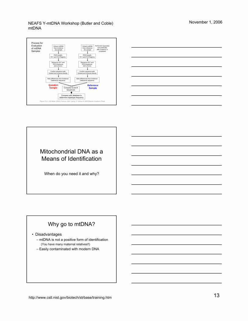
Extract mtDNA from evidence
(Q) sample
PCR Amplify HV1 and HV2 Regions
Sequence HV1 and HV2 Amplicons
(both strands)
Confirm sequence with forward and reverse strands
Note differences from Anderson (reference) sequence
Compare with database to determine haplotype frequency
Compare Q and K sequences
Question Sample
Reference Sample
Extract mtDNA from reference
(K) sample
PCR Amplify HV1 and HV2 Regions
Sequence HV1 and HV2 Amplicons
(both strands)
Confirm sequence with forward and reverse strands
Note differences from Anderson (reference) sequence
Performed separately and preferably
after evidence is completed
Process for Evaluation of mtDNA Samples
Figure 10.4, J.M. Butler (2005) Forensic DNA Typing, 2nd Edition © 2005 Elsevier Academic Press
Mitochondrial DNA as a Means of Identification
When do you need it and why?
Why go to mtDNA?
• Disadvantages– mtDNA is not a positive form of identification
(You have many maternal relatives!!)– Easily contaminated with modern DNA
NEAFS Y-mtDNA Workshop (Butler and Coble)mtDNA
November 1, 2006
http://www.cstl.nist.gov/biotech/strbase/training.htm 14
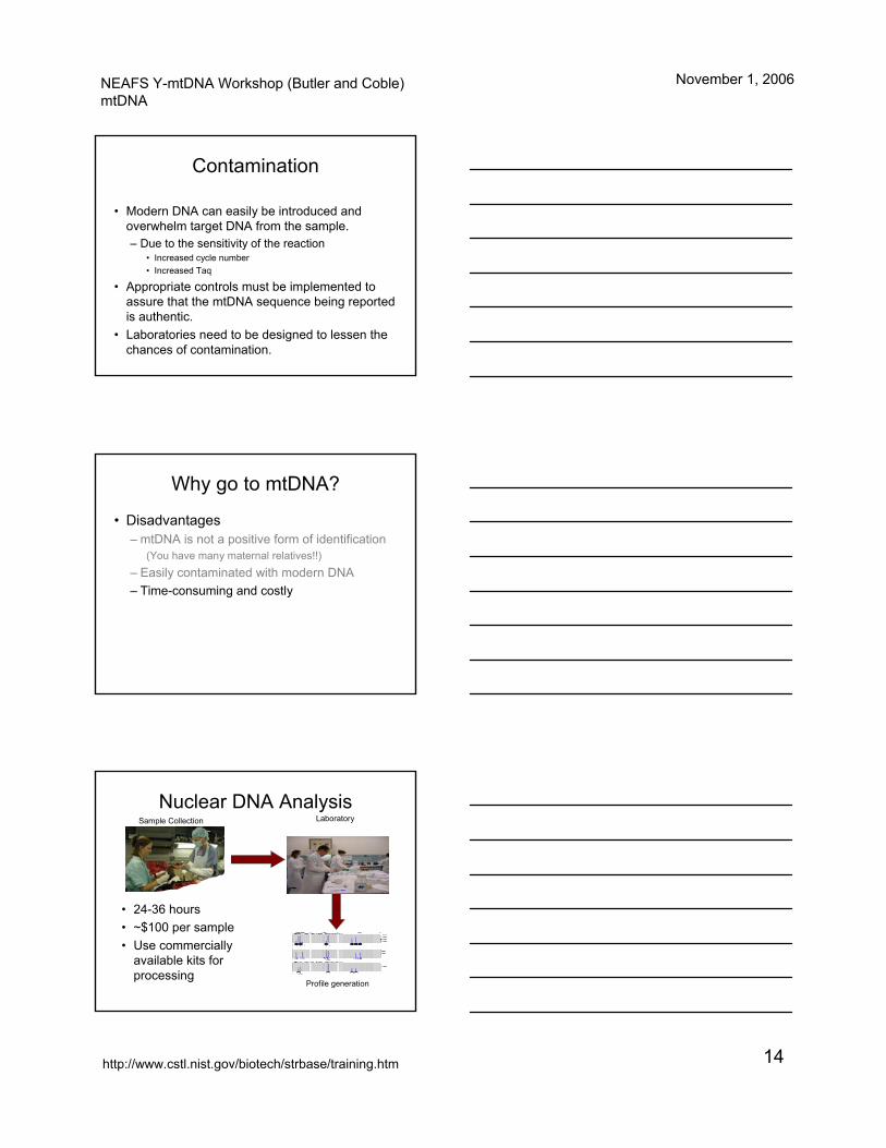
Contamination
• Modern DNA can easily be introduced and overwhelm target DNA from the sample.– Due to the sensitivity of the reaction
• Increased cycle number• Increased Taq
• Appropriate controls must be implemented to assure that the mtDNA sequence being reported is authentic.
• Laboratories need to be designed to lessen the chances of contamination.
Why go to mtDNA?
• Disadvantages– mtDNA is not a positive form of identification
(You have many maternal relatives!!)– Easily contaminated with modern DNA– Time-consuming and costly
Nuclear DNA Analysis
• 24-36 hours• ~$100 per sample• Use commercially
available kits for processing
Sample Collection Laboratory
Profile generation
NEAFS Y-mtDNA Workshop (Butler and Coble)mtDNA
November 1, 2006
http://www.cstl.nist.gov/biotech/strbase/training.htm 15
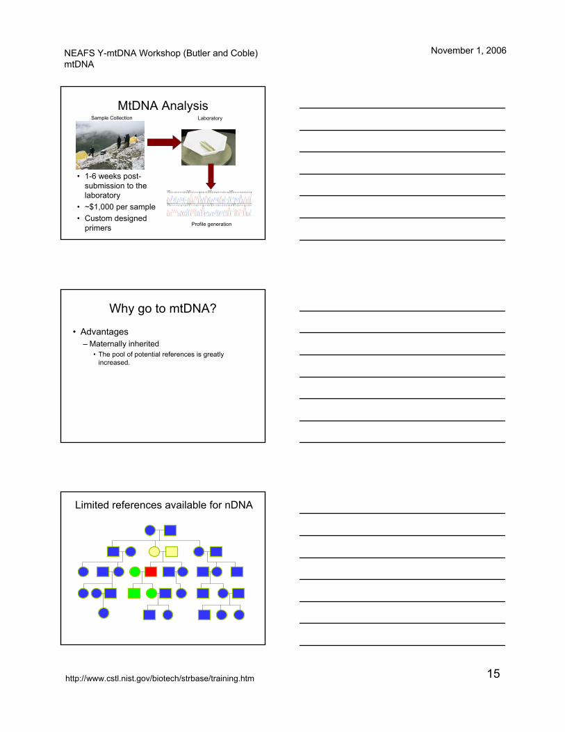
MtDNA Analysis
• 1-6 weeks post-submission to the laboratory
• ~$1,000 per sample• Custom designed
primers
Sample Collection Laboratory
Profile generation
Why go to mtDNA?
• Advantages– Maternally inherited
• The pool of potential references is greatly increased.
Limited references available for nDNA
NEAFS Y-mtDNA Workshop (Butler and Coble)mtDNA
November 1, 2006
http://www.cstl.nist.gov/biotech/strbase/training.htm 16
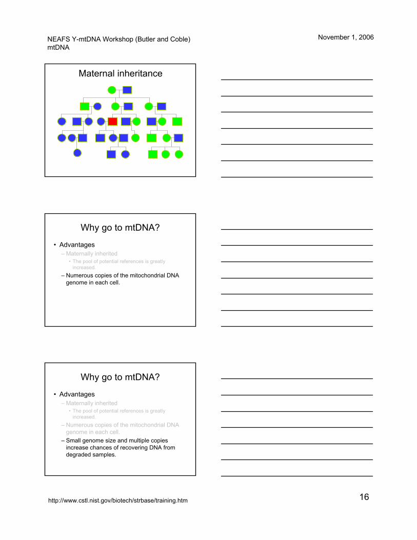
Maternal inheritance
Why go to mtDNA?
• Advantages– Maternally inherited
• The pool of potential references is greatly increased.
– Numerous copies of the mitochondrial DNA genome in each cell.
Why go to mtDNA?
• Advantages– Maternally inherited
• The pool of potential references is greatly increased.
– Numerous copies of the mitochondrial DNA genome in each cell.
– Small genome size and multiple copies increase chances of recovering DNA from degraded samples.
NEAFS Y-mtDNA Workshop (Butler and Coble)mtDNA
November 1, 2006
http://www.cstl.nist.gov/biotech/strbase/training.htm 17
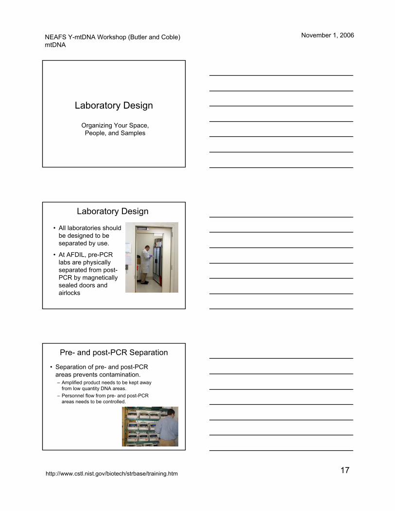
Laboratory Design
Organizing Your Space, People, and Samples
Laboratory Design
• All laboratories should be designed to be separated by use.
• At AFDIL, pre-PCR labs are physically separated from post-PCR by magnetically sealed doors and airlocks
Pre- and post-PCR Separation
• Separation of pre- and post-PCR areas prevents contamination.– Amplified product needs to be kept away
from low quantity DNA areas.– Personnel flow from pre- and post-PCR
areas needs to be controlled.
NEAFS Y-mtDNA Workshop (Butler and Coble)mtDNA
November 1, 2006
http://www.cstl.nist.gov/biotech/strbase/training.htm 18
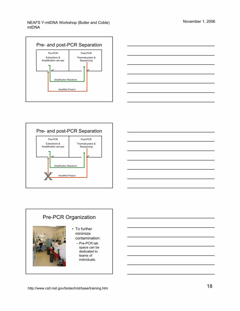
Pre- and post-PCR SeparationPre-PCR
Extractions & Amplification set-ups
Post-PCR
Thermalcyclers & Sequencing
Amplification Reactions
Amplified Product
Pre- and post-PCR SeparationPre-PCR
Extractions & Amplification set-ups
Post-PCR
Thermalcyclers & Sequencing
Amplification Reactions
Amplified Product
Pre-PCR Organization
• To further minimize contamination:– Pre-PCR lab
space can be dedicated to teams of individuals.
NEAFS Y-mtDNA Workshop (Butler and Coble)mtDNA
November 1, 2006
http://www.cstl.nist.gov/biotech/strbase/training.htm 19
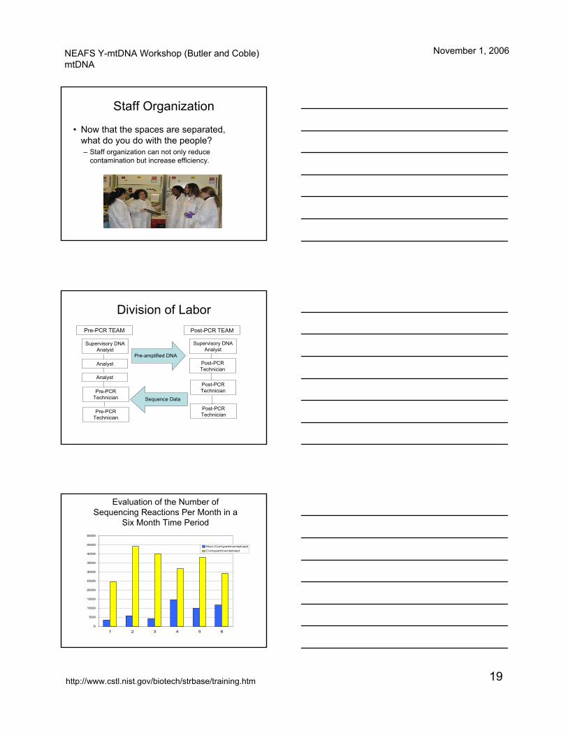
Staff Organization
• Now that the spaces are separated, what do you do with the people?– Staff organization can not only reduce
contamination but increase efficiency.
Division of Labor
Supervisory DNA Analyst
Analyst
Analyst
Pre-PCR Technician
Pre-PCR Technician
Pre-PCR TEAM
Supervisory DNA Analyst
Post-PCR Technician
Post-PCR Technician
Post-PCR Technician
Post-PCR TEAM
Pre-amplified DNA
Sequence Data
0
500
1000
1500
2000
2500
3000
3500
4000
4500
5000
1 2 3 4 5 6
Non-CompartmentalizedCompartmentalized
Evaluation of the Number of Sequencing Reactions Per Month in a
Six Month Time Period
NEAFS Y-mtDNA Workshop (Butler and Coble)mtDNA
November 1, 2006
http://www.cstl.nist.gov/biotech/strbase/training.htm 20
• With this type of sample volume comes the additional issues of tracking your samples and contamination.– Even if the lab is fairly small, chain of custody
issues and overall processing need to be tracked efficiently.
– Contamination needs to be tracked, found and eradicated before it becomes an issue.
Sample and Contamination Tracking
LIMS System
• An automated computer system is the most efficient method for accomplishing these goals.
• Many laboratory information management systems are available commercially.
• The name of our system is Laboratory Information Systems Application or LISA.
Case Accessioning
• Controls who has access to which samples based on set of ‘privileges’.
• Tracks Chain of Custody.• Every step requires a password even once you
are in the system.
• Names and identifies each piece of evidence that is received.
• Assigns a sequential case number.
NEAFS Y-mtDNA Workshop (Butler and Coble)mtDNA
November 1, 2006
http://www.cstl.nist.gov/biotech/strbase/training.htm 21
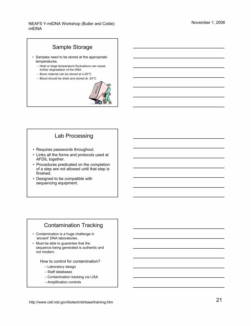
Sample Storage
• Samples need to be stored at the appropriate temperatures.– Heat or large temperature fluctuations can cause
further degradation of the DNA.– Bone material can be stored at ≤-20°C– Blood should be dried and stored at -20°C
Lab Processing
• Requires passwords throughout.• Links all the forms and protocols used at
AFDIL together.• Procedures predicated on the completion
of a step are not allowed until that step is finished.
• Designed to be compatible with sequencing equipment.
Contamination Tracking• Contamination is a huge challenge in
‘ancient’ DNA laboratories.• Must be able to guarantee that the
sequence being generated is authentic and not modern.
How to control for contamination?– Laboratory design– Staff databases– Contamination tracking via LISA– Amplification controls
NEAFS Y-mtDNA Workshop (Butler and Coble)mtDNA
November 1, 2006
http://www.cstl.nist.gov/biotech/strbase/training.htm 22
Control Databases• All members of the staff,
laboratory and administrative, at AFDIL have been profiled for both mitochondrial and nuclear DNA.
• The case management module of LISA has a separate database specifically for the sequences generated for contaminants
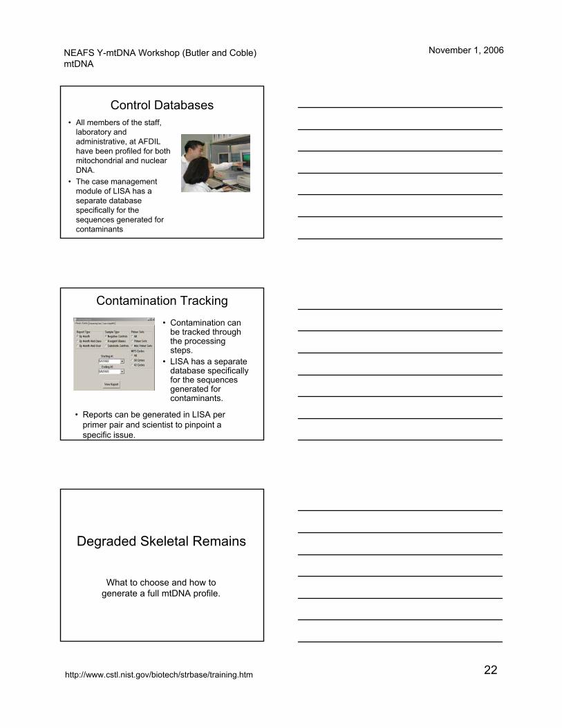
Contamination Tracking• Contamination can
be tracked through the processing steps.
• LISA has a separate database specifically for the sequences generated for contaminants.
• Reports can be generated in LISA per primer pair and scientist to pinpoint a specific issue.
Degraded Skeletal Remains
What to choose and how to generate a full mtDNA profile.
NEAFS Y-mtDNA Workshop (Butler and Coble)mtDNA
November 1, 2006
http://www.cstl.nist.gov/biotech/strbase/training.htm 23
Degraded Skeletal Remains
• Sample Selection• Extraction Methods• Amplification Strategies• Sequencing Strategies
Degraded Specimens
• In general terms all skeletal remains are degraded.
• Some are more degraded then others due to environmental stressors.
• Prudent sample selection will increase the rate of success.
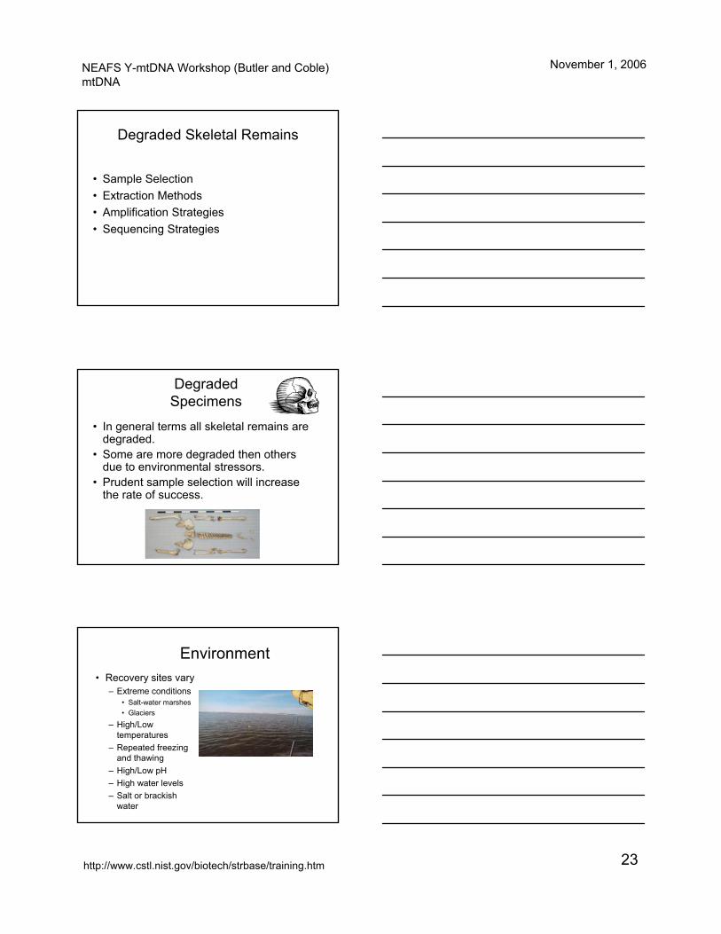
Environment• Recovery sites vary
– Extreme conditions• Salt-water marshes• Glaciers
– High/Low temperatures
– Repeated freezing and thawing
– High/Low pH– High water levels– Salt or brackish
water
NEAFS Y-mtDNA Workshop (Butler and Coble)mtDNA
November 1, 2006
http://www.cstl.nist.gov/biotech/strbase/training.htm 24
Environment• Remains may be
– On the surface– Buried in soil or other
substrates– Highly fragmented – Subjected to burning or high
heat– Exposed to fuel or other
chemicals– Disturbed or moved by humans
or animals– Animal destruction (feeding)
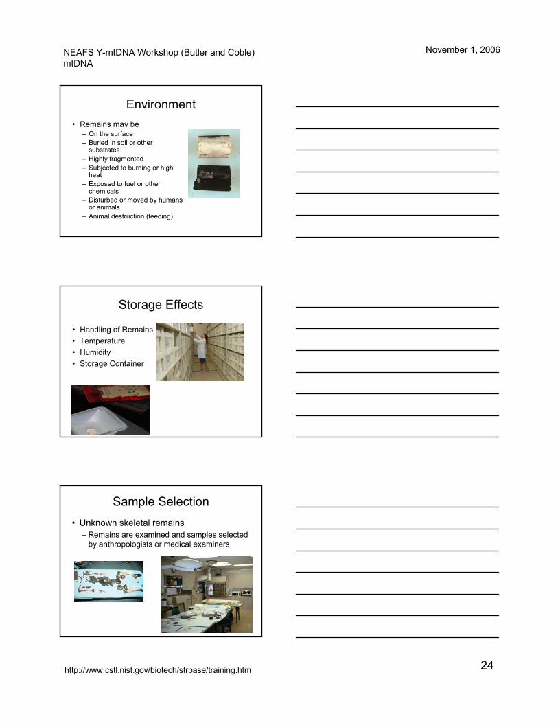
Storage Effects
• Handling of Remains• Temperature• Humidity• Storage Container
Sample Selection• Unknown skeletal remains
– Remains are examined and samples selected by anthropologists or medical examiners
NEAFS Y-mtDNA Workshop (Butler and Coble)mtDNA
November 1, 2006
http://www.cstl.nist.gov/biotech/strbase/training.htm 25
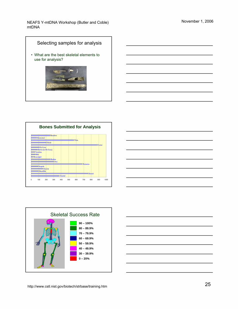
Selecting samples for analysis
• What are the best skeletal elements to use for analysis?
DentalMandible
ClavicleScapula
HumerusUlna
RadiusMetacarpal
RibVertebra
Pelvis (no Os Coxa)Os Coxa
FemurFibula
TibiaMetatarsal
Random
Cranial
0 100 200 300 400 500 600 700 800 900 1000
Bones Submitted for Analysis
90 – 100%
80 – 89.9%
70 – 79.9%
60 – 69.9%
50 – 59.9%
30 – 39.9%
40 – 49.9%
0 – 20%
Skeletal Success Rate
NEAFS Y-mtDNA Workshop (Butler and Coble)mtDNA
November 1, 2006
http://www.cstl.nist.gov/biotech/strbase/training.htm 26
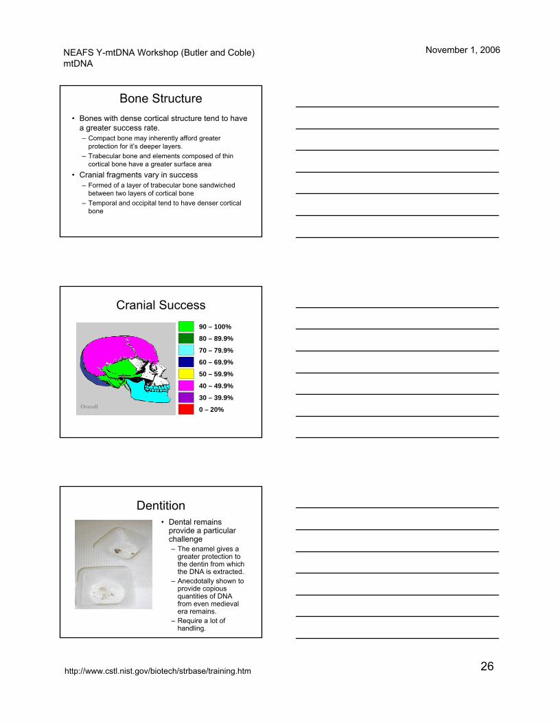
Bone Structure• Bones with dense cortical structure tend to have
a greater success rate.– Compact bone may inherently afford greater
protection for it’s deeper layers.– Trabecular bone and elements composed of thin
cortical bone have a greater surface area• Cranial fragments vary in success
– Formed of a layer of trabecular bone sandwiched between two layers of cortical bone
– Temporal and occipital tend to have denser cortical bone
Overall
90 – 100%
80 – 89.9%
70 – 79.9%
60 – 69.9%
50 – 59.9%
30 – 39.9%
40 – 49.9%
0 – 20%
Cranial Success
Dentition• Dental remains
provide a particular challenge– The enamel gives a
greater protection to the dentin from which the DNA is extracted.
– Anecdotally shown to provide copious quantities of DNA from even medieval era remains.
– Require a lot of handling.
NEAFS Y-mtDNA Workshop (Butler and Coble)mtDNA
November 1, 2006
http://www.cstl.nist.gov/biotech/strbase/training.htm 27
Extraction Methods
• Cleaning the samples – how much is too much?
• What protocols give the greatest yield of DNA?
• What method is right for you?• Trouble-shooting the extraction.
Cleaning the Sample
• The exterior of the bone fragment needs to be cleaned of any possible contaminants:
– Dirt– Plant material– Extraneous DNA– Dried Tissue
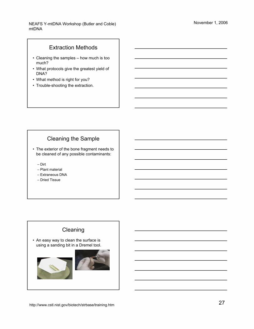
Cleaning
• An easy way to clean the surface is using a sanding bit in a Dremel tool.
NEAFS Y-mtDNA Workshop (Butler and Coble)mtDNA
November 1, 2006
http://www.cstl.nist.gov/biotech/strbase/training.htm 28
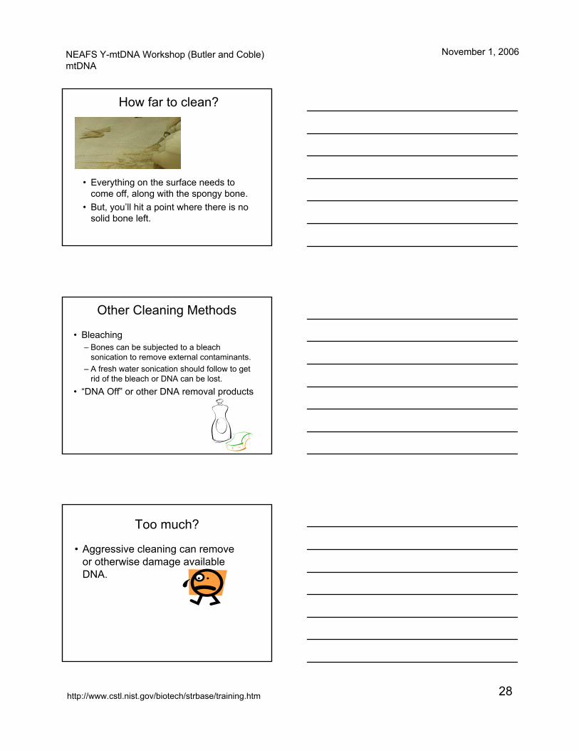
How far to clean?
• Everything on the surface needs to come off, along with the spongy bone.
• But, you’ll hit a point where there is no solid bone left.
Other Cleaning Methods
• Bleaching– Bones can be subjected to a bleach
sonication to remove external contaminants.– A fresh water sonication should follow to get
rid of the bleach or DNA can be lost.• “DNA Off” or other DNA removal products
Too much?
• Aggressive cleaning can remove or otherwise damage available DNA.

NEAFS Y-mtDNA Workshop (Butler and Coble)mtDNA
November 1, 2006
http://www.cstl.nist.gov/biotech/strbase/training.htm 29
Extraction Methods
• Numerous extraction methods available.• Involve different methods of –
– pulverizing the samples– removing the DNA from the samples
• Different starting quantities of bone can also be used.
Pulverization Methods• Freezer Mill
– Uses liquid nitrogen and a magnet to pulverize the bone into a very fine powder.
– Disadvantage: • Requires storage and handling of liquid
nitrogen.• Grinders and sample vials are reused –
potential contamination.
Pulverization Method
• Waring Blender Cup– Also grinds bone to a relatively fine powder– Disadvantage: Cups are reused, so there is
a possibility of contamination.
NEAFS Y-mtDNA Workshop (Butler and Coble)mtDNA
November 1, 2006
http://www.cstl.nist.gov/biotech/strbase/training.htm 30
“Freeing” the DNA
• Samples may be subjected to a decalcification step.– Demineralizes the bone matrix.
• Other chemical/physical treatments are commercially available to more easily acquire the DNA.– Silica gel– Charge Switch™– DNA IQ™
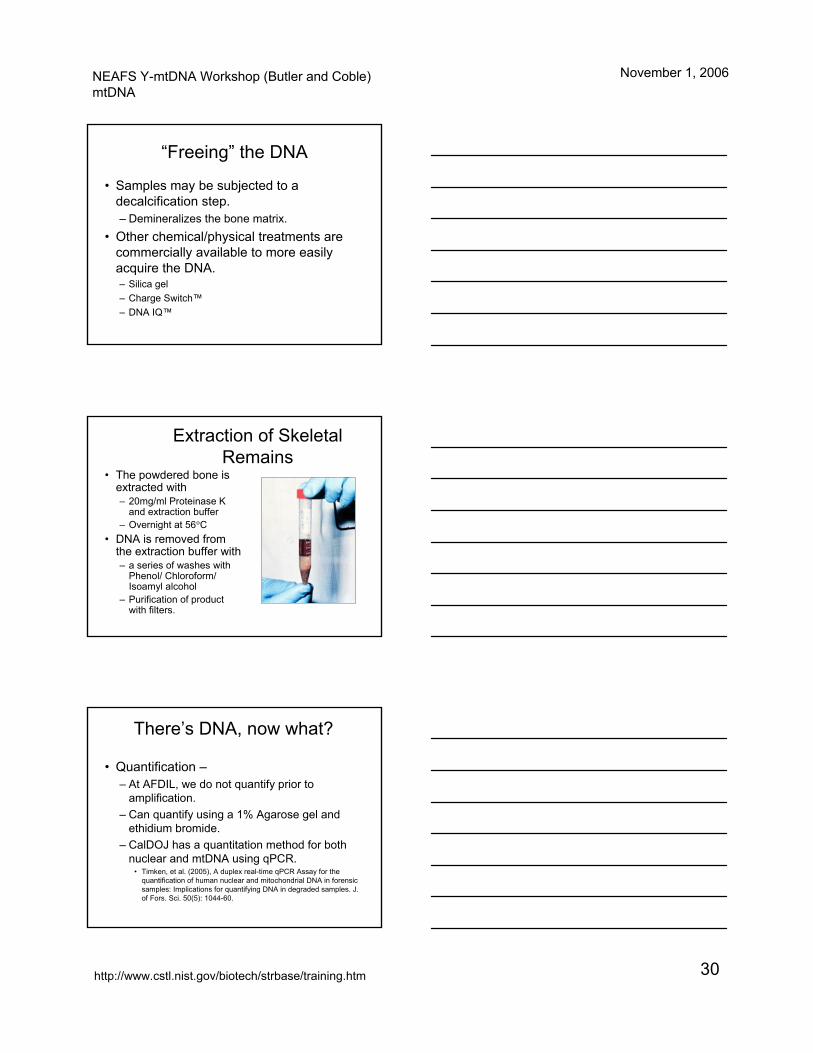
Extraction of Skeletal Remains
• The powdered bone is extracted with – 20mg/ml Proteinase K
and extraction buffer– Overnight at 56°C
• DNA is removed from the extraction buffer with– a series of washes with
Phenol/ Chloroform/ Isoamyl alcohol
– Purification of product with filters.
There’s DNA, now what?
• Quantification –– At AFDIL, we do not quantify prior to
amplification.– Can quantify using a 1% Agarose gel and
ethidium bromide.– CalDOJ has a quantitation method for both
nuclear and mtDNA using qPCR.• Timken, et al. (2005), A duplex real-time qPCR Assay for the
quantification of human nuclear and mitochondrial DNA in forensic samples: Implications for quantifying DNA in degraded samples. J. of Fors. Sci. 50(5): 1044-60.
NEAFS Y-mtDNA Workshop (Butler and Coble)mtDNA
November 1, 2006
http://www.cstl.nist.gov/biotech/strbase/training.htm 31
Amplification
• A standard program for amplification is used for the 9700’s.
• The basic program is modified based on the primer pair used (Gabriel, et al. 2001)– 10-minute soak at 96.0°C– Followed by 38 cycles of
• 20s at 94.0°C• 20s at 56.0°C• 30s at 72.0°C
– Final hold at 4°C
PCR Amplification of mtDNA
• Usually performed with 34-38 cycles
• Some protocols may go to 42 cycles for highly degraded specimens
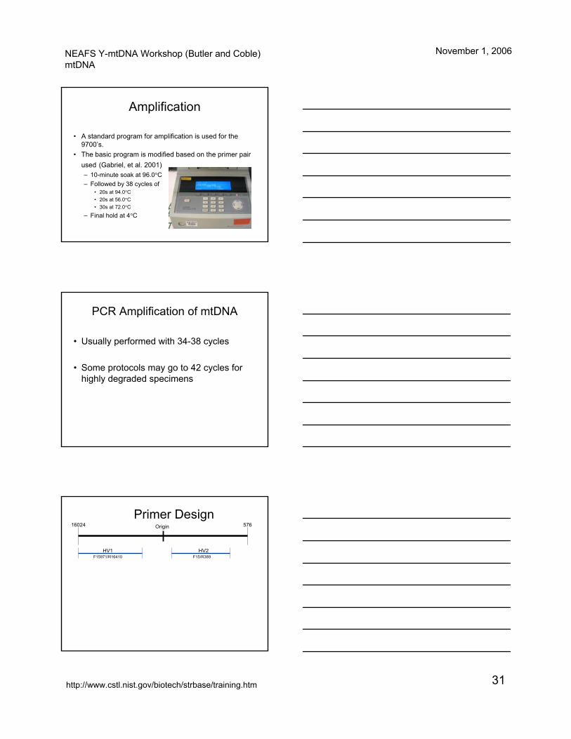
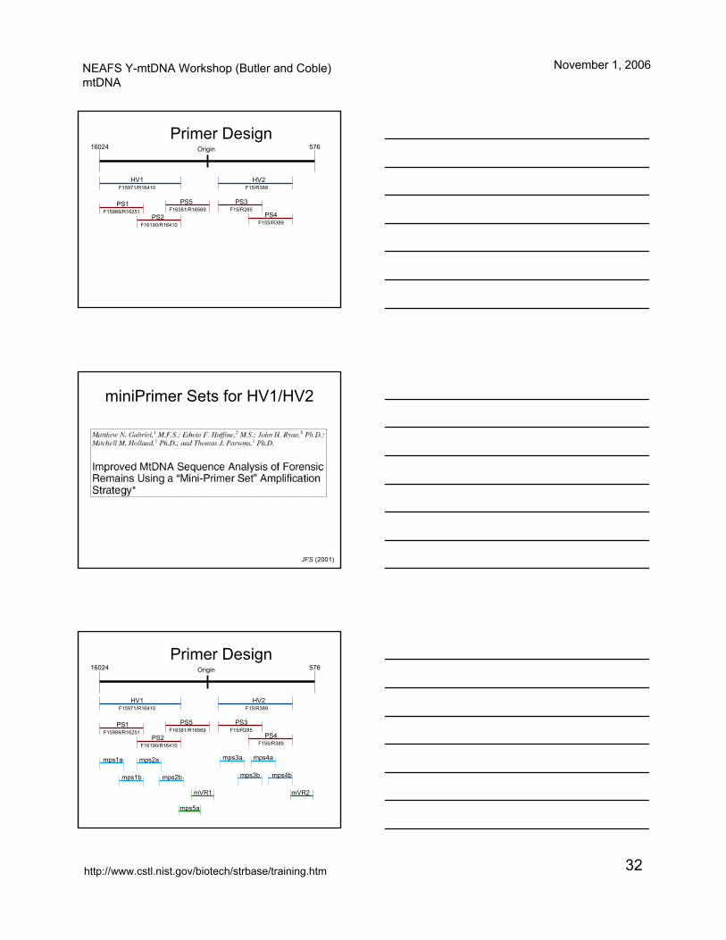
Primer DesignOrigin16024 576
HV1F15971/R16410
HV2F15/R389
NEAFS Y-mtDNA Workshop (Butler and Coble)mtDNA
November 1, 2006
http://www.cstl.nist.gov/biotech/strbase/training.htm 32
Primer DesignOrigin16024 576
HV1F15971/R16410
HV2F15/R389
PS1F15989/R16251
PS2F16190/R16410
PS3F15/R285
PS4F155/R389
PS5F16381/R16569
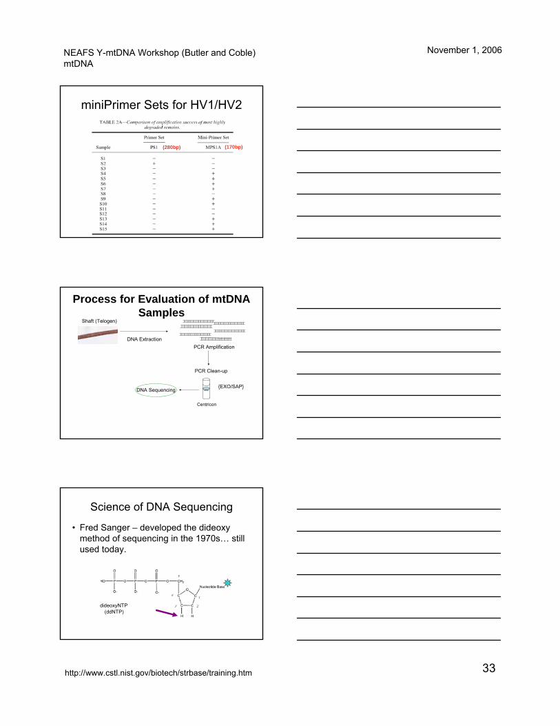
miniPrimer Sets for HV1/HV2
JFS (2001)
Primer DesignOrigin16024 576
HV1F15971/R16410
HV2F15/R389
PS1F15989/R16251
PS2F16190/R16410
PS3F15/R285
PS4F155/R389
mps1a mps2a
mps1b mps2b
mps3a mps4a
mps3b mps4b
mps5a
mVR1 mVR2
PS5F16381/R16569
NEAFS Y-mtDNA Workshop (Butler and Coble)mtDNA
November 1, 2006
http://www.cstl.nist.gov/biotech/strbase/training.htm 33
miniPrimer Sets for HV1/HV2
(280bp) (170bp)
Process for Evaluation of mtDNA Samples
Shaft (Telogen)
DNA ExtractionPCR Amplification
DNA Sequencing
PCR Clean-up
{EXO/SAP}
Centricon
Science of DNA Sequencing
dideoxyNTP(ddNTP)
• Fred Sanger – developed the dideoxymethod of sequencing in the 1970s… still used today.

NEAFS Y-mtDNA Workshop (Butler and Coble)mtDNA
November 1, 2006
http://www.cstl.nist.gov/biotech/strbase/training.htm 34
Sanger Sequencing3’-TAAATGATTCC-5’
ATT
ATTTACTAA
ATTTACT ATTTAC
ATTTATTTA
AT
ATTTACTA
ATTTACTAAGATTTACTAAGG
ADNA template5’ 3’
Primer anneals
Extension produces a series of ddNTP terminated products each one base different in length
Each ddNTP is labeled with a different color fluorescent dye
Sequence is read by noting peak color in electropherogram (possessing single base resolution)
Figure 10.5, J.M. Butler (2005) Forensic DNA Typing, 2nd Edition © 2005 Elsevier Academic Press
Primers Used for Control Region Amplification and Sequencing
13 primers – 11 for routine sequencing
Why the Redundancy?
• Homopolymeric stretches of Cytosines (C-stretches).
AAAACCCCCTCCCCATG
16189T
5 Cs 4 Cs
AAAACCCCCCCCCCATG
16189C
10 Cs
Strand slippage cancreate 11+ tandem Cs
HV1
NEAFS Y-mtDNA Workshop (Butler and Coble)mtDNA
November 1, 2006
http://www.cstl.nist.gov/biotech/strbase/training.htm 35
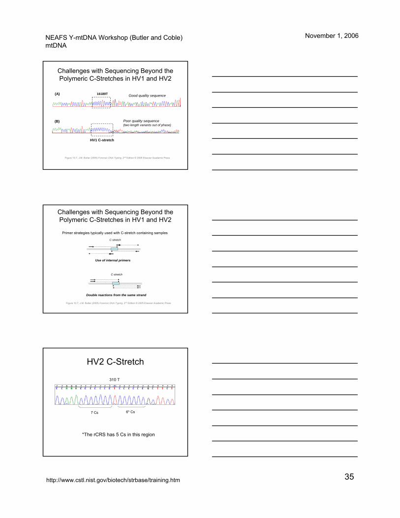
Challenges with Sequencing Beyond the Polymeric C-Stretches in HV1 and HV2
HV1 C-stretch
16189T
Poor quality sequence (two length variants out of phase)
Good quality sequence(A)
(B)
Figure 10.7, J.M. Butler (2005) Forensic DNA Typing, 2nd Edition © 2005 Elsevier Academic Press
Challenges with Sequencing Beyond the Polymeric C-Stretches in HV1 and HV2
Primer strategies typically used with C-stretch containing samples
C-stretch
C-stretch
Use of internal primers
Double reactions from the same strand
Figure 10.7, J.M. Butler (2005) Forensic DNA Typing, 2nd Edition © 2005 Elsevier Academic Press
HV2 C-Stretch
7 Cs 6* Cs
*The rCRS has 5 Cs in this region
310 T
NEAFS Y-mtDNA Workshop (Butler and Coble)mtDNA
November 1, 2006
http://www.cstl.nist.gov/biotech/strbase/training.htm 36
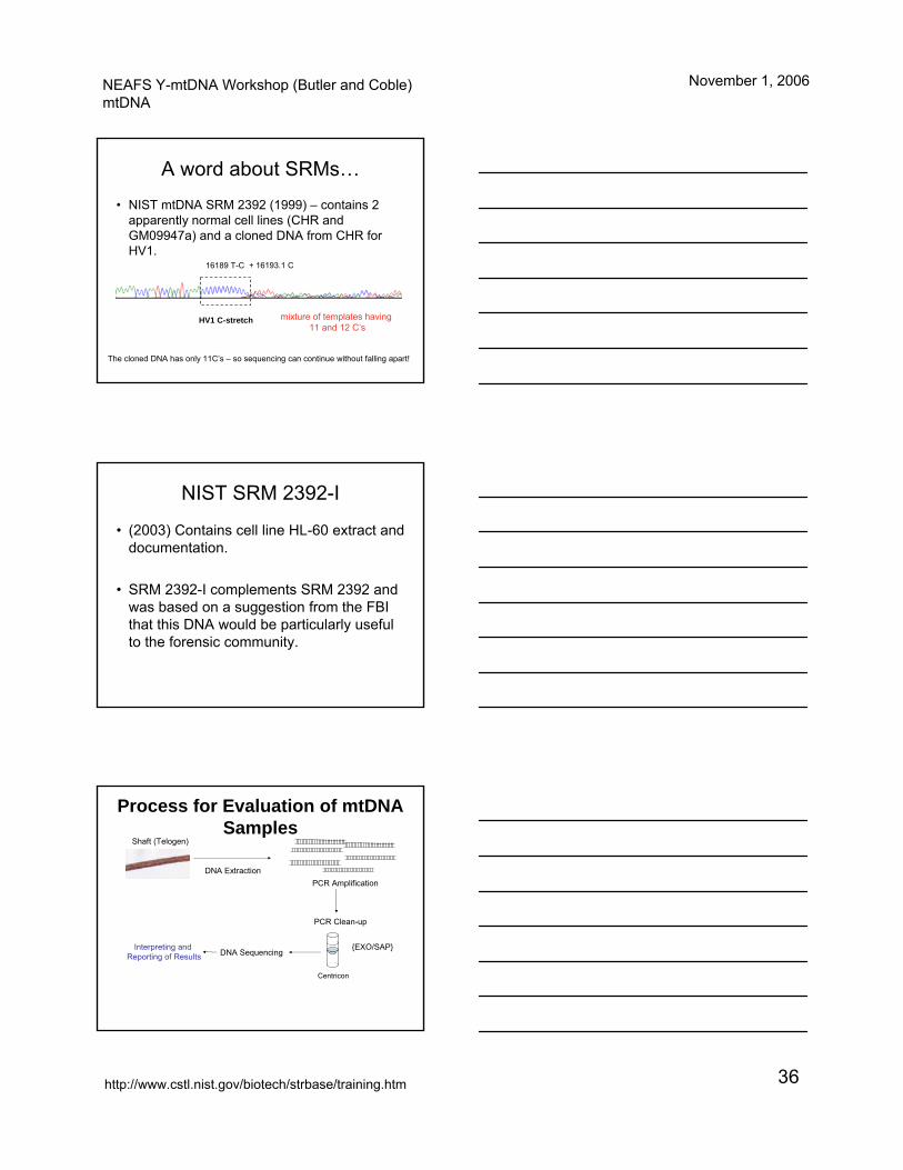
A word about SRMs…
• NIST mtDNA SRM 2392 (1999) – contains 2 apparently normal cell lines (CHR and GM09947a) and a cloned DNA from CHR for HV1.
HV1 C-stretch
16189 T-C + 16193.1 C
The cloned DNA has only 11C’s – so sequencing can continue without falling apart!
mixture of templates having 11 and 12 C’s
NIST SRM 2392-I
• (2003) Contains cell line HL-60 extract and documentation.
• SRM 2392-I complements SRM 2392 and was based on a suggestion from the FBI that this DNA would be particularly useful to the forensic community.
Process for Evaluation of mtDNA Samples
Shaft (Telogen)
DNA ExtractionPCR Amplification
DNA Sequencing
PCR Clean-up
{EXO/SAP}
Centricon
Interpreting and Reporting of Results