Embed Size (px)
Citation preview
8/7/2019 zl zimmerman
http://slidepdf.com/reader/full/zl-zimmerman 1/13
The Plant Cell, Vol. 5, 1411-1423, October 1993 O 1993 American Society of Plant Physiologists
Somatic Embryogenesis: A Model for Early Development inHigher Plants
J. Lynn Zimmerman
Department of Biological Sciences, University of Maryland Baltimore County, Baltimore, Maryland 21228
INTRODUCTION
The ability to produce morphologically and developmentallynormal embryos and, indeed, whole plants from undifferen-
tiated somatic cells n culture, through the process of somatic
embryogenesis, resides uniquely within the plant kingdom.Since the initial description of somatic embryo production from
carrot callus cells more than 35 years ago (Steward et al., 1958),this unique developmental potential has been recognized both
as an important pathway for the regeneration of plants from
cell culture systems and as a potential model or studying early
regulatory and morphogenetic events in plant embryogenesis.
The last 5 to 10 years have witnessed an explosion in the
number of species that can now be regenerated from cell cul-ture into whole plants through somatic embryogenesis. The
literature contains hundreds of references describing thespecific manipulations required to effect somatic embryo de-
velopment from a variety of agronomically and horticulturallyimportant plants. Although this is obviously an extremely im-
portant application of the process of somatic embryogenesis,
it is not the focus of this review. Rather, this review will focuson the use of somatic embryogenesis as a model system forunderstanding he regulation of gene expression required for
the earliest developmental events in the life of a higher plant:
the development of the fertilized zygote into a mature embryo.
The events of fertil ization and subsequent embryo devel-opment normally occur deep within maternal issues. The early
embryo is minute and is surrounded by both endosperm and
maternal cells. Although the morphological description of em-
bryo development has been extensively recorded through
microscopy, molecular and biochemical analyses of early em-bryogenesis have been hampered significantly by this physicalinaccessibility. As a consequence, we know very little aboutthe genes that are necessary or early embryogenesis n higher
plants and even less about their regulation. This is beginningto be remedied by recent intensive efforts o genetically iden-tify genes required for early embryogenesis n model systems
such as Arabidopsis and maize (see West and Harada, 1993,this issue); many interesting mutants have been identified hat
may provide entry points nto molecular analyses of major mor-phogenetic events in embryogenesis. These analyses wouldbe greatly enhanced by the availability of cell, tissue, and de-velopmental stage-specific markers of important events in hedifferentiationof cells and the establishment of the major tissue
systems of the plant, which occur early in embryogenesis. In
addition, once genes have been identified hat are essential
for embryogenesis, he subsequent analysis of their regulation
would be greatly facilitated by the availability of an appropri-
ate in vitro model system that is not limited n tissue quantityor accessibility. The somatic embryo system represents ust
such a model system. This review will summarize he process
of somatic embryogenesis and will address he strengths andlimitations of somatic embryos as potential models for study-
ing early events in plant embryo development.
SOMATIC EMBRYO INDUCTION AND DEVELOPMENT
The original descriptions of somatic embryogenesis came fromobservations of carrot cells in culture, and carrot has remainedthe primary experimental system for studying somatic embryo-
genesis. Recent studiesof Dudits et al. (1991) have highlightedthe utility of alfalfa microcallus cells as an alternative system,
particularly for studying the induction of embryo development
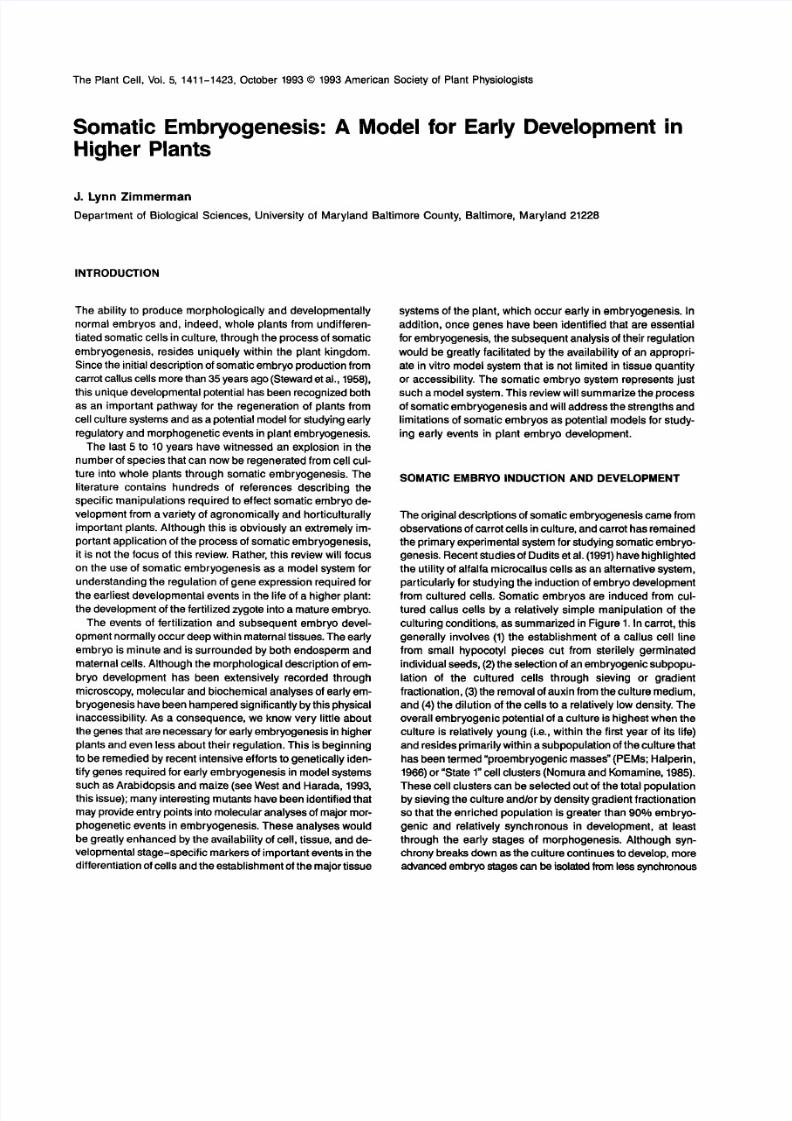
from cultured cells. Somatic embryos are induced from cul-tured callus cells by a relatively simple manipulation of the
culturingconditions, as summarized in Figure 1. In carrot, this
generally involves (1) the establishment of a callus cell linefrom small hypocotyl pieces cut from sterilely germinated
individual seeds, (2) the selection of an embryogenic subpopu-
lation of the cultured cells through sieving or gradientfractionation, (3) the removal of auxin from the culture medium,and (4) the dilution of the cells to a relatively ow density. Theoverall embryogenic potentialofa culture is highest when theculture is relatively young (i.e., within the first year of its life)
and resides primarily within a subpopulationof the culture thathas been termed “proembryogenic masses” (PEMs; Halperin,1966) or “State 1” cell clusters (Nomura and Komamine, 1985).These cell clusters can be selected out of the total populationby sieving the culture and/or by density gradient fractionationso that the enriched population is greater than 90% embryo-
genic and relatively synchronous in development, at leastthrough the early stages of morphogenesis. Although syn-chrony breaks downasthe culture continues to develop, moreadvanced embryo stagescan be solatedh m esssynchronous
8/7/2019 zl zimmerman
http://slidepdf.com/reader/full/zl-zimmerman 2/13
1412 The Plant Cell
Germinate sterile seeds Initiate callus culture
Globular
Remove
2,4-D
Heart
Torpedo
Figure 1. Summary of the Culturing of Carrot Somatic Embryos.
cultures by resieving the culture an d selecting fo r larger em -bryos. Using this basic procedure, gram quantities of any given
stage of embryogenesis can be easily isolated.
SOMATIC VERSUS ZYGOTIC EMBRYOGENESIS
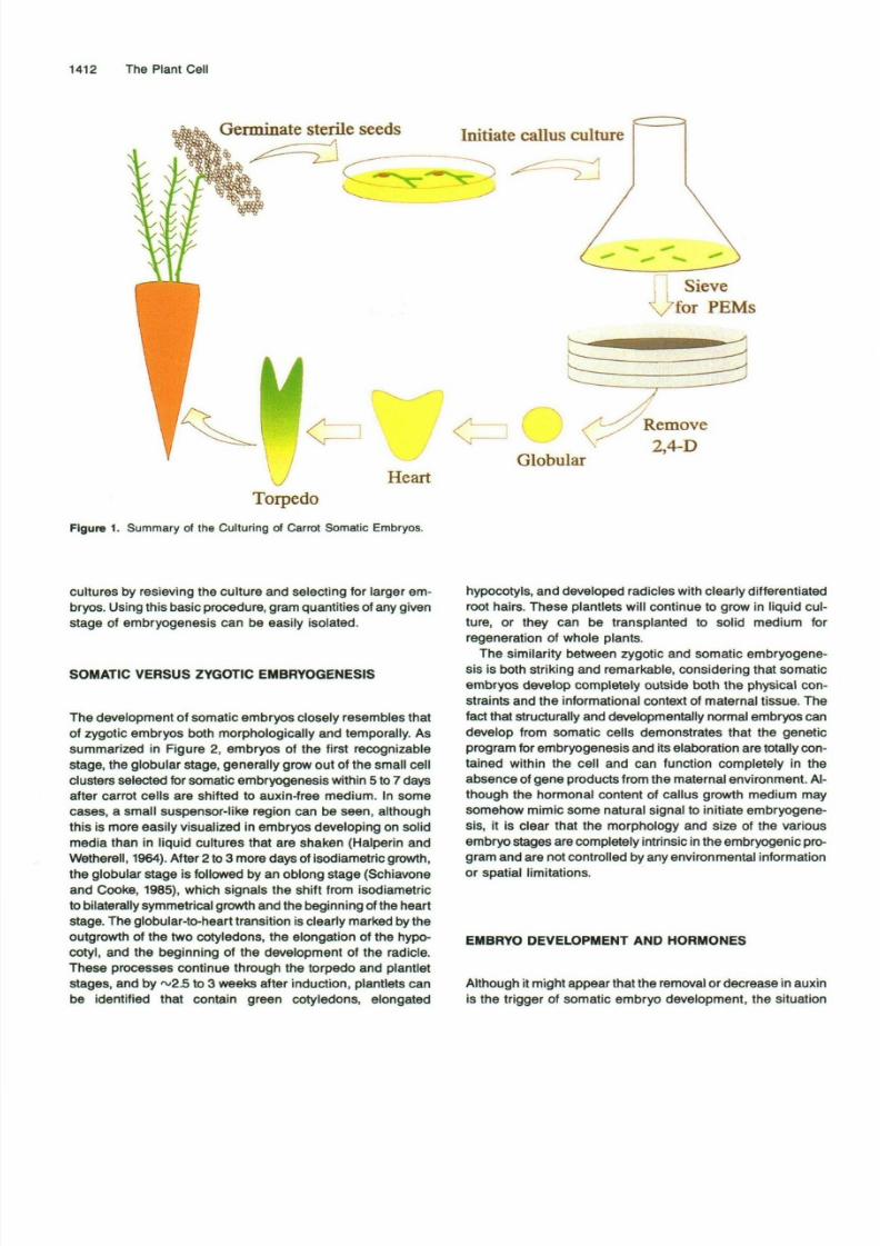
The development of somatic embryos closely resembles that
of zygotic embryos both morphologically and temporally. As
summarized in Figure 2, embryos of the first recognizable
stage, the globular stage, generally grow out of the small cell
clusters selected fo r somatic embryogenesis within 5 to 7 days
after carrot cells are shifted to auxin-free medium. In some
cases, a small suspensor-like region can be seen, although
this is more easily visualized in embryos developing on solid
media than in liquid cultures that are shaken (Halperin and
Wetherell, 1964). After 2 to 3 more days of isodiametric growth,
the globular stage is followed by an oblong stage (Schiavone
and Cooke, 1985), which signals the shift from isodiametric
to bilaterally symmetrical growthand the beginning of the heart
stage. The globular-to-heart transition is clearly marked by the
outgrowth of the two cotyledons, the elongation of the hypo-
cotyl, and the beginning of the development of the radicle.
These processes continue through the torpedo and plantlet
stages, and by ~2.5 to 3 weeks after induction, plantlets canbe identified that contain green cotyledons, elongated
hypocotyls, anddeveloped radicles with clearly differentiatedroot hairs. These plantlets will continue to grow in liquid cul-
ture, or they can be transplanted to solid medium for
regeneration of whole plants.
The similarity between zygotic and somatic embryogene-
sis is both striking and remarkable, considering that somatic
embryos develop completely outside both the physical con-
straints and the informational context of maternal tissue. The
fact that structurally and developmentally normal embryos can
develop from somatic cells demonstrates that the genetic
program forembryogenesis and its elaboration aretotally con-
tained within the cell and can function completely in the
absence of gene products from the maternal environment. Al-
though the hormonal content of callus growth medium may
somehow mimic some natural signal to initiate embryogene-
sis, it is clear that the morphology and size of the various
embryo stages are completely intrinsic in the embryogenic pro-
gram and are not controlled by any environmental information
or spatial limitations.
EMBRYO DEVELOPMENT AND HORMONES
Although it might appear that the removalor decrease in auxinis the trigger of somatic embryo development, the situation
8/7/2019 zl zimmerman
http://slidepdf.com/reader/full/zl-zimmerman 3/13
Somatic Embryogenesis 1413
Somatic Embryogenesis
r1
Calhis
Regenerationof
wholeplant
Plantlet
i Zygotic Embryogenesis |
ZygoteGlobular Heart/Toipedo
Cotyledon
Expansion
Maturationjr.
Desiccation
C-Dormancy
Germination[ a n d
Growth
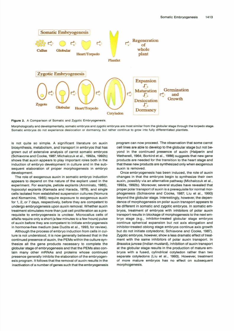
Figure 2. A Comparison of Somatic and Zygotic Embryogenesis.
Morphologically and developm entally, somatic em bryo s and zygotic emb ryos are most similar from the globular stage through the torpedo stage.
Somatic embryos do not experience desiccation or dormancy, but rather continue to grow into fully differentiated plantlets.
is no t quite so simple. A significant literature on auxin
biosynthesis, metabolism, and transport in embryo s that has
grown out of extensive analysis of carrot somatic embryos
(Schiavone and Cooke, 1987; Michalczuk et al., 1992a, 1992b)shows that auxin appears to play important roles both in the
induction of embryo development in culture and in the sub-
sequent elaboration of proper morphogenesis in embryo
development.
The role of exogenous auxin in somatic em bryo induction
appears to depend on the nature of the explant used in the
experiment. For example, petiole explants (Ammirato, 1985),
hypocotyl explants (Kamada and Harada, 1979), and single
cells isolated from established suspension cultures (Nomura
an d Komam ine, 1985) require exposu re to exogenous auxin
for 1, 2, or 7 days, respectively, before they are competent to
undergo embryo genesis upon auxin removal. Whether auxin
treatment stimulates m ore than just cell proliferation as a pre-requisite to embryogenesis is unclear. Microcallus cells of
alfalfa require only a short (a few minutes to a few hours) pulse
of auxin before they are competent to initiate em bryogenes is
in hormo ne-free medium (see Dudits et al., 1993, fo r review).
Although the process of embryo induction from cells in cul-
ture is not understood, it is now generally believed that in the
continued presence of auxin, the PEMs within the culture syn-
thesize all the gene products necessary to complete the
globular stage of em bryogenesis and that the PEMs also con-
tain many other mRNAs an d proteins whose continued
presence generally inhibits the elaboration of the embryogen-
esis program. It follows that the removal of auxin results in theinactivation of a numb er of genes such that the embryogenesis
program can now proceed. The observation that some carrot
cell lines are able to develop to the globular stage but no t be-
yond in the continued presence of auxin (Halperin an d
Wethere ll, 1964; Borkird et al., 1986) sugges ts that new geneproducts a re needed for the transition to the heart stage and
that these new products are synthesized only when exogenous
auxin is removed.
Once embryogenesis has been induced, the role of auxin
changes in that the embryos begin to synthesize their own
auxin, p ossibly via an alternative pathway (Michalczuk et al.,
1992a, 1992b). Moreover, several studies have revealed that
proper polar transport of auxin is a prerequisite for normal mor-
phogenesis (Schiavone and Cooke, 1987; Liu et al., 1993)
beyond the globu lar stage. Interestingly, however, the depen-
dence of morphogenesis on polar auxin transport appears to
be different in somatic an d zygotic em bryos. In somatic em -
bryos, treatment of embryos with inhibitors of polar auxin
transp ort results in blockage of morp hogenesis to the next em-bryo stage (e.g., inhibitor-treated globular stage embryoscontinue spherical expansion but not axis elongation and
inhibitor-treated oblong stage emb ryos continue axis growth
but do not initiate cotyledons; Schiavone and Cooke, 1987).
Zygotic embryos, however, show a less dramatic effect of treat-
ment with the same inhibitors of polar auxin transport. In
Brassica juncea (Indian mustard), inhibition of auxin transport
at the globular stage results in the production of mature em -
bryos with a fused, cylindrical cotyledon rather than two
separate cotyledons (Liu et al., 1993). However, treatment
of more mature embryos has no effect on subsequentmorphogenesis.
8/7/2019 zl zimmerman
http://slidepdf.com/reader/full/zl-zimmerman 4/13
1414 The Plant Cell
What is the basis of this difference in response betweenso-
matic and zygotic embryos? One suggestion (T. Cooke,
personal communication) s that in zygotic embryos, morpho-
genesis s actually regulated by two overlapping mechanisms,
one of which arises as a maternal effect oras a consequence
of the polarized position of the embryo in the embryo sac,whereas the other is intrinsic in the embryo itself. This latter,
intrinsic mechanism, which would be the only active mecha-nism in somatic embryos, would be dependent on polar auxin
transport, whereas the former mechanism, which would exist
in zygotic embryos, would promote some aspects of morpho-
genesis even in the absence of polar auxin transport. Although
the existence of a dual system for auxin regulation of embryo-
genesis remains speculative, these results illustrate the
potential of using somatic embryos to distinguish between n-
trinsic and extrinsic regulation of the process of embryogenesis.
This may also be useful in understanding the influence of an-
other plant hormone, abscisic acid (ABA), on the process of
embryogenesis, as discussed below.Clearly, the involvement of auxin in the induction and de-
velopment of zygotic and somatic plant embryos is complex,but what is obvious is that the products of both processes, a
globular embryo with the full potential for correct organogen-
esis, appear quite equivalent. It follows that the molecular
events that dictate the differentiation and propagation of all
of the major tissue systems in the developingembryo are likely
very comparable, if not equivalent, n somatic and zygotic em-
bryos. Because these events occur during the globular to
heartkorpedo stages, it is therefore at these stages that so -
matic embryos can most clearly serve as a good model forembryogenesis and as a source of materials for biochemical
and molecular analysis.
Beyond the torpedo stage, the processes of zygotic and so-
matic embryogenesis again diverge. Zygotic embryos move
into the cotyledon stage, followed by the maturation stage dur-ing which substantial storage protein synthesis occurs, followed
by preparation for desiccation and dormancy (see Thomas,
1993, this issue). Mature zygotic embryos dehydrate, a periodof quiescence ensues, and ultimately a new program of post-
germination development begins. A significant proportion of
this desiccation-dormancy program appears to be hormonally
regulated, primarily by ABA. In contrast, somatic embryos grow
and differentiate continuously, apparently activating the shoot
and root apical meristemswith no obvious quiescent state. Be-
cause of the divergencebetween somatic and zygotic embryos
at these later stages of development, the term “plantlet” em-bryo (versus “cotyledon” embryo) seems more appropriate todescribe the fully differentiated somatic embryo.
Although somatic embryos do not desiccate and become
dormant, they do synthesize and accumulate ABA (Hatzopouloset al., 199Oa), and they also express a number of genes that
have been shown to be ABA inducible and that are generally
associated with desiccation tolerance (e.g., LEA genes, dis-
cussed below). If these genes do, in fact, play a role in
desiccation tolerance, then it would logically follow thatthe signal to prepare for desiccation is an intrinsic and
anticipatory one, in that these genes are expressed n somatic
embryos that will never experience desiccation. In contrast,
the dormancy program is apparently not activated in somaticembryos, but rather appears to be induced extrinsically, pos-
sibly through a maternal signal, which could be simply a higher
ABA concentration. This would be consistent with the obser-vation that treatment of plantlet stage somatic embryos with
exogenous ABA can induce a quiescent state similar to the
dormancy of zygotic embryos (Ammirato, 1987). Clearly, our
understanding of the role of ABA in embryogenesis is incom-
plete, but further analysis of ABA metabolism and influence
in somatic embryo developmentshould help to clarify the con-
tribution of interna1 and externa1 sourcesof ABA in the proper
development of zygotic embryos.
GENE EXPRESSION DURING SOMATICEMBRYOGENESIS
Gene expressionduring somatic embryogenesiscan be evalu-
ated either by isolating genes expressed n somatic embryos
and subsequently identifying the function of those genes or
by studying the expression of avariety of other genes isolated
from nonembryo tissues in the hope that they may also playsome role in embryogenesis. A number of genes have been
identified that are enhanced in expression in developing em-
bryos, and severa1 of these are being used to analyze
mechanisms of gene regulation during embryogenesis.
Genes lsolated from Somatic Embryos
The dramatic transition from unorganized callus cell growth
to somatic embryo development, coupled with measurements
that indicate active RNA synthesis (Fujimura and Komamine,
1980) during this transition, suggested that a substantial
reprogramming of gene expression, presumably occurring at
the transcriptional level, dictates this developmental switch.
Early studies of Sung and coworkers showed that few changeswere apparent in the abundant proteins that were being syn-
thesized in somatic embryos compared to callus cells (Sung
and Okimoto, 1981). These results were optimistically inter-
preted as showing that at least a few changes in gene
expression could be observed by protein analysis, and it was
speculated hat even more changes may have occurred in the
production of less abundant proteins or mRNAs. Severalgroups took a similar approach to trying to identify “embryoenhanced genes” from carrot somatic embryos. The basic ex-
perimental strategy relied on a comparison between genes and
proteins being expressed in somatic embryos versus calluscells. Several of the genes that have been isolated from so-
matic embryos are summarized in Table l .In addition to these general differential screening ap-
proaches, other experimental strategies have allowed the
isolation of a small number of additional genes that are up
regulated in somatic embryos. For example, capitalizing on
8/7/2019 zl zimmerman
http://slidepdf.com/reader/full/zl-zimmerman 5/13
Somatic Embryogenesis 1415
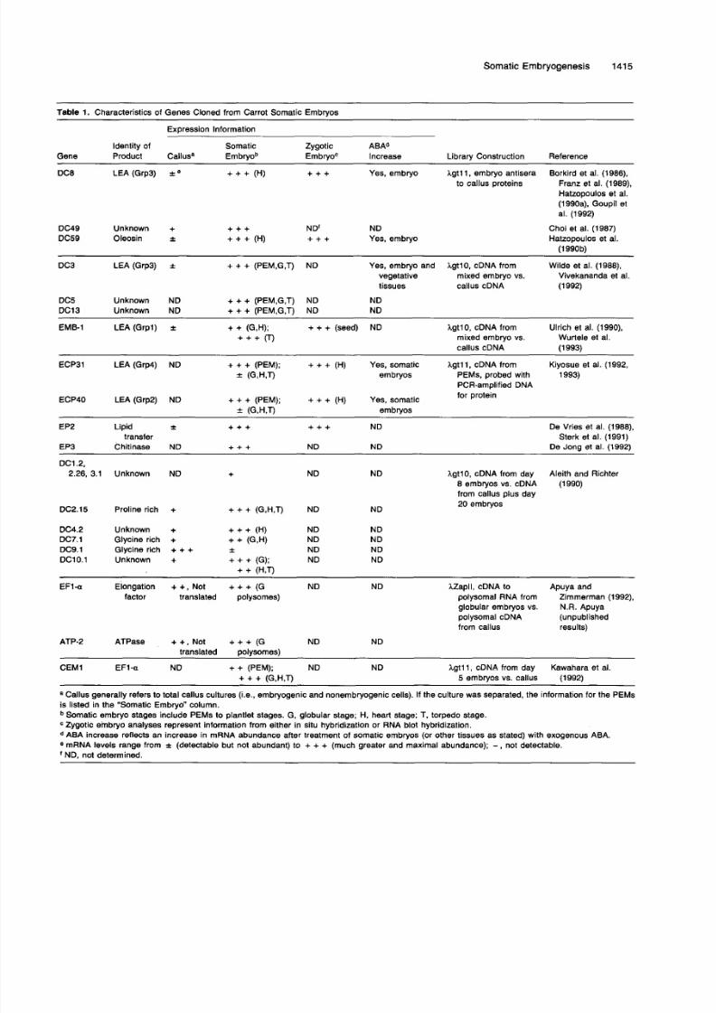
Table 1. Characteristics of Genes Cloned from Carrot Somatic Embryos
Expression lnformation
ldentity of Somatic Zygotic ABAd
Gene Product Callusa Embryob EmbryoC lncrease Library Construction Reference
DC8 LEA(Grp3) i e + + + (H) + + + Yes, embryo I g t l l , embryo antisera Borkird et al. (1986),
Franz et al. (1989),
Hatzopoulos et al.(1990a), Goupil et
al. (1992)
to callus proteins
DC49 Unknown + + + + ND NDDC59 Oleosin 2 + + + (H) + + + Yes, embryo
Choi et al. (1987)
Hatzopoulos et al.(1990b)
DC3 LEA (Grp3) f + + + (PEM,G,T) ND Yes, embryo and Igt lO , cDNA from Wilde et al. (1988),vegetative mixed embryo vs. Vivekananda et al.tissues callus cDNA (1992)
DC5 Unknown ND + + + (PEM,G,T) ND NDDC13 Unknown ND + + + (PEM,G,T) ND ND
EMB-1 LEA (Grpl)i
+ + (G,H); + + + (seed) ND+++o I g t lO, cDNA from Ulrich et al. (ISSO),Wurtele et al.ixed embryo vs.
callus cDNA (1993)
ECP31 LEA (Grp4) ND + + + (PEM); + + + (H) Yes, somatic I g t l l , cDNA from Kiyosue et al. (1992,
f (G,H,T) embryos PEMs, probed with 1993)PCR-amplified DNAfor protein
ECP40 LEA(Grp2) ND + + + (PEM); + + + (H) Yes, somatic
EP2 Lipid i + + + + + + ND De Vries et al. (1988),transfer Sterk et al. (1991)
EP3 Chitinase ND + + + ND ND De Jona et al. (19921
f (G,H,T) embryos
DCI 2,
2.26, 3.1 Unknown ND + ND ND
DC2.15 Proline rich + + + + (G,H,T) ND ND
Igt10, cDNA from day8 embryos vs. cDNA (1990)
from callus plus day20 embryos
Aleith and Richter
DC4.2 Unknown + + + + (H) ND NDDC7.1 Glycine rich + + + (G,W ND NDDC9.1 Glycine rich + + + f ND NDDC1O.l Unknown + + + + (G); ND ND
+ + (H,T)
EFI-Q Elongation + +, Not + + + (G ND ND Zapll , cDNA to Apuya andfactor translated polysomes) polysomal RNA from Zimmerman (1992),
globular embryos vs. N.R. Apuyapolysomal cDNA (unpublishedfrom callus results)
ATP-2 ATPase + + , Not + + + (G ND NDtranslated oolvsomes)
CEM1 EF1-a ND + + (PEM); ND ND I g t l l , cDNA from day Kawahara e1 al.
a Callus generally refers to total callus cultures (i.e., embryogenic and nonembryogenic cells). If the culture was separated, the information for the PEMsis listed in the "Somatic Embryo" column.
+ + + (G,H,T) 5 embryos vs. callus (1992)
Somatic embryo stages include PEMs to plantlet stages. G, globular stage; H, heart stage; T, torpedo stage.Zygotic embryo analyses represent information from either in situ hybridization or RNA blot hybridization.ABA increase reflects an increase in mRNA abundance after treatment of somatic embryos (or other tissues as stated) with exogenous ABA.
ND, not determined.* mRNA levels range from f (detectable but not abundant) to + + + (much greater and maximal abundance); - not detectable.
8/7/2019 zl zimmerman
http://slidepdf.com/reader/full/zl-zimmerman 6/13
1416 The Plant Cell
the observation that embryogenic cultures secrete some
unique proteins, Sterk et al. (1991) isolated genes encoding
two extracellular proteins (EP), EP2 and EP3, that are enhanced
during embryogenesis. Experiments designed to isolate post-
transcriptionally controlled genes yielded two other genes,
EF1-a and ATP2 (Apuya and Zimmerman, 1992). In total, 21genes have been described, some of which have been charac-
terized in greater detail than others. The identities or at least
the properties of severa1 of these genes have been described
and are summarized in Table 1. Each of the classes of pro-
teins that have been identified as being up regulated during
embryogenesis has added to our understanding of both the
process of somatic embryogenesis as well as the relationship
between somatic and zygotic embryogenesis, as described
below.
Late Embryogenesis Abundant (LEA) Gene Expression
in Somatic Embryos
Several genes that are preferentially expressed n somatic em-
bryos appear to have characteristicsof a class of proteins called
LEA proteins (Dure et al., 1981,1989; Galau et al., 1986). LEA
proteins are very hydrophilic proteins that are abundantly ex-
pressed ate n zygotic embryogenesis in many plant species,
includingcotton (in which they were originally described), bar-
ley, rice, oilseed rape, and wheat (Dure et al., 1989). The timing
of their expression n embryogenesis and their ABA inducibil-
ity have led to the suggestion that in zygotic embryos, they
play a role in protecting he embryo during desiccation. The
LEA genes lsolated from carrot somatic embryos include
representatives of all four described groups of LEA proteins,which are characterizedby significant homology n either amino
acid composition or general protein structure (Dure et al., 1989).
All of the carrot LEA genes that have been analyzed to date
show detectable expression n callus cells (some, like ECP31
and ECP40, are highly expressed in the PEMs of callus cell
cultures), and most of the LEA transcripts increase significantly
in abundance in somatic embryos at the heart stage (Choi et
al., 1987; Wilde et al., 1988; Franz et al., 1989; Kiyosue et al.,
1992, 1993; Wurtele et al., 1993). Indeed, we have found the
LEAs to be the most abundant and differentially expressed
mRNAs in somatic embryos, as compared to callus cells (X.
Lin, G-J. Hwang, and J.L. Zimmerman, unpublishedobserva-
tions), which probably explains why so manyLEA genes have
been isolated through differential screening approaches usingcarrot somatic embryos. All of the carrotLEAgenes tested can
be induced by ABA treatment of callus cells and somatic em-
bryos (Hatzopoulos et al., 1990a; Goupil et al., 1992; Kiyosue
et al., 1992, 1993; Vivekananda et al., 1992). However, it ap-
pears that only DC3, a group 3 LEA, can be induced in
nonembryonic tissues by either ABA or desiccation stress,
whereas the other LEAs that have been tested (DC8, DC59,
ECP31, and ECP4O) cannot be induced in nonembryonic cells.
A further discussion of such genes and their regulation by ABA
and other factors can be found in Thomas (1993, this issue).
In addition to the analysis of regulatory mechanisms gov-
erning the expression of LEA genes in embryogenesis, an
investigation of the distribution and timing of EMB-1LEA gene
expression n somatic and zygotic embryos of carrot has been
performed (Wurtele et al., 1993). In situ hybridization analysis
showed that EMB-1 mRNA begins o accumulate uniformly inboth somatic and zygotic globular embryos, which is signifi-
cantly earlier in development than the time of maximum
production of LEA proteins. As development proceeds to the
heart stage in both zygotic and somatic embryos, EMB-1 mRNA
accumulates to higher levels and begins to show a polarity
of distribution, with hybridization predominantly over the pe-
ripheral regions of the embryo. In mature zygotic embryos, very
high evels of EMB-1 mRNA are seen, primarily associated with
the procambium and shoot and root apical meristems. In
plantlet stage somatic embryos, EMB-1 also accumulates pre-
dominantly n the meristematic cells, but at lower levels. There
is no detectable EMB-1 mRNA in the endosperm, developing
seed coat, or developing carpels or fruit of zygotically producedseeds, and EMB-1 mRNA does not accumulate in any cell type
of young plants. The apparent difference n levels of accumu-
lation of this LEA mRNA in somatic and zygotic embryos could
support the idea that zygotic embryos experience a second-
ary signal (possibly a pulse of ABA) from the maternal
environment that could not only enhance LEA gene expres-
sion, but could also signal the beginning of the dormancy
program.
In situ localization experiments have thus shown that the
expression pattern of at least the EMB-1 gene in carrot somatic
embryos is analogous to its expression pattern in zygotic em-
bryos of carrot (wurtele et al., 1993). This result provides
important validation of the useof somatic embryos as a modelfor studying embryogenesis. The fact that LEA mRNAs can
be detected at much earlier stages in carrot somatic embryos
than in cotton zygotic embryos may be a reflection of the fact
that so much more young embryo tissue can be sampled in
somatic embryo systems in which laboriousdissection is not
necessary, or, possibly, that in situ hybridization allows a more
direct comparison between somatic and zygotic embryos. It
would be instructive to perform n situ hybridizationanalyses
of LEA gene expression in other species, such as cotton, for
which RNA blot analysis suggests expression ater in embryo
development.
Somatic embryos provide a useful backdrop for analysis of
the intrinsic hormonal nfluences on embryo nduction, morpho-
genesis, and maturation preceding desiccation and dormancy.If the entire process is hormonally regulated, the activation
of the entire signal transduction pathway must be triggered
by the embryo itself rather than from any maternal nteraction,
because this is totally lacking n developing somatic embryos.
Secreted Proteins Pmduced by Somatic Embryos
Several years ago, it was observed that conditioned medi-
um from somatic embryo cultures could promote somatic
8/7/2019 zl zimmerman
http://slidepdf.com/reader/full/zl-zimmerman 7/13
Somatic Embryogenesis 1417
embryogenesis Hari, 1980; Smith and Sung, 1985). In addition,
it was shown that secreted EPs could rescue embryogenesisin a temperaturesensitive ts) ariant carrotcell line (Lo Schiavo
et al., 1988; De Jong et al., 1992). Exploiting the observation
that important proteins appear to be secreted from carrot so-matic embryo cultures, clones encoding some of these EPswere isolated (De Vries et al., 1988; Sterk et al., 1991). One
of these clones, EP2, was identified as encoding a lipid trans-fer protein whose function was suggested to involve the
transport of l ipids or other apolar molecules out of cells. The
expression of carrot EP2 is spatially regulated, showing re-
striction to cells of the protoderm of somatic embryos and
epidermal cells of leaf primordia and floral organs (Sterk et
al., 1991). Thus, although the expression of EP2 is not em-bryo specific, this gene can serve as a useful marker for the
establishment of the epidermal cell layer. Other EPs, such as
EPl and EP3, have been similarly isolated from media of so-
matic embryo cultures (Van Engelen et al., 1991; De Jong et
al., 1992). EP3 encodes a glycosylated acidic endochitinase(De Jong et al., 1992) that is able to restore embryo develop-
ment to a tssomatic embryo defectivecell line, ts77 (see below).EP1, which has a region of homology with Bfassica S locus
glycoprotein genes (Van Engelen et al., 1991; Van Engelen and
De Vries, 1992), is not expressed n embryogenic cell clusters
or somatic embryos but rather is produced by the nonembryo-geniccellsof a callus culture. It has been suggested that these
secreted proteins likely play a role in the regulationo4 cell ex-pansion, which is critical to the maintenance of the integrityof the epidermal layer in embryos and other tissues, and tothe proper establishment of shape and form, which is largelycontrolled by cell expansion (Van Engelen and De Vries, 1992;
Sterk and De Vries, 1993).
Other Genes lsolatedfrom Somatic Embryos
Severa1 other genes have been isolated from cDNA libraries
constructed from mRNA isolated from somatic embryos, as
summarized in Table 1. Although none of these has been ex-
tensively characterized, the properties of a few of them arenoteworthy. The DC59 clone, originally described by Choi et
al. (1981), has been shown to encode a lipid body membraneprotein (also called oleosin) that, although distinct from theLEA prdeins, isalsoABA inducible (Hatzopoulosetai.,1990a).
Molecular analysis of this gene has identified regions within
its 5'end that interact with nuclear factors present only in em-
bryo extracts; these regions show sequence similarity to
regions5'to theDC8LEAgene ( m a ikely he ABA-responsiveregions; Hatzopolousetal., 1990b). Inaddition, several of theclones isolatedbyAleith and Richter (1990) have unusual amino
acid sequences. DC2.15 has a core of repeating Pro-X motifs,and DC7.1 and DC9.1 are glycine rich. The two glycine-rich
proteinsalsoappear to have the potential for interesting sec-
ondary structures, such as membranespanning a-helicaldomains at both the arnino and carboxyl termini and exten-sive p-pleated sheet structures n their cores.Although these
proline- and glycine-rich proteins bear resemblance to certain
cell wall proteins (Chen and Varner, 1985; Condit and Meagher,
1986; Keller et al., 1988), nothing is known about the specific
functions of these proteins n embryogenesis or at other times
in the development of plants. It would not be surprising f theseproteins are cell wall components, because cell division andconcomitant wall synthesis are very active during embryogen-
esis. Each of these genes, and the others described by Aleith
and Richter (1990), is regulated n abundance during sornaticembryogenesis. Their patternsof expression, where known,
are summarized in Table 1.
In addition to the isolation of several genes by virtue of their
enhanced transcription and abundance n total RNAof sornatic
embryos, a few genes have been identified that are transla-
tionally enhanced in somatic embryogenesis. These includethe translation elongation factor EF1-a and the f3 subunit of
ATP synthase, ATP-2 (N.R. Apuya and J.L. Zimmerman, un-
publisheddata). A clone encoding EF1-a was also isolated by
more standard differential hybridization by Kawahara et al.(1992). Recent efforts in my laboratory have resulted in theisolation of five more genes that appear to be translationally
controlled in somatic embryos of carrot (X. Lin, G.-J. Hwang,
and J.L. Zimmerman, unpublished data).Although the extent, significance, and mechanism(s) of post-
transcriptional regulation of gene expression in plant embryo
development are only beginning to be addressed, severalstudies suggest that there may be some similarities to animalembryogenesis, in which translational activation of stored or"maternal" mRNAs is the primary leve1 of gene regulation in
early development of nonmammalian embryos. StoredmRNAs
appear to support embryo development until the eight-cell
stage in the fern Marsilea vestita (Kuligowski et al., 1991), and
maternal mRNAs are activated in early zygote developmentin Fucus (Masters et al., 1992). In addition, Pramanik et al.
(1992) demonstrated that storage protein synthesis in alfalfa
somatic embryos is translationally regulated; the mRNA is pres-
ent but restricted to the nontranslated mRNP pool early in
embryogenesis (globular-tetorpedostages) and is then shifted
to the polysome pool at the cotyledon stage. Moreover, post-
transcriptional regulation, ncluding differential transcript sta-
bility, has been suggested to be an important component ofembryo mRNA accumulation in zygotic embryos (Walling et
al., 1986). It is likely hat further study will reveal that eachas-
pect of post-transcriptional regulation (transcript processing,stability, translatability, etc.) contributes significantly to the pro-
cess of early embryo development in plants.
Expmssionof 'Nonembryonic" Genes during Somatic
Embryogenesis
In addition to using somatic embryosas a means of isolatinggenes that are regulatedduring embryogenesis (and that rnaytherefore be important n that process), somatic embryos havealso been used to assess the expression of genes isolatedfrom nonembryonic tissues. Particular attention has focused
8/7/2019 zl zimmerman
http://slidepdf.com/reader/full/zl-zimmerman 8/13
1418 The Plant Cell
on both cell cycle genes and histone genes. Callus suspension
cultures of alfalfa and the somatic embryos derived rom them
are proving useful in studying the relationship among auxin,
the reactivation of rapid cell cycling, and the induction of genes
associated with the cell cycle (Hirt et al., 1991). In alfalfa, theinduction of embryogenesis is quite different from that in car-
rot, in that alfalfa cultures are subjected to a brief pulse of
relatively high concentrations (100 pM) of the synthetic auxin
2,4-D prior to being transferred to hormone-free medium for
the development of embryos. In alfalfacultures, his auxin pulse
induces both active cell division, and, for the first few days
of embryo development, the accumulation of mRNAs (two to
three different size transcripts) for the cdc2 protein kinase (Hirt
et al., 1991). These mRNAs hen decrease in abundance. Much
of this work has recently been reviewed by Dudits et al. (1991,
1993). No analogous studies have been conducted in carrot
in which induction of somatic embryogenesis does not require
this high auxin pulse.
GENERATING MOLECULAR MARKERS FOR CELL
DIFFERENTIATION IN EMBRYOGENESIS
One extremely important use of the somatic embryo system
in studying development is in the generation of molecular mark-ers for events in cell and tissue differentiation during the
formation and development of the embryo. Such markers are
essential for an understanding of the developmental conse-
quences of embryo mutations that are being identified and
characterized n other systems such as Arabidopsis. Some ofthe genes that have been cloned from carrot somatic embryos,
such asEP2, have urned out to be useful markers of differen-tiation (of the dermal tissue system, in the case of EP2). Indeed,
the EP2 gene serves o illustrate an important point: this gene
is not embryo specific, but it is enhanced inexpression n em-
bryogenesis and, due to its specific localization, it can still
represent a very useful molecular marker.
Early attempts at cloning genes expressed n somatic em-bryos were designed with the bias that genes that are important
for embryo development should either not be expressed at all
in callus cells or should be greatly reduced in mRNA abun-
dance in callus. It now appears, however, hat callus cells, and
particularly the PEMs contained in the callus, are already ex-
pressing many of the genes that will be expressed through
at least the globular stage of embryogenesis. Therefore, anygene cloning scheme that involves a differential or subtrac-
tive hybridization step comparing embryo cDNA with callus
cDNA will likely eliminate many gene sequences that could
represent potentially useful molecular markers, particularly forearly events in embryogenesis.
One alternative approach to the isolation of useful markers
in embryo development s simply to use genes that have been
cloned from other time points in development during which
expression of the gene appears to be tissue or cell-type spe-cific. For example, genes whose expression s localized to the
vascular system in leaf tissue might be useful in identifyingprovascular cells in early embryos. The characterizationof the
expression patterns of such genes by in situ hybridization to
embryo sections could be informative.
,Another alternative approach to gene isolation that my lab-oratory has taken recently sto dentify clones that are induced
in globular embryos as compared to Fday-old seedlings. Su-
perimposed upon this comparison was the use of only
polysome-associated mRNAs rather than total RNA. A “sub-
tracted probe” was made using seedling polysomal cDNA
hybridized to globular polysomal cDNA. This probe allowed
the isolation of 50 different clones (X. Lin, G.-J. Hwang, and
J.L. Zimmerman, unpublished data) that included many genesthat had been isolated previously and that are included in Ta-
blel,mong them DC8, DC59, DC3, EMB-l, EP2, and EF-la.
In addition, the screen identified several genes that had been
previously isolated and characterized in nonembryonic issues,
such as histone genes and other genes of metabolism.
Moreover, we were able to identify several undescribedor nove1sequences that are enhanced in embryos compared to seed-
lings. The location and timing of expression of each of these
genes during somatic embryogenesis are currently being
evaluated through n situ hybridization. t is our hope hat some
of them may prove to be useful markers or specific cell types
within the developing embryo.
SOMATIC EMBRYOS AS A GENETIC SYSTEM
The power of genetic approaches to understanding develop-
mental systems has been elegantly demonstrated in severalanimal systems. The generation of single gene mutations thatblock the expression of essential developmentally regulated
genes has revealed many of the basic principles of cell differ-
entiation and pattern formation in eukaryotes. Typical genetic
approaches involving mutagenesis and the identification of
defective phenotypes cannot be easily applied to the somaticembryo system for several reasons: (1) the proportion of cells
that actually enter and complete embryogenesis is naturally
somewhat variable and decreases with increasing time in cul-
ture; (2)prolonged ime in culture can lead to the accumulation
of mutations (somaclonal variation; Widholm, 1984) that may
at the least confound analysis of embryo mutations, f not ob-
scure them completely; (3) unusual plantlet phenotypes, such
as multiple cotyledons, are not unusual in any culture; and(4)most embryogenic cell lines have a limited embryogenic
lifespan (usually in the range of 1 to 2 years), after which it
may become impossible to generate any embryos. Thus, al-
though large numbers of individuals can be analyzed usingthe cell culture/somatic embryo system, the background of
genetic and epigenetic variation in somatic embryogenesisoften obscures interesting mutant phenotypes.
One genetic approach has, however, been shown to be
potentially useful in dissecting events in somatic embryogen-esis: the identification and characterization of conditional
8/7/2019 zl zimmerman
http://slidepdf.com/reader/full/zl-zimmerman 9/13
Somatic Embryogenesis 1419
mutants. These are mutant cell lines that are capable ofundergoing normal somatic embryogenesis at a permissive
condition but are blocked in development when exposed to
a restrictive condition (usually increased emperature).ts vari-
ants of carrot somatic embryogenesis have been used in two
different experimental approaches.
ts MutantsAllow Gene lsolation
The ts cell line ts77 facilitated the isolation of one embryo-
enhanced gene. This mutant cell l hewill grow but not develop
normally through somatic embryogenesis at 32OC (Giuliano
et al., 1984); at the restrictive temperature, this line produces
abnormal misshapen heartlike embryos that lack normal polar-
ity. However, normal development can be restored by inducing
embryogenesis n “conditioned medium” (i.e., medium that has
previously supported a normal embryogenic line’s develop-
ment). Fractionation and characterization revealed that the
component of conditioned media hat restored normal embryo-
genesis is a secreted protein, EP3, that is a glycosylated acidic
endochitinase (De Jong et al., 1992). Although this is the only
reported example of the use of ts mutants to assist in gene
isolation, the potential utility of this technique is high, so long
as the mutation is stable and the line remains embryogenic
at the permissive emperature. As described below, this is not
always the case.
ts Mutants Can Define lnteresting Developmental
Transitions
The possibility of isolating ts variants in carrot somatic em-
bryogenesis is highlighted by the identification of ts variants
that are defective or arrested at particular time points in em-
bryogenesis. Breton and Sung (1982) described three primary
classes of developmentalvariants that become apparent when
cells are induced to undergo somatic embryogenesis at the
restrictive temperature: those blocked for growth, those thatcannot initiate embryogenesis, and those that are blocked at
the globular stage. Furthermore, Breton and Sung (1982)
reported hat the tsphenotypes were generally stable in callus
culture for at least 12 months and that the phenotypes weremaintained after plantlet regeneration and subsequent recul-
turing. One of these variants, ts59, is blocked at the globularstage of development when cultured at the restrictive tem-
perature of 32OC but develops normally at 24OC. The develop-
mental block can be imposed by exposure to 32OC at two
separate time periods of development, he first during the early
globular stage and the second during the globular-to-heart ran-
sition. The only observable difference at the protein leve1
between the wild type and ts59 mutant was a variation in the
heat shock protein (hsp) profile(LoSchiavo et al., 1988). The
ts59 mutant failed to phosphorylate some hsps in the 36 to
45 kD range. However, it has not been determined whether
this difference in hsp profile has any direct relationship to the
ts embryo-arrested phenotype of this line.Other experiments, including those from my own laboratory,
have also shown a number of potentially signif icant relation-
ships among hsp gene regulation, somatic embryogenesis,
and tsmutations. For example, two-dimensional protein anal-
ysis of other ts variants of carrot somatic embryo development
(see below) revealed a number of reproducible deficiencies
in the constellation of low molecular weight hsps that areproduced by these embryo-arrested cell lines (Hwang and
Zimmerman, 1989). In the most extreme case, only six small
hsps remained in a ts variant, compared to the 20 different
low molecular weight hsps typically synthesized by normal car-
rot cell lines. However, it is not known whether the embryo-
defective phenotypes result from the absence of the hsps and
consequent decreased thermotolerance (thus rendering he
cell lines ts and embryo defective), or whether the embryo-
defective phenotypes result from an independent mutation in
an essential embryo gene, with the alteration in hsp produc-tion being coincidental and unrelated to the phenotypes.
The relationship between hsps and embryo development
(in both plants and animals) is complex. Carrot somatic em-
bryos exhibit an extreme sensitivity to heat shock during a
specific portion of the globular stage of carrot somatic embryo-
genesis (Zimmerman et al., 1989). Indeed, further development
is arrested by a relatively brief heat shock during the globular
stage, and heat shock at other time points in embryo develop-ment induces a higher proport ionof abnormal morphology at
subsequent embryo stages. There are many analogous ex-
amples of heat shock sensitivity n early animal embryogenesis
(see Zimmerman and Cohill, 1991, for a comparative review).
One of the conclusions that emerges from studies of both
plant and animal embryogenesis is that for an embryo to be
thermotolerant, most, if not all, heat shock genes must be tran-
scribed, f not translated, and generally his is not accomplished
until the zygotic genome is transcriptionally active. A second
general observation s hat heat shock exposure at certain spe-
cific periods of development can result in predictable,
nonheritable developmental defects; in Drosophila these
defects, termed “phenocopies” (see Peterson, 1990, for review),
mimic many developmentalmutations, such asbirhorax (Gloor,
1947) and forked (Mitchell and Peterson, 1982). In mammals,
heat shock of embryos has been linked with severa1 neural
and craniofacial defects (Walsh et al., 1987, 1989). Althoughthe precise molecular basis for all these developmentaldefects
remains o be determined, it is clear that heat shock can havea profound effect on the process of embryo development, and
at least some of these effects may relate to heat shock gene
regulation.
A related and more extensive description of ts variants insomatic embryogenesis was reported by Schnall et al. (1988).In addition to characterizing a larger number of variants, this
study defined the temperature-sensitive period(TSP)for each
of the variants to establish the timing of gene action duringembryogenesis. Although many of the variants exhibited
extended TSPs, some showed TSPs that were restricted to
8/7/2019 zl zimmerman
http://slidepdf.com/reader/full/zl-zimmerman 10/13
1420 The Plant Cell
one or two embryonic stages. In some cases, the TSP tem-
porally preceded the appearance of the variant phenotype,
indicating gene activity in advance of any obvious morpho-
logical change; inothers, the TSPwas essentially coincidentwith the visualization of aberrant development. Although this
study identified variants for every developmental stage in em-bryogenesis, approximately half of the variant embryos were
blocked prior to the globular-to-heart transition. This is simi-
lar to the preponderance of globular embryo defects observed
in the analysis of embryo defective mutants of Arabidopsis (see
West and Harada, 1993, this issue) and may imply that a num-
ber of new gene products are necessary or this developmental
transition to occur.
Two of these variants were further characterized by protein
analysis in an attempt to define some gene products that mightbe stage specific (Schnall ef al., 1991). Although many differ-
ences in protein profiles were apparent in each of the variants,
subsequent analysis revealed almost all of them reflected ei-
ther cell line differences, age differences, or random variations,rather than differences due todevelopmental changes in pro-
tein synthesis (Schnall et al., 1991). It was therefore concluded
that the changes that could be observed by protein analysis
were not reflective of the genetic alterations in any of these
lines. Unfortunately, all of the fs lines originally described by
Schnall et al. (1988) were subsequently lost, either because
they ultimately lost their embryogenicity at the permissive
condition or because they lost the specificity of the variant phe-
notype at the restrictive temperature (T Cooke, personal
communication).
Limitations of Somatic Embryos in Genetic Analyses
The results of the protein analysis, coupled with the ultimate
loss of thers lines (or their embryogenicity), serve to highlight
severa1 properties of somatic embryos that mustberecognized
in considerations of somatic embryosasa genetic system. First,by virtue of their prolonged propagation n cell culture, the lines
that produce somatic embryos likely contain and accumulate
mutations that may ultimately confound any other genetic
manipulations. Second, in most cases, the ephemeral nature
of the embryogenic potential of any given cell line will severely
restrict the time frame over which experiments can be con-
ducted. This can be partially overcome by regular cycling ofthe cell line through embryogenesis and selection of normal
plantlets or the reinitiationof
the lineas
well as by whole plantregeneration and maintenance through vegetative growthand/or zygotic embryogenesis. Although the problems are not
insurmountable, the propagation and maintenance of highlyembryogenic cell lines s very labor intensive, as s the regener-
ation of plantsfromcell culture, and requires both finesse andvigilance. It is clear that the somatic embryo system of carrot
can be manipulated genetically, but the questions one attempts
toanswer must be well considered and designed with an aware-ness of the vagaries and potential limitations of the somatic
embryo system for this analysis.
MICROSURGICAL ANALVSIS OF SOMATIC
EMBRYOGENESIS
Microsurgical manipulations have been used to characterize
a number of aspects of patterning processes n both plant andanimal systems (see Slack, 1983; Steeves and Sussex, 1989).
These techniques have the potential to reveal such basic in-
formation as the source of pattern information, the degree of
commitment or determination of particular cell or tissue types,
and the contribution of context or environment to the develop-
mental fate of a cell or tissue. Microsurgical manipulation has
been applied to the carrot somatic embryo system with some
interesting esults. Studies by Schiavone (1988) and Schiavoneand Racusen (1990,1991) have revealed that when carrot so-
matic embryos are manipulated microsurgically, by eitherremoving or grafting regions, the resulting embryo pieces or
grafts retain developmental competence. For example, sec-
tionsof
the shoot pole comprising25
to 90% of the originallength of the torpedo embryo regenerated he root pole in ap-
proximately half of the cases; the regenerating root could be
visualized within a few days of the surgery (Schiavone and
Racusen, 1990). The regeneration of the shoot pole was more
difficult to accomplish and was induced efficiently only when
at least 90% of the axial length was removed; sections that
contained a greater portion of the embryo length simply con-tinued to grow as roots. Removal of one cotyledon resultedin the regeneration of one, or usually multiple, cotyledons.
No growth of the removed cotyledon was observed. If both
cotyledons were removed from the same embryo, either the
cotyledons regenerated at the cut sites or the amputated re-
gion greened while a new shoot emerged along the surface
of the midhypocotyl.These experiments highlight the potential for developmen-
tal perturbation and recovery that exists in the somatic embryo+em. However, the small size of even torpedo-stageembryos
limits the util ity and reproducibility of microsurgical manipu-
lation. The types of manipulation that have been performed
are relatively crude (althoughstill remarkable), in that they have
been limited to the deletion of relatively large portions of the
embryos. Nonetheless, these experiments may lead the way
for other types of manipulations, such as laser surgery or cell
ablation experiments.
PROSPECTS FOR FUTURE STUDIES IN EMBRVODEVELOPMENT USlNG SOMATIC EMBRYOS
The ability to generate essentially unlimited quantities of stagedembryos through relatively simple manipulations n cell cul-
ture holds great promise for unravelling the complex processof plant embryo development. Perhaps the greatest potential
for advancing our understanding of embryogenesis lies at
the interface between somatic and zygotic embryogenesissystems. For example, although Arabidopsis remains an ideal
8/7/2019 zl zimmerman
http://slidepdf.com/reader/full/zl-zimmerman 11/13
Somatic Embryogenesis 1421
model system for the identificationof developmental mutants
in embryogenesis, subsequent molecular analysis of those mu-
tants will always be complicated by the very small size of the
embryo. Somatic embryos, by contrast, can supply milligram
quantities of mRNA for developmental RNA blot analyses. More
over, somatic embryos can allow analyses of transcriptional
versus post-transcriptional regulation through quantitative nu-
clear run-off measurements and analysis of polysomal versus
total RNAs (Zimmerman et al., 1989; ApuyaandZimmerman,
1992).Although these experimentsare possible n zygotic em-
bryos, through polymerase chain reaction amplificationand
other microtechniques, they are technically complicated
enough that they will likely be avoided; indeed, the history of
molecular analysis of embryogenesis suggests that this will
be the case. In addition to facilitating a biochemical and mo-
lecular approach to embryogenesis, an understanding of the
developmental consequences of any mutation inembryogen-
esis, such as disruption of epidermal development, defective
placement or timing of provascular differentiation, or failureto establish apical meristems, would be greatly expedited by
the availability of molecular markers for these differentiation
events. The somatic embryo system has only begun to reveal
its potential for generating such markers.
The availability of cell-specific markers could also allow the
application of cell ablation techniques, suchas those elegantly
applied to impose male sterility (Mariani et al., 1990), result-
ing in the precise elimination of specific cells. These altered
embryos could then be studied as they progress through de-
velopment to determine whether other cells can take over forthe ablated ones or the elimination of specific cells results in
embryo lethality. Such experimentscould initially be performed
in somatic embryos and subsequently confirmed in zygoticembryos.
In summary, the molecular and genetic analysis of plant
embryogenesiswill reveal he mechanismsat work in heestab-
lishment of the polarity, the differentiation of the tissue systems,
and the elaboration of the pattern that ultimately carries each
species into the next generation. The analysis of somatic em-
bryos can contribute significantly to this analysis. The system
is not without its limitations, but, well used, holds the poten-
tia1 for significantly expediting our understanding of plant
embryogenesis.
REFERENCES
Alelth, F., and Rlchter,0. (1990). Gene expression during inductionof somatic embryogenesis n carrot cell suspensions. Planta183,
Ammlrato, P.V. (1985). Patternsof development in culture. In TissueCulture in Forestry and Agriculture,R . R . Henke, K.W. Hughes, M.P.Constantin, andA. Hollaender, eds (New York: Plenum Press), pp.
Ammlrato, P.V. (1987).Organizational events during somatic embryc-genesis. In Plant Tissue and Cell Culture, C.E. Green, D.A. Somers,
17-24.
9-29.
W.P. Hackett, and D.D. Biesboer, eds (New York: Alan R . Liss, Inc.),
Apuya, N.R., and Zimmerman, J.L. (1992).Heat shock gene expres-sion is controlled primarily at the translational leve1 in carrot cellsand somatic embryos. Plant Cell 4, 657-665.
Borklrd, C Choi, J.H., and Sung, Z.R. (1986). Effect of 2,4-dichlorophenoxyaceticacid on the expression of embryogenic pro-gram in carrot. Plant Physiol. 81, 1143-1146.
Breton, A.M., and Sung, Z.R. (1982). Temperature-sensitive carrotvariants impaired n somatic embryogenesis. Dev. Biol.90,5&66.
Chen, J., and Varner, J.E. (1985). lsolation and characterization ofcDNA clones for carrot extensin and a prolinerich 33kD protein.Proc. Natl. Acad. Sci. USA 82, 4399-4403.
Chol, J.H., Liu, L.S., Borkird, C and Sung, Z.R. (1987). Cloningof genes developmentally regulated during plant embryogenesis.Proc. Natl. Acad. Sci. USA 84, 1906-1910.
Condlt, C.M., and Meagher, R.B. (1986). A gene encoding a nove1glycine-rich structural protein of petunia. Nature323, 178-181.
De Jong, A.K., Cordewener, J., Lo Schlavo, F., Terzi, M. ,Vandekerckhove, J., Van Kammen,A., andDe Vries, SC (1992).
A carrot somatic embryo mutant is rescued by chitinase. Plant Cell4, 425-433.
De Vries, S.C., Booij, H., Janssen, R., Vogels, R., Sarls, L., Lo
Schiavo, F., Terzi, M., and Van Kammen,A. (1988).Carrot somaticembryogenesis depends on the phyiohormonecontrolled expres-sion of correctly glycosylated extracellular proteins. Genes Dev.2,
Dudlts, D., Bogre, L., andGyorgyey, J. (1991). Molecular and cellu-lar approaches to the analysisof plant embryo development fromsomatic cells in vitro. J. Cell Sci. 99, 475-484.
Dudlts,D., Bogre, L., Balco, L., Dedeoglu. D., Magyar, Z., Kapros,
T., Felfoldi, F., and Gyorgyey, J. (1993). Key components of cell
cycle control during auxin-induced cell division. In Molecular andCell Biologyof the Plant Cell Cycle,J.Ormrod andD.Framcos, eds( Netherlands: Kluwer Academic Publishers), pp. 111-131.
Dure, L. 111, Greenway, S.G., and Galau, G.A. (1981).Developmentalbiochemistryof wltonseed embryogenesis and germination: Changing messenger ribonucleic acid populationsas shown by in vitroand in vivo protein synthesis. Biochemistry20, 4162-4168.
Dure,L. 111, Crouch, M., Harada,J., HqTrH.D., Mundy,A, Quatranq
R., Thomes, T., and Sung, Z.R. (1989). Common amino acid se-
quence domains among the LEA proteins of higher plants. PlantMOI. Biol. 12, 475-486.
Franz, G., Hatzopoulos, P., Jones, T.J., Krauss,M. , and Sung, Z.R.
(1989).Molecular and genetic analysisofan embryonic gene, DC8,from Daucus carota L. MOI. Gen. Genet. 218, 143-151.
Fujlmura, T., and Komamlne,A. (1980).The seria1 observationof em-bryogenesis in a carrot cell suspension culture. New Phyiol. 86,
Galau, G.A., Hughes, D.W., and Dure, L. 111 (1986). Abscisic acidinduction of cloned cotton late embryogenesis-abundant (Lea)mRNAs. Plant MOI. Biol. 7, 155-170.
Glullano, O., LoSchiavo, F., and Terzl, M. (1984). lsolation and de-velopmental characterization of temperaturesensitive carrot cellvariants. Theor. Appl. Genet. 87, 179-183.
Gloor, H. (1947).PhBnokopie-Versuche mit kher an Drosophils. Rev.Suisse Zool. 54, 637-708.
pp. 57-81.
462-476.
213-218.
8/7/2019 zl zimmerman
http://slidepdf.com/reader/full/zl-zimmerman 12/13
1422 The Plant Cell
Goupll, P., Hatzopoulos, P., Franz, G., Hempel, F.D., You, R., and
Sung, Z.R. (1992). Transcriptional regulation of a seed-specific carrot
gene, DC8. Plant MOI. Biol. 18, 1049-1063.
Halperln, W. (1966). Alternative morphogenetic events in cell suspen-
sions. Am. J. Bot. 53, 443-451.
Halperln, W., and Wetherell, D.F. (1964). Adventive embryony in tis-
sue cultures of the wild carrot, Daucus carota. Am. J. Bot. 512,
Hari,V (1980). Effect of cell density changes and conditioned media
of carrot cell embryogenesis. Z. Pflanzenphysiol. 96, 227-231.
Hatzopoulos,R, Fong,F., and Sung, Z.R. (199Oa). Abscisic acid regu-
lation of DC8, a carrot embryonic gene. Plant Physiol. 94,690-695.
Hatropoulos, P., Franr,G., Choy, L., and Sung, Z.R. (1990b). Inter-
action of nuclear factors with upstream sequences of a lipid body
membrane protein gene from carrot. Plant Cell 2, 457-467.
Hlrt, H., Pay, A., Gyorgyey, J., Bako, L., Nemeth, K., Bogre, L.,
Schweyen, R.J., Heberle-Bors, E., and Dudits, D. (1991). Com-
plementation of a yeast cell cycle mutant by an alfalfa cDNA encoding
a protein kinase homologous to ~ 3 4 % Proc. Natl. Acad. Sci. USA
Hwang, C.H., and Zimmerman, J.L. (1989). Heat shock proteins in
cultured carrot cells: Variations between cultured cell lines. Plant
Physiol. 91, 552-558.
Kamada, H., and Harada, H. (1979). Studies on organogenesis in carrot
tissue cultures. I.Effects of growth regulators on somatic embryo-
genesis and root formation. Z. Pflanzenphysiol. 91, 255-266.
Kawahara, R., Sunabori,S., Fukuda, H., and Komamlne, A. (1992).
A gene expressed preferentially n he globular stage of somatic em-
bryogenesis encodes elongation actor l a n carrot. Eur.J.Biochem.
Keller, B., Sauer, N., and Lamb, C.J. (1988). Glycine-rich cell wall
proteins n bean: Gene structure and association of the protein with
the vascular system. EMBO J. 7 3625-3633.Klyosue, T., Yamaguchi-Shinozaki, K., Shinozaki, K., Higashi,K. ,
Satoh,S., Kamada, H., and Harada, H. (1992). lsolation and char-
acterization of a cDNA that encodes ECP31, an embryogenic-cell
protein from carrot. Plant MOI. Biol. 19, 239-249.
Klyosue,T., Yamaguchi-Shinozaki, K., Shinozaki,K., Kamada, H.,
and Harada, H. (1993). cDNA cloning of ECP40, an embryogenic-
cell protein n carrot, and its expression during somatic and zygotic
embryogenesis. Plant Moi. Biol. 21, 1053-1068.
Kullgowskl, J., Ferrand, M ., and Chenou, E. (1991). Stored mRNA
in early embryos of a fern Marsilea vestita: A paternal and maternal
origin. MOI. Reprod. Dev. 30, 27-33.
Llu, C.-m., Xu, 2.-h., and Chua, N.-H. (1993). Auxin polar transport
is essential for the establishment of bilateral symmetry during early
plant embryogenesis. Plant Cell 5, 621-630.Lo Schlavo, F., Glullano,G., and Sung, Z.R. (1988). Characteriza-
tion of a temperature-sensitive cell mutant impaired in somatic
embryogenesis. Plant Sci. 54, 157-164.
Marianl, C., DeBeuckeleer, M., Treuttner, J., Leemans, J., and
Goldberg, R.B. (1990). lnduction of male sterility in plants by a
chimaer ic ribonuclease gene. Nature 347, 737-741.
Masters, A.K., Shirras, A.D., and Hetherington, A.M. (1992). Maternal
mRNA and early development in Fucus senatus. PlantJ.2,619-622.
Michalczuk, L., Cooke, T.J., and Cohen, J.D. (1992a). Auxin levels
at different stages of carrot somatic embryogenesis. Phytochem.
274-283.
88, 1636-1640.
209, 157-162.
31, 1097-1103.
Michalczuk,L., Ribnicky, D.M., Cooke, T.J., and Cohen, J.D. (1992b).
Regulation of indole-3-acetic acid biosynthetic pathways in carrot
cell cultures. Plant Physiol. 100, 1346-1353.
Mitchell, H.K., and Peterson, N.S. (1982). Developmental abnormal-
ities induced by heat shock. Dev. Biol. 3, 91-102.
Nomura, K., and Komamine, A. (1985). ldentification and isolation
of single cells that produce somatic embryos at a high frequency
in a carrot suspension culture. Plant Physiol. 79, 988-991.
Peterson, N.S. (1990). Effects of heat and chemical stress on devel-
opment. Adv. Genet. 28, 275-296.
Pramanik, S.K., Krochko, J.E., and Bewley, J.D. (1992). Distribu-
tion of cytosolic mRNAs between polysomal and ribonucleoprotein
complex fractions in alfalfa embryos: Stage-specific translational
repression of storage protein synthesis during early somatic em-
bryo development. Plant Physiol. 99, 1590-1596.
Schiavone, F.M. (1988). Micromanipulation of somatic embryos of the
domesticated carrot reveals apical control of axis elongation and
root regeneration. Development 103, 657-664.
Schiavone,F.M., and Cooke, T.J. (1985). A geometric analysis of so-matic embryo formation in carrot cell culture. Can. J. Bot. 63,
Schiavone, F.M., and Cooke, T.J. (1987). Unusual patterns of somatic
embryogenesis n the domesticated carrot: Developmental effects
of exogenous auxins and auxin transport inhibitors. Cell Differ. 21,
Schiavone, F.M., and Racusen, R.H. (1990). Microsurgery reveals
regional capabilities for pattern reestablishment in somatic carrot
embryos. Dev. Biol. 141, 211-219.
Schiavone, F.M., and Racusen, R.H. (1991). Regeneration of the root
pole in surgically transected carrot embryos occurs by position-
dependent, proximodistal replacement of missing tissues. Devel-
omen t 113, 1305-1313.
1573-1 578.
53-62.
Schnall, J.A., Cooke, T.J., and Cress, D.E. (1988). Genetic analysis
of somatic embryogenesis in carrot cell culture: lnitial character-
ization of six classes of temperature-sensitivevariants. Dev. Genet.
9, 49-67.
Schnall, J.A., Hwang, C.H., Cooke, T.J., and Zimmerman, J.L. (1991).
An evaluation of gene expression during somatic embryogenesis
of two temperature-sensitive carrot variants unable o complete em-
bryo development. Physiol. Plant. 82, 498-504.
Slack, J.M.W. (1983). From egg to embryo: Determinative events in
early development. (Cambridge: Cambridge University Press).
Smith, J.A., and Sung, Z.R. (1985). lncrease n regeneration of plant
cells by cross feeding with regenerating Daucus carota cells. In So-
matic Embryogenesis, M. Terzi, L.Pitto, andZ.R. Sung, eds (Rome:
IPRA), pp. 133-136.Steeves, T.A., and Sussex, I.M. (1989). Patterns in Plant Develop-
ment, 2nd ed. (Cambridge: Cambridge University Press).
Sterb, P., and De Vrles, S.C. (1993). Molecular markers for plant em-
bryos. In SynSeeds: Applications of Synthetic Seeds to Crop
Improvement, K. Redenbaugh, ed (Boca Raton, FL:CRC Press).
Sterk, P., Boolj, H., Schellekens, G.A., Van Kammen, A., and De
Vries, S.C. (1991). Cell-specific expression of the carrot EP2 lipid
transfer protein gene. Plant Cell 3, 907-921.
Steward, F.C., Mapes, M.O., and Smlth, J. (1958). Growth and orga-
nized development of cultured cells. I.Growth and division of freely
suspended cells. Am. J. Bot. 45, 693-703.
8/7/2019 zl zimmerman
http://slidepdf.com/reader/full/zl-zimmerman 13/13
Somatic Embryogenesis 1423
Sung, Z.R., and Okimoto, R. (1981). Embryogenic proteins n somatic
embryos of carrot. Proc. Natl. Acad. Sci. USA 78, 3683-3687.
Thomas, T.L. (1993). Gene expression during plant embryogenesis
and germination: An overview. Plant Cell 5, 1401-1410.
Ulrich, T.H., Wurtele, E.S., and Nikolau, B.J. (1990). Sequence of
EMB-1, an mRNA accumulating specifically in. embryos of carrot.
Nucl. Acids Res. 18, 2826.
Van Engelen, F.A., and De Vries, S.C. (1992). Extracellu lar proteins
in plant embryogenesis. Trends Genet. 8, 66-70.
Van Engelen, F.A., Sterk, P. Booij, H.,Cordewener, J.H.G., Rook,
W., Van Kammen, A., and De Vries, S.C. (1991). Heterogeneityand
cell type-specific localizationof a cell wall glycoprotein from carrot
suspension cells. Plant Physiol. 96, 705-712.
Vivekananda, J., Drew, M.C., and Thomas, T.L. (1992) Hormonal and
environmental regulation of the carrot leaclass gene Dc3. Plant Phys-
iol. 100, 576-581.
Walling, L., Drews, G.N., and Goldberg, R.B. (1986). Transcriptional
and post-transcriptional egulation of soybean seed protein mRNA
levels. Proc. Natl. Acad. Sci. USA. 83, 2123-2127.
Walsh, D.A., Klein, N.W., Hightower, L.E., and Edwards, M.J. (1987).
Heat shock and thermotolerance during early rat embryo develop-
ment. Teratology 36,181-191.
Walsh, D.A., Li, K., Speirs, J., Crowther, C.E., and Edwards, M.J.
(1989). Regulation of the inducible heat shock7l genes in early neural
development of cultured rat embryos. Teratology 40, 321-334.
West, M.A.L., and Harada, J.J. (1993). Embryogenesis n higherplants: An overview. Plant Cell 5, 1361-1369.
Widholm, J.M. (1984). Induction, selection and characterizationof mu-
tants in carrot cell cultures. In Cell Culture and Somatic Cell Genetics
of Plants, Vol. 1, I.K. Vasil, ed (New York: Academic Press), pp.
Wilde, H.D., Nelson, W.S., Booij, H., De Vries, S.C., and Thomas,
T.L. (1988). Gene expression programs in embryogenic and non-
embryogenic carrot cultures. Planta 176, 205-211.
Wurtele, E.S.,Wang, H., Durgerian, S.,Nikolau, B.J., and Ulrich,
T.H. (1993). Characterization of a gene that is expressed early in
somatic embryogenesis of Daucus carota. Plant Physiol. 102,
Zimmerman, J.L., and Cohill, P.R. (1991). Heat shock and thermotoler-
ance in plant and animal embryogenesis. New Biol. 3,641-650.
Zimmerman, J.L., Apuya, N., Darwish, K., and OCarroll , C. (1989).
Nove1 regulation of heat shock genes during carrot somatic embryo
development. Plant Cell 1, 113-1146.
563-570.
303-312.