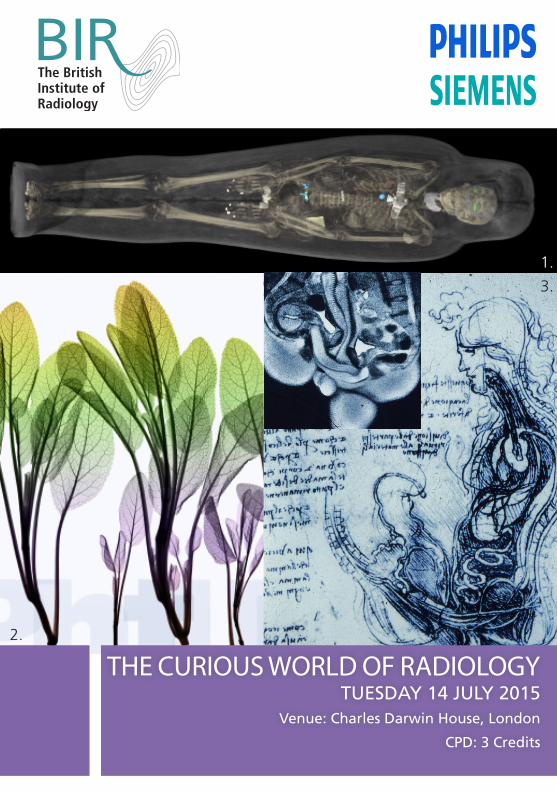
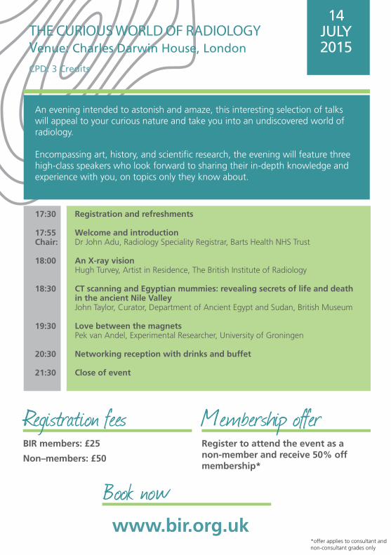
THE CURIOUS WORLD OF RADIOLOGYTUESDAY 14 JULY 2015
Venue: Charles Darwin House, London
CPD: 3 Credits
1.
2.
3.
Registration and refreshments
Welcome and introductionDr John Adu, Radiology Speciality Registrar, Barts Health NHS Trust
An X-ray visionHugh Turvey, Artist in Residence, The British Institute of Radiology
CT scanning and Egyptian mummies: revealing secrets of life and death in the ancient Nile ValleyJohn Taylor, Curator, Department of Ancient Egypt and Sudan, British Museum
Love between the magnetsPek van Andel, Experimental Researcher, University of Groningen
Networking reception with drinks and buffet
Close of event
17:30
17:55Chair:
18:00
18:30
19:30
20:30
21:30
THE CURIOUS WORLD OF RADIOLOGYVenue: Charles Darwin House, London
CPD: 3 Credits
An evening intended to astonish and amaze, this interesting selection of talks will appeal to your curious nature and take you into an undiscovered world of radiology.
Encompassing art, history, and scientific research, the evening will feature three high-class speakers who look forward to sharing their in-depth knowledge and experience with you, on topics only they know about.
14 JULY 2015
BIR members: £25
Non–members: £50
www.bir.org.uk
Book now
Registration fees Membership offerRegister to attend the event as a non-member and receive 50% off membership*
*offer applies to consultant and non-consultant grades only
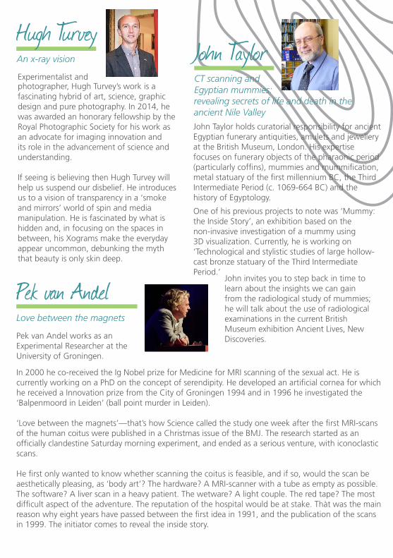
Hugh Turvey An x-ray vision
Pek van Andel Love between the magnets
John Taylor CT scanning and Egyptian mummies:
Pek van Andel works as an Experimental Researcher at the University of Groningen.
In 2000 he co-received the Ig Nobel prize for Medicine for MRI scanning of the sexual act. He is currently working on a PhD on the concept of serendipity. He developed an artificial cornea for which he received a Innovation prize from the City of Groningen 1994 and in 1996 he investigated the ‘Balpenmoord in Leiden’ (ball point murder in Leiden).
‘Love between the magnets’—that’s how Science called the study one week after the first MRI-scans of the human coitus were published in a Christmas issue of the BMJ. The research started as an officially clandestine Saturday morning experiment, and ended as a serious venture, with iconoclastic scans.
He first only wanted to know whether scanning the coitus is feasible, and if so, would the scan be aesthetically pleasing, as ‘body art’? The hardware? A MRI-scanner with a tube as empty as possible. The software? A liver scan in a heavy patient. The wetware? A light couple. The red tape? The most difficult aspect of the adventure. The reputation of the hospital would be at stake. Thàt was the main reason why eight years have passed between the first idea in 1991, and the publication of the scans in 1999. The initiator comes to reveal the inside story.
photographer, Hugh Turvey’s work is a fascinating hybrid of art, science, graphic design and pure photography. In 2014, he was awarded an honorary fellowship by the Royal Photographic Society for his work as an advocate for imaging innovation and its role in the advancement of science and understanding.
If seeing is believing then Hugh Turvey will help us suspend our disbelief. He introduces us to a vision of transparency in a ‘smoke and mirrors’ world of spin and media manipulation. He is fascinated by what is hidden and, in focusing on the spaces in between, his Xograms make the everyday appear uncommon, debunking the myth that beauty is only skin deep.
John Taylor holds curatorial responsibility for ancient Egyptian funerary antiquities, amulets and jewellery at the British Museum, London. His expertise focuses on funerary objects of the pharaonic period (particularly coffins), mummies and mummification, metal statuary of the first millennium BC, the Third Intermediate Period (c. 1069-664 BC) and the history of Egyptology.
One of his previous projects to note was ‘Mummy: the Inside Story’, an exhibition based on the non-invasive investigation of a mummy using 3D visualization. Currently, he is working on ‘Technological and stylistic studies of large hollow-cast bronze statuary of the Third Intermediate Period.’
revealing secrets of life and death in the ancient Nile Valley
John invites you to step back in time to learn about the insights we can gain from the radiological study of mummies; he will talk about the use of radiological examinations in the current British Museum exhibition Ancient Lives, New Discoveries.
Experimentalist and
48–50 St John Street, London, EC1M 4DG
www.bir.org.ukRegistered charity number: 215869
@BIR_News
/britishinstituteofradiology
The British Institute of Radiology
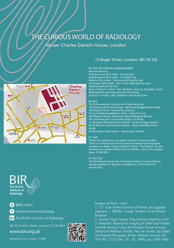
THE CURIOUS WORLD OF RADIOLOGYVenue: Charles Darwin House, London
12 Roger Street, London, WC1N 2JU
BY RAIL OR LONDON UNDERGROUND:Nearest stations: Chancery Lane (0.4 miles) - Central lineRussell Square (0.5 miles) - Piccadilly lineHolborn (0.6 miles) - Central and Piccadilly lineFarringdon (0.8 miles) - Rail, Circle, Metropolitan and Hammersmith & City lineKing’s Cross (1.1 miles) - Rail, Nothern, Victoria, Piccadilly, Circle, Metropolitan and Hammersmith & City lineEuston (1.3 miles) - Rail, Northern and Victoria line
BY BUS:The following bus routes serve Theobalds Road:19 (Finsbury Park Interchange - Battersea Bridge/Hester Road)38 (Clapton Pond – Victoria bus station)55 (Lea Bridge Road/Bakers Arms - Oxford Circus)243 (Redvers Road - Waterloo Station/Mepham Street)The following bus routes serve Gray’s Inn Road:17 (Archway Station/Junction Road - London Bridge Station)45 (St Pancras International Station - Atkins Road/New Park Road)46 (Lancaster Gate Station - Stonecutter Street).
BY CAR:Visitors are advised to use public transport where possible. There is a limited amount of on-street metered parking space available on Roger Street and North Mews. The nearest car park facilities are London Saffron Hill, EC1N 8XA, and 12 Farringdon Road, EC1M 3JB.
BY BICYCLE:Charles Darwin House has a limited number of secure parking spaces available for bicycles, available on a first come first served basis.
Images on front cover:1. ‘CT scan of the mummy of Tamut, an Egyptian priestess, c. 900 BC. Image: Trustees of the British Museum.2. Source: Hugh Turvey: http://www.x-rayartist.com/3. Magnetic Resonance Imaging of Male and Female Genitals during Coitus and Female Sexual Arousal,Willibrord Weijmar Schultz, Pek van Andel, Ida Sabeis and Eduard Mooyaart, British Medical Journal, Vol. 319, No. 7225 (Dec. 18 - 25, 1999), pp. 1596-1600.
Recommended