Learning Objectives✓ How is ATP generated in glycolysis?
✓ Why is the regeneration of NAD� crucial to fermentations?
✓ How is gluconeogenesis powered in the cell?
✓ How are glycolysis and gluconeogenesis coordinated?
We begin our study of metabolism by focusing on the processing of glucose,a fundamental fuel molecule for virtually all life forms. The first metabolic
pathway that we encounter is glycolysis, an ancient pathway employed by a hostof organisms. Glycolysis is the sequence of reactions that converts one moleculeof glucose into two molecules of pyruvate while generating ATP. Glycolysis servestwo major functions in the cell. First, this set of reactions generates ATP. Indeed,some tissues, such as the brain and red blood cells, rely solely on glucose as a fuel;consequently, glycolysis is especially important in these tissues. The second majorfunction of glycolysis is to provide building blocks for biosynthesis. For instance,the molecules formed in the metabolism of glucose in glycolysis are used as pre-cursors for amino acid and fatty acid synthesis.
SECTION
7Glycolysis and Gluconeogenesis
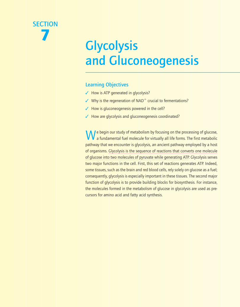
Because glucose is such a precious fuel, the end products of biochemicalpathways are salvaged to synthesize glucose in the process of gluconeogenesis.Gluconeogenesis is vital for ensuring that the brain and red blood cells haveadequate supplies of glucose even during a fast, such as during a night’s sleep.Although glycolysis and gluconeogenesis have some enzymes in common, thetwo pathways are not simply the reverse of each other. In particular, the highlyexergonic, irreversible steps of glycolysis are bypassed in gluconeogenesis. Thetwo pathways are reciprocally regulated so that glycolysis and gluconeogene-sis do not take place simultaneously in the same cell at the same time to asignificant extent, thereby preventing the waste in energy that would resultif glucose were being broken down at the same instant as it is being synthesized.
We start this section with glycolysis, paying special attention to the regulationof this pathway. We proceed to gluconeogenesis, again with a focus on regu-lation. We end this section by noting how glycolysis and gluconeogenesis areregulated within a cell as well as between tissues.
Chapter 15: Glycolysis
Chapter 16: Gluconeogenesis
226
CHAPTER
15 Glycolysis
15.1 Glycolysis Is an Energy-Conversion Pathway
15.2 NAD� Is Regenerated from theMetabolism of Pyruvate
15.3 Fructose and Galactose AreConverted into GlycolyticIntermediates
15.4 The Glycolytic Pathway Is TightlyControlled
15.5 Metabolism in Context:Glycolysis Helps Pancreatic� Cells Sense Glucose
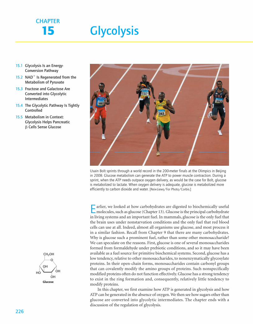
Earlier, we looked at how carbohydrates are digested to biochemically usefulmolecules, such as glucose (Chapter 13). Glucose is the principal carbohydrate
in living systems and an important fuel. In mammals, glucose is the only fuel thatthe brain uses under nonstarvation conditions and the only fuel that red bloodcells can use at all. Indeed, almost all organisms use glucose, and most process itin a similar fashion. Recall from Chapter 9 that there are many carbohydrates.Why is glucose such a prominent fuel, rather than some other monosaccharide?We can speculate on the reasons. First, glucose is one of several monosaccharidesformed from formaldehyde under prebiotic conditions, and so it may have beenavailable as a fuel source for primitive biochemical systems. Second, glucose has alow tendency, relative to other monosaccharides, to nonenzymatically glycosylateproteins. In their open-chain forms, monosaccharides contain carbonyl groupsthat can covalently modify the amino groups of proteins. Such nonspecificallymodified proteins often do not function effectively. Glucose has a strong tendencyto exist in the ring formation and, consequently, relatively little tendency tomodify proteins.
In this chapter, we first examine how ATP is generated in glycolysis and howATP can be generated in the absence of oxygen. We then see how sugars other thanglucose are converted into glycolytic intermediates. The chapter ends with adiscussion of the regulation of glycolysis.
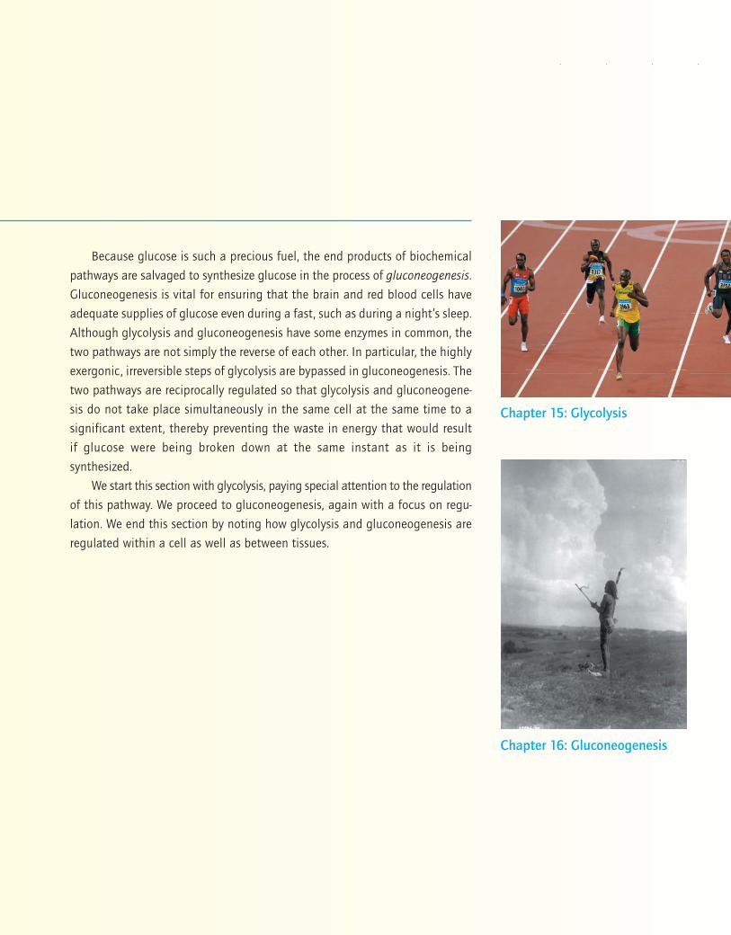
Usain Bolt sprints through a world record in the 200-meter finals at the Olimpics in Beijingin 2008. Glucose metabolism can generate the ATP to power muscle contraction. During asprint, when the ATP needs outpace oxygen delivery, as would be the case for Bolt, glucoseis metabolized to lactate. When oxygen delivery is adequate, glucose is metabolized moreefficiently to carbon dioxide and water. [Reix-Liews/For Photo/Corbis.]
Glucose
O
OH
CH2OH
OH
OH
HO
15.1 Glycolysis Is an Energy-Conversion PathwayWe now begin our consideration of the glycolytic pathway. This pathway iscommon to virtually all cells, both prokaryotic and eukaryotic. In eukaryotic cells,glycolysis takes place in the cytoplasm. Glucose is converted into two molecules ofpyruvate with the concomitant generation of two molecules of ATP.
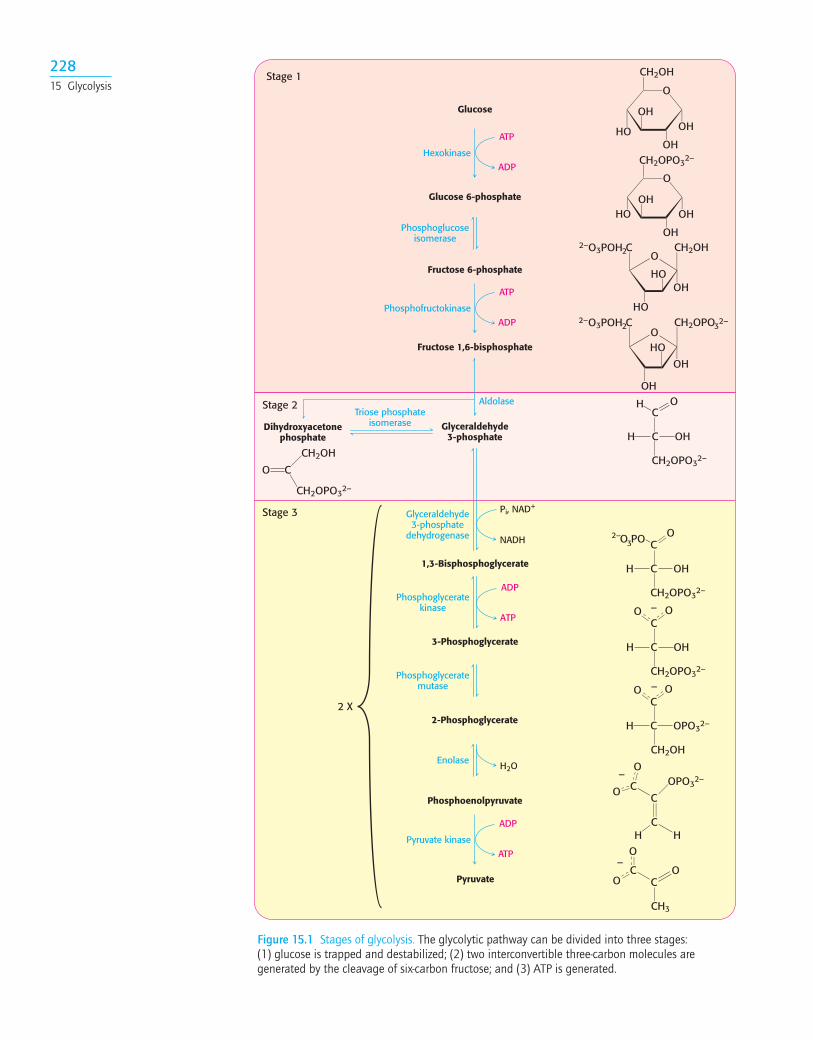
Glycolysis can be thought of as comprising three stages (Figure 15.1). Stage 1,which is the conversion of glucose into fructose 1,6-bisphosphate, consists of threesteps: a phosphorylation, an isomerization, and a second phosphorylation reac-tion. The strategy of these initial steps in glycolysis is to trap the glucose in the cell andform a compound that can be readily cleaved into phosphorylated three-carbon units.Stage 2 is the cleavage of the fructose 1,6-bisphosphate into two three-carbonfragments. These resulting three-carbon units are readily interconvertible. Instage 3,ATP is harvested when the three-carbon fragments are oxidized to pyruvate.
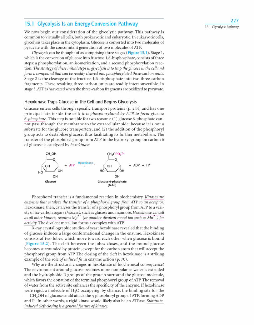
Hexokinase Traps Glucose in the Cell and Begins GlycolysisGlucose enters cells through specific transport proteins (p. 244) and has oneprincipal fate inside the cell: it is phosphorylated by ATP to form glucose6-phosphate. This step is notable for two reasons: (1) glucose 6-phosphate can-not pass through the membrane to the extracellular side, because it is not asubstrate for the glucose transporters, and (2) the addition of the phosphorylgroup acts to destabilize glucose, thus facilitating its further metabolism. Thetransfer of the phosphoryl group from ATP to the hydroxyl group on carbon 6of glucose is catalyzed by hexokinase.
22715.1 Glycolytic Pathway
+ ATP + ADP H++Hexokinase
Glucose Glucose 6-phosphate(G-6P)
O
OH
CH2OH
OH
OH
HO
O
OH
CH2OPO32–
OH
OHHO
Phosphoryl transfer is a fundamental reaction in biochemistry. Kinases areenzymes that catalyze the transfer of a phosphoryl group from ATP to an acceptor.Hexokinase, then, catalyzes the transfer of a phosphoryl group from ATP to a vari-ety of six-carbon sugars (hexoses), such as glucose and mannose. Hexokinase, as wellas all other kinases, requires Mg2� (or another divalent metal ion such as Mn2�) foractivity. The divalent metal ion forms a complex with ATP.
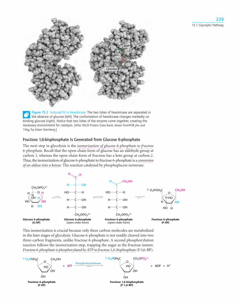
X-ray crystallographic studies of yeast hexokinase revealed that the bindingof glucose induces a large conformational change in the enzyme. Hexokinaseconsists of two lobes, which move toward each other when glucose is bound(Figure 15.2). The cleft between the lobes closes, and the bound glucosebecomes surrounded by protein, except for the carbon atom that will accept thephosphoryl group from ATP. The closing of the cleft in hexokinase is a strikingexample of the role of induced fit in enzyme action (p. 70).
Why are the structural changes in hexokinase of biochemical consequence?The environment around glucose becomes more nonpolar as water is extrudedand the hydrophobic R groups of the protein surround the glucose molecule,which favors the donation of the terminal phosphoryl group of ATP. The removalof water from the active site enhances the specificity of the enzyme. If hexokinasewere rigid, a molecule of H2O occupying, by chance, the binding site for the
CH2OH of glucose could attack the � phosphoryl group of ATP, forming ADPand Pi. In other words, a rigid kinase would likely also be an ATPase. Substrate-induced cleft closing is a general feature of kinases.
¬
22815 Glycolysis
Glucose
Glucose 6-phosphate
Fructose 6-phosphate
Fructose 1,6-bisphosphate
Dihydroxyacetonephosphate
Glyceraldehyde3-phosphate
1,3-Bisphosphoglycerate
3-Phosphoglycerate
2-Phosphoglycerate
Phosphoenolpyruvate
Pyruvate
2 X
Aldolase
Stage 1
Stage 2
Stage 3
O
OH
CH2OH
OH
OH
HO
O
OH
CH2OPO32–
OH
OHHO
OCH2OH
HO
HO
2–O3POH C2
OH
32–
OCH2OPO
HO
OH
2–O3POH C2
OH
C
C
CH2OPO32–
OHH
OO –
C
C OPO32–H
OO –
CH2OH
OPO32–
H H
O
O
–
CH3
OO
O–
CO
CH2OPO32–
CH2OH
C
C
CH2OPO32–
OHH
O PO32– O
C
C
CH2OPO32–
OHH
OH
ATP
ADPHexokinase
Phosphofructokinase
ATP
ADP
Pyruvate kinase
ADP
ATP
Phosphoglucoseisomerase
Triose phosphateisomerase
Pi, NAD+
NADH
Glyceraldehyde3-phosphate
dehydrogenase
Phosphoglyceratekinase
ADP
ATP
Phosphoglyceratemutase
H2OEnolase
C
CC
C
C
Figure 15.1 Stages of glycolysis. The glycolytic pathway can be divided into three stages:(1) glucose is trapped and destabilized; (2) two interconvertible three-carbon molecules aregenerated by the cleavage of six-carbon fructose; and (3) ATP is generated.
Fructose 1,6-bisphosphate Is Generated from Glucose 6-phosphateThe next step in glycolysis is the isomerization of glucose 6-phosphate to fructose6-phosphate. Recall that the open-chain form of glucose has an aldehyde group atcarbon 1, whereas the open-chain form of fructose has a keto group at carbon 2.Thus, the isomerization of glucose 6-phosphate to fructose 6-phosphate is a conversionof an aldose into a ketose. The reaction catalyzed by phosphoglucose isomerase.
22915.1 Glycolytic Pathway
Glucose 6-phosphate(G-6P)
Glucose 6-phosphate(open-chain form)
Fructose 6-phosphate(open-chain form)
Fructose 6-phosphate(F-6P)
O H
OH
CH2OPO32–
H
OH
OH
H
H
HO
H
C
C
C
C
C
CH2OPO32–
O H
OHH
HHO
OHH
OHH
C
C
C
C
CH2OPO32–
HHO
OHH
OHH
O CH2OH
OCH2OH
HO
HO
2–O3POH C2
H
HH OH
+ ATP + H++ADPPhosphofructokinase
Fructose 6-phosphate(F-6P)
Fructose 1,6-bisphosphate(F-1,6-BP)
OCH2OH
HO
2–O3POH C2
OH
32–
OCH2OPO
HO
OHOH
2–O3POH C2
OH
Glucose
ADP
Figure 15.2 Induced fit in hexokinase. The two lobes of hexokinase are separated inthe absence of glucose (left). The conformation of hexokinase changes markedly on
binding glucose (right). Notice that two lobes of the enzyme come together, creating thenecessary environment for catalysis. [After RSCB Protein Data Bank; drawn fromPDB yhx and1hkg/by Adam Steinberg.]
This isomerization is crucial because only three carbon molecules are metabolizedin the later stages of glycolysis. Glucose 6-phosphate is not readily cleaved into twothree-carbon fragments, unlike fructose 6-phosphate. A second phosphorylationreaction follows the isomerization step, trapping the sugar as the fructose isomer.Fructose 6-phosphate is phosphorylated by ATP to fructose 1,6-bisphosphate (F-l,6-BP).
This reaction is catalyzed by phosphofructokinase (PFK), an allosteric enzyme thatsets the pace of glycolysis (p. 241). As we will learn, this enzyme is the key regu-latory enzyme for glycolysis.
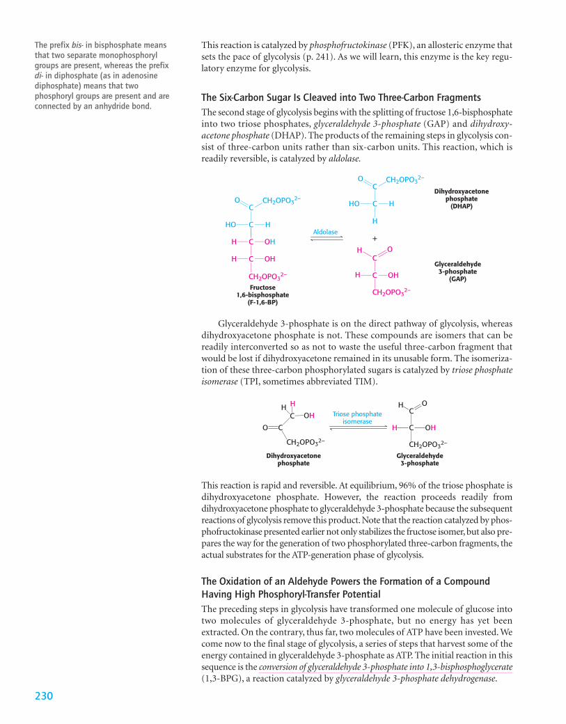
The Six-Carbon Sugar Is Cleaved into Two Three-Carbon FragmentsThe second stage of glycolysis begins with the splitting of fructose 1,6-bisphosphateinto two triose phosphates, glyceraldehyde 3-phosphate (GAP) and dihydroxy-acetone phosphate (DHAP). The products of the remaining steps in glycolysis con-sist of three-carbon units rather than six-carbon units. This reaction, which isreadily reversible, is catalyzed by aldolase.
The prefix bis- in bisphosphate meansthat two separate monophosphorylgroups are present, whereas the prefixdi- in diphosphate (as in adenosinediphosphate) means that twophosphoryl groups are present and areconnected by an anhydride bond.
230
+Aldolase
Fructose1,6-bisphosphate
(F-1,6-BP)
Dihydroxyacetonephosphate
(DHAP)
Glyceraldehyde3-phosphate
(GAP)
C
C
C
C
HHO
OHH
OHH
O CH2OPO32–
CH2OPO32–
CH2OPO32–
CH2OPO32–
C
C HHO
O
H
C
C OHH
OH
Triose phosphateisomerase
Dihydroxyacetonephosphate
Glyceraldehyde3-phosphate
C
C
O
CH2OPO32–
H H
OH C
C
CH2OPO32–
OHH
OH
Glyceraldehyde 3-phosphate is on the direct pathway of glycolysis, whereasdihydroxyacetone phosphate is not. These compounds are isomers that can bereadily interconverted so as not to waste the useful three-carbon fragment thatwould be lost if dihydroxyacetone remained in its unusable form. The isomeriza-tion of these three-carbon phosphorylated sugars is catalyzed by triose phosphateisomerase (TPI, sometimes abbreviated TIM).
This reaction is rapid and reversible. At equilibrium, 96% of the triose phosphate isdihydroxyacetone phosphate. However, the reaction proceeds readily fromdihydroxyacetone phosphate to glyceraldehyde 3-phosphate because the subsequentreactions of glycolysis remove this product. Note that the reaction catalyzed by phos-phofructokinase presented earlier not only stabilizes the fructose isomer, but also pre-pares the way for the generation of two phosphorylated three-carbon fragments, theactual substrates for the ATP-generation phase of glycolysis.
The Oxidation of an Aldehyde Powers the Formation of a CompoundHaving High Phosphoryl-Transfer PotentialThe preceding steps in glycolysis have transformed one molecule of glucose intotwo molecules of glyceraldehyde 3-phosphate, but no energy has yet beenextracted. On the contrary, thus far, two molecules of ATP have been invested. Wecome now to the final stage of glycolysis, a series of steps that harvest some of theenergy contained in glyceraldehyde 3-phosphate as ATP. The initial reaction in thissequence is the conversion of glyceraldehyde 3-phosphate into 1,3-bisphosphoglycerate(1,3-BPG), a reaction catalyzed by glyceraldehyde 3-phosphate dehydrogenase.
23115.1 Glycolytic Pathway
+ Pi+NAD+ + +NADH H+
Glyceraldehyde3-phosphate
(GAP)
1,3-Bisphosphoglycerate(1,3-BPG)
Glyceraldehyde3-phosphate
dehydrogenaseC
C
CH2OPO32–
OHH
H OC
C
CH2OPO32–
OHH
O PO32– O
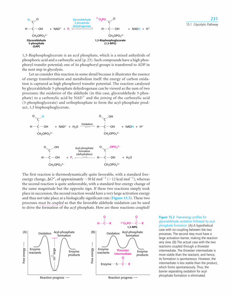
1,3-Bisphosphoglycerate is an acyl phosphate, which is a mixed anhydride ofphosphoric acid and a carboxylic acid (p. 23). Such compounds have a high phos-phoryl-transfer potential; one of its phosphoryl groups is transferred to ADP inthe next step in glycolysis.
Let us consider this reaction in some detail because it illustrates the essenceof energy transformation and metabolism itself: the energy of carbon oxida-tion is captured as high phosphoryl transfer potential. The reaction catalyzedby glyceraldehyde 3-phosphate dehydrogenase can be viewed as the sum of twoprocesses: the oxidation of the aldehyde (in this case, glyceraldehyde 3-phos-phate) to a carboxylic acid by NAD� and the joining of the carboxylic acid (3-phosphoglycerate) and orthophosphate to form the acyl-phosphate prod-uct, 1,3 bisphosphoglycerate.
+ H2O+NAD+ + H++NADHOxidation
C
C
CH2OPO32–
OHH
HOC
C
CH2OPO32–
OHH
O OH
+ Pi + H2O
Acyl-phosphateformation
(dehydration)C
C
CH2OPO32–
OHH
O OPO32–
C
C
CH2OPO32–
OH
OH
H
O
The first reaction is thermodynamically quite favorable, with a standard free-energy change, �G°�, of approximately �50 kJ mol�1 (�12 kcal mol�1), whereasthe second reaction is quite unfavorable, with a standard free-energy change ofthe same magnitude but the opposite sign. If these two reactions simply tookplace in succession, the second reaction would have a very large activation energyand thus not take place at a biologically significant rate (Figure 15.3). These twoprocesses must be coupled so that the favorable aldehyde oxidation can be usedto drive the formation of the acyl phosphate. How are these reactions coupled?
H C R
O O
2–O3PO C R
O
CEnzyme S R
Reaction progress
Oxidation(A)
Enzymereactants Enzyme
products
ΔG++
larg
e
Acyl-phosphateformation
Free
ene
rgy
Reaction progress
Oxidation(B)
Enzymereactants Enzyme
productsThioester
intermediate
Acyl-phosphateformation
Free
ene
rgy
1,3-BPG
Figure 15.3 Free-energy profiles forglyceraldehyde oxidation followed by acyl-phosphate formation. (A) A hypotheticalcase with no coupling between the twoprocesses. The second step must have alarge activation barrier, making the reactionvery slow. (B) The actual case with the tworeactions coupled through a thioesterintermediate. The thioester intermediate ismore stable than the reactant, and hence,its formation is spontaneous. However, theintermediate is less stable than the product,which forms spontaneously. Thus, thebarrier separating oxidation for acyl-phosphate formation is eliminated.

23215 Glycolysis
+ ATP+ ADP + H+
Phosphoglyceratekinase
1,3-Bisphosphoglycerate 3-Phosphoglycerate
C
C
CH2OPO32–
OHH
O OPO32–
C
C
CH2OPO32–
OHH
OO –
Phosphoglyceratemutase
3-Phosphoglycerate 2-Phosphoglycerate
H2O
Enolase
Phosphenolpyruvate Pyruvate
ADP+ H+ ATP
Pyruvatekinase
C
C
C
OHH
OO
H
H
OPO32–
–
C
C
C
OPO32–H
OO
H
H
OH
–
OPO32–
H H
O
O
–
CH3
OO
O–
C
CCC
C
The key is an intermediate that is linked to the enzyme by a thioester bond after thealdehyde has been oxidized. This intermediate reacts with orthophosphate toform the high-energy compound 1,3-bisphosphoglycerate. The thioester inter-mediate is a free-energy intermediate between the aldehyde and the free carboxylicacid. The favorable oxidation and unfavorable phosphorylation reactions arecoupled by the thioester intermediate, which preserves much of the free energyreleased in the oxidation reaction (see Figure 15.3B).
ATP Is Formed by Phosphoryl Transfer from 1,3-Bisphosphoglycerate1,3-Bisphosphoglycerate is an energy-rich molecule with a greater phosphoryl-transfer potential than that of ATP (p. 206). Thus, 1,3-BPG can be used to powerthe synthesis of ATP from ADP and orthophosphate. Phosphoglycerate kinasecatalyzes the transfer of the phosphoryl group from the acyl phosphate of1,3-bisphosphoglycerate to ADP. ATP and 3-phosphoglycerate are the products.
The formation of ATP in this manner is referred to as substrate-level phosphoryla-tion because the phosphate donor, 1,3-BPG, is a kinase substrate with highphosphoryl-transfer potential. We will contrast this manner of ATP formationwith the formation of ATP from ionic gradients in Chapters 19 and 20.
Thus, we now start to see a return on our initial investment of two moleculesof ATP in stage 1. Going backward in the pathway one step to glyceraldehyde3-phosphate, we find the outcomes of the reactions catalyzed by glyceraldehyde3-phosphate dehydrogenase and phosphoglycerate kinase to be as follows:
1. Glyceraldehyde 3-phosphate, an aldehyde, is oxidized to 3-phosphoglycerate,a carboxylic acid.
2. NAD� is concomitantly reduced to NADH.
3. ATP is formed from Pi and ADP at the expense of carbon-oxidation energy.
In essence, the energy released in the oxidation of glyceraldehyde 3-phosphate to3-phosphoglycerate is temporarily trapped as 1,3-bisphosphoglycerate. Thisenergy powers the transfer of a phosphoryl group from 1,3-bisphosphoglycerateto ADP to yield ATP. Keep in mind that, because of the actions of aldolase andtriose phosphate isomerase on fructose 1,6-bisphosphate at the end of stage 1, twomolecules of glyceraldehyde 3-phosphate were formed and, hence, two moleculesof ATP were generated. These ATP molecules make up for the two molecules ofATP consumed in the first stage of glycolysis.
Additional ATP Is Generated with the Formation of PyruvateIn the remaining steps of glycolysis, 3-phosphoglycerate is converted into pyru-vate, and a second molecule of ATP is formed from ADP.
The first reaction is a rearrangement. 3-phosphoglycerate is converted into2-phosphoglycerate by phosphoglycerate mutase, which shifts the position of thephosphoryl group. In general, a mutase is an enzyme that catalyzes the intramol-ecular shift of a chemical group, such as a phosphoryl group.
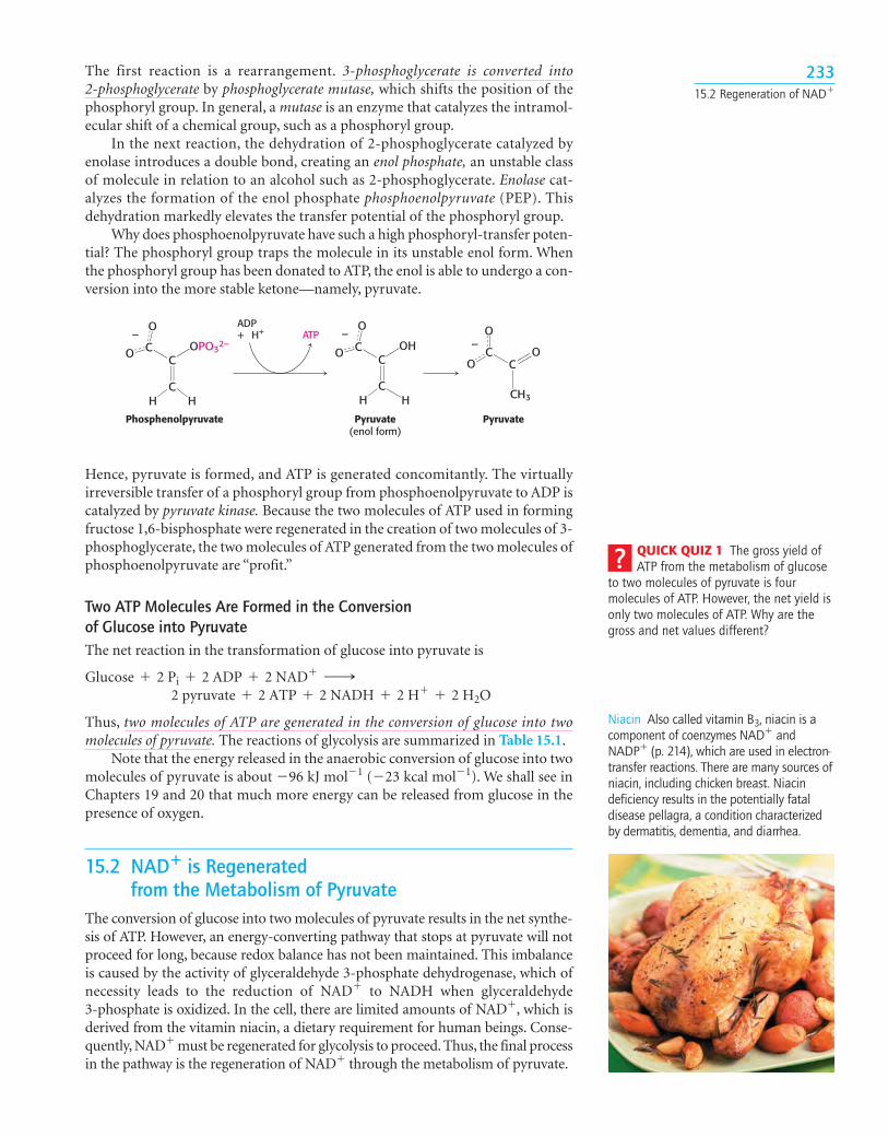
In the next reaction, the dehydration of 2-phosphoglycerate catalyzed byenolase introduces a double bond, creating an enol phosphate, an unstable classof molecule in relation to an alcohol such as 2-phosphoglycerate. Enolase cat-alyzes the formation of the enol phosphate phosphoenolpyruvate (PEP). Thisdehydration markedly elevates the transfer potential of the phosphoryl group.
Why does phosphoenolpyruvate have such a high phosphoryl-transfer poten-tial? The phosphoryl group traps the molecule in its unstable enol form. Whenthe phosphoryl group has been donated to ATP, the enol is able to undergo a con-version into the more stable ketone—namely, pyruvate.
23315.2 Regeneration of NAD�
Phosphenolpyruvate PyruvatePyruvate(enol form)
OPO32–
H H CH3
OOH
H H
ADP+ H+ ATP
CC
O
O
–
CC
O
O
–
CC
O
O–
CC
Hence, pyruvate is formed, and ATP is generated concomitantly. The virtuallyirreversible transfer of a phosphoryl group from phosphoenolpyruvate to ADP iscatalyzed by pyruvate kinase. Because the two molecules of ATP used in formingfructose 1,6-bisphosphate were regenerated in the creation of two molecules of 3-phosphoglycerate, the two molecules of ATP generated from the two molecules ofphosphoenolpyruvate are “profit.”
Two ATP Molecules Are Formed in the Conversion of Glucose into PyruvateThe net reaction in the transformation of glucose into pyruvate is
Thus, two molecules of ATP are generated in the conversion of glucose into twomolecules of pyruvate. The reactions of glycolysis are summarized in Table 15.1.
Note that the energy released in the anaerobic conversion of glucose into twomolecules of pyruvate is about �96 kJ mol�1 (�23 kcal mol�1). We shall see inChapters 19 and 20 that much more energy can be released from glucose in thepresence of oxygen.
15.2 NAD� is Regenerated from the Metabolism of Pyruvate
The conversion of glucose into two molecules of pyruvate results in the net synthe-sis of ATP. However, an energy-converting pathway that stops at pyruvate will notproceed for long, because redox balance has not been maintained. This imbalanceis caused by the activity of glyceraldehyde 3-phosphate dehydrogenase, which ofnecessity leads to the reduction of NAD� to NADH when glyceraldehyde3-phosphate is oxidized. In the cell, there are limited amounts of NAD�, which isderived from the vitamin niacin, a dietary requirement for human beings. Conse-quently, NAD� must be regenerated for glycolysis to proceed. Thus, the final processin the pathway is the regeneration of NAD� through the metabolism of pyruvate.
2 pyruvate + 2 ATP + 2 NADH + 2 H+ + 2 H2OGlucose + 2 Pi + 2 ADP + 2 NAD+ ¡
QUICK QUIZ 1 The gross yield ofATP from the metabolism of glucose
to two molecules of pyruvate is fourmolecules of ATP. However, the net yield isonly two molecules of ATP. Why are thegross and net values different?
Niacin Also called vitamin B3, niacin is acomponent of coenzymes NAD� andNADP� (p. 214), which are used in electron-transfer reactions. There are many sources ofniacin, including chicken breast. Niacindeficiency results in the potentially fataldisease pellagra, a condition characterizedby dermatitis, dementia, and diarrhea.
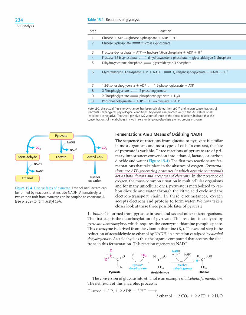
Fermentations Are a Means of Oxidizing NADHThe sequence of reactions from glucose to pyruvate is similarin most organisms and most types of cells. In contrast, the fateof pyruvate is variable. Three reactions of pyruvate are of pri-mary importance: conversion into ethanol, lactate, or carbondioxide and water (Figure 15.4) The first two reactions are fer-mentations that take place in the absence of oxygen. Fermenta-tions are ATP-generating processes in which organic compoundsact as both donors and acceptors of electrons. In the presence ofoxygen, the most-common situation in multicellular organismsand for many unicellular ones, pyruvate is metabolized to car-bon dioxide and water through the citric acid cycle and theelectron-transport chain. In these circumstances, oxygenaccepts electrons and protons to form water. We now take acloser look at these three possible fates of pyruvate.
1. Ethanol is formed from pyruvate in yeast and several other microorganisms.The first step is the decarboxylation of pyruvate. This reaction is catalyzed bypyruvate decarboxylase, which requires the coenzyme thiamine pyrophosphate.This coenzyme is derived from the vitamin thiamine (B1). The second step is thereduction of acetaldehyde to ethanol by NADH, in a reaction catalyzed by alcoholdehydrogenase. Acetaldehyde is thus the organic compound that accepts the elec-trons in this fermentation. This reaction regenerates NAD�.
CO2 CO2
NADH
NAD+
NADH
NAD+
Furtheroxidation
Pyruvate
LactateAcetaldehyde
Ethanol
Acetyl CoA
Figure 15.4 Diverse fates of pyruvate. Ethanol and lactate canbe formed by reactions that include NADH. Alternatively, atwo-carbon unit from pyruvate can be coupled to coenzyme A(see p. 269) to form acetyl CoA.
Table 15.1 Reactions of glycolysis
Step Reaction
1 Glucose � ATP S glucose 6-phosphate � ADP � H�
2 Glucose 6-phosphate fructose 6-phosphate
3 Fructose 6-phosphate � ATP S fructose 1,6-bisphosphate � ADP � H�
4 Fructose 1,6-bisphosphate dihydroxyacetone phosphate � glyceraldehyde 3-phosphate5 Dihydroxyacetone phosphate glyceraldehyde 3-phosphate
6 Glyceraldehyde 3-phosphate � Pi � NAD� 1,3-bisphosphoglycerate � NADH � H�
7 1,3-Bisphosphoglycerate � ADP 3-phosphoglycerate � ATP8 3-Phosphoglycerate 2-phosphoglycerate9 2-Phosphoglycerate phosphoenolpyruvate � H2O
10 Phosphoenolpyruvate � ADP � H� S pyruvate � ATP
Note: �G, the actual free-energy change, has been calculated from �G°� and known concentrations ofreactants under typical physiological conditions. Glycolysis can proceed only if the �G values of allreactions are negative. The small positive �G values of three of the above reactions indicate that theconcentrations of metabolities in vivo in cells undergoing glycolysis are not precisely known.
ΔΔ
Δ
Δ
ΔΔ
Δ
H+ CO2 + H+NADH
NAD+
Pyruvate Acetaldehyde
Pyruvatedecarboxylase
Alcoholdehydrogenase
Ethanol
CH3
H O
CH3
H OHH
CH3
OC CO
O–
CC
The conversion of glucose into ethanol is an example of alcoholic fermentation.The net result of this anaerobic process is
2 ethanol + 2 CO2 + 2 ATP + 2 H2OGlucose + 2 Pi + 2 ADP + 2 H+ ¡
23415 Glycolysis
2. Lactate is formed from pyruvate in a variety of microorganisms in a processcalled lactic acid fermentation. The reaction also takes place in the cells of higherorganisms when the amount of oxygen is limiting, as in skeletal-muscle cells dur-ing intense activity. Pyruvate accepts the electrons from NADH to form lactate ina reaction catalyzed by lactate dehydrogenase.
235
�G°� in kJ mol�1 �G in kJ mol�1
Enzyme Reaction type (kcal mol�1) (kcal mol�1)
Hexokinase Phosphoryl transfer �16.7 (�4.0) �33.5 (�8.0)Phosphoglucose Isomerization �1.7 (�0.4) �2.5 (�0.6)isomerasePhosphofructokinase Phosphoryl transfer �14.2 (�3.4) �22.2 (�5.3)Aldolase Aldol cleavage �23.8 (�5.7) �1.3 (�0.3)Triose phosphate Isomerization �7.5 (�1.8) �2.5 (�0.6)isomeraseGlyceraldehyde Phosphorylation �6.3 (�1.5) �1.7 (�0.4)3-phosphate coupleddehydrogenase to oxidationPhosphoglycerate kinase Phosphoryl transfer �18.8 (�4.5) �1.3 (�0.3)Phosphoglycerate mutase Phosphoryl shift �4.6 (�1.1) �0.8 (�0.2)Enolase Dehydration �1.7 (�0.4) �3.3 (�0.8)Pyruvate kinase Phosphoryl transfer �31.4 (�7.5) �16.7 (�4.0)
AU: Add credit
Glyceraldehyde3-phosphate
1,3-Bisphosphoglycerate(1,3-BPG)
Glyceraldehyde3-phosphate
dehydrogenase
H+ CO2 NAD+
Pyruvate Acetaldehyde Ethanol
Alcoholdehydrogenase
NADH+ H+
NADH+ H+NAD+PiC
C
CH2OPO32–
OHH
HOC
C
CH2OPO32–
OHH
OPO32–O
CH3
OO
O–
CH3
H
CH3
H OHH
CC
OC C
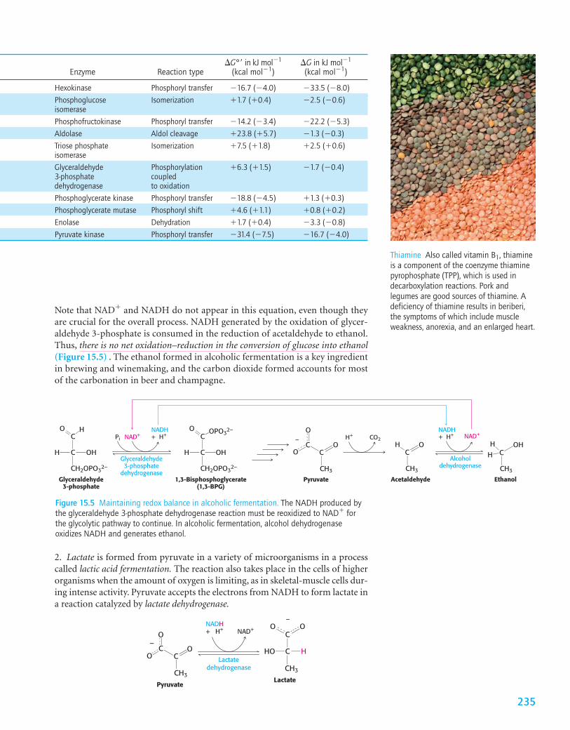
Figure 15.5 Maintaining redox balance in alcoholic fermentation. The NADH produced bythe glyceraldehyde 3-phosphate dehydrogenase reaction must be reoxidized to NAD� forthe glycolytic pathway to continue. In alcoholic fermentation, alcohol dehydrogenaseoxidizes NADH and generates ethanol.
PyruvateLactate
Lactatedehydrogenase
CH3
O
CH3
C
C HHO
OO–
+ H+NADH
NAD+
O
O–
CC
Note that NAD� and NADH do not appear in this equation, even though theyare crucial for the overall process. NADH generated by the oxidation of glycer-aldehyde 3-phosphate is consumed in the reduction of acetaldehyde to ethanol.Thus, there is no net oxidation–reduction in the conversion of glucose into ethanol(Figure 15.5) . The ethanol formed in alcoholic fermentation is a key ingredientin brewing and winemaking, and the carbon dioxide formed accounts for mostof the carbonation in beer and champagne.
Thiamine Also called vitamin B1, thiamineis a component of the coenzyme thiaminepyrophosphate (TPP), which is used indecarboxylation reactions. Pork andlegumes are good sources of thiamine. Adeficiency of thiamine results in beriberi,the symptoms of which include muscleweakness, anorexia, and an enlarged heart.
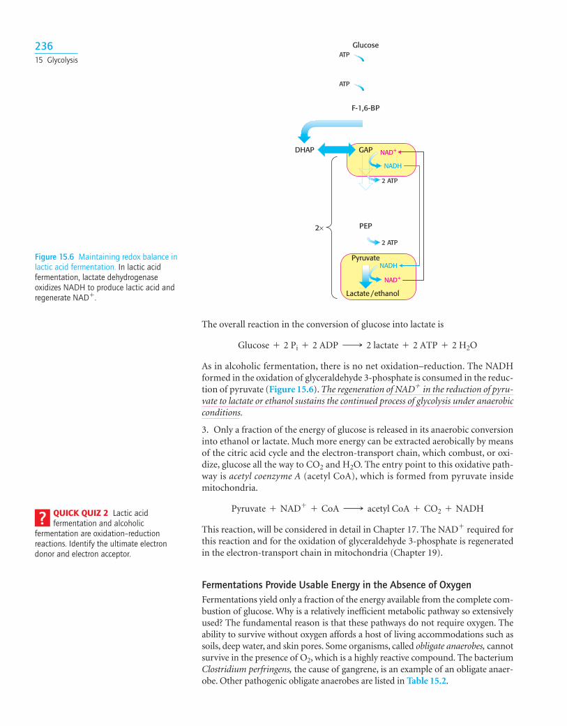
The overall reaction in the conversion of glucose into lactate is
As in alcoholic fermentation, there is no net oxidation–reduction. The NADHformed in the oxidation of glyceraldehyde 3-phosphate is consumed in the reduc-tion of pyruvate (Figure 15.6). The regeneration of NAD� in the reduction of pyru-vate to lactate or ethanol sustains the continued process of glycolysis under anaerobicconditions.
3. Only a fraction of the energy of glucose is released in its anaerobic conversioninto ethanol or lactate. Much more energy can be extracted aerobically by meansof the citric acid cycle and the electron-transport chain, which combust, or oxi-dize, glucose all the way to CO2 and H2O. The entry point to this oxidative path-way is acetyl coenzyme A (acetyl CoA), which is formed from pyruvate insidemitochondria.
This reaction, will be considered in detail in Chapter 17. The NAD� required forthis reaction and for the oxidation of glyceraldehyde 3-phosphate is regeneratedin the electron-transport chain in mitochondria (Chapter 19).
Fermentations Provide Usable Energy in the Absence of OxygenFermentations yield only a fraction of the energy available from the complete com-bustion of glucose. Why is a relatively inefficient metabolic pathway so extensivelyused? The fundamental reason is that these pathways do not require oxygen. Theability to survive without oxygen affords a host of living accommodations such assoils, deep water, and skin pores. Some organisms, called obligate anaerobes, cannotsurvive in the presence of O2, which is a highly reactive compound. The bacteriumClostridium perfringens, the cause of gangrene, is an example of an obligate anaer-obe. Other pathogenic obligate anaerobes are listed in Table 15.2.
Pyruvate + NAD+ + CoA ¡ acetyl CoA + CO2 + NADH
Glucose + 2 Pi + 2 ADP ¡ 2 lactate + 2 ATP + 2 H2O
QUICK QUIZ 2 Lactic acidfermentation and alcoholic
fermentation are oxidation–reductionreactions. Identify the ultimate electrondonor and electron acceptor.
Figure 15.6 Maintaining redox balance inlactic acid fermentation. In lactic acidfermentation, lactate dehydrogenaseoxidizes NADH to produce lactic acid andregenerate NAD�.
Glucose
F-1,6-BP
NADH
NAD+
NADH
NAD+
2 ATP
2 ATP
PEP
Pyruvate
Lactate /ethanol
ATP
ATP
DHAP GAP
2×
23615 Glycolysis
Skeletal muscles in most animals can function anaerobically for short peri-ods. For example, when animals perform bursts of intense exercise, their ATPneeds rise faster than the ability of the body to provide oxygen to the muscle. Themuscle functions anaerobically until fatigue sets in, which is caused, in part, bylactate buildup. The burning sensation in muscles that occurs during an intensebout of exercise is due to lactic acid accumulation.
Although we have considered only lactic acid and alcoholic fermentation,microorganisms are capable of generating a wide array of molecules as endpoints to fermentation (Table 15.3). Indeed, many food products, includingsour cream, yogurt, various cheeses, beer, wine, and sauerkraut, result fromfermentation.
15.3 Fructose and Galactose Are Converted into Glycolytic Intermediates
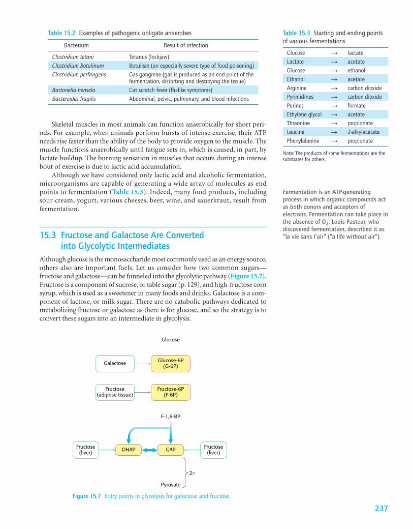
Although glucose is the monosaccharide most commonly used as an energy source,others also are important fuels. Let us consider how two common sugars—fructose and galactose—can be funneled into the glycolytic pathway (Figure 15.7).Fructose is a component of sucrose, or table sugar (p. 129), and high-fructose cornsyrup, which is used as a sweetener in many foods and drinks. Galactose is a com-ponent of lactose, or milk sugar. There are no catabolic pathways dedicated tometabolizing fructose or galactose as there is for glucose, and so the strategy is toconvert these sugars into an intermediate in glycolysis.
Fermentation is an ATP-generatingprocess in which organic compounds actas both donors and acceptors ofelectrons. Fermentation can take place inthe absence of O2. Louis Pasteur, whodiscovered fermentation, described it as“la vie sans l’air” (“a life without air”).
Table 15.2 Examples of pathogenic obligate anaerobes
Bacterium Result of infection
Clostridium tetani Tetanus (lockjaw)Clostridium botulinum Botulism (an especially severe type of food poisoning)Clostridium perfringens Gas gangrene (gas is produced as an end point of the
fermentation, distorting and destroying the tissue)Bartonella hensela Cat scratch fever (flu-like symptoms)Bacteroides fragilis Abdominal, pelvic, pulmonary, and blood infections
237
Table 15.3 Starting and ending pointsof various fermentations
Glucose S lactateLactate S acetateGlucose S ethanolEthanol S acetateArginine S carbon dioxidePyrimidines S carbon dioxidePurines S formateEthylene glycol S acetateThreonine S propionateLeucine S 2-alkylacetatePhenylalanine S propionate
Note: The products of some fermentations are thesubstrates for others.
Fructose-6P(F-6P)
Fructose(liver)
Fructose(liver)
Fructose(adipose tissue)
Galactose
F-1,6-BP
Glucose-6P(G-6P)
Glucose
GAP
Pyruvate
2×
DHAP
Figure 15.7 Entry points in glycolysis for galactose and fructose.
Galactose 1-phosphate then acquires a uridyl group from uridine diphosphate glu-cose (UDP-glucose), which activates the sugar phosphate so that it can be con-verted into glucose (Figure 15.9) UDP-monosaccharides are another example ofan activated intermediate (p. 129) and are formed as an intermediate in the
Galactose Galactose 1-phosphate
Galactokinase
ATP H++ADP
O
OH
CH2OH
OH
OH
HO
OP
O
OO
O
CH2OH
OH
OH
HO
2–
Figure 15.9 Galactose metabolism.Galactose 1-phosphate reacts withactivated glucose (UDP-glucose) to formUDP-galactose, which is subsequentlyconverted into UDP-glucose.
Galactose1-phosphate
UDP-glucose
uridine+
Galactose 1-phosphateuridyl transferase
UDP-galactose4-epimerase
O
O
CH2OH
OH
OH
HO O
O
CH2OH
OH
OHHO
PO
PO
O O O O
PO
O O
2–
uridine
UDP-galactose
+P
O
O O
O
O
CH2OH
OH
OH
HO
PO
O O– –
uridine
UDP-glucose
PO
O O
O
O
CH2OH
OH
OH
PO
O O– –
HO
Glucose1-phosphate
O
O
CH2OH
OH
OHHO
PO
O O
2–
– –
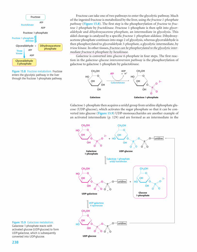
Fructose can take one of two pathways to enter the glycolytic pathway. Muchof the ingested fructose is metabolized by the liver, using the fructose 1-phosphatepathway (Figure 15.8). The first step is the phosphorylation of fructose to fruc-tose 1-phosphate by fructokinase. Fructose 1-phosphate is then split into glycer-aldehyde and dihydroxyacetone phosphate, an intermediate in glycolysis. Thisaldol cleavage is catalyzed by a specific fructose 1-phosphate aldolase. Dihydroxy-acetone phosphate continues into stage 2 of glycolysis, whereas glyceraldehyde isthen phosphorylated to glyceraldehyde 3-phosphate, a glycolytic intermediate, bytriose kinase. In other tissues, fructose can be phosphorylated to the glycolytic inter-mediate fructose 6-phosphate by hexokinase.
Galactose is converted into glucose 6-phosphate in four steps. The first reac-tion in the galactose–glucose interconversion pathway is the phosphorylation ofgalactose to galactose 1-phosphate by galactokinase.
Fructose
Fructose 1-phosphate
Glyceraldehyde + Dihydroxyacetonephosphate
Glyceraldehyde3-phosphate
Fructose 1-phosphatealdolase
FructokinaseATP
ADP
Triosekinase
ATP
ADP
Figure 15.8 Fructose metabolism. Fructoseenters the glycolytic pathway in the liverthrough the fructose 1-phosphate pathway.
238
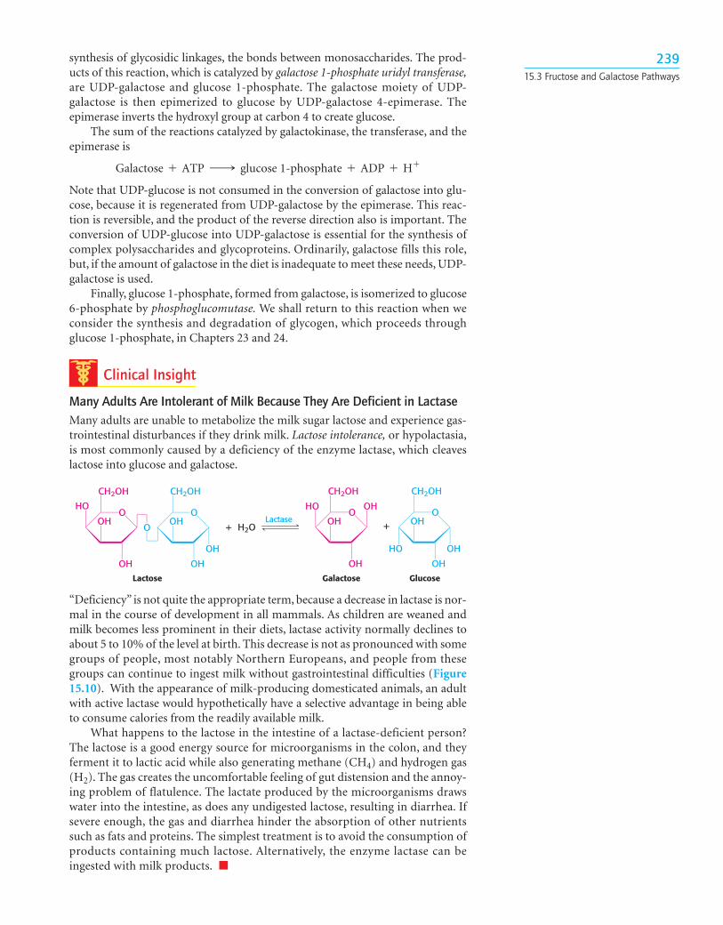
synthesis of glycosidic linkages, the bonds between monosaccharides. The prod-ucts of this reaction, which is catalyzed by galactose 1-phosphate uridyl transferase,are UDP-galactose and glucose 1-phosphate. The galactose moiety of UDP-galactose is then epimerized to glucose by UDP-galactose 4-epimerase. Theepimerase inverts the hydroxyl group at carbon 4 to create glucose.
The sum of the reactions catalyzed by galactokinase, the transferase, and theepimerase is
Note that UDP-glucose is not consumed in the conversion of galactose into glu-cose, because it is regenerated from UDP-galactose by the epimerase. This reac-tion is reversible, and the product of the reverse direction also is important. Theconversion of UDP-glucose into UDP-galactose is essential for the synthesis ofcomplex polysaccharides and glycoproteins. Ordinarily, galactose fills this role,but, if the amount of galactose in the diet is inadequate to meet these needs, UDP-galactose is used.
Finally, glucose 1-phosphate, formed from galactose, is isomerized to glucose6-phosphate by phosphoglucomutase. We shall return to this reaction when weconsider the synthesis and degradation of glycogen, which proceeds throughglucose 1-phosphate, in Chapters 23 and 24.
Clinical Insight
Many Adults Are Intolerant of Milk Because They Are Deficient in LactaseMany adults are unable to metabolize the milk sugar lactose and experience gas-trointestinal disturbances if they drink milk. Lactose intolerance, or hypolactasia,is most commonly caused by a deficiency of the enzyme lactase, which cleaveslactose into glucose and galactose.
Galactose + ATP ¡ glucose 1-phosphate + ADP + H+
23915.3 Fructose and Galactose Pathways
+ H2O +Lactase
Lactose Galactose Glucose
O OHO
CH2OH
OH
OH
O
CH2OH
OH
OH
OHO
HO
CH2OH
OH
OH
OHO
CH2OH
OH
OH
OH
HO
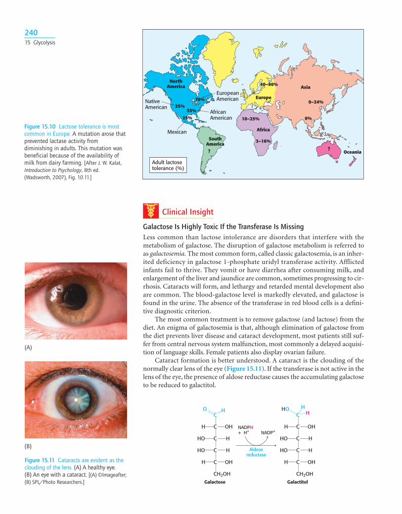
“Deficiency” is not quite the appropriate term, because a decrease in lactase is nor-mal in the course of development in all mammals. As children are weaned andmilk becomes less prominent in their diets, lactase activity normally declines toabout 5 to 10% of the level at birth. This decrease is not as pronounced with somegroups of people, most notably Northern Europeans, and people from thesegroups can continue to ingest milk without gastrointestinal difficulties (Figure15.10). With the appearance of milk-producing domesticated animals, an adultwith active lactase would hypothetically have a selective advantage in being ableto consume calories from the readily available milk.
What happens to the lactose in the intestine of a lactase-deficient person?The lactose is a good energy source for microorganisms in the colon, and theyferment it to lactic acid while also generating methane (CH4) and hydrogen gas(H2). The gas creates the uncomfortable feeling of gut distension and the annoy-ing problem of flatulence. The lactate produced by the microorganisms drawswater into the intestine, as does any undigested lactose, resulting in diarrhea. Ifsevere enough, the gas and diarrhea hinder the absorption of other nutrientssuch as fats and proteins. The simplest treatment is to avoid the consumption ofproducts containing much lactose. Alternatively, the enzyme lactase can beingested with milk products. ■
Clinical Insight
Galactose Is Highly Toxic If the Transferase Is MissingLess common than lactose intolerance are disorders that interfere with themetabolism of galactose. The disruption of galactose metabolism is referred toas galactosemia. The most common form, called classic galactosemia, is an inher-ited deficiency in galactose 1-phosphate uridyl transferase activity. Afflictedinfants fail to thrive. They vomit or have diarrhea after consuming milk, andenlargement of the liver and jaundice are common, sometimes progressing to cir-rhosis. Cataracts will form, and lethargy and retarded mental development alsoare common. The blood-galactose level is markedly elevated, and galactose isfound in the urine. The absence of the transferase in red blood cells is a defini-tive diagnostic criterion.
The most common treatment is to remove galactose (and lactose) from thediet. An enigma of galactosemia is that, although elimination of galactose fromthe diet prevents liver disease and cataract development, most patients still suf-fer from central nervous system malfunction, most commonly a delayed acquisi-tion of language skills. Female patients also display ovarian failure.
Cataract formation is better understood. A cataract is the clouding of thenormally clear lens of the eye (Figure 15.11). If the transferase is not active in thelens of the eye, the presence of aldose reductase causes the accumulating galactoseto be reduced to galactitol.
24015 Glycolysis
Figure 15.11 Cataracts are evident as theclouding of the lens. (A) A healthy eye. (B) An eye with a cataract. [(A) ©Imageafter;(B) SPL/Photo Researchers.]
NADPH+ H+ NADP+
Galactose Galactitol
Aldosereductase
C
C
C
C
C
CH2OH
O H
OHH
HHO
HHO
OHH
C
C
C
C
C
CH2OH
HOH
OHH
HHO
HHO
OHH
H
Figure 15.10 Lactose tolerance is mostcommon in Europe. A mutation arose thatprevented lactase activity fromdiminishing in adults. This mutation wasbeneficial because of the availability ofmilk from dairy farming. [After J. W. Kalat,Introduction to Psychology, 8th ed.(Wadsworth, 2007), Fig. 10.11.]
NativeAmerican
AfricanAmerican
EuropeanAmerican
Mexican
NorthAmerica
SouthAmerica
Europe
Asia
Africa
Oceania??
10–25%
25%
25%
35%
78%
40–90%
0–24%
0%
3–16%
Adult lactosetolerance (%)
(B)
(A)
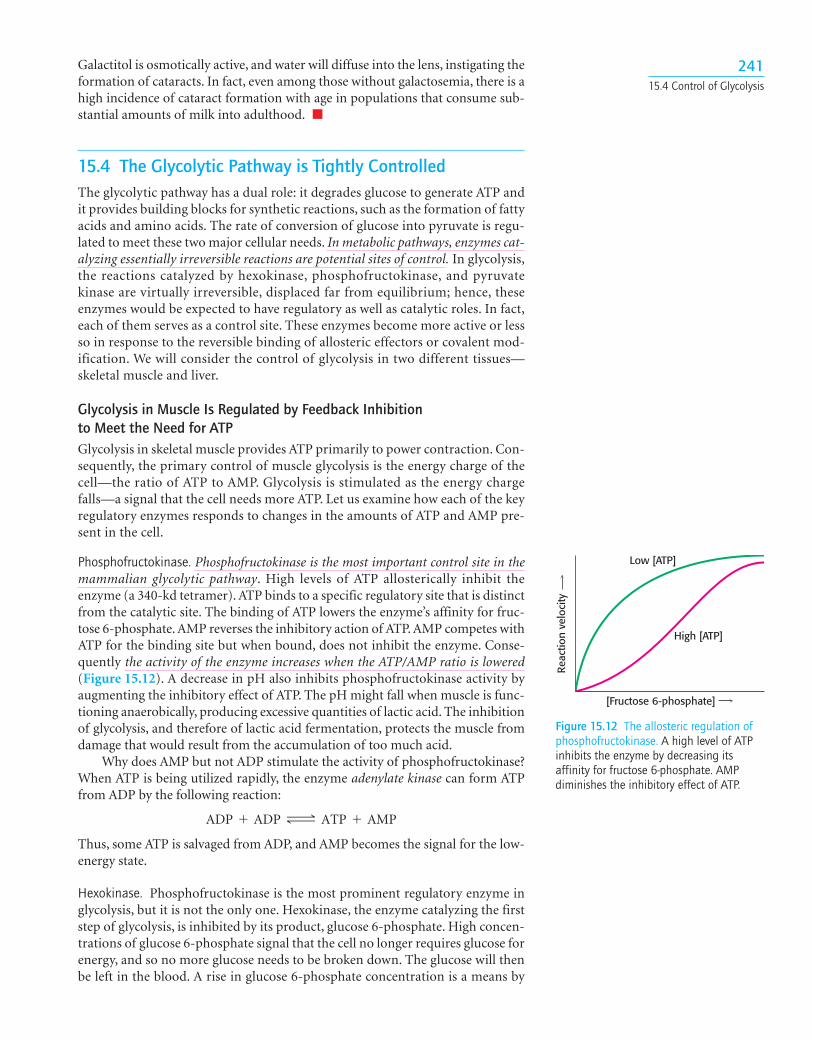
Galactitol is osmotically active, and water will diffuse into the lens, instigating theformation of cataracts. In fact, even among those without galactosemia, there is ahigh incidence of cataract formation with age in populations that consume sub-stantial amounts of milk into adulthood. ■
15.4 The Glycolytic Pathway is Tightly ControlledThe glycolytic pathway has a dual role: it degrades glucose to generate ATP andit provides building blocks for synthetic reactions, such as the formation of fattyacids and amino acids. The rate of conversion of glucose into pyruvate is regu-lated to meet these two major cellular needs. In metabolic pathways, enzymes cat-alyzing essentially irreversible reactions are potential sites of control. In glycolysis,the reactions catalyzed by hexokinase, phosphofructokinase, and pyruvatekinase are virtually irreversible, displaced far from equilibrium; hence, theseenzymes would be expected to have regulatory as well as catalytic roles. In fact,each of them serves as a control site. These enzymes become more active or lessso in response to the reversible binding of allosteric effectors or covalent mod-ification. We will consider the control of glycolysis in two different tissues—skeletal muscle and liver.
Glycolysis in Muscle Is Regulated by Feedback Inhibition to Meet the Need for ATPGlycolysis in skeletal muscle provides ATP primarily to power contraction. Con-sequently, the primary control of muscle glycolysis is the energy charge of thecell—the ratio of ATP to AMP. Glycolysis is stimulated as the energy chargefalls—a signal that the cell needs more ATP. Let us examine how each of the keyregulatory enzymes responds to changes in the amounts of ATP and AMP pre-sent in the cell.
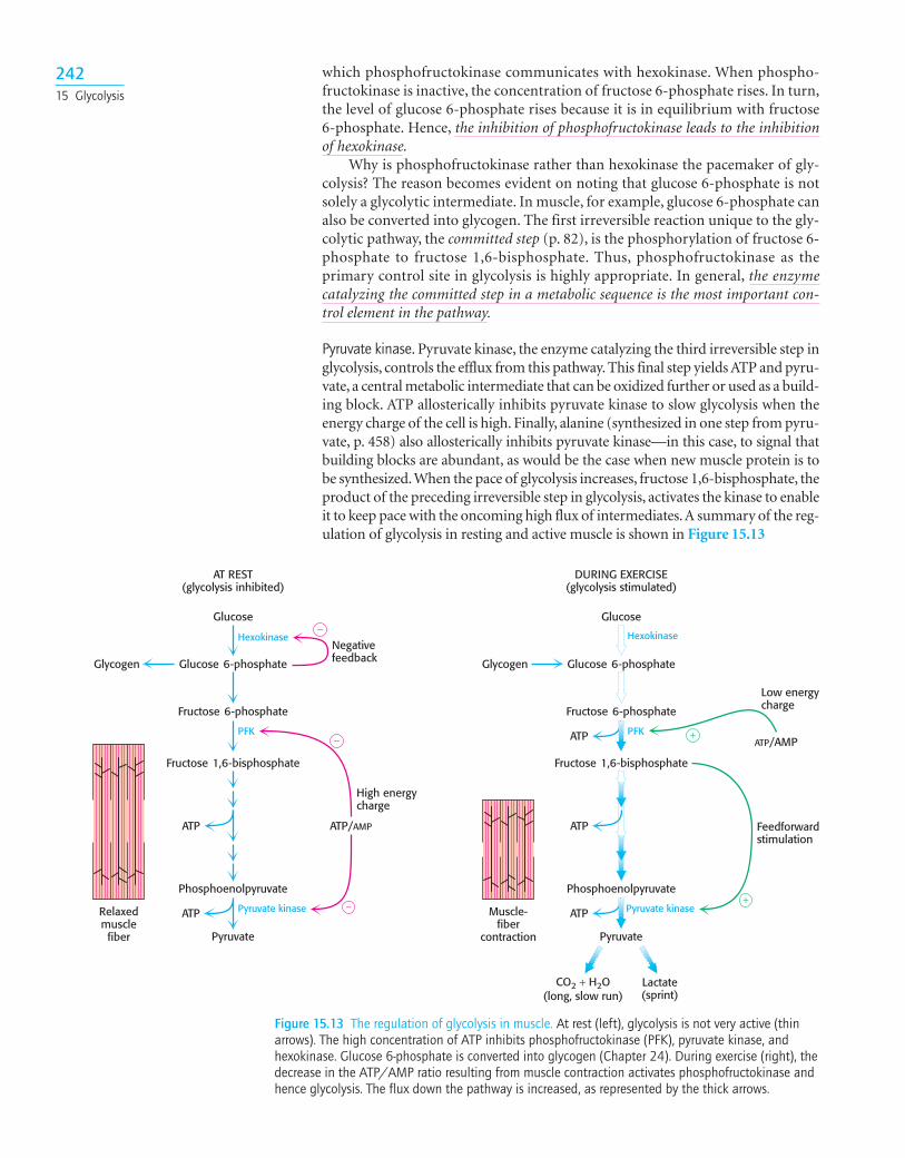
Phosphofructokinase. Phosphofructokinase is the most important control site in themammalian glycolytic pathway. High levels of ATP allosterically inhibit theenzyme (a 340-kd tetramer). ATP binds to a specific regulatory site that is distinctfrom the catalytic site. The binding of ATP lowers the enzyme’s affinity for fruc-tose 6-phosphate. AMP reverses the inhibitory action of ATP. AMP competes withATP for the binding site but when bound, does not inhibit the enzyme. Conse-quently the activity of the enzyme increases when the ATP/AMP ratio is lowered(Figure 15.12). A decrease in pH also inhibits phosphofructokinase activity byaugmenting the inhibitory effect of ATP. The pH might fall when muscle is func-tioning anaerobically, producing excessive quantities of lactic acid. The inhibitionof glycolysis, and therefore of lactic acid fermentation, protects the muscle fromdamage that would result from the accumulation of too much acid.
Why does AMP but not ADP stimulate the activity of phosphofructokinase?When ATP is being utilized rapidly, the enzyme adenylate kinase can form ATPfrom ADP by the following reaction:
Thus, some ATP is salvaged from ADP, and AMP becomes the signal for the low-energy state.
Hexokinase. Phosphofructokinase is the most prominent regulatory enzyme inglycolysis, but it is not the only one. Hexokinase, the enzyme catalyzing the firststep of glycolysis, is inhibited by its product, glucose 6-phosphate. High concen-trations of glucose 6-phosphate signal that the cell no longer requires glucose forenergy, and so no more glucose needs to be broken down. The glucose will thenbe left in the blood. A rise in glucose 6-phosphate concentration is a means by
ADP + ADP Δ ATP + AMP
24115.4 Control of Glycolysis
[Fructose 6-phosphate]
Low [ATP]
High [ATP]
Reac
tion
velo
city
Figure 15.12 The allosteric regulation ofphosphofructokinase. A high level of ATPinhibits the enzyme by decreasing itsaffinity for fructose 6-phosphate. AMPdiminishes the inhibitory effect of ATP.
which phosphofructokinase communicates with hexokinase. When phospho-fructokinase is inactive, the concentration of fructose 6-phosphate rises. In turn,the level of glucose 6-phosphate rises because it is in equilibrium with fructose6-phosphate. Hence, the inhibition of phosphofructokinase leads to the inhibitionof hexokinase.
Why is phosphofructokinase rather than hexokinase the pacemaker of gly-colysis? The reason becomes evident on noting that glucose 6-phosphate is notsolely a glycolytic intermediate. In muscle, for example, glucose 6-phosphate canalso be converted into glycogen. The first irreversible reaction unique to the gly-colytic pathway, the committed step (p. 82), is the phosphorylation of fructose 6-phosphate to fructose 1,6-bisphosphate. Thus, phosphofructokinase as theprimary control site in glycolysis is highly appropriate. In general, the enzymecatalyzing the committed step in a metabolic sequence is the most important con-trol element in the pathway.
Pyruvate kinase. Pyruvate kinase, the enzyme catalyzing the third irreversible step inglycolysis, controls the efflux from this pathway. This final step yields ATP and pyru-vate, a central metabolic intermediate that can be oxidized further or used as a build-ing block. ATP allosterically inhibits pyruvate kinase to slow glycolysis when theenergy charge of the cell is high. Finally, alanine (synthesized in one step from pyru-vate, p. 458) also allosterically inhibits pyruvate kinase—in this case, to signal thatbuilding blocks are abundant, as would be the case when new muscle protein is tobe synthesized.When the pace of glycolysis increases, fructose 1,6-bisphosphate, theproduct of the preceding irreversible step in glycolysis, activates the kinase to enableit to keep pace with the oncoming high flux of intermediates.A summary of the reg-ulation of glycolysis in resting and active muscle is shown in Figure 15.13
24215 Glycolysis
Glycogen
ATP ATP/AMP
Negativefeedback
High energycharge
Low energycharge
Glucose 6-phosphate
Fructose 6-phosphate
Fructose 1,6-bisphosphate
Pyruvate Pyruvate
Muscle-fiber
contraction
Relaxedmusclefiber
Hexokinase
PFK
ATP
Phosphoenolpyruvate
Pyruvate kinase
+
+
−
−
−
Glucose
AT REST(glycolysis inhibited)
Glycogen
ATP
ATPATP/AMP
Feedforwardstimulation
Glucose 6-phosphate
Fructose 6-phosphate
Fructose 1,6-bisphosphate
Lactate(sprint)
Hexokinase
PFK
ATP
Phosphoenolpyruvate
Pyruvate kinase
Glucose
DURING EXERCISE(glycolysis stimulated)
CO2 + H2O(long, slow run)
Figure 15.13 The regulation of glycolysis in muscle. At rest (left), glycolysis is not very active (thinarrows). The high concentration of ATP inhibits phosphofructokinase (PFK), pyruvate kinase, andhexokinase. Glucose 6-phosphate is converted into glycogen (Chapter 24). During exercise (right), thedecrease in the ATP/AMP ratio resulting from muscle contraction activates phosphofructokinase andhence glycolysis. The flux down the pathway is increased, as represented by the thick arrows.
The Regulation of Glycolysis in the Liver Corresponds to the Biochemical Versatility of the LiverThe liver has a greater diversity of biochemical functions than muscle. Signifi-cantly, the liver maintains blood-glucose levels: it stores glucose as glycogen whenglucose is plentiful, and it releases glucose when supplies are low. It also uses glu-cose to generate reducing power for biosynthesis (p. 231) as well as to synthesizea host of building blocks for other biomolecules. So, although the liver has manyof the regulatory features of muscle glycolysis, the regulation of glycolysis in theliver is more complex.
Phosphofructokinase. Although the regulation of phosphofructokinase withrespect to ATP is the same in the liver as in muscle, it is not as important in liver,because the ATP levels in liver do not fluctuate as they do in skeletal muscle. LowpH is not a metabolic signal for the liver enzyme, because lactate is not normallyproduced in the liver. Indeed, as we will see, lactate is converted into glucose inthe liver.
Glycolysis also furnishes carbon skeletons for biosyntheses, and so a signalindicating whether building blocks are abundant or scarce should also regulatephosphofructokinase. In the liver, phosphofructokinase is inhibited by citrate, anearly intermediate in the citric acid cycle (p. 280). A high level of citrate in thecytoplasm means that biosynthetic precursors are abundant, and so there is noneed to degrade additional glucose for this purpose. In this way, citrate enhancesthe inhibitory effect of ATP on phosphofructokinase.
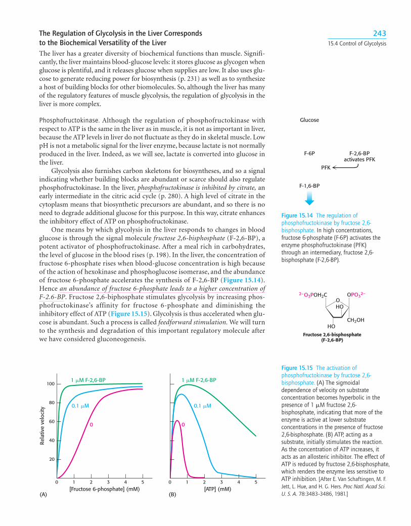
One means by which glycolysis in the liver responds to changes in bloodglucose is through the signal molecule fructose 2,6-bisphosphate (F-2,6-BP), apotent activator of phosphofructokinase. After a meal rich in carbohydrates,the level of glucose in the blood rises (p. 198). In the liver, the concentration offructose 6-phosphate rises when blood-glucose concentration is high becauseof the action of hexokinase and phosphoglucose isomerase, and the abundanceof fructose 6-phosphate accelerates the synthesis of F-2,6-BP (Figure 15.14).Hence an abundance of fructose 6-phosphate leads to a higher concentration ofF-2.6-BP. Fructose 2,6-biphosphate stimulates glycolysis by increasing phos-phofructokinase’s affinity for fructose 6-phosphate and diminishing theinhibitory effect of ATP (Figure 15.15). Glycolysis is thus accelerated when glu-cose is abundant. Such a process is called feedforward stimulation. We will turnto the synthesis and degradation of this important regulatory molecule afterwe have considered gluconeogenesis.
Figure 15.14 The regulation ofphosphofructokinase by fructose 2,6-bisphosphate. In high concentrations,fructose 6-phosphate (F-6P) activates theenzyme phosphofructokinase (PFK)through an intermediary, fructose 2,6-bisphosphate (F-2,6-BP).
Glucose
F-6P F-2,6-BPactivates PFK
PFK
F-1,6-BP
Figure 15.15 The activation ofphosphofructokinase by fructose 2,6-bisphosphate. (A) The sigmoidaldependence of velocity on substrateconcentration becomes hyperbolic in thepresence of 1 �M fructose 2,6-bisphosphate, indicating that more of theenzyme is active at lower substrateconcentrations in the presence of fructose2,6-bisphosphate. (B) ATP, acting as asubstrate, initially stimulates the reaction.As the concentration of ATP increases, itacts as an allosteric inhibitor. The effect ofATP is reduced by fructose 2,6-bisphosphate,which renders the enzyme less sensitive toATP inhibition. [After E. Van Schaftingen, M. F.Jett, L. Hue, and H. G. Hers. Proc Natl. Acad Sci.U. S. A. 78:3483–3486, 1981.]
1 �M F-2,6-BP
Rela
tive
velo
city
0
[Fructose 6-phosphate] (mM) [ATP] (mM)
0.1 �M
1 �M F-2,6-BP
0
0.1 �M
(A) (B)
100
80
60
40
20
0 1 2 3 4 5 0 1 2 3 4 5
Fructose 2,6-bisphosphate(F-2,6-BP)
OOPO
CH2OH
32–
HO
HO
2–O3POH2C
24315.4 Control of Glycolysis
Hexokinase. In the liver as well as in the muscle, hexokinase is an important reg-ulatory molecule. The hexokinase reaction in the liver is controlled as in themuscle. However, the enzyme primarily responsible for phosphorylating glu-cose in the liver is not hexokinase, but glucokinase (hexokinase D). Glucokinasephosphorylates glucose only when glucose is abundant, as would be the caseafter a meal. The reason is that glucokinase’s affinity for glucose is about 50-foldlower than that of hexokinase, which means that glucokinase binds only to theglucose molecules that are in excess of what hexokinase can bind. Furthermore,glucokinase is not inhibited by its product, glucose 6-phosphate, as hexokinaseis. The role of glucokinase is to provide glucose 6-phosphate for the synthesis ofglycogen and for the formation of fatty acids. The low affinity of glucokinase forglucose in the liver gives the brain and muscles first call for glucose when its sup-ply is limited, and it ensures that glucose will not be wasted when it is abundant.
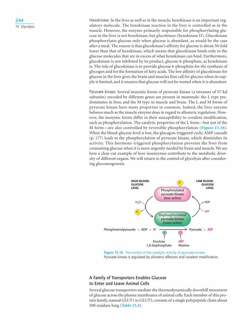
Pyruvate kinase. Several isozymic forms of pyruvate kinase (a tetramer of 57-kdsubunits) encoded by different genes are present in mammals: the L type pre-dominates in liver, and the M type in muscle and brain. The L and M forms ofpyruvate kinase have many properties in common. Indeed, the liver enzymebehaves much as the muscle enzyme does in regard to allosteric regulation. How-ever, the isozymic forms differ in their susceptibility to covalent modification,such as phosphorylation. The catalytic properties of the L form—but not of theM form—are also controlled by reversible phosphorylation (Figure 15.16).When the blood-glucose level is low, the glucagon-triggered cyclic AMP cascade(p. 177) leads to the phosphorylation of pyruvate kinase, which diminishes itsactivity. This hormone-triggered phosphorylation prevents the liver fromconsuming glucose when it is more urgently needed by brain and muscle. We seehere a clear-cut example of how isoenzymes contribute to the metabolic diver-sity of different organs. We will return to the control of glycolysis after consider-ing gluconeogenesis.
24415 Glycolysis
Figure 15.16 The control of the catalytic activity of pyruvate kinase.Pyruvate kinase is regulated by allosteric effectors and covalent modification.
HIGH BLOOD-GLUCOSELEVEL
Dephosphorylatedpyruvate kinase(more active)
ADPH2O
ATP
Phosphorylatedpyruvate kinase
(less active)
Phosphoenolpyruvate + ADP + H+
+ −Pyruvate + ATP
Fructose1,6-bisphosphate
ATPAlanine
LOW BLOOD-GLUCOSE
LEVEL
Pi
Pi
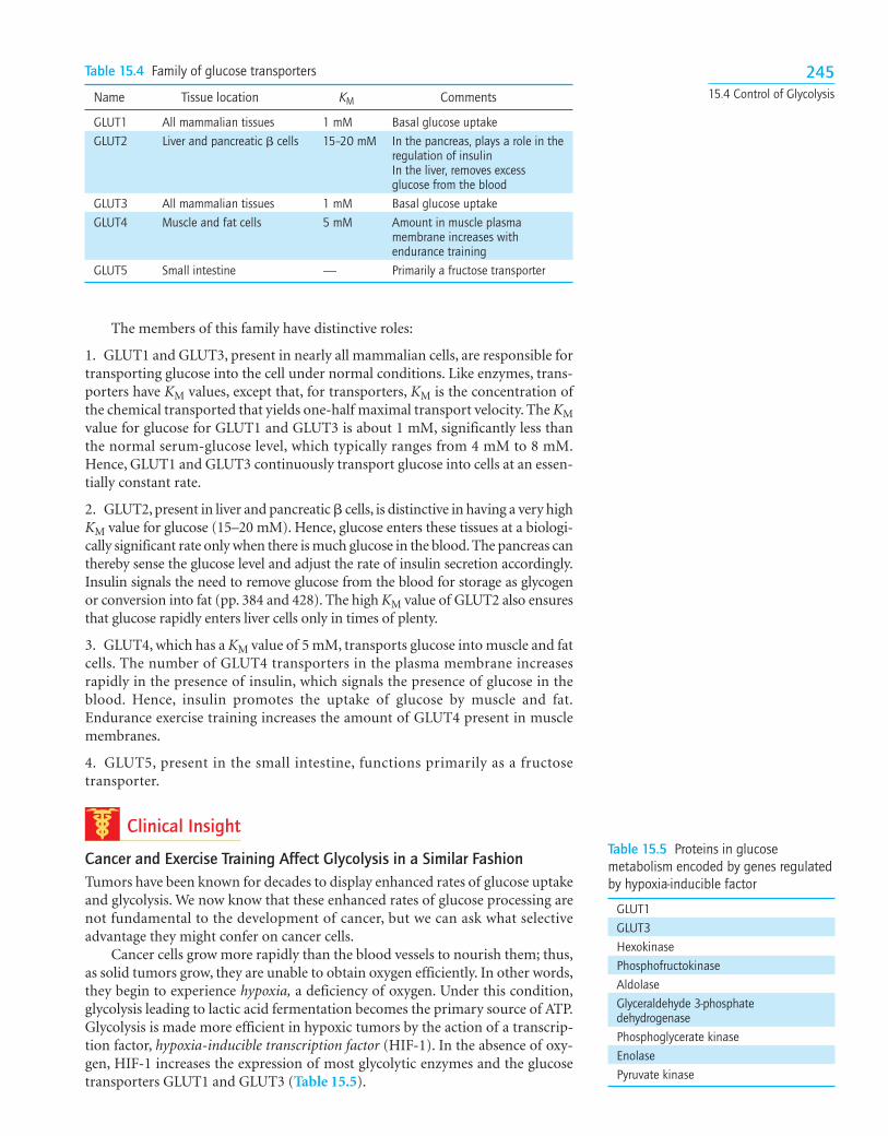
A Family of Transporters Enables Glucose to Enter and Leave Animal CellsSeveral glucose transporters mediate the thermodynamically downhill movementof glucose across the plasma membranes of animal cells. Each member of this pro-tein family, named GLUT1 to GLUT5, consists of a single polypeptide chain about500 residues long (Table 15.4).
The members of this family have distinctive roles:
1. GLUT1 and GLUT3, present in nearly all mammalian cells, are responsible fortransporting glucose into the cell under normal conditions. Like enzymes, trans-porters have KM values, except that, for transporters, KM is the concentration ofthe chemical transported that yields one-half maximal transport velocity. The KMvalue for glucose for GLUT1 and GLUT3 is about 1 mM, significantly less thanthe normal serum-glucose level, which typically ranges from 4 mM to 8 mM.Hence, GLUT1 and GLUT3 continuously transport glucose into cells at an essen-tially constant rate.
2. GLUT2, present in liver and pancreatic � cells, is distinctive in having a very highKM value for glucose (15–20 mM). Hence, glucose enters these tissues at a biologi-cally significant rate only when there is much glucose in the blood. The pancreas canthereby sense the glucose level and adjust the rate of insulin secretion accordingly.Insulin signals the need to remove glucose from the blood for storage as glycogenor conversion into fat (pp. 384 and 428). The high KM value of GLUT2 also ensuresthat glucose rapidly enters liver cells only in times of plenty.
3. GLUT4, which has a KM value of 5 mM, transports glucose into muscle and fatcells. The number of GLUT4 transporters in the plasma membrane increasesrapidly in the presence of insulin, which signals the presence of glucose in theblood. Hence, insulin promotes the uptake of glucose by muscle and fat.Endurance exercise training increases the amount of GLUT4 present in musclemembranes.
4. GLUT5, present in the small intestine, functions primarily as a fructosetransporter.
Clinical Insight
Cancer and Exercise Training Affect Glycolysis in a Similar FashionTumors have been known for decades to display enhanced rates of glucose uptakeand glycolysis. We now know that these enhanced rates of glucose processing arenot fundamental to the development of cancer, but we can ask what selectiveadvantage they might confer on cancer cells.
Cancer cells grow more rapidly than the blood vessels to nourish them; thus,as solid tumors grow, they are unable to obtain oxygen efficiently. In other words,they begin to experience hypoxia, a deficiency of oxygen. Under this condition,glycolysis leading to lactic acid fermentation becomes the primary source of ATP.Glycolysis is made more efficient in hypoxic tumors by the action of a transcrip-tion factor, hypoxia-inducible transcription factor (HIF-1). In the absence of oxy-gen, HIF-1 increases the expression of most glycolytic enzymes and the glucosetransporters GLUT1 and GLUT3 (Table 15.5).
24515.4 Control of Glycolysis
Table 15.4 Family of glucose transporters
Name Tissue location KM Comments
GLUT1 All mammalian tissues 1 mM Basal glucose uptakeGLUT2 Liver and pancreatic � cells 15–20 mM In the pancreas, plays a role in the
regulation of insulinIn the liver, removes excess glucose from the blood
GLUT3 All mammalian tissues 1 mM Basal glucose uptakeGLUT4 Muscle and fat cells 5 mM Amount in muscle plasma
membrane increases with endurance training
GLUT5 Small intestine — Primarily a fructose transporter
Table 15.5 Proteins in glucosemetabolism encoded by genes regulatedby hypoxia-inducible factor
GLUT1GLUT3HexokinasePhosphofructokinaseAldolaseGlyceraldehyde 3-phosphate dehydrogenasePhosphoglycerate kinaseEnolasePyruvate kinase
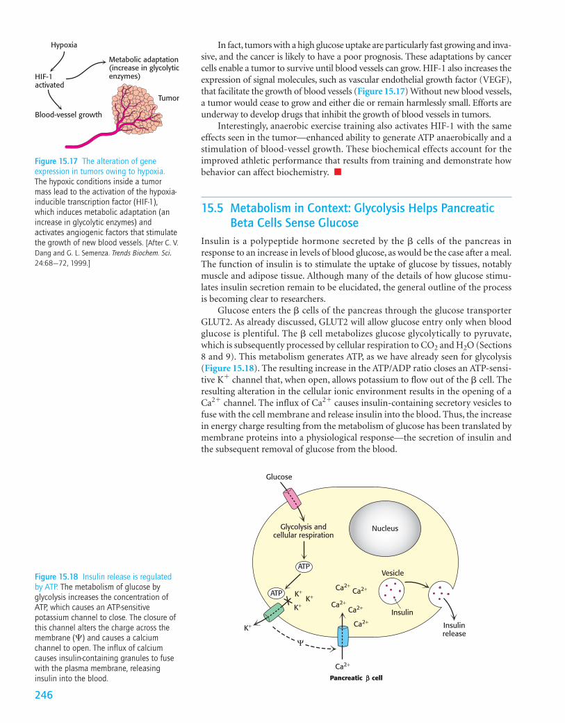
In fact, tumors with a high glucose uptake are particularly fast growing and inva-sive, and the cancer is likely to have a poor prognosis. These adaptations by cancercells enable a tumor to survive until blood vessels can grow. HIF-1 also increases theexpression of signal molecules, such as vascular endothelial growth factor (VEGF),that facilitate the growth of blood vessels (Figure 15.17) Without new blood vessels,a tumor would cease to grow and either die or remain harmlessly small. Efforts areunderway to develop drugs that inhibit the growth of blood vessels in tumors.
Interestingly, anaerobic exercise training also activates HIF-1 with the sameeffects seen in the tumor—enhanced ability to generate ATP anaerobically and astimulation of blood-vessel growth. These biochemical effects account for theimproved athletic performance that results from training and demonstrate howbehavior can affect biochemistry. ■
15.5 Metabolism in Context: Glycolysis Helps PancreaticBeta Cells Sense Glucose
Insulin is a polypeptide hormone secreted by the � cells of the pancreas inresponse to an increase in levels of blood glucose, as would be the case after a meal.The function of insulin is to stimulate the uptake of glucose by tissues, notablymuscle and adipose tissue. Although many of the details of how glucose stimu-lates insulin secretion remain to be elucidated, the general outline of the processis becoming clear to researchers.
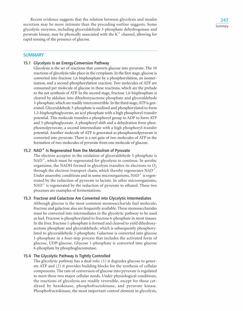
Glucose enters the � cells of the pancreas through the glucose transporterGLUT2. As already discussed, GLUT2 will allow glucose entry only when bloodglucose is plentiful. The � cell metabolizes glucose glycolytically to pyruvate,which is subsequently processed by cellular respiration to CO2 and H2O (Sections8 and 9). This metabolism generates ATP, as we have already seen for glycolysis(Figure 15.18). The resulting increase in the ATP/ADP ratio closes an ATP-sensi-tive K� channel that, when open, allows potassium to flow out of the � cell. Theresulting alteration in the cellular ionic environment results in the opening of aCa2� channel. The influx of Ca2� causes insulin-containing secretory vesicles tofuse with the cell membrane and release insulin into the blood. Thus, the increasein energy charge resulting from the metabolism of glucose has been translated bymembrane proteins into a physiological response—the secretion of insulin andthe subsequent removal of glucose from the blood.
Tumor
Metabolic adaptation(increase in glycolyticenzymes)HIF-1
activated
Hypoxia
Blood-vessel growth
Figure 15.17 The alteration of geneexpression in tumors owing to hypoxia.The hypoxic conditions inside a tumormass lead to the activation of the hypoxia-inducible transcription factor (HIF-1),which induces metabolic adaptation (anincrease in glycolytic enzymes) andactivates angiogenic factors that stimulatethe growth of new blood vessels. [After C. V.Dang and G. L. Semenza. Trends Biochem. Sci.24:68–72, 1999.]
Figure 15.18 Insulin release is regulatedby ATP. The metabolism of glucose byglycolysis increases the concentration ofATP, which causes an ATP-sensitivepotassium channel to close. The closure ofthis channel alters the charge across themembrane () and causes a calciumchannel to open. The influx of calciumcauses insulin-containing granules to fusewith the plasma membrane, releasinginsulin into the blood.
Ca2+
Ca2+Ca2+
Ca2+
Ca2+Ca2+
K+
K+
K+K+
Glycolysis andcellular respiration
Insulinrelease
Vesicle
Glucose
Pancreatic cell
Nucleus
ATP
ATP
Insulin
Ψ
246
Recent evidence suggests that the relation between glycolysis and insulinsecretion may be more intimate than the preceding outline suggests. Someglycolytic enzymes, including glyceraldehyde 3-phosphate dehydrogenase andpyruvate kinase, may be physically associated with the K� channel, allowing forrapid sensing of the presence of glucose.
SUMMARY
15.1 Glycolysis Is an Energy-Conversion PathwayGlycolysis is the set of reactions that converts glucose into pyruvate. The 10reactions of glycolysis take place in the cytoplasm. In the first stage, glucose isconverted into fructose 1,6-bisphosphate by a phosphorylation, an isomer-ization, and a second phosphorylation reaction. Two molecules of ATP areconsumed per molecule of glucose in these reactions, which are the preludeto the net synthesis of ATP. In the second stage, fructose 1,6-bisphosphate iscleaved by aldolase into dihydroxyacetone phosphate and glyceraldehyde3-phosphate, which are readily interconvertible. In the third stage,ATP is gen-erated. Glyceraldehyde 3-phosphate is oxidized and phosphorylated to form1,3-bisphosphoglycerate, an acyl phosphate with a high phosphoryl-transferpotential. This molecule transfers a phosphoryl group to ADP to form ATPand 3-phosphoglycerate. A phosphoryl shift and a dehydration form phos-phoenolpyruvate, a second intermediate with a high phosphoryl-transferpotential. Another molecule of ATP is generated as phosphoenolpyruvate isconverted into pyruvate. There is a net gain of two molecules of ATP in theformation of two molecules of pyruvate from one molecule of glucose.
15.2 NAD� Is Regenerated from the Metabolism of PyruvateThe electron acceptor in the oxidation of glyceraldehyde 3-phosphate isNAD�, which must be regenerated for glycolysis to continue. In aerobicorganisms, the NADH formed in glycolysis transfers its electrons to O2through the electron-transport chain, which thereby regenerates NAD�.Under anaerobic conditions and in some microorganisms, NAD� is regen-erated by the reduction of pyruvate to lactate. In other microorganisms,NAD� is regenerated by the reduction of pyruvate to ethanol. These twoprocesses are examples of fermentations.
15.3 Fructose and Galactose Are Converted into Glycolytic IntermediatesAlthough glucose is the most common monosaccharide fuel molecule,fructose and galactose also are frequently available. These monosaccharidesmust be converted into intermediates in the glycolytic pathway to be usedas fuel. Fructose is phosphorylated to fructose 6-phosphate in most tissues.In the liver, fructose 1-phosphate is formed and cleaved to yield dihydroxy-acetone phosphate and glyceraldehyde, which is subsequently phosphory-lated to glyceraldehyde 3-phosphate. Galactose is converted into glucose1-phosphate in a four-step process that includes the activated form ofglucose, UDP-glucose. Glucose 1-phosphate is converted into glucose6-phosphate by phosphoglucomutase.
15.4 The Glycolytic Pathway Is Tightly ControlledThe glycolytic pathway has a dual role: (1) it degrades glucose to gener-ate ATP and (2) it provides building blocks for the synthesis of cellularcomponents. The rate of conversion of glucose into pyruvate is regulatedto meet these two major cellular needs. Under physiological conditions,the reactions of glycolysis are readily reversible, except for those cat-alyzed by hexokinase, phosphofructokinase, and pyruvate kinase.Phosphofructokinase, the most important control element in glycolysis,
247Summary
is inhibited by high levels of ATP and citrate, and it is activated by AMPand fructose 2,6-bisphosphate. In the liver, this bisphosphate signals thatglucose is abundant. Hence, phosphofructokinase is active when eitherenergy or building blocks are needed. Hexokinase is inhibited by glucose6-phosphate, which accumulates when phosphofructokinase is inactive.ATP and alanine allosterically inhibit pyruvate kinase, the other controlsite, and fructose 1,6-bisphosphate activates the enzyme. Consequently,pyruvate kinase is maximally active when the energy charge is low andglycolytic intermediates accumulate.
15.5 Metabolic Integration: Glycolysis Helps Pancreatic Beta Cells SenseGlucoseThe increase in the ratio of ATP/ADP that results from the metabolism ofglucose to pyruvate closes K� channels in the membrane of � cells of thepancreas. The resulting change in the cellular ionic environment causes aninflux of Ca2�, which, in turn, leads to insulin secretion. Insulin stimulatesthe uptake of glucose by muscle and adipose tissue.
24815 Glycolysis
Key Terms
glycolysis (p. 227)hexokinase (p. 227)kinase (p. 227)phosphofructokinase (PFK) (p. 230)thioester intermediate (p. 232)
substrate-level phosphorylation (p. 232)
mutase (p. 233)enol phosphate (p. 233)pyruvate kinase (p. 233)
alcoholic fermentation (p. 234)lactic acid fermentation (p. 235)obligate anaerobe (p. 236)committed step (p. 242)feedforward stimulation (p. 243)
Answers to QUICK QUIZZES
1. Two molecules of ATP are produced per glyceraldehyde3-phosphate and, because two molecules of GAP are pro-duced per glucose, the total ATP yield is four. However, twomolecules of ATP are required to convert glucose into fruc-tose 1,6-bisphosphate. Thus, the net yield is only two mol-ecules of ATP.
2. In both cases, the electron donor is glyceraldehyde 3-phosphate. In lactic acid fermentation, the electron accep-tor is pyruvate, converting it into lactate. In alcoholic ferme-nation, acetaldehyde is the electron acceptor, formingethanol.
1. ATP yield. Each of the following molecules is processedby glycolysis to lactate. How much ATP is generated fromeach molecule?
(a) Glucose 6-phosphate(b) Dihydroxyacetone phosphate(c) Glyceraldehyde 3-phosphate
4. Recommended daily allowance. The recommended dailyallowance for the vitamin niacin is 15 mg per day. Howwould glycolysis be affected by niacin deficiency?
5. Who’s on first? Although both hexokinase and phospho-fructokinase catalyze irreversible steps in glycolysis and thehexokinase-catalyzed step is first, phosphofructokinase isnonetheless the pacemaker of glycolysis.What does this infor-mation tell you about the fate of the glucose 6-phosphateformed by hexokinase?
6. The tortoise and the hare. Why is the regulation of phos-phofructokinase by energy charge not as important in theliver as it is in muscle?
Problems
(d) Fructose(e) Sucrose
2. Enzyme redundancy? Why is it advantageous for theliver to have both hexokinase and glucokinase to phospho-rylate glucose?
3. Corporate sponsors. Some of the early research on glycolysis was supported by the brewing industry. Whywould the brewing industry be interested in glycolysis?
7. State function. Fructose 2,6-bisphosphate is a potentstimulator of phosphofructokinase. Explain how fructose2,6-bisphosphate might function in the concerted model forallosteric enzymes.
8. Running in reverse.Why can’t the reactions of the glycolyticpathway simply be run in reverse to synthesize glucose?
9. Road blocks. What reactions of glycolysis are not read-ily reversible under intracellular conditions?
10. No pickling. Why is it in the muscle’s best interest toexport lactic acid into the blood during intense exercise?
11. Après vous. Why is it physiologically advantageous forthe pancreas to use GLUT2, with a high KM, as the trans-porter that allows glucose entry into � cells?
12. Bypass. In the liver, fructose can be converted into glyc-eraldehyde 3-phosphate and dihydroxyacetone phosphatewithout passing through the phosphofructokinase-regulated reaction. Show the reactions that make this con-version possible. Why might ingesting high levels of fructosehave deleterious physiological effects?
13. Trouble ahead. Suppose that a microorganism that wasan obligate anaerobe suffered a mutation that resulted in theloss of triose phosphate isomerase activity. How would thisloss affect the ATP yield of fermentation? Could such anorganism survive?
14. Kitchen chemistry. Sucrose is commonly used to pre-serve fruits. Why is glucose not suitable for preservingfoods?
15. Tracing carbon atoms. Glucose labeled with l4C at C-1 isincubated with the glycolytic enzymes and necessary cofactors.
(a) What is the distribution of 14C in the pyruvate that isformed? (Assume that the interconversion of glyceraldehyde3-phosphate and dihydroxyacetone phosphate is very rapidcompared with the subsequent step.)(b) If the specific activity of the glucose substrate is 10 mCimmol�1, what is the specific activity of the pyruvate that isformed?
16. Lactic acid fermentation. (a) Write a balanced equationfor the conversion of glucose into lactate. (b) Calculate thestandard free-energy change of the formation of lactatefrom glucose by using the data given in Table 15.1 and thefact that �G°� is �25 kJ mol�1 (�6 kcal mol�1) for the fol-lowing reaction:
(c) What is the free-energy change (�G, not �G°�) of this reac-tion when the concentrations of reactants are: glucose, 5 mM;lactate, 0.05 mM; ATP, 2 mM; ADP, 0.2 mM; and Pi, 1 mM?
Pyruvate + NADH + H+ Δ lactate + NAD+
17. High potential. What is the equilibrium ratio of phos-phoenolpyruvate to pyruvate under standard conditionswhen [ATP]/[ADP] 10?
18. Hexose–triose equilibrium. What are the equilibriumconcentrations of fructose 1,6-bisphosphate, dihydroxyace-tone phosphate, and glyceraldehyde 3-phosphate when1 mM fructose 1,6-bisphosphate is incubated with aldolaseunder standard conditions?
19. An informative analog. Xylose has the same structure asthat of glucose except that xylose has a hydrogen atom atC-5 in place of a hydroxymethyl group. The rate of ATPhydrolysis by hexokinase is markedly enhanced by the addi-tion of xylose. Why?
20. Distinctive sugars. The intravenous infusion of fructoseinto healthy volunteers leads to a two- to fivefold increase inthe level of lactate in the blood, a far greater increase thanthat observed after the infusion of the same amount ofglucose.
(a) Why is glycolysis more rapid after the infusion of fructose?(b) Fructose has been used in place of glucose for intra-venous feeding. Why is this use of fructose unwise?
21. Metabolic mutants. Predict the effect of each of the fol-lowing mutations on the pace of glycolysis in liver cells:
(a) Loss of the allosteric site for ATP in phosphofructokinase.(b) Loss of the binding site for citrate in phosphofructokinase.(c) Loss of the phosphatase domain of the bifunctionalenzyme that controls the level of fructose 2,6-bisphosphate.(d) Loss of the binding site for fructose 1,6-bisphosphate inpyruvate kinase.
22. Arsenate poisoning. Arsenate (AsO43�) closely resem-
bles Pi in structure and reactivity. In the reaction catalyzedby glyceraldehyde 3-phosphate dehydrogenase, arsenatecan replace phosphate in attacking the energy-richthioester intermediate. The product of this reaction,1-arseno-3-phosphoglycerate, is unstable. It and other acylarsenates are rapidly and spontaneously hydrolyzed. Whatis the effect of arsenate on energy generation in a cell?
23. Reduce, reuse, recycle. In the conversion of glucose intotwo molecules of lactate, the NADH generated earlier in thepathway is oxidized to NAD�. Why is it not to the cell’sadvantage to simply make more NAD� so that the regener-ation would not be necessary? After all, the cell would savemuch energy because it would no longer need to synthesizelactic acid dehydrogenase.
24. Quick-change artist. Adenylate kinase is responsible forinterconverting the adenylate nucleotide pool:
ADP + ADP Δ ATP + AMP
Problems 249

(a) How does the P. furiosus phosphofructokinase differ fromthe phosphofructokinase considered in this chapter?(b) What effects do AMP and ATP have on the reactionwith ADP?
The equilibrium constant for this reaction is close to 1, inas-much as the number of phosphoanhydride bonds is thesame on each side of the equation. Using the equation forthe equilibrium constant for this reaction, show whychanges in [AMP] are a more effective indicator of theadenylate pool than those in [ATP].
Chapter Integration Problem
25. Not just for energy. People with galactosemia displaycentral nervous system abnormalities even if galactose iseliminated from the diet. The precise reason for it is notknown. Suggest a plausible explanation.
Data Interpretation Problem
26. Now, that’s unusual. Phosphofructokinase has recentlybeen isolated from the hyperthermophilic archaeonPyrococcus furiosus. It was subjected to standard biochemi-cal analysis to determine basic catalytic parameters. Theprocesses under study were of the form
fructose 1,6-biphosphate + (x)Fructose 6-Phosphate + (x - Pi) ¡
The assay measured the increase in fructose 1,6-bisphosphate.Selected results are shown in the adjoining graph.
[ADP], mM
+ 5 mM ATP+ 5 mM AMP
Enzy
me
activ
ity
120
100
80
60
40
20
00 0.2 0.4 0.6 0.8 1 1.2
250 15 Glycolysis
[Data from J. E. Tuininga et al. J. Biol. Chem.274:21023–21028, 1999.]
Selected readings for this chapter can be found online at www.whfreeman.com/Tymoczko
Recommended