
A class of its ownPhilips Ingenuity Elite with IMR specifications
Computed tomography
32
Table of contents
1 Introduction 3
2 User interface 42.1 iPatient key benefits 4
2.2 ExamCards 4
2.3 ScanRuler 4
3 DoseWise 53.1 DoseRight Index 5
3.2 CT Dose Check 5
3.3 DICOM structured reporting/IHE REM profile 5
3.4 DoseRight automatic current selection 5
3.5 DoseRight angular dose modulation 5
3.6 DoseRight Z-DOM 5
(longitudinal dose modulation)
3.7 3D-DOM 5
3.8 Dedicated pediatric protocols 5
3.9 Locking protocols 5
3.10 Dose display and reports 5
3.11 Dose performance data 5
4 Gantry 64.1 Gantry 6
4.2 Gantry control panels 6
4.3 Operator’s console control panel 6
4.4 AutoVoice 6
5 Patient table 6
6 Accessories 76.1 Standard accessories 7
6.2 Optional accessories 7
7 Imaging chain 87.1 Generator 8
7.2 X-ray tube 8
7.3 NanoPanel Elite detector 8
8 Image quality 98.1 Spatial resolution 9
8.2 Low-contrast resolution 9
8.3 Other 9
9 Reconstruction 109.1 Reconstruction speed 10
9.2 IMR Platinum 10
9.3 iDose4 Premium Package 10
9.4 HyperSight IMR reconstructor 10
9.5 Cone Beam Reconstruction Algorithm 10
– COBRA
9.6 ClearRay reconstruction 10
9.7 Adaptive filtering 10
9.8 Adaptive multicycle reconstruction 10
9.9 Reconstruction field of view 10
9.10 Image matrix 10
9.11 Off-line reconstruction 10
9.12 Preview images 10
10 Clinical enhancements 1110.1 SyncRight (optional) 11
10.2 Bolus tracking 11
10.3 Spiral Auto Start (SAS) 11
10.4 Patient centering on surview 11
10.5 Clinical applications, standard 11
10.6 Clinical applications (optional) 11
10.7 Pulmonary Toolkit (optional) 11
10.8 Pulmonary Toolkit for Oncology (optional) 11
10.9 RateResponsive CV Toolkit for Ingenuity 11
10.10 Step & Shoot Complete 11
10.11 Jog Scan (optional) 11
10.12 CT Interventional (optional) 11
11 Networking and storage 1211.1 Networking 12
11.2 DICOM 12
11.3 DICOM connectivity 12
11.4 DICOM DVD/CD writer 12
11.5 Filming 12
12 Site planning 1312.1 Power requirements 13
12.2 Console Uninterrupted Power 13
Supply (UPS) (optional)
12.3 Environmental requirements 13
12.4 System requirements, standard 14
and bariatric tables
12.5 Dimensions and weights, standard 14
and bariatric tables
12.6 System requirements, long table 15
12.7 Dimensions and weights, long table 15
32
1. Introduction
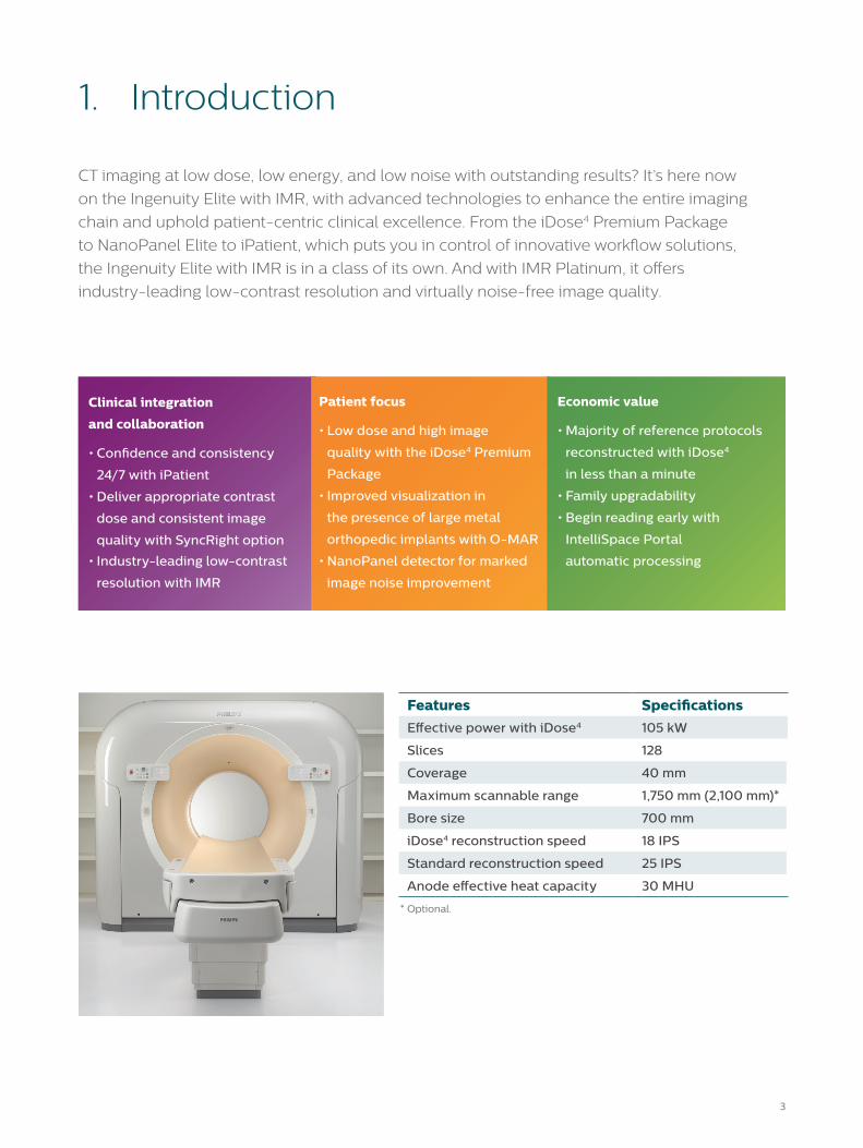
CT imaging at low dose, low energy, and low noise with outstanding results? It’s here now
on the Ingenuity Elite with IMR, with advanced technologies to enhance the entire imaging
chain and uphold patient-centric clinical excellence. From the iDose4 Premium Package
to NanoPanel Elite to iPatient, which puts you in control of innovative workflow solutions,
the Ingenuity Elite with IMR is in a class of its own. And with IMR Platinum, it offers
industry-leading low-contrast resolution and virtually noise-free image quality.
* Optional.
Economic value
• Majority of reference protocols
reconstructed with iDose4
in less than a minute
• Family upgradability
• Begin reading early with
IntelliSpace Portal
automatic processing
Clinical integration
and collaboration
• Confidence and consistency
24/7 with iPatient
• Deliver appropriate contrast
dose and consistent image
quality with SyncRight option
• Industry-leading low-contrast
resolution with IMR
Patient focus
• Low dose and high image
quality with the iDose4 Premium
Package
• Improved visualization in
the presence of large metal
orthopedic implants with O-MAR
• NanoPanel detector for marked
image noise improvement
Features Specifications
Effective power with iDose4 105 kW
Slices 128
Coverage 40 mm
Maximum scannable range 1,750 mm (2,100 mm)*
Bore size 700 mm
iDose4 reconstruction speed 18 IPS
Standard reconstruction speed 25 IPS
Anode effective heat capacity 30 MHU
54
2. User interface
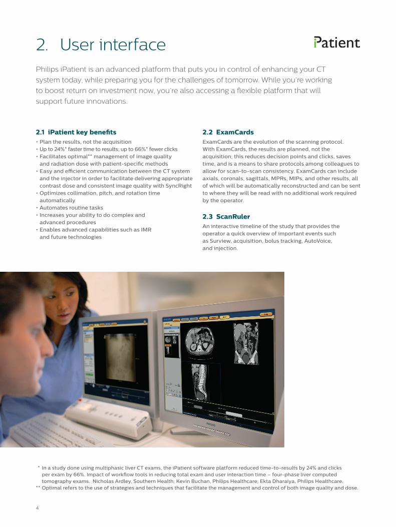
2.1 iPatient key benefits• Plan the results, not the acquisition• Up to 24%* faster time to results; up to 66%* fewer clicks• Facilitates optimal** management of image quality
and radiation dose with patient-specific methods• Easy and efficient communication between the CT system
and the injector in order to facilitate delivering appropriate contrast dose and consistent image quality with SyncRight
• Optimizes collimation, pitch, and rotation time automatically
• Automates routine tasks• Increases your ability to do complex and
advanced procedures• Enables advanced capabilities such as IMR
and future technologies
Philips iPatient is an advanced platform that puts you in control of enhancing your CT
system today, while preparing you for the challenges of tomorrow. While you’re working
to boost return on investment now, you’re also accessing a flexible platform that will
support future innovations.
2.2 ExamCardsExamCards are the evolution of the scanning protocol. With ExamCards, the results are planned, not the acquisition; this reduces decision points and clicks, saves time, and is a means to share protocols among colleagues to allow for scan-to-scan consistency. ExamCards can include axials, coronals, sagittals, MPRs, MIPs, and other results, all of which will be automatically reconstructed and can be sent to where they will be read with no additional work required by the operator.
2.3 ScanRulerAn interactive timeline of the study that provides the operator a quick overview of important events such as Surview, acquisition, bolus tracking, AutoVoice, and injection.
* In a study done using multiphasic liver CT exams, the iPatient software platform reduced time-to-results by 24% and clicks per exam by 66%. Impact of workflow tools in reducing total exam and user interaction time – four-phase liver computed tomography exams. Nicholas Ardley, Southern Health; Kevin Buchan, Philips Healthcare; Ekta Dharaiya, Philips Healthcare. ** Optimal refers to the use of strategies and techniques that facilitate the management and control of both image quality and dose.
54
3.1 DoseRight IndexDoseRight Index (DRI) is a single number used to specify the image quality required for the diagnostic task at hand. DRI includes organ-specific DRI for the liver and the head/neck to provide appropriate dose and image quality within a single acquisition. 11 weight-based protocols can be generated for ExamCards, including 1 infant, 7 child, and 3 adult reference sizes.
3.2 CT Dose CheckSupports an operator notification in each ExamCard that will be shown if an acquisition is planned that exceeds a specified CTDIvol or DLP. In addition, an alert is available such that, if an acquisition is planned and the total exam will exceed a specified CTDIvol or DLP, the operator will be required to enter his or her name and (if configured) a password to proceed, or the operator can adjust the scan parameters. Compliant with NEMA XR-25 and XR-29.
3.3 DICOM structured reporting/IHE REM profileDICOM radiation dose structured report that can be transferred to external systems such as HIS/RIS, PACS, or dose registries.
3.4 DoseRight automatic current selectionPersonalizes dose for each patient by automatically suggesting tube current settings according to the estimated patient diameter in the scan region.
3.5 DoseRight angular dose modulationAngular dose modulation varies the tube current during helical scans according to changes in patient shape (eccentricity) and tissue attenuation as the tube rotates. For each rotation, projections are processed to determine the maximum and minimum patient diameter. The tube current for the next rotation is then modulated between these limits.
3. DoseWise
3.6 DoseRight Z-DOM
(longitudinal dose modulation)Longitudinal dose modulation (Z-DOM) aids in adapting dose to an individual patient’s size and shape. In particular, Z-DOM adjusts the tube current-time product (mAs) in the craniocaudal or caudocranial (z-axis) direction based on the Surview by comparing the actual patient’s attenuation at each longitudinal location to a reference.
3.7 3D-DOM3D-DOM combines angular and longitudinal information to modulate dose in three dimensions.
3.8 Dedicated pediatric protocolsIn the iPatient approach, size-specific ExamCards can be easily generated. ExamCards can be based on one of eight (1 infant, 7 child) midpoint reference diameters that are directly related to weight based intervals. iPatient includes reference pediatric protocols for a number of clinical indications.
3.9 Locking protocolsUnauthorized protocol modifications may be prevented through password-protected access.
3.10 Dose display and reportsPhilips CT scanners include intuitive reporting and recording of estimated dose indices, dose reduction, and dose efficiency. Dose estimates are displayed on the operator’s console for all scan protocols prior to and throughout the examination. Volume computed tomography dose index (CTDIvol) and dose-length product (DLP) are automatically updated as the operator plans the scan. Also, a dose report may be included as a DICOM dose structured report and/or DICOM secondary capture with the reconstructed data set.
3.11 Dose performance data CTDIvol Measurement Head 12.9 mGy/100 mAs Body 6.6 mGy/100 mAsMeasured on head and body CTDI phantoms (IEC 60601-2-44 ed.3)
at 120 kVp.
Philips DoseWise is a holistic approach to dose management that is active in every level
of product design. It encompasses a set of techniques, programs and practices based
on the ALARA (As Low As Reasonably Achievable) principle and supports outstanding
image quality at low dose.
76
4. Gantry
4.2 Gantry control panels• Multi-directional control
for fast movement• Fine movement in/out control• Start button
Audio notification 10 seconds before X-ray On so that operator and staff can exit room before X-ray On.
4.3 Operator’s console control panel• Table in/out/up/down• Emergency stop• X-ray indicator• Start button• Pause button
• Pause button• Visual countdown• Zero table location• Lasers
4.4 AutoVoiceA standard set of commands for patient communication before, during, and after scanning in the following languages:• Arabic• Chinese –
standard Mandarin• Danish• Dutch• English• French
• Georgian• German• Greek• Hebrew• Italian• Japanese• Norwegian
• Romanian• Russian• Spanish• Swedish• Thai• Turkish• Vietnamese
Additional languages will continue to be added in the future. Support of some languages may vary by configuration. Customized messages can also be created.
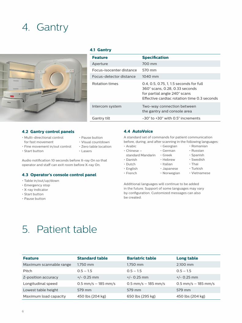
4.1 Gantry
Feature SpecificationAperture 700 mm
Focus-isocenter distance 570 mm
Focus-detector distance 1040 mm
Rotation times 0.4, 0.5, 0.75, 1, 1.5 seconds for full 360° scans, 0.28, 0.33 seconds for partial angle 240° scans Effective cardiac rotation time 0.3 seconds
Intercom system Two-way connection between the gantry and console area
Gantry tilt -30° to +30° with 0.5° increments
5. Patient table
Feature Standard table Bariatric table Long table
Maximum scannable range 1,750 mm 1,750 mm 2,100 mm
Pitch 0.5 – 1.5 0.5 – 1.5 0.5 – 1.5
Z-position accuracy +/- 0.25 mm +/- 0.25 mm +/- 0.25 mm
Longitudinal speed 0.5 mm/s – 185 mm/s 0.5 mm/s – 185 mm/s 0.5 mm/s – 185 mm/s
Lowest table height 579 mm 579 mm 579 mm
Maximum load capacity 450 lbs (204 kg) 650 lbs (295 kg) 450 lbs (204 kg)
76
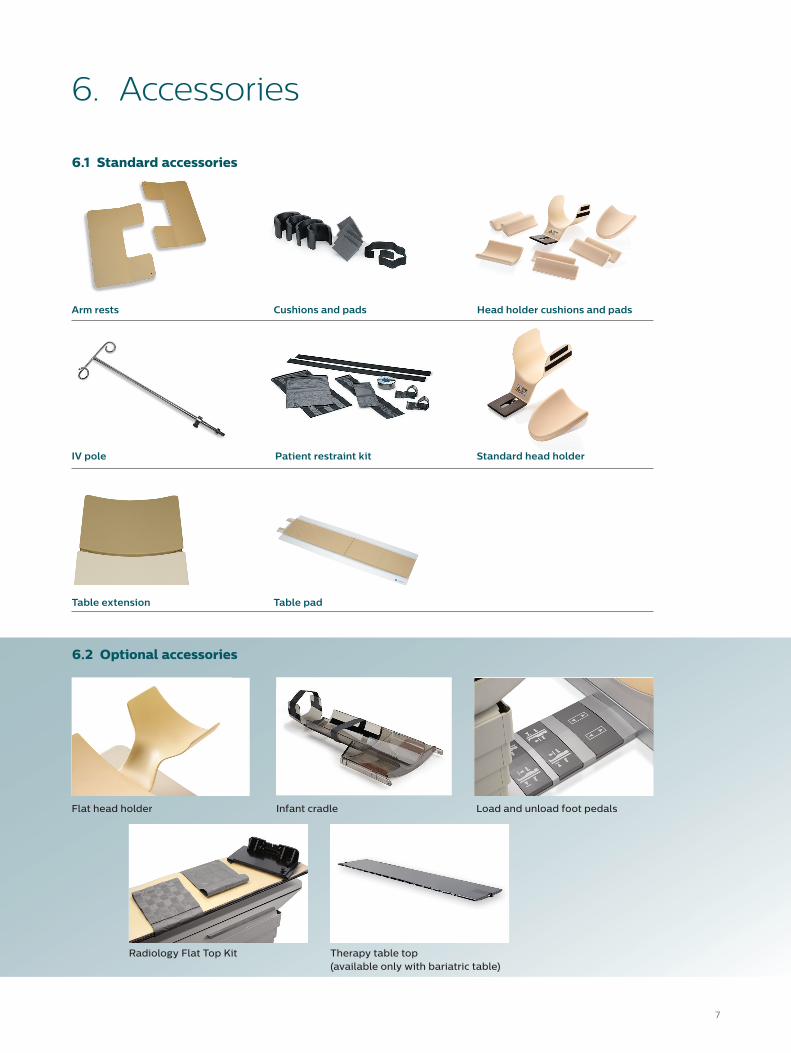
Table extension Table pad
6. Accessories
6.2 Optional accessories
Flat head holder
Radiology Flat Top Kit Therapy table top (available only with bariatric table)
Infant cradle Load and unload foot pedals
6.1 Standard accessories
Patient restraint kit
Head holder cushions and pads
Standard head holder
Cushions and padsArm rests
IV pole
4.1 Gantry
Feature SpecificationAperture 700 mm
Focus-isocenter distance 570 mm
Focus-detector distance 1040 mm
Rotation times 0.4, 0.5, 0.75, 1, 1.5 seconds for full 360° scans, 0.28, 0.33 seconds for partial angle 240° scans Effective cardiac rotation time 0.3 seconds
Intercom system Two-way connection between the gantry and console area
Gantry tilt -30° to +30° with 0.5° increments
98
7. Imaging chain
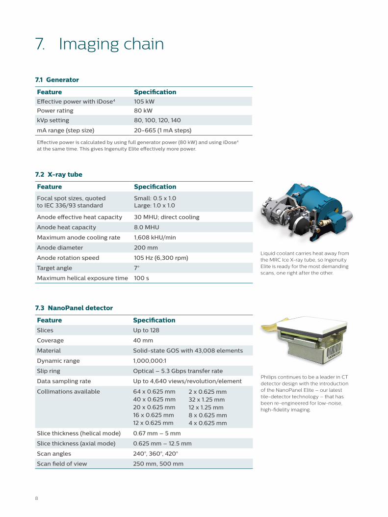
Philips continues to be a leader in CT detector design with the introduction of the NanoPanel Elite – our latest tile-detector technology – that has been re-engineered for low-noise, high-fidelity imaging.
Liquid coolant carries heat away from the MRC Ice X-ray tube, so Ingenuity Elite is ready for the most demanding scans, one right after the other.
7.1 Generator
Feature SpecificationEffective power with iDose4 105 kW
Power rating 80 kW
kVp setting 80, 100, 120, 140
mA range (step size) 20-665 (1 mA steps)
Effective power is calculated by using full generator power (80 kW) and using iDose4 at the same time. This gives Ingenuity Elite effectively more power.
7.2 X-ray tube
Feature Specification
Focal spot sizes, quoted to IEC 336/93 standard
Small: 0.5 x 1.0Large: 1.0 x 1.0
Anode effective heat capacity 30 MHU; direct cooling
Anode heat capacity 8.0 MHU
Maximum anode cooling rate 1,608 kHU/min
Anode diameter 200 mm
Anode rotation speed 105 Hz (6,300 rpm)
Target angle 7°
Maximum helical exposure time 100 s
7.3 NanoPanel detector
Feature SpecificationSlices Up to 128
Coverage 40 mm
Material Solid-state GOS with 43,008 elements
Dynamic range 1,000,000:1
Slip ring Optical – 5.3 Gbps transfer rate
Data sampling rate Up to 4,640 views/revolution/element
Collimations available 64 x 0.625 mm 40 x 0.625 mm 20 x 0.625 mm 16 x 0.625 mm12 x 0.625 mm
Slice thickness (helical mode) 0.67 mm – 5 mm
Slice thickness (axial mode) 0.625 mm – 12.5 mm
Scan angles 240°, 360°, 420°
Scan field of view 250 mm, 500 mm
2 x 0.625 mm32 x 1.25 mm 12 x 1.25 mm8 x 0.625 mm 4 x 0.625 mm
98
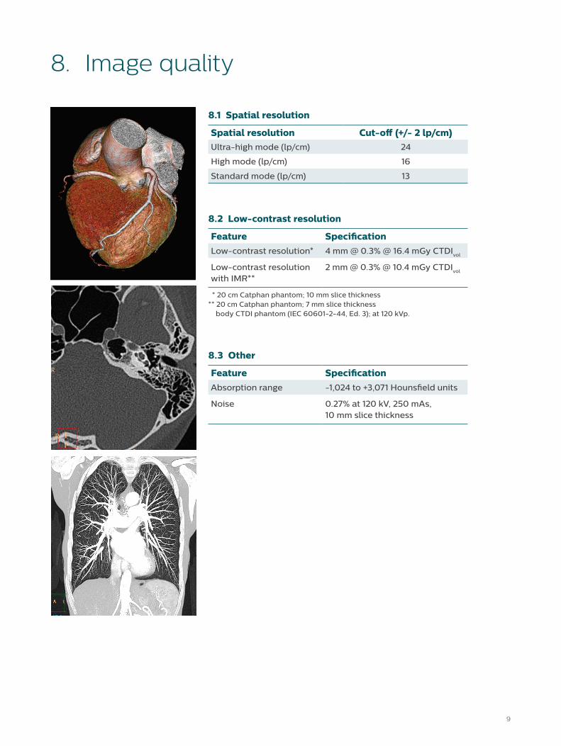
8. Image quality
8.2 Low-contrast resolution
Feature SpecificationLow-contrast resolution* 4 mm @ 0.3% @ 16.4 mGy CTDIvol
Low-contrast resolutionwith IMR**
2 mm @ 0.3% @ 10.4 mGy CTDIvol
* 20 cm Catphan phantom; 10 mm slice thickness** 20 cm Catphan phantom; 7 mm slice thickness body CTDI phantom (IEC 60601-2-44, Ed. 3); at 120 kVp.
8.1 Spatial resolution
Spatial resolution Cut-off (+/- 2 lp/cm)Ultra-high mode (lp/cm) 24
High mode (lp/cm) 16
Standard mode (lp/cm) 13
8.3 Other
Feature SpecificationAbsorption range -1,024 to +3,071 Hounsfield units
Noise 0.27% at 120 kV, 250 mAs, 10 mm slice thickness
1110
9. Reconstruction
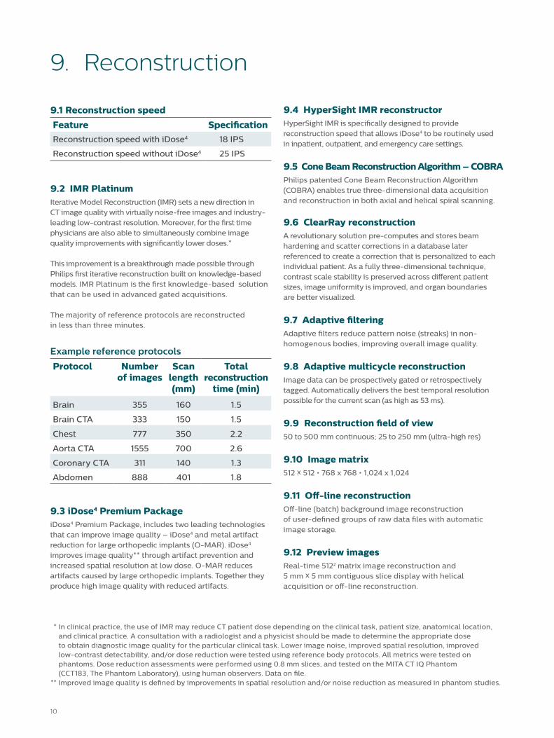
9.2 IMR Platinum Iterative Model Reconstruction (IMR) sets a new direction in CT image quality with virtually noise-free images and industry-leading low-contrast resolution. Moreover, for the first time physicians are also able to simultaneously combine image quality improvements with significantly lower doses.*
This improvement is a breakthrough made possible through Philips first iterative reconstruction built on knowledge-based models. IMR Platinum is the first knowledge-based solution that can be used in advanced gated acquisitions. The majority of reference protocols are reconstructed in less than three minutes.
Example reference protocols
Protocol Number of images
Scan length (mm)
Total reconstruction
time (min)
Brain 355 160 1.5
Brain CTA 333 150 1.5
Chest 777 350 2.2
Aorta CTA 1555 700 2.6
Coronary CTA 311 140 1.3
Abdomen 888 401 1.8
9.3 iDose4 Premium PackageiDose4 Premium Package, includes two leading technologies that can improve image quality – iDose4 and metal artifact reduction for large orthopedic implants (O-MAR). iDose4 improves image quality** through artifact prevention and increased spatial resolution at low dose. O-MAR reduces artifacts caused by large orthopedic implants. Together they produce high image quality with reduced artifacts.
9.4 HyperSight IMR reconstructorHyperSight IMR is specifically designed to provide reconstruction speed that allows iDose4 to be routinely used in inpatient, outpatient, and emergency care settings.
9.5 Cone Beam Reconstruction Algorithm – COBRAPhilips patented Cone Beam Reconstruction Algorithm (COBRA) enables true three-dimensional data acquisition and reconstruction in both axial and helical spiral scanning.
9.6 ClearRay reconstructionA revolutionary solution pre-computes and stores beam hardening and scatter corrections in a database later referenced to create a correction that is personalized to each individual patient. As a fully three-dimensional technique, contrast scale stability is preserved across different patient sizes, image uniformity is improved, and organ boundaries are better visualized.
9.7 Adaptive filteringAdaptive filters reduce pattern noise (streaks) in non-homogenous bodies, improving overall image quality.
9.8 Adaptive multicycle reconstructionImage data can be prospectively gated or retrospectively tagged. Automatically delivers the best temporal resolution possible for the current scan (as high as 53 ms).
9.9 Reconstruction field of view50 to 500 mm continuous; 25 to 250 mm (ultra-high res)
9.10 Image matrix512 x 512 • 768 x 768 • 1,024 x 1,024
9.11 Off-line reconstructionOff-line (batch) background image reconstruction of user-defined groups of raw data files with automatic image storage.
9.12 Preview imagesReal-time 5122 matrix image reconstruction and 5 mm x 5 mm contiguous slice display with helical acquisition or off-line reconstruction.
9.1 Reconstruction speed
Feature SpecificationReconstruction speed with iDose4 18 IPS
Reconstruction speed without iDose4 25 IPS
* In clinical practice, the use of IMR may reduce CT patient dose depending on the clinical task, patient size, anatomical location, and clinical practice. A consultation with a radiologist and a physicist should be made to determine the appropriate dose to obtain diagnostic image quality for the particular clinical task. Lower image noise, improved spatial resolution, improved low-contrast detectability, and/or dose reduction were tested using reference body protocols. All metrics were tested on phantoms. Dose reduction assessments were performed using 0.8 mm slices, and tested on the MITA CT IQ Phantom (CCT183, The Phantom Laboratory), using human observers. Data on file. ** Improved image quality is defined by improvements in spatial resolution and/or noise reduction as measured in phantom studies.
1110
10. Clinical enhancements
Optional10.1 SyncRightThe Philips CT SyncRight option enables easy and efficient communication between the CT system and the injector in order to facilitate delivering appropriate contrast dose and consistent image quality.
10.2 Bolus trackingAn automated injection planning technique to monitor actual contrast enhancement and initiate scanning at a predetermined level.
10.3 Spiral Auto Start (SAS)Spiral Auto Start allows the injector to communicate with the scanner. This allows the technologist tomonitor the contrast injection and to start the scan (with a predetermined delay) while in the scan room.
10.4 Patient centering on surviewTraditionally, patients are centered using the gantry laser lights; with this feature it is possible to improve patient centering using the lateral surview with real-time feedback.
10.5 Clinical applications, standard• Calcium Scoring • Cardiac Viewer• CT Reporting
Optional10.6 Clinical applications, optional• Advanced Brain Perfusion • Bone Mineral Analysis• Dental Analysis
10.7 Pulmonary ToolkitPhilips Pulmonary Toolkit enables the user to trigger a scan at a particular breath level, reducing artifacts caused by respiratory motion. This allows enhanced chest imaging of patients who cannot hold their breath. The Philips Bellows device is included.
10.8 Pulmonary Toolkit for OncologyPhilips Pulmonary Toolkit for Oncology includes the features found in the Philips Pulmonary Toolkit, and also includes Retrospective Spiral (4D CT) capabilities and support for the Varian RPMTM (device not included).
10.7 RateResponsive CV toolkit for IngenuityEnables cardiac imaging and includes an ECG monitor, Retrospective Tagging, Prospective Gating, Cardiac Viewer, Heartbeat-CS, and CT Reporting. Uses Philips exclusive Adaptive Multicycle Reconstruction algorithm to enhance temporal resolution – as high as 53 ms – and uses Philips patented Beat-to-Beat Algorithm to automatically find the best phase for cardiac imaging. Includes automatic arrhythmia detection and management.
10.8 Step & Shoot CompleteStep & Shoot Complete enables low-dose, prospectively ECG-triggered, axial thoracic imaging. Step & Shoot Complete allows gated, submillimeter, isotropic imaging of the entire thorax (up to 50 cm transaxial field of view), including the coronary arteries.
Step & Shoot Complete is well suited for patients with heart rates below 65 bpm. Arrhythmias are managed in real-time using proprietary, prospective-detection algorithms to pause acquisition during unstable heart rhythms.
Optional10.9 Jog ScanProvides up to 80 mm of organ coverage for perfusion studies. An axial scan is taken in one location, the couch translates to another location within a few seconds, and another axial scan is taken. These multiple datasets are registered automatically to provide the extended coverage.
10.11 CT InterventionalCT Interventional includes enhanced interventional capabilities to increase throughput and control of interventional procedures. With the option of either cart-mount or ceiling-mount solutions, the system helps you in your efforts to drive clinical confidence and consistency with flexible displays (1:1, 3:1, or volumetric) and allows the user to adjust the viewing convention or scan parameters and to switch scan modes on the fly. Reference series display enhances intra-procedural needle guidance. Both the single and continuous interventional scan modes support iDose4 and are DoseRight- and DRI-capable.
The Philips interventional table control option enhances operational efficiency during CT-guided interventional procedures.
• CT Viewer• Filming• Functional CT
1312
11. Networking and storage
11.1 NetworkingSupports 10/100/1000 Mbps (10/100/1000 BaseT) networks. For optimal performance, Philips recommends a minimum 100 Mbps network (1 Gbps preferred) and for the CT network to be segmented from the rest of the hospital network.
11.2 DICOMDICOM 3.0-compliant image format. Lossless image compression/decompression is used during image storage/retrieval to/from all local storage areas. Images can be auto-stored to selected archive media.
Includes the following DICOM functionality:• Service class user and profile (CT, MR, NM,
Secondary Capture)• DICOM Print• DICOM Modality Worklist• Query/Retrieve User and Provider• Modality Performed Procedure Step User• Storage Commitment User• Removable Media• Structured Reports
11.3 DICOM connectivityFull implementation of the DICOM 3.0 communications protocol allows connectivity to DICOM 3.0-compliant scanners, workstations, and printers; supports IHE requirements for DICOM connectivity. Further details on connectivity and interoperability are provided within the DICOM Conformance statement.
11.4 DICOM DVD/CD writerStores DICOM images and associated image viewing software on DVD/CD media. Images on these DVD/CDs can be viewed and manipulated on PCs meeting the minimum specifications. Suited for individual result storage and referring physician support.
11.5 FilmingThis function allows the user to set up and store filming parameters. Pre-stored protocols can be set to include auto-filming. The operator can film immediately after each image, at the end of a series, or after the end of a study, and review images before printing. The operator can also automatically film the study at three different windows and incorporate “Combine Images” functionality to manage large datasets. Basic monochrome and color DICOM print capability are supported.
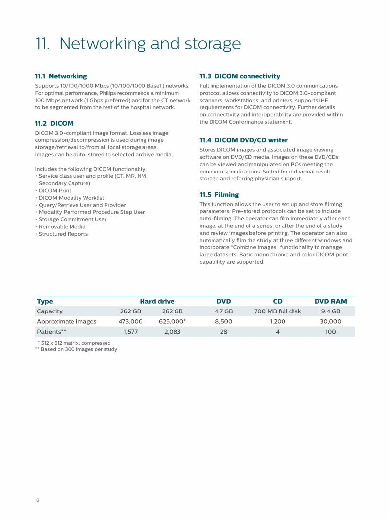
Type Hard drive DVD CD DVD RAMCapacity 262 GB 262 GB 4.7 GB 700 MB full disk 9.4 GB
Approximate images 473,000 625,000* 8,500 1,200 30,000
Patients** 1,577 2,083 28 4 100
* 512 x 512 matrix; compressed** Based on 300 images per study
1312
12. Site planning
12.1 Power requirements• 200/208/240/380/400/415/480/500 VAC• 50/60 Hz • 112.5 kVA supply (150 kVA preferred) • Three-phase distribution source
12.3 Environmental requirementsTemperatureGantry room 18° to 24° C (64° to 75° F)Control room 15° to 24° C (59° to 75° F)
HumidityGantry/Control 35% to 70% non-condensing
Heat dissipationGantry 18,000 BTU/hourIsotran LM 2,210 BTU/hourHost 2,484 BTU/hourDual server 8,226 BTU/hour
Optional12.2 Console Uninterrupted Power Supply (UPS)Provides up to 30 minutes of backup power for host and reconstruction system.
1514
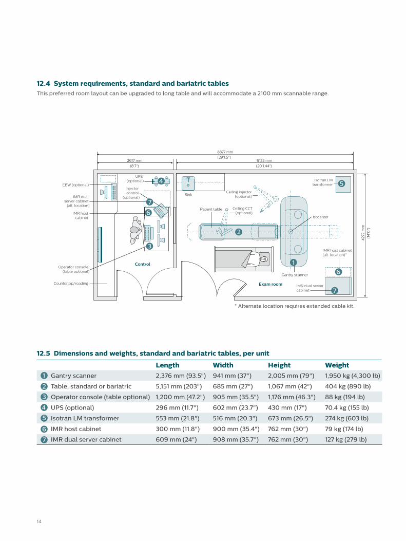
12.4 System requirements, standard and bariatric tablesThis preferred room layout can be upgraded to long table and will accommodate a 2100 mm scannable range.
8877 mm
(29'1.5")2617 mm
(8'7")
6133 mm
(20'1.44")
Control
Exam room
Isocenter
Sink
Countertop/reading
42
72 m
m
(14
'0")
IMR host cabinet(alt. location)*
Ceiling CCT(optional)
IMR dual servercabinet
Isotran LMtransformer
Ceiling injector(optional)
Patient table
Gantry scanner
Operator console (table optional)
IMR hostcabinet
IMR dual server cabinet
(alt. location)
EBW (optional)
UPS (optional)
Injector control
(optional)
* Alternate location requires extended cable kit.
7
6
1
2
3
4 5
6
7
12.5 Dimensions and weights, standard and bariatric tables, per unit
Length Width Height Weight1 Gantry scanner 2,376 mm (93.5") 941 mm (37") 2,005 mm (79") 1,950 kg (4,300 lb)
2 Table, standard or bariatric 5,151 mm (203") 685 mm (27") 1,067 mm (42") 404 kg (890 lb)
3 Operator console (table optional) 1,200 mm (47.2") 905 mm (35.5") 1,176 mm (46.3") 88 kg (194 lb)
UPS (optional) 296 mm (11.7") 602 mm (23.7") 430 mm (17") 70.4 kg (155 lb)
Isotran LM transformer 553 mm (21.8") 516 mm (20.3") 673 mm (26.5") 274 kg (603 lb)
IMR host cabinet 300 mm (11.8") 900 mm (35.4") 762 mm (30") 79 kg (174 lb)
IMR dual server cabinet 609 mm (24") 908 mm (35.7") 762 mm (30") 127 kg (279 lb)
6
7
4
5
1514
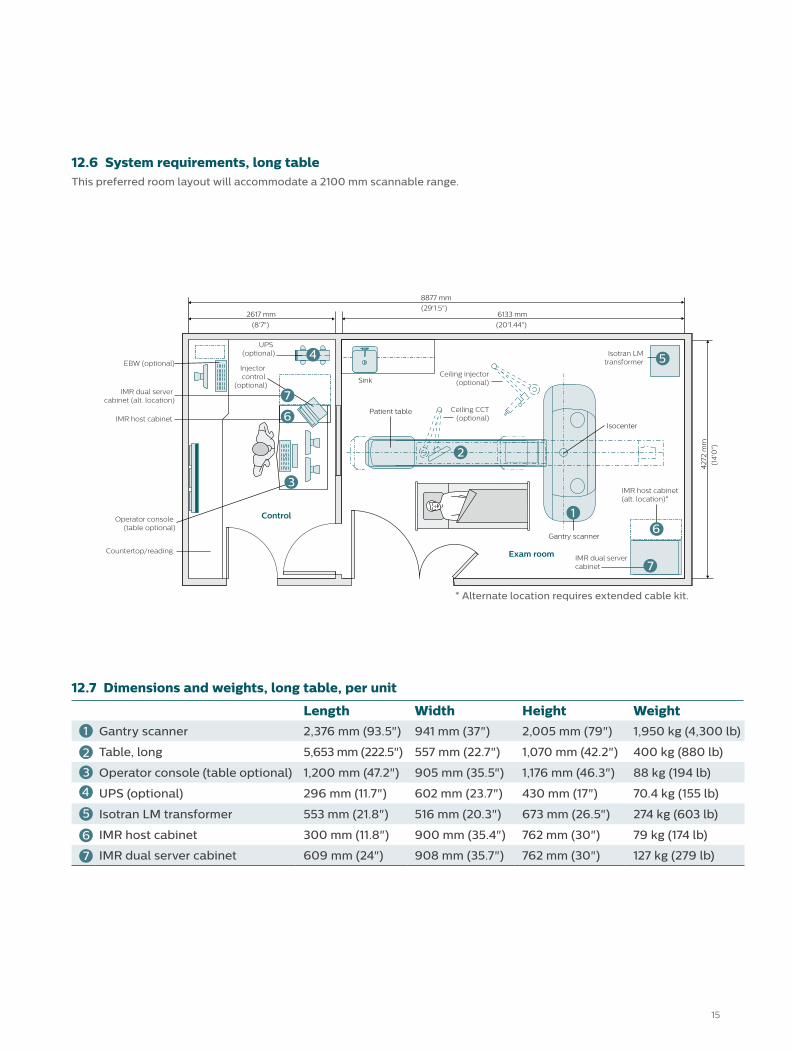
12.6 System requirements, long tableThis preferred room layout will accommodate a 2100 mm scannable range.
8877 mm
(29'1.5")2617 mm
(8'7")
6133 mm
(20'1.44")
42
72 m
m
(14
'0")
Control
Exam roomCountertop/reading
Operator console (table optional)
IMR host cabinet
IMR dual server cabinet (alt. location)
Sink
Ceiling CCT(optional)
Isotran LMtransformer
Ceiling injector(optional)
Patient table
Isocenter
IMR dual servercabinet
Gantry scanner
EBW (optional)
UPS (optional)
Injector control
(optional)
IMR host cabinet(alt. location)*
* Alternate location requires extended cable kit.
7
7
1
2
3
4 5
6
7
6
12.7 Dimensions and weights, long table, per unit
Length Width Height Weight1 Gantry scanner 2,376 mm (93.5") 941 mm (37") 2,005 mm (79") 1,950 kg (4,300 lb)
2 Table, long 5,653 mm (222.5") 557 mm (22.7") 1,070 mm (42.2") 400 kg (880 lb)
3 Operator console (table optional) 1,200 mm (47.2") 905 mm (35.5") 1,176 mm (46.3") 88 kg (194 lb)
UPS (optional) 296 mm (11.7") 602 mm (23.7") 430 mm (17") 70.4 kg (155 lb)
Isotran LM transformer 553 mm (21.8") 516 mm (20.3") 673 mm (26.5") 274 kg (603 lb)
IMR host cabinet 300 mm (11.8") 900 mm (35.4") 762 mm (30") 79 kg (174 lb)
IMR dual server cabinet 609 mm (24") 908 mm (35.7") 762 mm (30") 127 kg (279 lb)
6
7
4
5
© 2014 Koninklijke Philips N.V. All rights are reserved.
Philips Healthcare reserves the right to make changes in specifications and/or to discontinue any product at any time without notice or obligation and will not be liable for any consequences resulting from the use of this publication.
Please visit www.philips.com/IngenuityCT
Printed in The Netherlands.4522 991 05591 * DEC 2014
The images and descriptions contained herein provide technical specifications and optional features which may not be included with the standard system configuration. Contact your local Philips Representative for complete specific system details.
Some or all of the products, features, and accessories shown or described herein may not be available in your market. Please contact your local Philips Representative for availability.
The Ingenuity Elite with IMR is a configuration of the Ingenuity CT.
Optimize your system’s performance both now and in the future with regular
and ongoing updates, including functionality improvements and remote technical support.
Enhance your equipment with regular technology upgrades, and take advantage
of the newest features and capabilities.
Transform your investment at the end of your system’s life by transitioning
seamlessly to a next-generation solution or refurbished option.
Enhancing the capabilities of your existing iCT and Ingenuity CT
family scanners, the SmartPath upgrade offers easy access
to knowledge-based iterative reconstruction.
Recommended


