European Journal of Academic Essays 4(8): 202-223, 2017 ISSN (online): 2183-1904 ISSN (print): 2183-3818 www.euroessays.org
Cryptococcosis in Animals and Birds: A Review
Mohamed K. Refai 1, Mahmoud El-Hariri
1and Randa Alarousy
2,3
1 Department of Microbiology, Faculty of Veterinary Medicine Cairo University 2 Department of Microbiology and Immunology, National Research Center, Dokki, Giza, Egypt.
3 Department of Medical Laboratories, College of Applied Medical Sciences, Majmaah University, KSA
Abstract: Cryptococcus infections in bovines was mostly reported in association with mastitis, mainly in cattle. Cryptococcus
neoformans was recognized as the cause of severe outbreaks of mastitis in cattle and also in sporadic cases in buffaloes. Bovine
systemic cryptococcosis was rarely diagnosed. In camels, cryptococcosis is very rare. It has been reported in South American
camelids (llamas, alpacas and vicunas). In equines, cryptococcosis is uncommon. The nasal cavity is the most common site of
infection. Sporadic cases have been associated with granulomatous pneumonia, nasal granuloma, endometritis and placentitis with
neonatal cryptococcal pneumonia, abortion and mesenteric lymph node abscesses. Cryptococcal meningitis in equines is almost
associated with pneumonia and disseminated infection. Cryptococcus species were reported in sheep and goats as causes of
mastitis. Experimental mastitis in goats was induced by unilateral intramammary inoculation of Cryptococcus neoformans. Nasal
cryptococcosis is frequently seen as clinical signs in cats and dogs. With time, infections involving the nasal cavity can spread to
adjacent structures and disseminate to the brain and other organ and even the skin. Cryptococcal pneumonia and meningitis due to
Cryptococcus gatti was reported also in goats. In wild animals, cryptococcosis occurs in different animals and Cryptococcus
species. can affect the gastrointestinal and respiratory systems, nasal cavity and eyes.
Keywords: Cryptococcosis, Cryptococcus neoformans, Cryptococcus gatti, bovines, equines, camels, sheep and
goats, cats and dogs, wild animals, birds.
Corresponding author: Randa [email protected] -
1. Introduction
Cryptococcosis in animals is a systemic fungal infection of
worldwide significance that usually initially infects the nasal
cavity, paranasal tissues, or lungs. It can then disseminate,
most commonly to the skin, eyes, or central nervous system.
Cryptococcosis is a fungal disease caused by C. neoformans
and C. gattii, which are ubiquitous, saprophytic, round,
basidiomycetous yeasts (5 to 10 μm) with a large
heteropolysaccharide capsule (1 to 30 μm) that does not take
up common cytologic stains.
According to the current classification, the species complex
comprises two species, namely C. neoformans and C.
gattii [1& 2] with serotypes A, D and AD for the former,
and B and C for the latter species. Cryptococcus
neoformans currently consists of two varieties: C.
neoformans variety grubii (serotype A) [3] and C.
neoformans variety neoformans (serotype D) [4]
Recently, Hagen et al. [5] proposed the
genus Cryptococcus to include 3 separate species, C.
European Journal of Academic Essays 4(8): 202-223, 2017
204
neoformans (Cryptococcus neoformans var. grubii)
represented by genotypes VNI and VNII, C. deneoformans
(Cryptococcus neoformans var. neoformans) represented by
genotypes VNIII and VNIV, and Cryptococcus
gattii (represented by genotypes VGI, VGII, VGIII, and
VGIV).
The environmental reservoir of C. neoformans is usually
related to bird faeces, particularly pigeon droppings.
However, this yeast has also been found in decaying trees,
wood and plant debris, waterways and soil, all usually
contaminated with bird excrement [6, 7, 8, 9, 10, 11, 12].
The epidemiology of clinical disease depends largely on the
species of infecting organism. Cryptococcus neoformans
var. grubii (serotype A) and C. neoformans var. neoformans
(serotype D) are globally distributed and infect
predominantly immunocompromised hosts. Cryptococcus
gattii (serotypes B and C) has recently been recognized as a
species distinct from C. neoformans based on molecular and
mating type characteristics.
2. Cryptococcosis in domestic animals
2.1. Cryptococcosis in bovines:
Cryptococcus infections in bovines was mostly reported in
association with mastitis, mainly in bovines. Klein [13] was
the first to isolate a yeast from a case of mastitis, which he
reported to be identical with strains of Cryptococcus
neoformans of human and plant origin. Almost 50 years
later, Cryptococcus neoformans was recognized as the cause
of severe outbreaks of mastitis in cattle [14, 15, 16, 17].
Pounden et al. [18] reported the clinical aspects of an
outbreak in which 106 cows were affected in a 235 cow-
herd. They stated that during the outbreak, Cryptococcus has
been isolated from samples where no visible changes were
noted in either the gland or milk, and the cases with visible
signs varied from mild and transient swelling of one or more
quarters of the udder to severe swelling and distention of the
affected glands
Cryptococcal mastitis was detected also in sporadic cases by
Abdel Ghani et al. [19], Rahman et al., [20], Moawad,
[21], Hassan et al. [22], mostly, following treatment with
antibiotics [23, 24, 25, 26]. On the other hand, Rippon [27]
emphasized that cryptococcal mastitis in dairy cows is
worldwide in distribution.
Cryptococcus neoformans was the most commonly recorded
species in cows [28, 29, 8]. Cr .neoformans was also
recorded as a cause of mastitis in buffaloes [30, 31]. Other
species like C. albidus, C. laurentii, C. flavus, C.
lactativorus, C. luteolos, C. terreus, C. uniguttulatus and
Cryptococcus species were also reported in few reports [28,
32, 33, 34, 35, 36, 37, 38, Table 1].
Cryptococcal pneumonia was infrequently reported in
bovines [39, 22]. On the other hand, bovine systemic
cryptococcosis was rarely diagnosed. Only two reports could
be found in the available literature [40, 41]. Similar findings
were obtained in case of abortion caused by Cryptococcus
species [40, 41].
European Journal of Academic Essays 4(8): 202-223, 2017
205
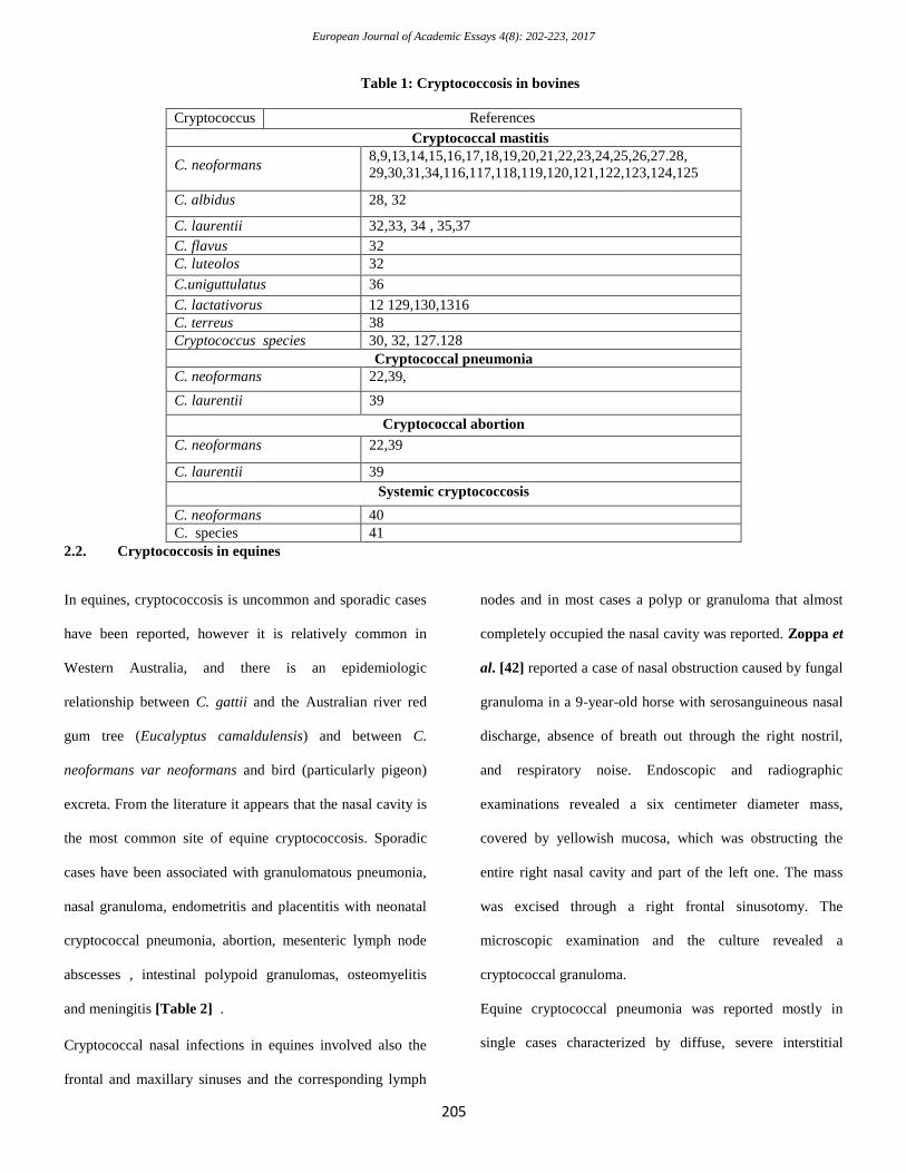
Table 1: Cryptococcosis in bovines
Cryptococcus References
Cryptococcal mastitis
C. neoformans
8,9,13,14,15,16,17,18,19,20,21,22,23,24,25,26,27.28,
29,30,31,34,116,117,118,119,120,121,122,123,124,125
C. albidus 28, 32
C. laurentii 32,33, 34 , 35,37
C. flavus 32
C. luteolos 32
C.uniguttulatus 36
C. lactativorus 12 129,130,1316
C. terreus 38
Cryptococcus species 30, 32, 127.128
Cryptococcal pneumonia
C. neoformans 22,39,
C. laurentii 39
Cryptococcal abortion
C. neoformans 22,39
C. laurentii 39
Systemic cryptococcosis
C. neoformans 40
C. species 41
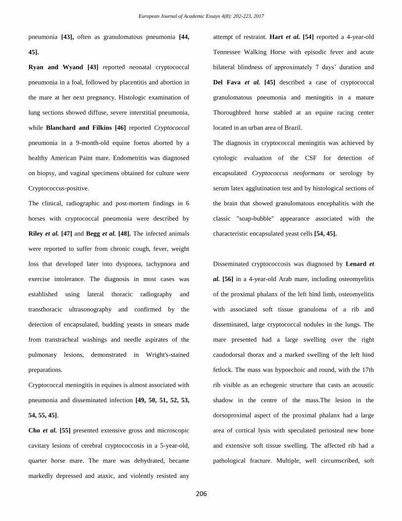
2.2. Cryptococcosis in equines
In equines, cryptococcosis is uncommon and sporadic cases
have been reported, however it is relatively common in
Western Australia, and there is an epidemiologic
relationship between C. gattii and the Australian river red
gum tree (Eucalyptus camaldulensis) and between C.
neoformans var neoformans and bird (particularly pigeon)
excreta. From the literature it appears that the nasal cavity is
the most common site of equine cryptococcosis. Sporadic
cases have been associated with granulomatous pneumonia,
nasal granuloma, endometritis and placentitis with neonatal
cryptococcal pneumonia, abortion, mesenteric lymph node
abscesses , intestinal polypoid granulomas, osteomyelitis
and meningitis [Table 2] .
Cryptococcal nasal infections in equines involved also the
frontal and maxillary sinuses and the corresponding lymph
nodes and in most cases a polyp or granuloma that almost
completely occupied the nasal cavity was reported. Zoppa et
al. [42] reported a case of nasal obstruction caused by fungal
granuloma in a 9-year-old horse with serosanguineous nasal
discharge, absence of breath out through the right nostril,
and respiratory noise. Endoscopic and radiographic
examinations revealed a six centimeter diameter mass,
covered by yellowish mucosa, which was obstructing the
entire right nasal cavity and part of the left one. The mass
was excised through a right frontal sinusotomy. The
microscopic examination and the culture revealed a
cryptococcal granuloma.
Equine cryptococcal pneumonia was reported mostly in
single cases characterized by diffuse, severe interstitial
European Journal of Academic Essays 4(8): 202-223, 2017
206
pneumonia [43], often as granulomatous pneumonia [44,
45].
Ryan and Wyand [43] reported neonatal cryptococcal
pneumonia in a foal, followed by placentitis and abortion in
the mare at her next pregnancy. Histologic examination of
lung sections showed diffuse, severe interstitial pneumonia,
while Blanchard and Filkins [46] reported Cryptococcal
pneumonia in a 9-month-old equine foetus aborted by a
healthy American Paint mare. Endometritis was diagnosed
on biopsy, and vaginal specimens obtained for culture were
Cryptococcus-positive.
The clinical, radiographic and post-mortem findings in 6
horses with cryptococcal pneumonia were described by
Riley et al. [47] and Begg et al. [48]. The infected animals
were reported to suffer from chronic cough, fever, weight
loss that developed later into dyspnoea, tachypnoea and
exercise intolerance. The diagnosis in most cases was
established using lateral thoracic radiography and
transthoracic ultrasonography and confirmed by the
detection of encapsulated, budding yeasts in smears made
from transtracheal washings and needle aspirates of the
pulmonary lesions, demonstrated in Wright's-stained
preparations.
Cryptococcal meningitis in equines is almost associated with
pneumonia and disseminated infection [49, 50, 51, 52, 53,
54, 55, 45].
Cho et al. [55] presented extensive gross and microscopic
cavitary lesions of cerebral cryptococcosis in a 5-year-old,
quarter horse mare. The mare was dehydrated, became
markedly depressed and ataxic, and violently resisted any
attempt of restraint. Hart et al. [54] reported a 4-year-old
Tennessee Walking Horse with episodic fever and acute
bilateral blindness of approximately 7 days’ duration and
Del Fava et al. [45] described a case of cryptococcal
granulomatous pneumonia and meningitis in a mature
Thoroughbred horse stabled at an equine racing center
located in an urban area of Brazil.
The diagnosis in cryptococcal meningitis was achieved by
cytologic evaluation of the CSF for detection of
encapsulated Cryptococcus neoformans or serology by
serum latex agglutination test and by histological sections of
the brain that showed granulomatous encephalitis with the
classic "soap-bubble" appearance associated with the
characteristic encapsulated yeast cells [54, 45].
Disseminated cryptococcosis was diagnosed by Lenard et
al. [56] in a 4-year-old Arab mare, including osteomyelitis
of the proximal phalanx of the left hind limb, osteomyelitis
with associated soft tissue granuloma of a rib and
disseminated, large cryptococcal nodules in the lungs. The
mare presented had a large swelling over the right
caudodorsal thorax and a marked swelling of the left hind
fetlock. The mass was hypoechoic and round, with the 17th
rib visible as an echogenic structure that casts an acoustic
shadow in the centre of the mass.The lesion in the
dorsoproximal aspect of the proximal phalanx had a large
area of cortical lysis with speculated periosteal new bone
and extensive soft tissue swelling. The affected rib had a
pathological fracture. Multiple, well circumscribed, soft
European Journal of Academic Essays 4(8): 202-223, 2017
207
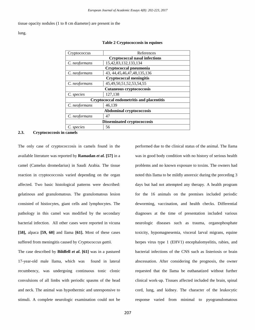
tissue opacity nodules (1 to 8 cm diameter) are present in the
lung.
Table 2 Cryptococcosis in equines
Cryptococcus References
Cryptococcal nasal infections
C. neoformans 15,42,83,132,133,134
Cryptococcal pneumonia
C. neoformans 43, 44,45,46,47,48,135,136
Cryptococcal meningitis
C. neoformans 45,49,50,51,52,53,54,55
Cutaneous cryptococcosis
C. species 127,138
Cryptococcal endometritis and placentitis
C. neoformans 46,139
Abdominal cryptococcosis
C. neoformans 47
Disseminated cryptococcosis
C. species 56
2.3. Cryptococcosis in camels
The only case of cryptococcosis in camels found in the
available literature was reported by Ramadan et al. [57] in a
camel (Camelus dromedarius) in Saudi Arabia. The tissue
reaction in cryptococcosis varied depending on the organ
affected. Two basic histological patterns were described:
gelatinous and granulomatous. The granulomatous lesion
consisted of histiocytes, giant cells and lymphocytes. The
pathology in this camel was modified by the secondary
bacterial infection. All other cases were reported in vicuna
[58], alpaca [59, 60] and llama [61]. Most of these cases
suffered from meningitis caused by Cryptococcus gattii.
The case described by Bildfell et al. [61] was in a pastured
17-year-old male llama, which was found in lateral
recumbency, was undergoing continuous tonic clonic
convulsions of all limbs with periodic spasms of the head
and neck. The animal was hypothermic and unresponsive to
stimuli. A complete neurologic examination could not be
performed due to the clinical status of the animal. The llama
was in good body condition with no history of serious health
problems and no known exposure to toxins. The owners had
noted this llama to be mildly anorexic during the preceding 3
days but had not attempted any therapy. A health program
for the 16 animals on the premises included periodic
deworming, vaccination, and health checks. Differential
diagnoses at the time of presentation included various
neurologic diseases such as trauma, organophosphate
toxicity, hypomagnesemia, visceral larval migrans, equine
herpes virus type 1 (EHV1) encephalomyelitis, rabies, and
bacterial infections of the CNS such as listeriosis or brain
abscessation. After considering the prognosis, the owner
requested that the llama be euthanatized without further
clinical work-up. Tissues affected included the brain, spinal
cord, lung, and kidney. The character of the leukocytic
response varied from minimal to pyogranulomatous
European Journal of Academic Essays 4(8): 202-223, 2017
208
meningitis with intralesional yeast that were bordered by a non-staining halo.
2.4. Cryptococcosis in sheep and goats
Cryptococcus species were reported in sheep and goats as
causes of mastitis, pneumonia, meningitis and abortion
[Table 3].
Experimental mastitis in goats was reported by unilateral
intramammary inoculation of 10 goats with 2 x 10⁶ cells of
Cryptococcus neoformans [62]. The infection resulted in the
development of mastitis, with gross and microscopic lesions
being restricted to the infected udder halves only and there
was no dissemination of infection to the opposite uninfected
udder halves as well as to other organs of the body. The
experiment was continued for 40 days, with 2 animals, each
from the infected and control groups being killed on 5th,
10th, 20th, 30th and 40th day post-inoculation (DPI). Initial
enlargement of the infected udder halves was followed by
marked decrease in size leading to very small, firm and
nodular udder halves. After infection, there was also sharp
fall in the milk yield. Cryptococcal organisms were
demonstrated in the mastitic milk and udder impression
smears with special stains. C. neoformans was re isolated
from the milk of the only infected udder halves up to 25th
DPI. Microscopically, there was initially acute diffuse
purulent mastitis which later on became chronic,
characterized by marked infiltration of lymphocytes,
macrophages, extensive fibrosis and development of
multiple granulomas. The cryptococcal organisms could be
demonstrated in the udder sections only up to 30th DPI.
Mycotic pneumonia in goats due to Cryptococcus gatti was
reported in Spain by Baro et al. [63]. The strains were
isolated from lung (10 samples), liver (1 sample), and brain
(2 samples) tissue specimens from six goats suffering from
predominantly severe pulmonary disease that were
autopsied. Biotyping was performed by culturing the isolates
on L-canavanine-glycine-bromothymol blue medium and
testing them for the assimilation of D-proline and D-
tryptophan. Serotyping by agglutination tests confirmed the
characterization of all strains as C. gattii.
C. gattii was reported to cause 5 epidemic outbreaks of
cryptococcosis in goats grazing freely in west Spain
grasslands [64] . The goats belonged to various milking breeds and
were grazing with variable status of health and husbandry. Goats affected
by cryptococcosis showed similar respiratory symptoms,
consisting of mucopurulent nasal discharge, cough, dyspnea
and progressive cachexia, causing death in a period of 2 to 4
weeks. In three outbreaks many animals also showed ataxia,
midriasis, blindness and progressive paralysis. Clinical
prevalence varied from 2 to 12% in the different outbreaks.
On the other hand, Gutiérrez and García Marin [65]
presented an adult Blanca-Celtibérica doe originating from a
goat herd with a high prevalence of tuberculosis with
respiratory signs. At necropsy, this goat had a diffuse and
severe mycotic pneumonia associated with the presence of
Cryptococcus neoformans concomitant with pulmonary
focal caseous nodules from which Mycobacterium bovis was
European Journal of Academic Essays 4(8): 202-223, 2017
209
isolated. Microscopically, the mycotic lesion was a
granulomatous pneumonia with many large foamy
macrophages containing intracellular yeast bodies. The
extensive mycotic changes, their granulomatous nature, and
the lack of positive response to different immunologic tests
for mycobacterial infection suggested an impaired immune
status in this animal.
Cryptococcal meningitis in goats was described by
Luvizotto et al. [66], who euthanized four-year-old male
goat with a history of neurological disorder. It presented
uncommon nodules in the brain and lungs associated with
multiple abscesses, predominantly in the spleen and liver.
Histological examination of brain and lung sections revealed
yeast forms confirmed to be Cryptococcus gattii. On the
other hand, Stilwell and Pissarra [67] described a case of a
five year old buck showing severe neurological signs,
including paraplegia and strong pain reaction to touch of the
hindquarters region. Postmortem examination revealed
lumbar meningitis, lung nodules and caseous lymphadenitis
lesions. Encapsulated Cryptococcus neoformans were
identified from the lungs and meninges, showing that
cryptococcal meningitis should be included in the
differential diagnosis of goats showing paresis and
hyperesthesia. Both Cryptococcus gattii genotype
AFLP4/VGI and Cryptococcus neoformans var. neoformans
genotype AFLP2/VNIV were incriminated as causes of
meningoencephalitis in goats by Maestrale et al. [68].
Cryptococcal meningitis in goats was described by da Silva
et al. [69], who performed a study aimed to report a 5-year-
old goat showing intermittent dry cough, ruminal tympany,
anorexia, fever, tachycardia and tachypnea in State of São
Paulo, Brazil. Postmortem examination revealed numerous
2.0-6.0 cm diameter yellow gelatinous pulmonary masses.
Tissues were evaluated by a combination of pathological,
mycological, and molecular diagnostic techniques.
Microscopically, pneumonia granulomatous, multifocal to
coalescing, moderate, with many intralesional carminophilic
yeasts was observed. The immunohistochemistry and
mycological culture confirmed Cryptococcus spp. Internal
transcribed spacers and orotidine monophosphate
pyrophosphorylase nucleotide differentiation demonstrated
that the isolate corresponds to the C. gattii VGII molecular
subtype.
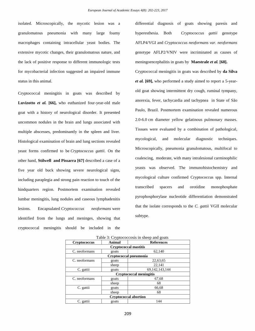
Table 3: Cryptococcosis in sheep and goats
Cryptococcus Animal References
Cryptococcal mastitis
C. neoformans goats 62,140
Cryptococcal pneumonia
C. neoformans
goats 22,63,65
sheep 22,141
C. gattii goats 69,142,143,144
Cryptococcal meningitis
C. neoformans goats 67,68
sheep 68
C. gattii goats 66,68
sheep 68
Cryptococcal abortion
C. gattii goats 144
European Journal of Academic Essays 4(8): 202-223, 2017
210
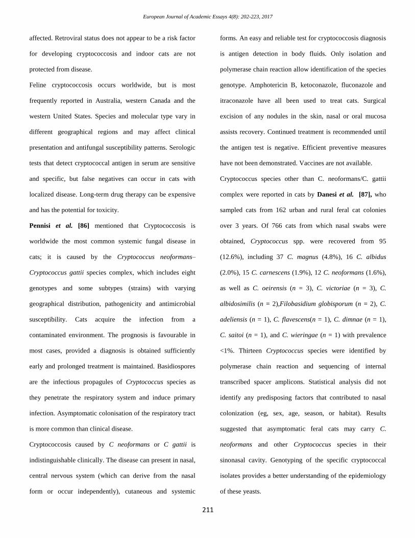
2.5. Cryptococcosis in cats and dogs
In cats and dogs, cryptococcosis can be either focal or
disseminated, affecting a single organ system or many
[Table 4]. It can begin insidiously, and may gradually
become more severe over weeks or months. Fever may be
absent, and if present, is often mild. Other nonspecific signs
can include lethargy, anorexia and weight loss. The most
common site of localized infections is the nasal cavity. Nasal
cryptococcosis is frequently seen clinical signs including
sneezing, snoring or snorting, dyspnea, nasal deformities
and/ or a mucopurulent, serous or sero-sanguineous nasal
discharge. Polyp-like masses sometimes protrude from one
or both nostrils. Cutaneous or subcutaneous swellings and
nodules may be seen on the face, particularly the bridge of
the nose, side of the face, upper lip or nostril. Some of these
lesions may ulcerate. In addition, the submandibular lymph
nodes are often enlarged. Rhinitis in cats was reported to be
caused by C. neoformans var. neoformans [70, 71, 72, 73].
With time, infections involving the nasal cavity can spread
to adjacent structures and disseminate to other organs [74,
75, 76, 77, 78], including the brain [79, 80] and eyes [79,
81] and even the skin [82]. Cutaneous involvement usually
appears as fluctuant or firm papules and nodules. Some skin
lesions may ulcerate, but there is little or no pruritus. Direct
inoculation of organisms into the skin can occasionally
cause solitary lesions.
Cryptococcus gattii has emerged since 1999 as an important
pathogen of humans and animals in southwestern British
Columbia. Historically thought to be restricted to the tropics
and subtropics, C. gattii has posed new diagnostic and
treatment challenges to veterinary practitioners working
within the recently identified endemic region. Clinical
reports of canine and feline cryptococcosis caused by C.
gattii diagnosed between January 1999 and December 2003
were reported. The most common manifestations of disease
were respiratory and central nervous system signs.
Multivariate survival analysis revealed that the only
significant predictor of mortality was the presence of central
nervous system signs upon presentation or during therapy.
Case fatality rates in both species were high. [70, 73, 83,
84].
Trivedi et al. [85] mentioned that Cryptococcosis,
principally caused by Cryptococcus neoformans and
Cryptococcus gattii, is the most common systemic mycosis
of cats worldwide. Cats may be infected following inhalation
of spores from the environment, with the nasal cavity
suspected as being the initial site of colonization and
subsequent infection. Other sites of infection in cats are the
skin, lungs, lymph nodes, central nervous system (CNS),
eyes and, occasionally, periarticular connective tissue.
Cryptococcosis can be diagnosed using serology (antigen
testing), cytologic examination of smears, histopathology or
culture. Treatment of localized disease is generally
successful using azole antifungal drugs; however, cats with
CNS involvement or disseminated disease require additional
treatment with amphotericin B, with or without flucytosine.
The prognosis is variable, depending on host and pathogen
factors. Some cats require long-term (>1 year) treatment or
indefinite therapy. Cats of any breed, gender and age may be
European Journal of Academic Essays 4(8): 202-223, 2017
211
affected. Retroviral status does not appear to be a risk factor
for developing cryptococcosis and indoor cats are not
protected from disease.
Feline cryptococcosis occurs worldwide, but is most
frequently reported in Australia, western Canada and the
western United States. Species and molecular type vary in
different geographical regions and may affect clinical
presentation and antifungal susceptibility patterns. Serologic
tests that detect cryptococcal antigen in serum are sensitive
and specific, but false negatives can occur in cats with
localized disease. Long-term drug therapy can be expensive
and has the potential for toxicity.
Pennisi et al. [86] mentioned that Cryptococcosis is
worldwide the most common systemic fungal disease in
cats; it is caused by the Cryptococcus neoformans–
Cryptococcus gattii species complex, which includes eight
genotypes and some subtypes (strains) with varying
geographical distribution, pathogenicity and antimicrobial
susceptibility. Cats acquire the infection from a
contaminated environment. The prognosis is favourable in
most cases, provided a diagnosis is obtained sufficiently
early and prolonged treatment is maintained. Basidiospores
are the infectious propagules of Cryptococcus species as
they penetrate the respiratory system and induce primary
infection. Asymptomatic colonisation of the respiratory tract
is more common than clinical disease.
Cryptococcosis caused by C neoformans or C gattii is
indistinguishable clinically. The disease can present in nasal,
central nervous system (which can derive from the nasal
form or occur independently), cutaneous and systemic
forms. An easy and reliable test for cryptococcosis diagnosis
is antigen detection in body fluids. Only isolation and
polymerase chain reaction allow identification of the species
genotype. Amphotericin B, ketoconazole, fluconazole and
itraconazole have all been used to treat cats. Surgical
excision of any nodules in the skin, nasal or oral mucosa
assists recovery. Continued treatment is recommended until
the antigen test is negative. Efficient preventive measures
have not been demonstrated. Vaccines are not available.
Cryptococcus species other than C. neoformans/C. gattii
complex were reported in cats by Danesi et al. [87], who
sampled cats from 162 urban and rural feral cat colonies
over 3 years. Of 766 cats from which nasal swabs were
obtained, Cryptococcus spp. were recovered from 95
(12.6%), including 37 C. magnus (4.8%), 16 C. albidus
(2.0%), 15 C. carnescens (1.9%), 12 C. neoformans (1.6%),
as well as C. oeirensis (n = 3), C. victoriae (n = 3), C.
albidosimilis (n = 2),Filobasidium globisporum (n = 2), C.
adeliensis (n = 1), C. flavescens(n = 1), C. dimnae (n = 1),
C. saitoi (n = 1), and C. wieringae (n = 1) with prevalence
<1%. Thirteen Cryptococcus species were identified by
polymerase chain reaction and sequencing of internal
transcribed spacer amplicons. Statistical analysis did not
identify any predisposing factors that contributed to nasal
colonization (eg, sex, age, season, or habitat). Results
suggested that asymptomatic feral cats may carry C.
neoformans and other Cryptococcus species in their
sinonasal cavity. Genotyping of the specific cryptococcal
isolates provides a better understanding of the epidemiology
of these yeasts.
European Journal of Academic Essays 4(8): 202-223, 2017
212
Table 4: Cryptococcosis in cats and dogs
Cryptococcus Animal References
Cryptococcal rhinitis
C. neoformans var. neoformans cats 70,71,72,73
dogs 71,145
C. gattii cats 70,73,83, 146,147,148
dogs 83,145
C. species Cats 149
Disseminated cryptococcosis
C. neoformans var. neoformans dogs 74
C. gattii cats 78
dogs 76
C. albidus cats 75
C. magnus 77
Cryptococcal meningitis
C. neoformans var. grubii cats 80
dogs 80
C. gattii cats 80
dogs 80
C. species cats 79, 150
Cryptococcal skin infection
C. species cats 82
Cryptococcal ophthalmitis
C. neoformans var. neoformans cats 81
C. species 79
Cryptococcal urinary tract infection
C. species cats 150.
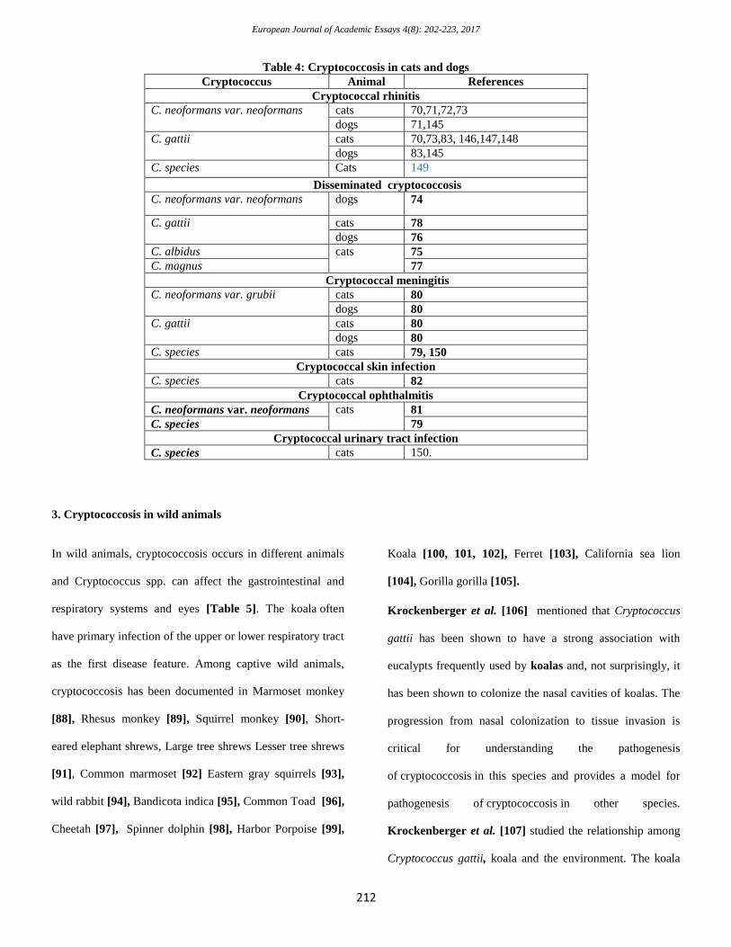
3. Cryptococcosis in wild animals
In wild animals, cryptococcosis occurs in different animals
and Cryptococcus spp. can affect the gastrointestinal and
respiratory systems and eyes [Table 5]. The koala often
have primary infection of the upper or lower respiratory tract
as the first disease feature. Among captive wild animals,
cryptococcosis has been documented in Marmoset monkey
[88], Rhesus monkey [89], Squirrel monkey [90], Short-
eared elephant shrews, Large tree shrews Lesser tree shrews
[91], Common marmoset [92] Eastern gray squirrels [93],
wild rabbit [94], Bandicota indica [95], Common Toad [96],
Cheetah [97], Spinner dolphin [98], Harbor Porpoise [99],
Koala [100, 101, 102], Ferret [103], California sea lion
[104], Gorilla gorilla [105].
Krockenberger et al. [106] mentioned that Cryptococcus
gattii has been shown to have a strong association with
eucalypts frequently used by koalas and, not surprisingly, it
has been shown to colonize the nasal cavities of koalas. The
progression from nasal colonization to tissue invasion is
critical for understanding the pathogenesis
of cryptococcosis in this species and provides a model for
pathogenesis of cryptococcosis in other species.
Krockenberger et al. [107] studied the relationship among
Cryptococcus gattii, koala and the environment. The koala
European Journal of Academic Essays 4(8): 202-223, 2017
213
was used as a natural biological sampler in an attempt to
understand the dynamics of Cryptococcus gattii in
Australian environments. Evidence of asymptomatic nasal
and skin colonization for extended periods by large numbers
of Cryptococcus gattii was obtained and geographical
factors assessed. The key finding was the ability of koalas to
amplify numbers of Cryptococcus gattii in certain
environments. Koalas were not found to be obligatory for
the survival of the organism in all environments.
Geographical factors alone could not explain differing rates
of nasal and skin colonization in koalas in different
environments. A strong association between healthy koalas
and Cryptococcus gattii was confirmed and Cryptococcus
gattii was isolated from novel sources, including the
turpentine gum tree (Syncarpia glomulifera), tallowwood
(Eucalyptus microcorys) and flooded gum (E. grandis).
Table 5. Cryptococcosis in wild animals
Cryptococcus species Animals References
C. neoformans
var. neoformans
Koalas 102,151
monkeys 89
Shrews 91
common toad 96
C. neoformans
var grubii
Ferrets 80,106
bandicoot 95
Gorilla 105
C. gattii Cheetah 97,152,155
Koalas 101,106,107,154
Ferrets 80,103,106
Dolphins 156,157
Porpoises 99,158
C. albidus Sea lion 104
C. yokohamensis Koalas 100
C. species Ferrets 103
Monkeys 88,90
Koalas 153
Marmosets 92
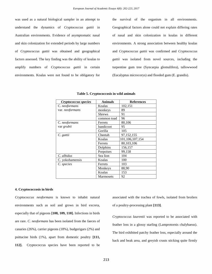
4. Cryptococcosis in birds
Cryptococcus neoformans is known to inhabit natural
environments such as soil and grows in bird excreta,
especially that of pigeons [108, 109, 110]. Infections in birds
are rare. C. neoformans has been isolated from the faeces of
canaries (26%), carrier pigeons (18%), budgerigars (2%) and
psittacine birds (1%), apart from domestic poultry [111,
112]. Cryptococcus species have been reported to be
associated with the trachea of fowls, isolated from broilers
of a poultry-processing plant [113].
Cryptococcus laurentii was reported to be associated with
feather loss in a glossy starling (Lamprotornis chalybaeus).
The bird exhibited patchy feather loss, especially around the
back and beak area, and greyish crusts sticking quite firmly
European Journal of Academic Essays 4(8): 202-223, 2017
214
to the underlying skin. The feathers had a greasy appearance
and disseminated a musty odour.
Malik et al. [114] analyzed clinical and laboratory findings
in 15 unreported cases of avian cryptococcosis from
Australia contrasted with 11 cases recorded in the literature.
Cryptococcus species produced localized invasive disease of
the upper respiratory tract of captive parrots living in
Australia. This resulted in signs referable to mycotic rhinitis
or to involvement of structures contiguous with the nasal
cavity, such as the beak, sinuses, choana, retrobulbar space
and palate. Cryptococcus appeared to behave as a primary
pathogen of immunocompetent hosts. One tissue specimen
was available from an Australian racing pigeon with
minimally invasive subcutaneous disease; immunohistology
demonstrated a Cryptococcus infection, presumably
subsequent to traumatic inoculation of yeast cells into the
subcutis.
Cryptococcus neoformans var. grubii was isolated from a
tissue specimen from an Australian racing pigeon with
minimally invasive subcutaneous disease; and was
demonstrated immunohistologically in the subcutis tissues
[114].
Cryptococcus gattii produced localized invasive disease of
the upper respiratory tract of captive parrots living in
Australia. This resulted in signs referable to mycotic rhinitis
or to involvement of structures contiguous with the nasal
cavity, such as the beak, sinuses, choana, retrobulbar space
and palate. Cryptococcus appeared to behave as a primary
pathogen of immunocompetent hosts [114]. Cryptococcus
gattii was reported in a 14-yr-old female Pesquet's parrot
(Psittrichas fulgidus) with lethargy and decreased ability to
fly. Radiographs revealed an irregular osteolytic lesion
isolated to the distal right humerus. Bone biopsy of the
lesion, cytology, and histopathology were diagnostic for
osteomyelitis with intralesional yeasts confirmed to be on
fungal culture [115]. Most of the studies on Cryptococcus in
avian species are concerned with bird droppings as seen in
[Table 6].
Table 6: Cryptococcus species isolated from avian droppings
Cryptococcus species References
C. neoformans
var neoformans
6,7,8,11,110,159,160,161,162,163,164,165,166,167,168,169,170,171,17
2,173,174,175,176
C. neoformans var. grubii 177,178,179,180
Cryptococcus gattii 159,170,174
Cryptococcus laurentii 6,162,165,166,174,181
Cryptococcus luteolus 174
Cryptococcus ater 174
Cryptococcus species 182
References
1. Kwon-Chung K.J., T. Boekhout, J.W. Fell, M.
Diaz. Proposal to conserve the
name Cryptococcus gattii against C.
hondurianus and C
European Journal of Academic Essays 4(8): 202-223, 2017
215
bacillisporus (Basidiomycota,
Hymenomycetes, Tremellomycetidae) Taxon,
51 (4) (2002), pp. 804–806
2. Kwon-Chung K.J., A. Varma. Do major species
concepts support one, two or more species
within Cryptococcus neoformans? FEMS
Yeast Res., 6 (4) (2006), pp. 574–587
3. Franzot, S.P. I.F. Salkin, A. Casadevall.
Cryptococcus neoformans var. grubii: separate
varietal status for Cryptococcus
neoformans serotype A isolatesJ. Clin.
Microbiol., 37 (3) (1999), pp. 838–8407
4. Kwon-Chung K.J., T. Boekhout, B.L. Wickes,
J.W. Fell. Systematics of the
genus Cryptococcus and its type species C.
neoformans. J. Heitman, T.R. Kozel, K.J.
Kwon-Chung, J.R. Perfect, A. Casadevall
(Eds.), Cryptococcus: From Human Pathogen
to Model Yeast, ASM Press (2011), pp. 3–16
(Chapter 1)
5. Hagen,F., K. Khayhan, B. Theelen et al.,
“Recognition of seven species in
the Cryptococcus
gattii/Cryptococcus neoformans species
complex,” Fungal Genetics and Biology, vol.
78, pp. 16–48, 2015.
6. Refai M, Taha M, Selim SA, Elshabourii F,
Yousseff HH (1983) Iolation of Cryptococcus
neoformans, Candida albicans and other yeasts
from pigeon droppings in Egypt. Sabouraudia
21: 163-165.
7. Abo El-Yazeed H, Ezz-Eldin N, Tawakkol W,
El-Hariri M, Kotb M, et al. (2006) Isolation
and identification of Cryptococcus neoformans
from bird droppings, with special reference to
newly formulated differential media based on
development of brown colonies. J Egy Vet
Med Assoc 66: 165-179.
8. Abou-Elmagd,Sh. Hosam Kotb, Khaled Abdalla
and Mohamed Refai Prevalence of Candida
albicans and Cryptococcus neoformans in
Animals from Quena Governorate with Special
Reference to RAPD-PCR Patterns] Journal of
American Science 2011; 7(12): 20-31]. (ISSN:
1545-1003).
9. Saleh, H., Amgad A. Moawad, Mahmoud El-
Hariri, Mohamed K. Refai (2011): Prevalence
of Yeasts in Human, Animals and soil sample
in El-Fayoum Governorate in Egypt. Int, J.
Microbiol. 2 (3): 233-239, 2011
10. Refai, M. K., M El-Hariri, R Alarousy
Monograph on Cryptococcus and
cryptococcosis., 2014
http://scholar.cu.edu.eg/?q=hanem/book/
11. El-Hariri M, D. Hamza, R. Elhelw, and M.
Refai, “Lovebirds and cockatiels risk reservoir
of Cryptococcus neoformans, a potential
hazard to human health,” Journal of Veterinary
Science & Medical Diagnosis, vol. 4, no. 4,
2015.
12. El-Hariri M, Dalia Hamza, Rehab Elhelw, and
Mohamed Refai, “Eucalyptus Tree: A
Potential Source of Cryptococcus
neoformans in Egyptian
Environment,” International Journal of
Microbiology, vol. 2016, Article ID 4080725,
5 pages, 2016. doi:10.1155/2016/4080725
13. Klein E (1901) Cité by Loftsgard G, Lindquist K
(1960) Bovine mycotic mastitis. Acta. Vet.
Scand 1: 201-220.
14. Carter S. H and Young J. L (1950): Note on the
isolation of Cryptococcus neoformans from a
sample of milk. J. Path. Bacteriol. 62: 271-273.
15. Stuart, P. An outbreak of bovine mastitis from
which yeasts were isolated and attempts to
reproduce the condition experimentally. Vet.
Rec. 63: 314. 1951.
16. Emmons, C. W.: Cryptococcus neojormans
strains from a severe outbreak of bovine
mastitis. Mycopath. et mycol. appl., 6: 231-
234, 1952
17. Hulse, E. C. An outbreak of mastitis in cattle
caused by yeasts and the experimental
reproduction of the condition. Vet. Rec. 64:
210-211. 1952.
18. Pounden,W.D., Amberson J.M. and Jaegeer R.F.
1952. A severe mastitis problem associated
with C. neoformans in a Large Dairy Herd.
Amer. J. Vet. Res. 13 :121–128.
19. Abd El-Ghany, M,. M. T. El-Sherif and S. Abd
El-Hamid (1978). Prevalence of mycotic
mastitis among sheep and goats in Egypt. J.
Egypt. Vet. Med. Ass. 38, 2, 85-90.
20. Rahman, H., Patgiri, G.P., Boro, B.R..,1983.
Isolation of Cryptococcus neoformans from a
case of mastitis in a buffalo (Bubalus bubalis).
Veterinary Record, 112, 16.
21. Moawad, A. Studies on yeasts isolated from milk
and faeces of cattle. M.S., Faculty of
Veterinary Medicine, Cairo University, Cairo
1991.
22. Hassan, A. Atef; Rashid, M.A. Noha H. Oraby S.
El-Araby Kammal and M.M., Minshawy,
Using of molecular biology techniques for
detection of Cryptococcus neoformans in dairy
cow with references to its control by
nanoparticles of iron oxides (Fe2O3). Egypt. J.
Appl. Sci. 28, 433-448 (2013)
European Journal of Academic Essays 4(8): 202-223, 2017
216
23. Simon, J., R.E. Nichols and E.V. Morse. 1953.
An Outbreak of bovine cryptococcosis. Jour.
A.V.M.A. :31–35.
24. Galli, G. Observations on studies on mastitis
caused by Cryptococcus albidins. Vet. ital. 16:
238- 247. 1965.
25. Monga, D.P. and Kalra (1971). Prevalence of
mycotic mastitis among animals in Hariana.
Indian. J. Anim. Sci. 41 (9): 813-816.
26. Sipka, M.; Petrović, D., 1975: High incidence of
mycotic mastitis in cattle. Zentralblatt für
Veterinarmedizin. Reihe B. Journal of
Veterinary Medicine. Series B 22(5): 353-361
27. Rippon, J. W. 1982. Medical mycology. The
pathogenic fungi and the pathogenic
actinomycetes, 2nd ed. Saunders, Philadelphia,
p. 532-558.
28. El-Far, F., Hammad, H. and Refai, M. :
Cryptococcus neoformans as a cause of bovine
mastitis in Egypt. J. Egypt. Vet. Med. Ass. 47,
303-208 (1987)
29. Saleh, H.A.E. (2005): Mycological studies on
Cr. neoformans and other yeasts isolated from
clinical cases and environment.
M.V.Sc.thesis,Department of Microbiology,
Faculty of Veterinary Medicine, Cairo
University.
30. Jand, S.K. and Dhillon, J.S. (1975). Mastitis
caused by fungi. Indian. Vet. J. 52 (2): 125-
128.
31. Pal, M. and. Mehrotra B.S, 1983. Cryptococcal
mastitis in dairy animals. Mykosen, 26: 615-
616.
32. Costa EO, Gandra CR, Pires MF, Coutinho SD,
Castilho W, Teixeira CM. Survey of bovine
mycotic mastitis in dairy herds in the state of
Sao Paulo, Brazil. Mycopathologia 1993; 124:
13–17.
33. Klimaite, J. et al. Etiology yeasts and other
microorganisms of subclinical mastitis in
cows. Veterinarija ir Zootechnika, v.23, p.5-9,
2003.
34. Türkyılmaz, S. and Kaynarca, S., 2010. The
slime production by yeasts isolated from
subclinical mastitic cows. Acta Veterinaria
Brno, 79, 581–586.
35. Wawron,W. Mariola Bochniarz, and Tomasz
Piech. Yeast Mastitis In Dairy Cows In The
Middle-Eastern Part Of Poland. Bull Vet Inst
Pulawy 54, 201-204, 2010 Wikse, S. E.; Fox,
J. G.; Kovatch, R. M. Candidiasis in simian
primates. Laboratory Animal Care 1970
Vol.20 pp.957-963
36. Fadda ME, Pisano MB, Scaccabarozzi L, Mossa
V, Deplano M, Moroni P, Liciardi
M, Cosentino S. Use of PCR-restriction
fragment length polymorphism analysis for
identification of yeast species isolated from
bovine intramammary infection. J. Dairy
Sci. 2013;96(12):7692-7.
37. Zhou, Y., Ren, Y., Fan, C. et al. Survey of
mycotic mastitis in dairy cows from
Heilongjiang Province, China. Trop Anim
Health Prod (2013) 45: 1709.
38. Akodouch, L., Aissi, M., Zenia, S., Saadi, A.
2014. Importance of Yeasts in the Mammary
Infection of the Cattle in the Region of Sidi
MÂ’Hamed Ben Ali, Wilaya of Relizane,
Algeria. J. Vet. Sci. Technol. 5: 2-7.
39. Pal M., 2007.Veterinary and Medical
Mycology,.1stEd.Indian Council of Agricultural
Research, New Delhi, India
40. Riet-Correa F, Krockenberger M, Dantas AF and
Oliveira DM. 2011. Bovine cryptococcal
meningoencephalitis. J Viet Diagn Invest 23:
1056-1060.
41. Akange EN, Kwanashie CN, Bisalla M, Useh
NM and Ngbede EO (2013) Evidence of
Cryptococcosis in cattle in Zaria Kaduna state,
Nigeria, Vet World 6(2): 64-67. doi:
10.5455/vetworld.2013.64-67
42. Zoppa AL, Crispim R, Sinhorini IL, Benites NR,
Silva LC, Baccarin RY. Nasal obstruction
caused by fungal granuloma in a horse: case
report. Arq Bras Med Vet Zootec 2008;60:315-
21.
43. Ryan MJ, Wyand DS. Cryptococcus as a cause
of neonatal pneumonia and abortion in two
horses. Vet Pathol 1981;18:270-2.
44. Kommers GD, Souza TM, Souto MA, La Corte
FD, Barros CS. Granulomatous cryptococcal
pneumonia in a horse. Cienc Rural
2005;35:938-40.
45. Del Fava, Claudia, Flávia L. Levy, Eloisa M.
Scannapieco, Maria do Carmo C.S.H. Lara,
Eliana M.C. Villalobos, Alessandra F.C.
Nassar, Mariana S. Cunha, Elenice Cunha.
Cryptococcal Pneumonia and Meningitis in a
Horse. Journal of Equine Veterinary Science
31 (2011) 693-695
46. Blanchard PC, Filkins M. Cryptococcal
pneumonia and abortion in an equine fetus. J
Am Vet Med Assoc. 1992 Nov
15;201(10):1591-2.
47. Riley CB , Bolton JR, Mills JN, Thomas JB.
Cryptococcosis in seven horses. Aust Vet J.
1992 Jun;69(6):135-9.
European Journal of Academic Essays 4(8): 202-223, 2017
217
48. Begg LM, Hughes KJ, Kessell A, Krockenberger
MB, Wigney DI, Malik R. Successful
treatment of cryptococcal pneumonia in a pony
mare. Aust Vet J 2004;82:686-92.
49. Banton, M.P.; Mcknight, I.: Cryptococcal
meningitis of a horse. Aust Vet J 48:534, 1972
50. Barklay WP, deLahunta A: Cryptococcal
meningitis in a horse. J Am Vet Med Assoc
174: 1236, 1974
51. Steckel RR, Adams SB, Long GG, Rebar AH:
Antemortem diagnosis and treatment of
cryptococcoal meningitis in a horse. J Am Vet
Med Assoc 180:1085, 1982
52. Teuscher E, Vrins A, Lemaire T. A vestibular
syndrome associated with Cryptococcus
neoformans in a horse. Zentbl
Vet1984;31:132–139.
53. Welsh R, Stair E. Cryptococcal meningitis in a
horse. J Equine Vet Sci 1995;15:80–82.
54. Hart, K.A. , M.J.B.F. Flaminio, B.E. LeRoy,
C.O. Williams, U.M. Dietrich, and M.H.
Barton. Successful Resolution of Cryptococcal
Meningitis and Optic Neuritis in an Adult
Horse with Oral Fluconazole. Journal of
Veterinary Internal MedicineVolume 22, Issue
6, Article first published online: 24 SEP 2008 \
55. Cho, D.-Y. , L. W. Pace, And R. E. Beadle
Cerebral Cryptococcosis in a Horse. Vet.
Pathol. 23:207-209 (1 986)
56. Lenard ZM , Lester NV, O'hara AJ, Hopper BJ,
Lester GD. . Disseminated cryptococcosis
including osteomyelitis in a horse. Aust Vet J.
2007 Jan-Feb;85(1-2):51-5
57. Ramadan, R.O.Fayed, A.A.El-Hassan, A.M.
Cryptococcosis in a camel (Camelus
dromedarius) [1989]
58. Griner LA. Order Columbiformes. In: Griner LA
(Ed.). Pathology of Zoo Animals. Zoological
Society of San Diego, 1983, 205-214.
59. Goodchild LM, Dart AJ, Collins MB, Dart CM,
Hodgson JL, Hodgson DR. Cryptococcal
meningitis in an alpaca. Aust Vet J.
1996;74:428–430.
60. Aboellail,T.(2012) Tracking spread of
Cryptococcus gattii into Colorado
camelids.http://csu-
cvmbs.colostate.edu/vdl/Pages/cryptococcus-
gattii-colorado-camelids.aspx
61. Bildfell RJ, Long P, Sonn R. Cryptococcosis in a
llama (Lama glama) J Vet Diagn Invest.
2002;14:337–339.
62. Singh M, Gupta PP, Rana JS, Jand SK. Clinico-
pathological studies on experimental
cryptococcal mastitis in goats.
Mycopathologia. 1994 Jun;126(3):147-55.
63. Baro T, Torres-Rodríguez JM, De Mendoza M,
Morera Y, Alía C: First identification of
autochthonous Cryptococcus neoformans var.
gattii isolated from goats with predominantly
severe pulmonary disease in Spain. J Clin
Microbiol 1998, 36:458–461.
64. Torres-Rodríguez,J. M., Miguel Hermoso de
Mendoza, Eidi Alvarado-Ramírez& Gemma
Segura-Roca. Cryptococcosis by Cryptococcus
gattii in immunocompetent goats in Spain and
review of the literature. Acta Scientiae
Veterinariae. 34(3): 245-253, 2006.
65. Gutiérrez M, García Marin JF: Cryptococcus
neoformans and Mycobacterium bovis causing
granulomatous pneumonia in a goat. Vet
Pathol 1999, 36:445–448.
66. Luvizotto MCR, Carreira VS, Ferrari HF,
Ribeiro D, Vallim MA, Azevedo V et al .
Brain and lung cryptococcoma and concurrent
corynebacterium pseudotuberculosis infection
in a goat: a case report. J. Venom. Anim.
Toxins incl. Trop. Dis [Internet]. 2009 ; 15( 3
): 553-561.
67. Stilwell G, Pissarra H. Cryptococcal meningitis
in a goat—a case report. BMC Vet Res
2014;10:84.
68. Maestrale C, Masia M, Pintus D, Lollai S, Kozel
TR, Gates-Hollingsworth MA, Cancedda MG,
Cabras P, Pirino S, D'Ascenzo V, Ligios C.
Genetic and pathological characteristics of
Cryptococcus gattii and Cryptococcus
neoformans var. neoformans from
meningoencephalitis in autochthonous goats
and mouflons, Sardinia, Italy. Vet Microbiol.
2015 Jun 12;177(3-4):409-13.
69. Da Silva EC, Guerra JM, Torres LN, Lacerda
AM, Gomes RG, Rodrigues DM, Réssio RA,
Melville PA, Martin CC, Benesi FJ, de Sá LR,
Cogliati B. Cryptococcus gattii molecular type
VGII infection associated with lung disease in
a goat. BMC Vet Res. 2017 Feb 7;13(1):41.
70. Malik R, Dill‐Macky E, Martin P, Wigney DI,
Muir DB, Love DN. Cryptococcosis in dogs: a
retrospective study of 20 consecutive cases. J
Med Vet Mycol. 1995;33:291–297.
71. Malik R , Wigney DI, Muir DB, Love DN.
Asymptomatic carriage of Cryptococcus
neoformans in the nasal cavity of dogs and
cats. J Med Vet Mycol. 1997 Jan-
Feb;35(1):27-31.
72. Barrs, V., Martin, P., Nicoll, R., Beatty, J. And
Malik, R. (2000), Pulmonary cryptococcosis
European Journal of Academic Essays 4(8): 202-223, 2017
218
and Capillaria aerophila infection in an FIV-
positive cat. Australian Veterinary Journal, 78:
154–158.
73. Beatty , J A , V R Barrs, G R Swinney, P A
Martin, R Malik . Peripheral Vestibular
Disease Associated with Cryptococcosis in
Three Cats. Journal of Feline Medicine &
SurgeryVolume 2, Issue 1, March 2000, Pages
29–34
74. Honsho, C.S., Mine, S.Y., Oriá, A.P., Benato,
N., Camacho, A.A., Alessi, A.C., & Laus, J.L..
(2003). Generalized systemic cryptococcosis in
a dog after immunosuppressive corticotherapy.
Arquivo Brasileiro de Medicina Veterinária e
Zootecnia, 55(2), 155-159.
75. Kano R , Kitagawat M, Oota S, Oosumi T,
Murakami Y, Tokuriki M, Hasegawa A. First
case of feline systemic Cryptococcus albidus
infection. Med Mycol. 2008 Feb;46(1):75-7.
76. Byrnes EJ, Bildfell RJ, Frank SA, Mitchell TG,
Marr KA, Heitman J (2009) Molecular
evidence that the range of the Vancouver
Island outbreak of Cryptococcus gattii
infection has expanded into the Pacific
Northwest in the United States. J Infect Dis
199:1081–1086
77. Poth T , Seibold M, Werckenthin C, Hermanns
W. First report of a Cryptococcus magnus
infection in a cat. Med Mycol. 2010
Nov;48(7):1000-4. doi:
10.3109/13693786.2010.489584
78. Meng H C, Lu C H, Wang H C, Chen H L, Tsai
N W, Li S H, Hsu N W, Lin W M, Kung C T
and Lin W C 2015: Long-Term
Neuropsychological Sequelae in HIV-
Seronegative Cryptococcal
Meningoencephalitis Patients with and without
Ventriculoperitoneal Shunts: A Cine MRI
Study. Behavioural Neurology. 2015: 1-10.
http://dx.doi.org/10.1155/2015/356476
79. Gerds-Grogan S , Dayrell-Hart B. Feline
cryptococcosis: a retrospective
evaluation.Journal of the American Animal
Hospital Association [1997, 33(2):118-122]
80. Lester , S. J. , Natalie J. Kowalewich, Karen H.
Bartlett, Mark B. Krockenberger, Theyne M.
Fairfax, Richard Malik, Clinicopathologic
features of an unusual outbreak of
cryptococcosis in dogs, cats, ferrets, and a
bird: 38 cases (January to July 2003). J Am Vet
Med Assoc 2004;225:1716–1722 .
81. Pimenta , Paulo, Sofia Alves-Pimenta, João
Barros, Maria J Pereira, Luís Maltez, A Paula
Maduro, Luís Cardoso,and Ana C Coelho.
Blepharitis due to Cryptococcus neoformans in
a cat from northern PortugalJournal of Feline
Medicine and Surgery Open Reports July-
December 2015 1:2055116915593963, first
published on July 6, 2015
doi:10.1177/2055116915593963
82. Medleau L, Hall EJ, Goldschmidt MH, Irby N.
Cutaneous cryptococcosis in three cats. J Am
Vet Med Assoc. 1985 Jul 15;187(2):169-70.
83. Duncan C , Stephen C, Lester S, Bartlett KH.
Follow-up study of dogs and cats with
asymptomatic Cryptococcus gattii infection or
nasal colonization. Med Mycol. 2005
Nov;43(7):663-6.
84. McGill S , Malik R, Saul N, Beetson S, Secombe
C, Robertson I, Irwin P. Cryptococcosis in
domestic animals in Western Australia: a
retrospective study from 1995-2006. Med
Mycol. 2009;47(6):625-39. doi:
10.1080/13693780802512519.
85. Trivedi SR , Malik R, Meyer W, Sykes JE.
Feline cryptococcosis: impact of current
research on clinical management. J Feline Med
Surg. 2011 Mar;13(3):163-72.
86. Pennisi MG , Hartmann K, Lloret A, Ferrer L,
Addie D, Belák S, Boucraut-Baralon C,
Egberink H, Frymus T, Gruffydd-Jones T,
Hosie MJ, Lutz H, Marsilio F, Möstl K,
Radford AD, Thiry E, Truyen U, Horzinek
MC. Cryptococcosis in cats: ABCD guidelines
on prevention and management.J Feline Med
Surg. 2013 Jul;15(7):611-8. doi:
10.1177/1098612X13489224.
87. Danesi P , Furnari C , Granato A , Schivo A ,
Otranto D , Capelli G , Cafarchia C, Molecular
identity and prevalence of Cryptococcus spp.
nasal carriage in asymptomatic feral cats in
Italy. Med Mycol. 2014 Oct;52(7):667-73.
88. Takoms J, Eltonn W: Spontaneous
cryptococcosis of marmoset monkeys in
Panama. AMA Arch Pathol55:403-407, 1953.
89. Pal M, Dubey GD, Mehrotra BS. Pulmonary
cryptococcosis in a rhesus monkey (Macca
mulatta ). Mykosen 1984; 27: 309_ 312.
90. Roussilhon C, Postal JM, Ravisse P.
Spontaneous cryptococcosis of a squirrel
monkey (Saimiri sciureus) in French Guyana. J
Med Primatol. 1987;16:39–47.
91. Tell, L. A., Nichols, D. K., Fleming, P. & Bush,
M. 1997. Cryptococcosis in tree shrews
(Tupaia tana and Tupaia minor) and elephant
shrews (Macroscelides proboscideus). Journal
of Zoo and Wildlife Medicine, 28, 175-181.
European Journal of Academic Essays 4(8): 202-223, 2017
219
92. Juan‐Salles C, Marco A, Domingo M. Intestinal
cryptococco‐sis in a common marmoset
(Callithrix jacchus) J Med
Primatol. 1998;27:298–302
93. Duncan CG, Stephen C, Campbell J. Evaluation
of risk factors for Cryptococcus gattii infection
in dogs and cats. J Am Vet Med Assoc. 2006
Feb 1;228(3):377-82.
94. Shin Kee-Sun, Hee-Mock Oh, Yong-Ha Park,
Kang Hyun Lee, Haryoung Poo, Gi-Seok
Kwon and O-Yu Kwon. Cryptococcus
mujuensis sp. nov. and Cryptococcus cuniculi
sp. nov., basidiomycetous yeasts isolated from
wild rabbit faeces. Int J Syst Evol
Microbiol. 2006 Sep;56(Pt 9):2241-4.
95. Singh SM, Naidu J, Sharma A, Nawange
SR, Singh K. First case of cryptococcosis in
a new species of bandicoot (Bandicota indica)
caused by Cryptococcus neoformans var.
grubii. Med Mycol. 2007 Feb;45(1):89-93.
96. Seixas, Fernanda, Maria da Luz Martins, Maria
de Lurdes Pinto, Paulo Jorge Travassos,
Manuela Miranda, and Maria dos Anjos Pires.
A Case of Pulmonary Cryptococcosis in a
Free-living Toad (Bufo bufo). Journal of
Wildlife Diseases, 44(2):460-463
97. Polo-Leal, J. L., T. Boekhout,2C. H. Klaassen4
and J. F. Meis. Reactivation of a Cryptococcus
gattii infection in a cheetah (Acinonyx jubatus)
held in the National Zoo, Havana, Cuba.
Mycoses 54, e889–e892 doi:10.1111/j.1439-
0507.2011.02046.x
98. Rotstein DS, West K, Levine G, Lockhart SR,
Raverty S, Morshed MG, Rowles
T. 2010. Cryptococcus gattii VGI in a spinner
dolphin (Stenella longirostris) from Hawaii. J
Zoo Wildl Med 41:181–183
99. Norman SA, Raverty S, Zabek E, Etheridge S, F
ord JK, Hoang LM,Morshed M. 2011. Materna
l-fetal transmission of Cryptococcus gattii in
harbor porpoise. Emerg. Infect. Dis. 17:304–
305. 10.3201/eid1702.101232.
100. Alshahni MM, Makimura K, Satoh K,
Nishiyama Y, Kido N, Sawada T
(2011) Cryptococcus yokohamensis sp. nov., a
basidiomycetous yeast isolated from trees and
Queensland koala kept in a Japanese
zoological park. Int J Syst Evol Microbiol
61:3068–3071
101. Kido N, Makimura K, Kamegaya C, Shindo
I, Shibata E, Omiya T, Yamamoto Y. Long-
term surveillance and treatment of
subclinical cryptococcosis and nasal
colonization by Cryptococcus neoformans and
C. gattii species complex in captive koalas
(Phascolarctes cinereus). Med Mycol. 2012
Apr;50(3):291-8.
102. Satoh K, Maeda M, Umeda Y, Sugamata
M, Makimura K. Cryptococcus lacticolor sp.
nov. and Rhodotorula oligophaga sp. nov.,
novel yeasts isolated from the nasal smear
microbiota of Queensland koalas kept in
Japanese zoological parks.
103. Morera N., Hagen F., Juan-Sallés C., Artigas
C., Patricio R., Serra J.I., Colom M.F. 2014.
Ferrets as sentinels of the presence of pathogenic
Cryptococcus species in the Mediterranean
environment. Mycopathologia, 178 (1–2)
(2014), pp. 145-151.
104. Mcleland, Shannon , Colleen G Duncan, Terry
Spraker, Frances Gulland. Cryptococcus
albidus Infection in a California Sea Lion
(Zalophus californianus). Journal of wildlife
diseases 48(4):1030-4 · October 2012.
105. Mischnik A., Stockklausner J., Hohneder N.,
Jensen H.E., Zimmermann S., Reuss D.E.,
Rickerts V., Tintelnot K., Stockklausner C.
First case of disseminated cryptococcosis in a
Gorilla gorilla. Mycoses, 57 (11) (2014), pp.
664-671.
106. Krockenberger MB, Canfield PJ, Barnes J,
Vogelnest L, Connolly J, Ley C, Malik R
(2002a) Cryptococcus
neoformans var. gattii in the koala
(Phascolarctos cinereus): serological evidence
for subclinical cryptococcosis. Med Mycol
40:273–282
107. Krockenberger MB, Canfield PJ, Malik R
(2002b) Cryptococcus neoformans in the
koala (Phascolarctos cinereus): colonization
by C. n. var. gattii and investigation of
environmental sources. Med Mycol 40:263–
272
108. Ajello, L. 1958. Ocurrence of Cryptococcus
neoformans in soil. Am J Hyg 67:72-77.
109. Denton, J.F., Di Salvo, A.F. The prevalence
of Cryptococcus neo-formans in various
natural habitats. Sabouraudia. 1968;6:213
110. Yamamoto Y, Kohno S, Noda T, Kakeya H,
Yanagihara K, Ohno H, Ogawa K, Kawamura
S, Ohtsubo T, Tomono K, et al. Isolation of
Cryptococcus neoformans from environments
(pigeon excreta) in Nagasaki. Kansenshogaku
Zasshi. 1995 Jun;69(6):642-5.
111. Saremi, F., J.L. Go and C.S. Zee,
2004. Imaging in CNS Cryptococcosis.
http://emedicine.medscape.com/article/339576-
overview.
European Journal of Academic Essays 4(8): 202-223, 2017
220
112. Singh, S.D. and B.B. Dash, 2008. Spontaneous
lesions of cryptococcosis in White Leghorn
chicken. Indian J. Vet. Pathol., 32: 68-69.
113. Laubscher W.D.F., Viljoen B.C., Albertyn J.,
(2000). The yeasts flora occurring in the
trachea of Broiler Chicken. Food techno.
Biotechnol. 38:77-80.
114. Malik R., Krockenberger M.B., Cross G.,
Doneley R., Madill D.N., Black D., Mcwhirter
P, Rozenwax A, Rose K, Alley M, Forshaw D,
Russel-Brown I, Johnstone A.C., Martin P.,
O'Brien C.R. & Love D.N. 2003. Avian
cryptococcosis. Med. Mycol. 41:115-24.
115. Molter CM, Zuba JR, Papendick R.
Cryptococcus gattii osteomyelitis and
compounded itraconazole treatment failure in a
Pesquet's parrot (Psittrichas fulgidus). J Zoo
Wildl Med. 2014 Mar;45(1):127-33.
116. Al-Ameed, A. I. 2013. Isolation and
identification of fungi from infected milk
samples obtained from cattle with mastitis and
studying the antifungal activity of Rosemary
Ethanolic extract agaimst the main strains.
Diyala Agricultural Sci. J. 5 (2) 1-13.
117. Asfour, H.A.E.; El-Metwally, A.E. and Kotb,
M. H. (2009): Yeast as a cause of bovine
mastitis and their Histopathological effect on
the mammary gland tissues.J. Egypt. vet. med.
Assoc., 69(4): 41 – 72.
118. Innes Jr, Seibold Hr, Arentzen Wp. The
pathology of bovine mastitis caused by
cryptococcus neoformans. Am J Vet Res. 1952
Oct;13(49):469-75.
119. Kotb, M. H. R. (1990): Mycological and
immunological studies on Cryptococcus
neoformans Ph. D. thesis, Microbiology,
Faculty of Veterinary Medicine, Cairo
Univeristy, Giza, Egypt.
120. Menhnert, B., K. Ernst and J. Gedek. Yeasts as
a cause of mastitis in cattle. Sonderdruck aus
Zentrablat fur veterainedizen, pp. 96-121,
1964.
121. Pal M. Mastitis in a water buffalo (Bubalus
Bubalis) due to Cr. neoformans var.
neoformans. Rev Iberoamericana Micologia
1991; 8: 89–91.
122. Pengov, A., Prevalence of mycotic mastitis in
cows. Acta veterinaria 52(2) · January 2008
123. Moshref, B. Studies on microbial causes of
mastitis in buffaloes Ph.D., Faculty of
Veterinary Medicine, Cairo University, 2004
124. Redaelli, G. Research about mycotic mastitis.
Archo vet ital. 8: 97-121. 1957.
125. Sharma, N., A.K. Srivastava, G. Bacic, D.K.
Jeong and R.K. Sharma, 2012. Epidemiology.
In: Bovine Mastitis. 1st Edn., Satish Serial
Publishing House, Delhi, India, pp: 231-312.
126. Richard JL, McDonald JS, Fichtner
RE, Anderson AJ. Identification of yeasts from
infected bovine mammary glands and their
experimental infectivity in cattle. Am J Vet
Res. 1980 Dec;41(12):1991-4.
127. Kirk JH, Bartlett PC. Bovine mycotic mastitis.
Comp Food Anim 1986; 8: 106-110
128. Spanamberg, Andréia, Sanches, Edna Maria
Cavallini, Santurio, Janio Morais, & Ferreiro,
Laerte. (2009). Mastite micótica em
ruminantes causada por leveduras. Ciência
Rural, 39(1), 282-290.
129. Al-Khalidi, A., M. J. and M. K. Faraj. Isolation
and identification of fungi associated with
chronic respiratory infections in human and
bovine. Al-Anbar J. Vet. Sci., Vol.: 5 No. (2),
2012 ISSN: 1999-6527 85
130. El-Metwally, Abeer, E., Hanaa, A. El-
Hallawany and M.H.R. Kotb . New Diagnostic
Tool for Mycotic Pneumonia in Calves Using
Autofluorescence Character of Fungi. Journal
of Reproduction and Infertility 2 (2): 13-23,
2011
131. Hassan, A. A., Hammad, A. M. and Manal A.
Hassan. Prevalence of some dermatophytes
and yeasts infections jn cattle and their
sensitivity to some antimycotics. Minufiya
Vet, J, 5, 2, 2008
132. Corrier DE, Wilson SR, Scrutchfield WL.
Equine cryptococcal rhinitis. Compend Contin
Educ Pract Vet 1984;6:S556-8.
133. Krogh, P., A. Basseb, M. Hesselholtc &
Annelise Bach. Equine cryptococcosis: A case
of rhinitis caused by Cryptococcus neoformans
serotype A. Journal of Medical and Veterinary
Mycology/ 12, 2, 1974, 272-278
134. Scott EA, Duncan JR, McCormack JE:
Cryptococcosis involving the paraorbital area
and frontal sinus in a horse. J Am Vet Med
Assoc 165:626, 1974
135. Hilbert BJ, Huxtable CR, Pawley SE:
Cryptococcal pneumonia in a horse. Aust Vet J
56:391, 1980
136. Pearson EG, Watrous BJ, Schmitz JA, Sonn
RJ: Cryptococcol pneumonia in a horse. J Am
Vet Med Assoc 183:577, 1983
137. Westhues, M.; Engelhardt, W.:
Hautblastomykose beim Pferd von Typus
BusseBuschke. Dtsch Tieraerztl Wochenschr
38:492-497, 1930
European Journal of Academic Essays 4(8): 202-223, 2017
221
138. Wile, U.J.: Cutaneous torulosis. Arch
Derrnatol Syphil. l31:58-66, 1935
139. Petrites-Murphy MB , Robbins LA, Donahue
JM, Smith B. Equine cryptococcal
endometritis and placentitis with neonatal
cryptococcal pneumonia. Vet Diagn Invest.
1996 Jul;8(3):383-6.
140. Pal M, Randhawa HS: Caprine mastitis due to
Cryptococcus neoformans. Sabouraudia 1976,
14:261–163.
141. Lemos LS, Siqueira de O dos Santos A,
Vieira-da-Motta O, Texeira GN, Queiroz de
Carvalho EC. Vet Microbiol. 2007 Dec
15;125(3-4):350-4.
142. Alvarado-Ramírez, E. , Josep M. Torres-
Rodríguez , Maite Sellart1 and Valerio Vidotto
Laccase activity in Cryptococcus gattii strains
isolated from goats. Rev Iberoam Micol 2008;
25: 150-153
143. Torres-Rodríguez,J. M., Miguel Hermoso de
Mendoza, Eidi Alvarado-Ramírez& Gemma
Segura-Roca. Cryptococcosis by Cryptococcus
gattii in immunocompetent goats in Spain and
review of the literature. Acta Scientiae
Veterinariae. 34(3): 245-253, 2006.
144. Villarroe A, Maggiulli TR: Rare Cryptococus
gattii infection in an immunocompetent dairy
goat following a cesarean section. Med Mycol
2012, 1:91–94.
145. Bowles, D. and Fry, D. Nasal cryptococcosis
in two dogs in New Zealand. New Zealand
veterinary journal 57(1):53-7 · March 2009
146. Cardoso, P.,Baroni, F.,Silva, S. et al. Feline
nasal granuloma due to Cryptoccocus
gattii type VGII. Mycopathologia
October 2013, Volume 176, Issue 3–4, pp
303–307
147. McGill S , Malik R, Saul N, Beetson S,
Secombe C, Robertson I, Irwin P.
Cryptococcosis in domestic animals in Western
Australia: a retrospective study from 1995-
2006. Med Mycol. 2009;47(6):625-39.
148. Livet, V., Javard, R., Alexander, K., Girard, C.
and Dunn, M., 2015. Cryptococcal
nasopharyngeal polypoid mass in a cat. Journal
of Feline Medicine and Surgery Open Reports,
1(2), p.205511691559723
149. Sutton, R. H. (1981), Cryptococcosis in dogs: A
report on 6 cases. Australian Veterinary Journal,
57: 558–564.
150. Chapman, Tara L and Simon E. Kirk (2008) An
Isolated Cryptococcal Urinary Tract Infection in
a Cat. Journal of the American Animal Hospital
Association: September/October 2008, Vol. 44,
No. 5, pp. 262-265.
151. Backhouse TC, Bolliger A. 1960.
Cryptococcosis in the koala Phascolarctos
cinereus. Aust. J. Sci. 23:86 –87.
152. Bolton LA, Lobetti RG, Evezard DN, Picard
JA, Nesbit JW, van Heerden J, Burroughs RE.
Cryptococcosis in captive cheetah (Acinonyx
jubatus): two cases. J S Afr Vet Assoc. 1999
Mar;70(1):35-9.
153. Bradley W. Bellenger C.R. Lamb W.A. Barrs
V.R. Foster S. Hemsley S. Canfield P.J. Love
D.N. (1997) Nasopharyngeal cryptococcosis.
Australian Veterinary Journal. 75: 7, 483-488.
154. Connolly, J. H., M. B. Krockenberger, R.
Malik, P. J. Canfield, D. I. Wigney, and D. B.
Muir. 1999. Asymptomatic carriage of
Cryptococcus neoformansin the nasal cavity of
the koala (Phascolarctos cinereus). Medical
Mycology 37: 331–338.
155. Illnait-Zaragozí MT, Hagen F, Fernández-
Andreu CM, Martínez-Machín GF, Polo-Leal
JL, Boekhout T, Klaassen CH, Meis JF.
Reactivation of a Cryptococcus gattii infection
in a cheetah (Acinonyx jubatus) held in the
National Zoo, Havana, Cuba. Mycoses. 2011
Nov;54(6):e889-92.
156. Miller WG, Padhye AA, Van Bonn W, Jensen
E, Brandt ME, Ridgway
SH. 2002. Cryptococcosis in a bottlenose
dolphin (Tursiops truncatus) by Cryptococcus
neoformans var. gattii. J Clin
Microbiol 40:721–724.
157. Rotstein DS, West K, Levine G, Lockhart SR,
Raverty S, Morshed MG, Rowles
T. 2010. Cryptococcus gattii VGI in a spinner
dolphin (Stenella longirostris) from Hawaii. J
Zoo Wildl Med 41:181–183
158. Stephen C, Lester S, Black W, Fyfe M,
Raverty S. British Columbia: Multispecies
outbreak of cryptococcosis on southern
Vancouver Island, British Columbia. The
Canadian Veterinary Journal. 2002;43(10):792-
794
159. Abegy M. A., Cella F.L., Faganello J., Valente
P., Schrank A., Vainstein M.H. (2006).
Cryptococcus neoformans e Cryptococcus gattii
isolated from excreta of psittaciformes in a
Southern Brazilian Zoological garden.
Mycopathologia. 161 (2):83-91.
160. Abou-Gabal M, Atia M. Study of the role of
pigeons in the dissemination of Cryptococcus
neoformans in nature. Sabouraudia. 1978
Mar;16(1):63-8.
European Journal of Academic Essays 4(8): 202-223, 2017
222
161. Baroni, F.A., C.R. Paula, E.G. Silva, F.C.
Viani, I.N.G. Rivera, M.T.B. Oliveira and W.
Gambale, 2006. Cryptococcus neoformans
strains isolated from church towers in Rio de
Janeiro city, RJ. Brazil. Rev. Inst. Med. Trop. S.
Paulo, 48: 71-75
162. Costa AK, Sidrim JJ, Cordeiro RA, Brilhante
RS, Monteiro AJ, Rocha MF. Urban pigeons
(Columba livia) as a potential source of
pathogenic yeasts: a focus on antifungal
susceptibility of Cryptococcus strains in
Northeast Brazil. Mycopathologia. 2010
Mar;169(3):207-13.
163. Faria RO, Nascente Pda S, Meinerz AR, et al.
Occurrence of Cryptococcus neoformans in
pigeon excrement in the city of Pelotas, State of
Rio Grande do Sul. Rev Soc Bras Med Trop.
2010 Mar-Apr;43(2):198-200.
164. Filiu W.F., Wanke B., Anguena S.M., et al.
(2002). Avian habitats as a source of
Cryptococcus neoformans in the city of Campo
Grande, Mato Grussu do Sul, Brazil. Rev. Sol.
Brazil Medicine Tropical. 35:591-595.
165. Ferreira-Paim K, Andrade-Silva L, Mora DJ, et
al. Genotyping of Cryptococcus neoformans
isolated from captive birds in Uberaba, Minas
Gerais, Brazil. Mycoses. 2011 Sep;54(5):e294-
300.
166. Kemoi, E K. Isolation and characterization of
yeast from gallus gallus droppings in kabigeriet
village, olenguruone. Master of Science
(microbiology) in the school of pure and
applied sciences, Kenyatta University, October,
2012
167. Kemoi EK, Okemo P, Bii CC. Presence of
cryptococcus species in domestic chicken
(gallus gallus) droppings and the possible risk it
posed to humans in kabigeriet village, nakuru
county, Kenya. East Afr Med J. 2013
Jun;90(6):202-6.
168. Khosravi AR. Isolation of Cryptococcus
neoformans from pigeon (Columbia livia
droppings in northern Iran. Mycopathologia.
1997;139(2):93-5.
169. Kuchak. A., Afshari S, Shokohi T, Aghili R,
Badali H. Epidemiology and molecular
characterization of Cryptococcus neoformans
isolated from pigeon excreta in Mazandaran
province, northern Iran. J Mycol Med. 2012
Jun;22(2):160-6.
170. Kangogo M, Boga H, Wanyoike W, Bii C.
Isolation and characterisation of cryptococcus
neoformans and cryptococcus gattii from
environmental sources in nairobi, Kenya. East
Afr Med J. 2014 Aug;91(8):281-5.
171. Ior, C.A, G. Ayanbimpe, L.D. Ior, J. Okechalu,
G. O.Chris-Otubor. Environmental isolation of
Cryptococcus neoformans IN JOS. Scientific
Research Journal (SCIRJ), Volume III, Issue
VI, June 2015
172. Takahara DT, Lazéra Mdos S, Wanke B, et al.
First report on Cryptococcus neoformans in
pigeon excreta from public and residential
locations in the metropolitan area of Cuiabá,
State of Mato Grosso, Brazil. Rev Inst Med
Trop Sao Paulo. 2013 Nov-Dec;55(6):371-6.
173. Tangwattanachuleeporn M, Somparn P,
Poolpol K, et al. Prevalence and antifungal
susceptibility of Cryptococcus neoformans
isolated from pigeon excreta in Chon Buri
Province, Eastern Thailand. Med Mycol J.
2013;54(3):303-7.
174. Teodoro VL, Gullo FP, Sardi Jde C, et al.
Environmental isolation, biochemical
identification, and antifungal drug susceptibility
of Cryptococcus species. Rev Soc Bras Med
Trop. 2013 Nov-Dec;46(6):759-64.
175. Xavier, T. Francis, A.Auxilia, et al. Isolation
and Identification of Cryptococcus neoformans
from pigeon droppings in Tiruchirappalli
district of Tamil Nadu, South India.
Int.J.Curr.Microbiol.App.Sci(2013) 2(11): 404-
409
176. Zarrin, M , Masoomeh Jorfi , Nasrin
Amirrajab , Morad Rostam Isolation of
Cryptococcus neoformans from pigeon
droppings in Ahwaz, Iran. Turk J Med Sci
2010; 40 (2): 313-316
177. Carvalho, V.G., M.S. Terceti, et al., 2007.
Serotype and Mating type characterization of
Cryptococcus neoformans by multiplex PCR.
Rev. Inst. Med. Trop. S. Paulo, 49: 207-210.
178. Chee HY, Lee KB. Isolation of Cryptococcus
neoformans var. grubii (serotype A) from
pigeon droppings in Seoul, Korea. J Microbiol.
2005 Oct;43(5):469-72.
179. Granados DP, Castañeda E. Isolation and
characterization of Cryptococcus neoformans
varieties recovered from natural sources in
Bogotá, Colombia, and study of ecological
conditions in the area. Microb Ecol. 2005
Feb;49(2):282-90.
180. Kuchak. A., Afshari S, Shokohi T, Aghili R,
Badali H. Epidemiology and molecular
characterization of Cryptococcus neoformans
isolated from pigeon excreta in Mazandaran
European Journal of Academic Essays 4(8): 202-223, 2017
223
province, northern Iran. J Mycol Med. 2012
Jun;22(2):160-6.
181. Mendes JF, Albano AP, Coimbra MA, et al.
Fungi isolated from the excreta of wild birds in
screening centers in Pelotas, RS, Brazil. Rev
Inst Med Trop Sao Paulo. 2014 Nov-
Dec;56(6):525-8.
182. Hamasha AM, Yildiran ST, Gonlum A, Saracli
MA, Doganci L. Cryptococcus neoformans
varieties from material under the canopies of
eucalyptus trees and pigeon dropping samples
from four major cities in Jordan.
Mycopathologia. 2004 Aug;158(2):195-9.
Dr. Mohamed K. Refai Professor Emeritus, Deparetment of Microbiology, Faculty
of Vet. Med. Cairo Univ.e-mail [email protected]
Dr.mahmoud D. El-Haeiri, Ass. Prof
Deparetment of Microbiology, Faculty of Vet. Med. Cairo
Univ.e-mail [email protected]
.
Dr. Randa M. Alarousy
Researcher of Microbiology and Immunology, National
Research Center. .e-mail [email protected]
Recommended