Differential Ability of Pandemic and Seasonal H1N1 InfluenzaA Viruses To Alter the Function of Human Neutrophils
Natalia Malachowa,a Brett Freedman,a Daniel E. Sturdevant,b Scott D. Kobayashi,a Vinod Nair,c Friederike Feldmann,d
Tregei Starr,a Olivia Steele-Mortimer,a John C. Kash,e Jeffery K. Taubenberger,e Heinz Feldmann,f Frank R. DeLeoa
aLaboratory of Bacteriology, Rocky Mountain Laboratories, National Institute of Allergy and Infectious Diseases,National Institutes of Health, Hamilton, Montana, USA
bGenomics Unit, Research Technologies Section, Rocky Mountain Laboratories, National Institute of Allergyand Infectious Diseases, National Institutes of Health, Hamilton, Montana, USA
cElectron Microscopy Unit, Research Technologies Section, Rocky Mountain Laboratories, National Institute ofAllergy and Infectious Diseases, National Institutes of Health, Hamilton, Montana, USA
dRocky Mountain Veterinary Branch, Rocky Mountain Laboratories, National Institute of Allergy and InfectiousDiseases, National Institutes of Health, Hamilton, Montana, USA
eLaboratory of Infectious Diseases, National Institute of Allergy and Infectious Diseases, National Institutes ofHealth, Bethesda, Maryland, USA
fLaboratory of Virology, Rocky Mountain Laboratories, National Institute of Allergy and Infectious Diseases,National Institutes of Health, Hamilton, Montana, USA
ABSTRACT Neutrophils are essential cells of host innate immunity. Although therole of neutrophils in defense against bacterial and fungal infections is well charac-terized, there is a relative paucity of information about their role against viral infec-tions. Influenza A virus (IAV) infection can be associated with secondary bacterialcoinfection, and it has long been posited that the ability of IAV to alter normal neu-trophil function predisposes individuals to secondary bacterial infections. To betterunderstand this phenomenon, we evaluated the interaction of pandemic or seasonalH1N1 IAV with human neutrophils isolated from healthy persons. These viruses wereingested by human neutrophils and elicited changes in neutrophil gene expressionthat are consistent with an interferon-mediated immune response. The viability ofneutrophils following coculture with either pandemic or seasonal H1N1 IAV was sim-ilar for up to 18 h of culture. Notably, neutrophil exposure to seasonal (but not pan-demic) IAV primed these leukocytes for enhanced functions, including production ofreactive oxygen species and bactericidal activity. Taken together, our results are atvariance with the universal idea that IAV impairs neutrophil function directly to pre-dispose individuals to secondary bacterial infections. Rather, we suggest that somestrains of IAV prime neutrophils for enhanced bacterial clearance.
IMPORTANCE A long-standing notion is that IAV inhibits normal neutrophil functionand thereby predisposes individuals to secondary bacterial infections. Here we re-port that seasonal H1N1 IAV primes human neutrophils for enhanced killing ofStaphylococcus aureus. Moreover, we provide a comprehensive view of the changesin neutrophil gene expression during interaction with seasonal or pandemic IAV andreport how these changes relate to functions such as bactericidal activity. This studyexpands our knowledge of IAV interactions with human neutrophils.
KEYWORDS H1N1, Staphylococcus aureus, immune response, influenza A virus,neutrophils
Influenza A virus (IAV) remains a common cause of human infections worldwide. TheCenters for Disease Control and Prevention estimates that influenza and influenza-
related illnesses have accounted for 12,000 to 56,000 deaths in the United States
Received 30 November 2017 Accepted 4December 2017 Published 3 January 2018
Citation Malachowa N, Freedman B,Sturdevant DE, Kobayashi SD, Nair V,Feldmann F, Starr T, Steele-Mortimer O, KashJC, Taubenberger JK, Feldmann H, DeLeo FR.2018. Differential ability of pandemic andseasonal H1N1 influenza A viruses to alterthe function of human neutrophils. mSphere3:e00567-17. https://doi.org/10.1128/mSphereDirect.00567-17.
Editor Michael J. Imperiale, University ofMichigan—Ann Arbor
This is a work of the U.S. Government and isnot subject to copyright protection in theUnited States. Foreign copyrights may apply.
Address correspondence to Frank R. DeLeo,[email protected].
Solicited external reviewers: Al Jesaitis,Montana State University; Jay Kolls, TulaneSchool of Medicine.
This paper was submitted via themSphereDirect™ pathway.
RESEARCH ARTICLEHost-Microbe Biology
crossm
January/February 2018 Volume 3 Issue 1 e00567-17 msphere.asm.org 1
on July 16, 2020 by guesthttp://m
sphere.asm.org/
Dow
nloaded from
annually since 2010 (1). Importantly, the vast majority of these deaths are associatedwith or caused by secondary bacterial infections (2–5). IAV belongs to the Orthomyxo-viridae family of RNA viruses, which are characterized by relatively high mutation ratesand reassortment of genetic material. Such antigenic drift and shift promotes theemergence of new IAV variants and viruses and subsequently leads to annual epidem-ics or recurrent pandemics (6). One of the most recent influenza pandemics was causedby swine origin H1N1 IAV (7). As with previous IAV pandemics, bacterial coinfectionscontributed to morbidity and mortality during the 2009 H1N1 IAV pandemic (2, 4).Streptococcus pneumoniae, Streptococcus pyogenes, Haemophilus influenzae, and Staph-ylococcus aureus were among the most prevalent bacteria recovered from individu-als with antecedent IAV infections (8, 9). In a recent retrospective study, Shah et al.demonstrated that bacterial coinfections remain a problem among patients admittedto intensive care units with severe H1N1 influenza virus infection (10). Patients withbacterial coinfections had an ~14% higher mortality rate and nearly double the lengthof hospital stay (10). S. aureus was the most prevalent bacterial species and accountedfor 36.5% of all bacterial coinfections in that study (10).
IAV infects epithelial cells of the respiratory tract, where it can replicate and produceprogeny virions. Epithelial cells orchestrate the pulmonary inflammatory response byproducing numerous molecules that cause an influx of neutrophils and monocytes tothe site of infection (11). Infections with pandemic H1N1 A/Mexico/4108/2009 (Mex09)IAV produce more severe upper and lower respiratory tract pathology than thosecaused by seasonal H1N1 A/Kawasaki/UTK-4/2009 (Kaw09) IAV in nonhuman primateinfection models (12–14). 2009 pandemic H1N1 IAV strains elicit greater production ofcytokines from epithelial cells and trigger greater neutrophil influx into the lung thanseasonal IAV strains do (13–15). Inasmuch as neutrophils are essential for defenseagainst bacterial pathogens, there is a seeming disconnect between the increasedneutrophil influx into the lung during these IAV infections and the occurrence ofsecondary bacterial infections. Many previous studies have reported that IAV impairsneutrophil function directly (16–25), and such a phenomenon could explain, in part,host susceptibility to secondary bacterial infections. Here, we tested the notion thatdifferences in clinical symptoms of upper respiratory tract infections caused by thesetwo viruses are linked to their direct effect on neutrophil function and viability.
RESULTS AND DISCUSSIONNeutrophil uptake of IAV. The exact mechanism of IAV binding and/or uptake by
neutrophils is incompletely determined, but previous studies indicate that binding ofvirus to the surface of neutrophils involves sialylated membrane molecules such asCD43, CD45, CDw65, and sialyl Lewisx (26–28). Using an in vitro microarray assaysystem, Childs et al. demonstrated that Mex09 and Kaw09 differ in host surface receptorbinding specificity (29). Pandemic Mex09 can bind �2,3- and �2,6-linked sialic acidreceptors, whereas seasonal Kaw09 IAV binds predominantly an �2,6-linked sialic acidreceptor (29). We first evaluated the binding and uptake of purified IAV by humanneutrophils by using fluorescence microscopy (Fig. 1). IAV particles were associatedwith human neutrophils within 30 min (Fig. 1A). On the basis of multiple experiments,there was no difference in the ability of Mex09 or Kaw09 to bind neutrophils. Althoughthere was a difference in the percentage of neutrophils that had ingested Mex09 orKaw09 at 30 min (40.5% � 5.7% for Mex09 versus 69.0% � 8.3% for Kaw09), it was notstatistically significant (P � 0.07) (Fig. 1B). Interestingly, ingested IAV was typicallyfound within a membrane-bound vacuole (Fig. 1C), results consistent with studiespublished in the 1980s (19, 30).
Synthesis of transcripts encoding viral molecules. We next used a real-timequantitative reverse transcriptase PCR (qRT-PCR) approach to evaluate whether neu-trophils synthesize transcripts encoding viral proteins. Although neutrophils are termi-nally differentiated cells, they have the capacity to synthesize new transcripts andproteins, albeit to a limited extent compared to cells that replicate. Following coculturewith neutrophils, there was a limited increase in IAV matrix gene transcripts over time,
Malachowa et al.
January/February 2018 Volume 3 Issue 1 e00567-17 msphere.asm.org 2
on July 16, 2020 by guesthttp://m
sphere.asm.org/
Dow
nloaded from
especially compared to the increase in viral RNA transcripts in MDCK cells infected atthe same multiplicity of infection (MOI) (Fig. 2A). Inasmuch as uptake of IAV by MDCKcells (see Fig. S1 in the supplemental material) was more efficient than that byneutrophils (Fig. 1B), infected MDCK cells were used as a positive control for productiveviral replication. Consistent with the limited increase in IAV RNA during coculture withneutrophils, we were unable to detect the production of viral proteins in infectedneutrophils (Fig. 2B). These data suggest that there is no productive infection ofhuman neutrophils. These findings are at variance with a previous study by Cassidy etal. (31), who reported that human neutrophils synthesize IAV proteins. It is possible thatdifferences in the sensitivity of protein detection between the two methods—radiola-
FIG 1 Uptake of IAV by human neutrophils. (A) Purified human neutrophils were cocultured with Kaw09virus for up to 4 h. Association of virus with neutrophils was monitored by immunofluorescence micros-copy. Neutrophil F-actin was detected by labeling with Alexa Fluor 488-conjugated phalloidin (green). IAVwas detected with an anti-HA antibody (red). DAPI was used to visualize neutrophil nuclei (blue). (B) Uptakeof Mex09 and Kaw09 (each at an MOI of 5) by human PMNs was determined by fluorescence microscopyas described in Materials and Methods. The upper panels are representative images of Kaw09 uptake at30 min. The left panel is a merged image of the middle and right panels. Particles that stained red only areingested. The middle panel depicts extracellular (EC) associated viral particles (green). The right panelshows a combination of extracellular and ingested (intracellular [IC]) viral particles (red). The bar graph isa quantitation of the microscopy data. Data are presented as the mean � the standard error of the meanfrom three experiments. (C) TEM analysis of Kaw09 uptake by a human neutrophil. Direct magnification inthe upper panels is at �11,000. Each rectangle indicates the area enlarged in the panel below (�98,000).The arrowheads indicate IAV.
Influenza A Virus Alters Human Neutrophil Function
January/February 2018 Volume 3 Issue 1 e00567-17 msphere.asm.org 3
on July 16, 2020 by guesthttp://m
sphere.asm.org/
Dow
nloaded from
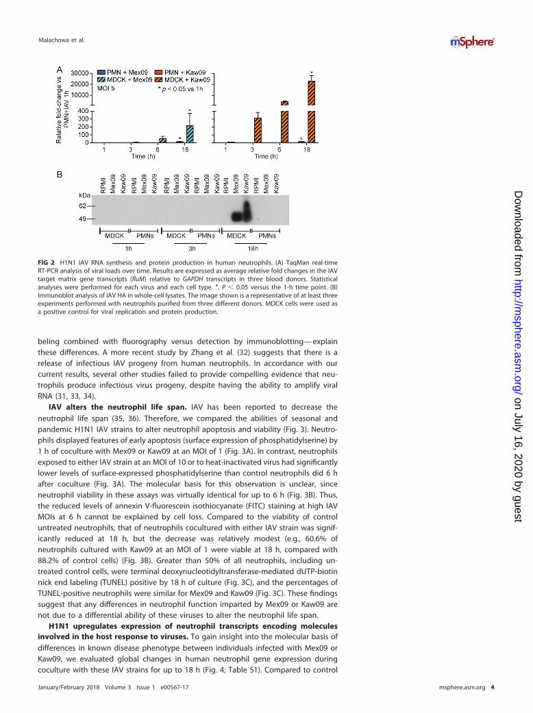
beling combined with fluorography versus detection by immunoblotting— explainthese differences. A more recent study by Zhang et al. (32) suggests that there is arelease of infectious IAV progeny from human neutrophils. In accordance with ourcurrent results, several other studies failed to provide compelling evidence that neu-trophils produce infectious virus progeny, despite having the ability to amplify viralRNA (31, 33, 34).
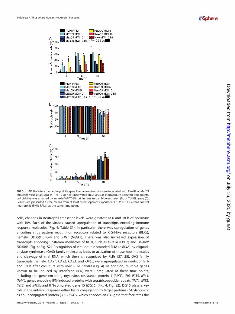
IAV alters the neutrophil life span. IAV has been reported to decrease theneutrophil life span (35, 36). Therefore, we compared the abilities of seasonal andpandemic H1N1 IAV strains to alter neutrophil apoptosis and viability (Fig. 3). Neutro-phils displayed features of early apoptosis (surface expression of phosphatidylserine) by1 h of coculture with Mex09 or Kaw09 at an MOI of 1 (Fig. 3A). In contrast, neutrophilsexposed to either IAV strain at an MOI of 10 or to heat-inactivated virus had significantlylower levels of surface-expressed phosphatidylserine than control neutrophils did 6 hafter coculture (Fig. 3A). The molecular basis for this observation is unclear, sinceneutrophil viability in these assays was virtually identical for up to 6 h (Fig. 3B). Thus,the reduced levels of annexin V-fluorescein isothiocyanate (FITC) staining at high IAVMOIs at 6 h cannot be explained by cell loss. Compared to the viability of controluntreated neutrophils, that of neutrophils cocultured with either IAV strain was signif-icantly reduced at 18 h, but the decrease was relatively modest (e.g., 60.6% ofneutrophils cultured with Kaw09 at an MOI of 1 were viable at 18 h, compared with88.2% of control cells) (Fig. 3B). Greater than 50% of all neutrophils, including un-treated control cells, were terminal deoxynucleotidyltransferase-mediated dUTP-biotinnick end labeling (TUNEL) positive by 18 h of culture (Fig. 3C), and the percentages ofTUNEL-positive neutrophils were similar for Mex09 and Kaw09 (Fig. 3C). These findingssuggest that any differences in neutrophil function imparted by Mex09 or Kaw09 arenot due to a differential ability of these viruses to alter the neutrophil life span.
H1N1 upregulates expression of neutrophil transcripts encoding moleculesinvolved in the host response to viruses. To gain insight into the molecular basis ofdifferences in known disease phenotype between individuals infected with Mex09 orKaw09, we evaluated global changes in human neutrophil gene expression duringcoculture with these IAV strains for up to 18 h (Fig. 4; Table S1). Compared to control
FIG 2 H1N1 IAV RNA synthesis and protein production in human neutrophils. (A) TaqMan real-timeRT-PCR analysis of viral loads over time. Results are expressed as average relative fold changes in the IAVtarget matrix gene transcripts (fluM) relative to GAPDH transcripts in three blood donors. Statisticalanalyses were performed for each virus and each cell type. *, P � 0.05 versus the 1-h time point. (B)Immunoblot analysis of IAV HA in whole-cell lysates. The image shown is a representative of at least threeexperiments performed with neutrophils purified from three different donors. MDCK cells were used asa positive control for viral replication and protein production.
Malachowa et al.
January/February 2018 Volume 3 Issue 1 e00567-17 msphere.asm.org 4
on July 16, 2020 by guesthttp://m
sphere.asm.org/
Dow
nloaded from
cells, changes in neutrophil transcript levels were greatest at 6 and 18 h of coculturewith IAV. Each of the viruses caused upregulation of transcripts encoding immuneresponse molecules (Fig. 4; Table S1). In particular, there was upregulation of genesencoding virus pattern recognition receptors related to RIG-I-like receptors (RLRs),namely, DDX58 (RIG-I) and IFIH1 (MDA5). There was also increased expression oftranscripts encoding upstream mediators of RLRs, such as DHX58 (LPG2) and DDX60/DDX60L (Fig. 4; Fig. S2). Recognition of viral double-stranded RNA (dsRNA) by oligoad-enylate synthetase (OAS) family molecules leads to activation of these host moleculesand cleavage of viral RNA, which then is recognized by RLRs (37, 38). OAS familytranscripts, namely, OAS1, OAS2, OAS3, and OASL, were upregulated in neutrophils 6and 18 h after coculture with Mex09 or Kaw09 (Fig. 4). In addition, multiple genesknown to be induced by interferon (IFN) were upregulated at these time points,including the gene encoding myxovirus resistance protein 1 (MX1), IFI6, IFI35, IFI44,IFI44L, genes encoding IFN-induced proteins with tetratricopeptide repeats (IFIT1, IFIT2,IFIT3, and IFIT5), and IFN-stimulated gene 15 (ISG15) (Fig. 4; Fig. S2). ISG15 plays a keyrole in the antiviral response either by its conjugation to target proteins (ISGylation) oras an unconjugated protein (39). HERC5, which encodes an E3 ligase that facilitates the
FIG 3 H1N1 IAV alters the neutrophil life span. Human neutrophils were incubated with Kaw09 or Mex09influenza virus at an MOI of 1 to 10 or heat-inactivated (h.i.) virus as indicated. At selected time points,cell viability was assessed by annexin V-FITC-PI staining (A), trypan blue exclusion (B), or TUNEL assay (C).Results are presented as the means from at least three separate experiments. *, P � 0.05 versus controlneutrophils (PMN RPMI) at the same time point.
Influenza A Virus Alters Human Neutrophil Function
January/February 2018 Volume 3 Issue 1 e00567-17 msphere.asm.org 5
on July 16, 2020 by guesthttp://m
sphere.asm.org/
Dow
nloaded from
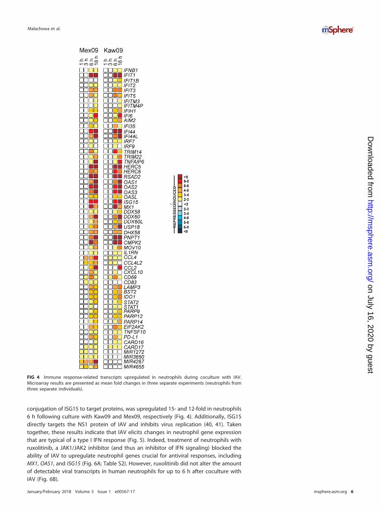
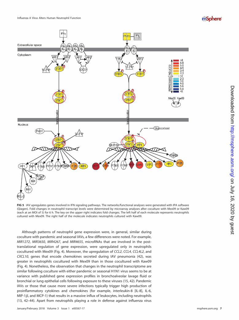
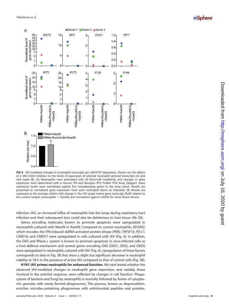
conjugation of ISG15 to target proteins, was upregulated 15- and 12-fold in neutrophils6 h following culture with Kaw09 and Mex09, respectively (Fig. 4). Additionally, ISG15directly targets the NS1 protein of IAV and inhibits virus replication (40, 41). Takentogether, these results indicate that IAV elicits changes in neutrophil gene expressionthat are typical of a type I IFN response (Fig. 5). Indeed, treatment of neutrophils withruxolitinib, a JAK1/JAK2 inhibitor (and thus an inhibitor of IFN signaling) blocked theability of IAV to upregulate neutrophil genes crucial for antiviral responses, includingMX1, OAS1, and ISG15 (Fig. 6A; Table S2). However, ruxolitinib did not alter the amountof detectable viral transcripts in human neutrophils for up to 6 h after coculture withIAV (Fig. 6B).
FIG 4 Immune response-related transcripts upregulated in neutrophils during coculture with IAV.Microarray results are presented as mean fold changes in three separate experiments (neutrophils fromthree separate individuals).
Malachowa et al.
January/February 2018 Volume 3 Issue 1 e00567-17 msphere.asm.org 6
on July 16, 2020 by guesthttp://m
sphere.asm.org/
Dow
nloaded from
Although patterns of neutrophil gene expression were, in general, similar duringcoculture with pandemic and seasonal IAVs, a few differences were noted. For example,MIR1272, MIR3650, MIR4267, and MIR4655, microRNAs that are involved in the post-translational regulation of gene expression, were upregulated only in neutrophilscocultured with Mex09 (Fig. 4). Moreover, the upregulation of CCL2, CCL4, CCL4L2, andCXCL10, genes that encode chemokines secreted during IAV pneumonia (42), wasgreater in neutrophils cocultured with Mex09 than in those cocultured with Kaw09(Fig. 4). Nonetheless, the observation that changes in the neutrophil transcriptome aresimilar following coculture with either pandemic or seasonal H1N1 virus seems to be atvariance with published gene expression profiles in bronchoalveolar lavage fluid orbronchial or lung epithelial cells following exposure to these viruses (15, 42). PandemicIAVs or those that cause more severe infections typically trigger high production ofproinflammatory cytokines and chemokines (for example, interleukin-8 [IL-8], IL-6,MIP-1�, and MCP-1) that results in a massive influx of leukocytes, including neutrophils(13, 42–44). Apart from neutrophils playing a role in defense against influenza virus
FIG 5 IAV upregulates genes involved in IFN signaling pathways. The networks/functional analyses were generated with IPA software(Qiagen). Fold changes in neutrophil transcript levels were determined by microarray analyses after coculture with Mex09 or Kaw09(each at an MOI of 5) for 6 h. The key on the upper right indicates fold changes. The left half of each molecule represents neutrophilscultured with Mex09. The right half of the molecule indicates neutrophils cultured with Kaw09.
Influenza A Virus Alters Human Neutrophil Function
January/February 2018 Volume 3 Issue 1 e00567-17 msphere.asm.org 7
on July 16, 2020 by guesthttp://m
sphere.asm.org/
Dow
nloaded from
infection (45), an increased influx of neutrophils into the lungs during respiratory tractinfection and their subsequent lysis could also be deleterious to host tissue (46–50).
Genes encoding molecules known to promote apoptosis were upregulated inneutrophils cultured with Mex09 or Kaw09. Compared to control neutrophils, EIF2AK2,which encodes the IFN-induced dsRNA-activated protein kinase (PKR); TNFSF10; PD-L1;CARD16; and CARD17 were upregulated in cells cultured with IAV (Fig. 4). In addition,the OAS and RNase L system is known to promote apoptosis in virus-infected cells asa host defense mechanism and several genes encoding OAS (OAS1, OAS2, and OAS3)were upregulated in neutrophils cultured with IAV (Fig. 4). Upregulation of these factorscorresponds to data in Fig. 3B that show a slight but significant decrease in neutrophilviability at 18 h in the presence of active IAV compared to that of control cells (Fig. 3B).
H1N1 IAV primes neutrophils for enhanced function. We next tested whether theobserved IAV-mediated changes in neutrophil gene expression, and notably thoseinvolved in the antiviral response, were reflected by changes in cell function. Phago-cytosis of bacteria and fungi by neutrophils is normally followed by fusion of cytoplas-mic granules with newly formed phagosomes. This process, known as degranulation,enriches microbe-containing phagosomes with antimicrobial peptides and proteins.
FIG 6 IAV-mediated changes in neutrophil transcripts are JAK/STAT dependent. Shown are the effectsof a JAK1/JAK2 inhibitor on the levels of expression of selected neutrophil antiviral transcripts (A) andviral loads (B). (A) Neutrophils were pretreated with 20 µM ruxolitinib, and changes in geneexpression were determined with a Human IFN and Receptor RT2 Profiler PCR Array (Qiagen). Geneexpression levels were normalized against five housekeeping genes in the array panel. Results arepresented as normalized gene expression from each neutrophil donor as indicated. (B) Results areexpressed as the average relative fold change in the IAV target matrix gene transcript (fluM) relative tothe control sample (neutrophils � Kaw09) and normalized against GAPDH for three blood donors.
Malachowa et al.
January/February 2018 Volume 3 Issue 1 e00567-17 msphere.asm.org 8
on July 16, 2020 by guesthttp://m
sphere.asm.org/
Dow
nloaded from
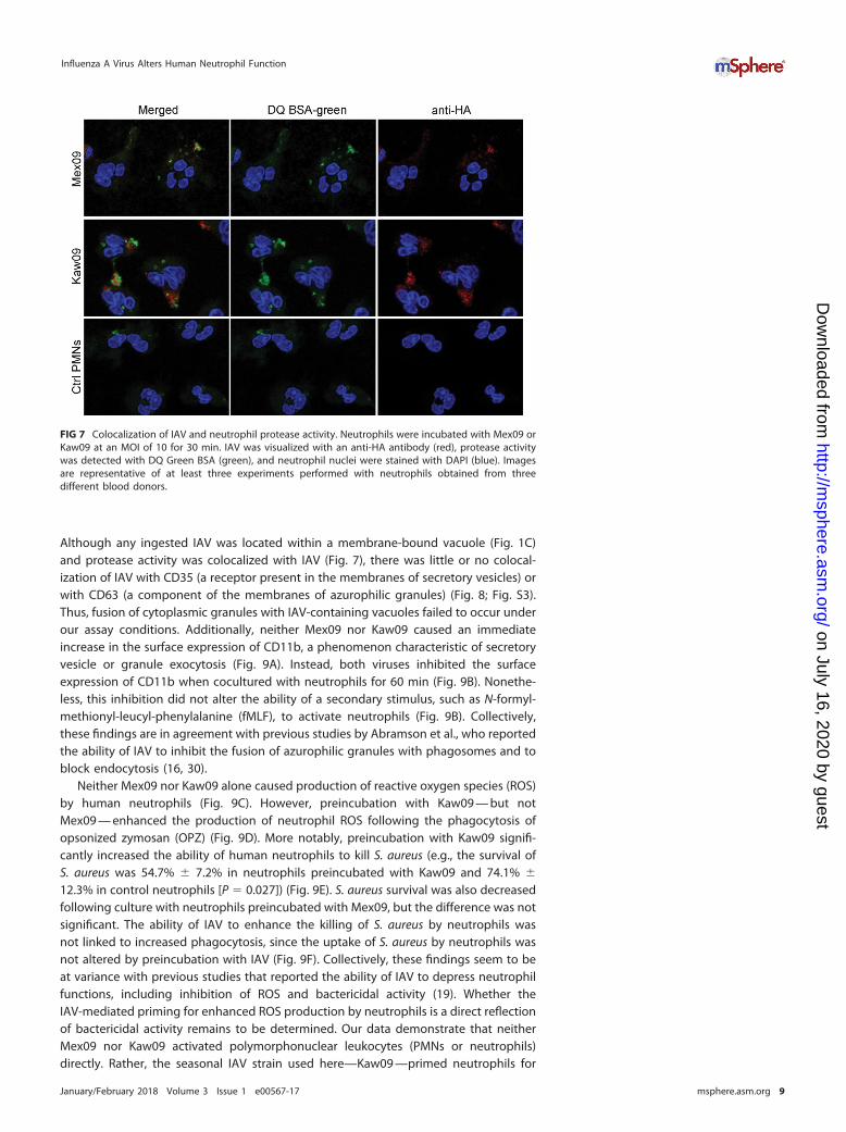
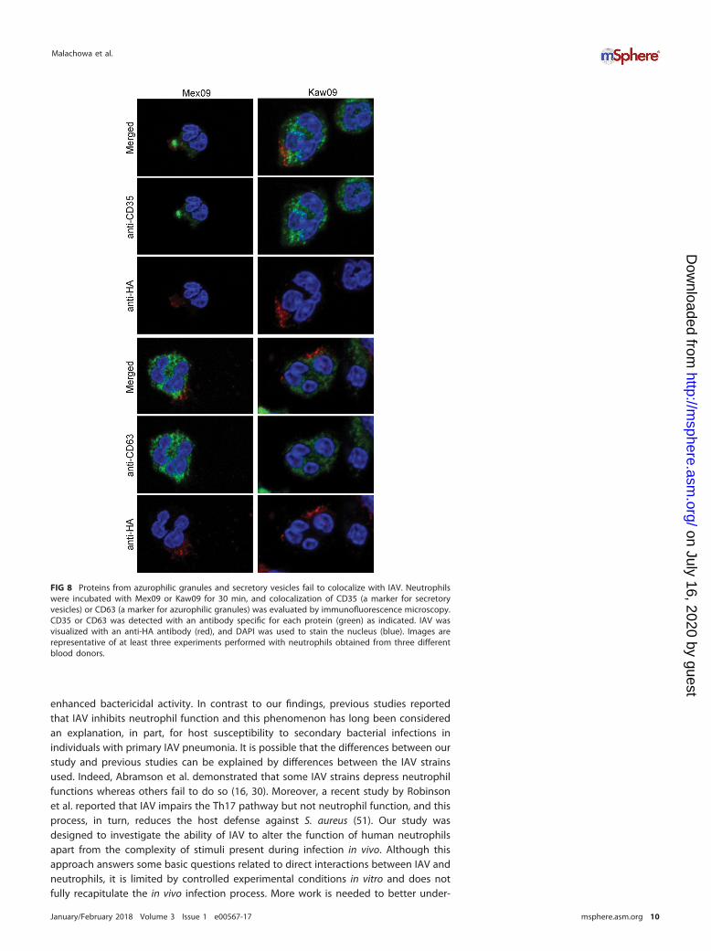
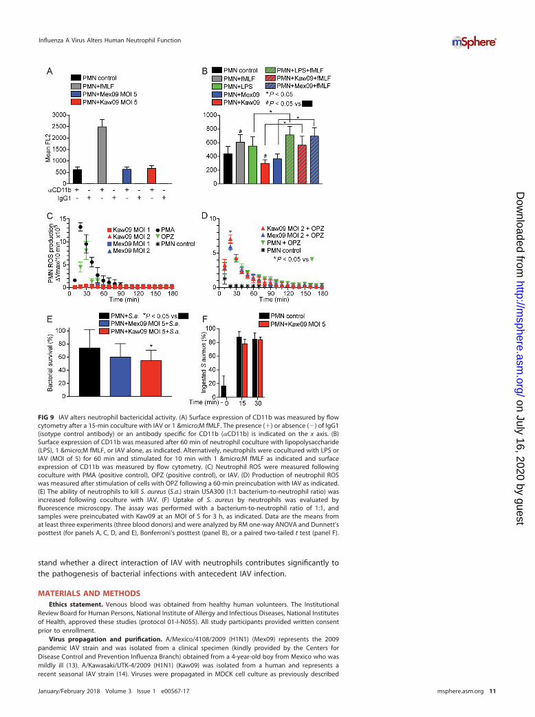
Although any ingested IAV was located within a membrane-bound vacuole (Fig. 1C)and protease activity was colocalized with IAV (Fig. 7), there was little or no colocal-ization of IAV with CD35 (a receptor present in the membranes of secretory vesicles) orwith CD63 (a component of the membranes of azurophilic granules) (Fig. 8; Fig. S3).Thus, fusion of cytoplasmic granules with IAV-containing vacuoles failed to occur underour assay conditions. Additionally, neither Mex09 nor Kaw09 caused an immediateincrease in the surface expression of CD11b, a phenomenon characteristic of secretoryvesicle or granule exocytosis (Fig. 9A). Instead, both viruses inhibited the surfaceexpression of CD11b when cocultured with neutrophils for 60 min (Fig. 9B). Nonethe-less, this inhibition did not alter the ability of a secondary stimulus, such as N-formyl-methionyl-leucyl-phenylalanine (fMLF), to activate neutrophils (Fig. 9B). Collectively,these findings are in agreement with previous studies by Abramson et al., who reportedthe ability of IAV to inhibit the fusion of azurophilic granules with phagosomes and toblock endocytosis (16, 30).
Neither Mex09 nor Kaw09 alone caused production of reactive oxygen species (ROS)by human neutrophils (Fig. 9C). However, preincubation with Kaw09 — but notMex09 — enhanced the production of neutrophil ROS following the phagocytosis ofopsonized zymosan (OPZ) (Fig. 9D). More notably, preincubation with Kaw09 signifi-cantly increased the ability of human neutrophils to kill S. aureus (e.g., the survival ofS. aureus was 54.7% � 7.2% in neutrophils preincubated with Kaw09 and 74.1% �
12.3% in control neutrophils [P � 0.027]) (Fig. 9E). S. aureus survival was also decreasedfollowing culture with neutrophils preincubated with Mex09, but the difference was notsignificant. The ability of IAV to enhance the killing of S. aureus by neutrophils wasnot linked to increased phagocytosis, since the uptake of S. aureus by neutrophils wasnot altered by preincubation with IAV (Fig. 9F). Collectively, these findings seem to beat variance with previous studies that reported the ability of IAV to depress neutrophilfunctions, including inhibition of ROS and bactericidal activity (19). Whether theIAV-mediated priming for enhanced ROS production by neutrophils is a direct reflectionof bactericidal activity remains to be determined. Our data demonstrate that neitherMex09 nor Kaw09 activated polymorphonuclear leukocytes (PMNs or neutrophils)directly. Rather, the seasonal IAV strain used here—Kaw09 —primed neutrophils for
FIG 7 Colocalization of IAV and neutrophil protease activity. Neutrophils were incubated with Mex09 orKaw09 at an MOI of 10 for 30 min. IAV was visualized with an anti-HA antibody (red), protease activitywas detected with DQ Green BSA (green), and neutrophil nuclei were stained with DAPI (blue). Imagesare representative of at least three experiments performed with neutrophils obtained from threedifferent blood donors.
Influenza A Virus Alters Human Neutrophil Function
January/February 2018 Volume 3 Issue 1 e00567-17 msphere.asm.org 9
on July 16, 2020 by guesthttp://m
sphere.asm.org/
Dow
nloaded from
enhanced bactericidal activity. In contrast to our findings, previous studies reportedthat IAV inhibits neutrophil function and this phenomenon has long been consideredan explanation, in part, for host susceptibility to secondary bacterial infections inindividuals with primary IAV pneumonia. It is possible that the differences between ourstudy and previous studies can be explained by differences between the IAV strainsused. Indeed, Abramson et al. demonstrated that some IAV strains depress neutrophilfunctions whereas others fail to do so (16, 30). Moreover, a recent study by Robinsonet al. reported that IAV impairs the Th17 pathway but not neutrophil function, and thisprocess, in turn, reduces the host defense against S. aureus (51). Our study wasdesigned to investigate the ability of IAV to alter the function of human neutrophilsapart from the complexity of stimuli present during infection in vivo. Although thisapproach answers some basic questions related to direct interactions between IAV andneutrophils, it is limited by controlled experimental conditions in vitro and does notfully recapitulate the in vivo infection process. More work is needed to better under-
FIG 8 Proteins from azurophilic granules and secretory vesicles fail to colocalize with IAV. Neutrophilswere incubated with Mex09 or Kaw09 for 30 min, and colocalization of CD35 (a marker for secretoryvesicles) or CD63 (a marker for azurophilic granules) was evaluated by immunofluorescence microscopy.CD35 or CD63 was detected with an antibody specific for each protein (green) as indicated. IAV wasvisualized with an anti-HA antibody (red), and DAPI was used to stain the nucleus (blue). Images arerepresentative of at least three experiments performed with neutrophils obtained from three differentblood donors.
Malachowa et al.
January/February 2018 Volume 3 Issue 1 e00567-17 msphere.asm.org 10
on July 16, 2020 by guesthttp://m
sphere.asm.org/
Dow
nloaded from
stand whether a direct interaction of IAV with neutrophils contributes significantly tothe pathogenesis of bacterial infections with antecedent IAV infection.
MATERIALS AND METHODSEthics statement. Venous blood was obtained from healthy human volunteers. The Institutional
Review Board for Human Persons, National Institute of Allergy and Infectious Diseases, National Institutesof Health, approved these studies (protocol 01-I-N055). All study participants provided written consentprior to enrollment.
Virus propagation and purification. A/Mexico/4108/2009 (H1N1) (Mex09) represents the 2009pandemic IAV strain and was isolated from a clinical specimen (kindly provided by the Centers forDisease Control and Prevention Influenza Branch) obtained from a 4-year-old boy from Mexico who wasmildly ill (13). A/Kawasaki/UTK-4/2009 (H1N1) (Kaw09) was isolated from a human and represents arecent seasonal IAV strain (14). Viruses were propagated in MDCK cell culture as previously described
FIG 9 IAV alters neutrophil bactericidal activity. (A) Surface expression of CD11b was measured by flowcytometry after a 15-min coculture with IAV or 1 µM fMLF. The presence (�) or absence (�) of IgG1(isotype control antibody) or an antibody specific for CD11b (�CD11b) is indicated on the x axis. (B)Surface expression of CD11b was measured after 60 min of neutrophil coculture with lipopolysaccharide(LPS), 1 µM fMLF, or IAV alone, as indicated. Alternatively, neutrophils were cocultured with LPS orIAV (MOI of 5) for 60 min and stimulated for 10 min with 1 µM fMLF as indicated and surfaceexpression of CD11b was measured by flow cytometry. (C) Neutrophil ROS were measured followingcoculture with PMA (positive control), OPZ (positive control), or IAV. (D) Production of neutrophil ROSwas measured after stimulation of cells with OPZ following a 60-min preincubation with IAV as indicated.(E) The ability of neutrophils to kill S. aureus (S.a.) strain USA300 (1:1 bacterium-to-neutrophil ratio) wasincreased following coculture with IAV. (F) Uptake of S. aureus by neutrophils was evaluated byfluorescence microscopy. The assay was performed with a bacterium-to-neutrophil ratio of 1:1, andsamples were preincubated with Kaw09 at an MOI of 5 for 3 h, as indicated. Data are the means fromat least three experiments (three blood donors) and were analyzed by RM one-way ANOVA and Dunnett’sposttest (for panels A, C, D, and E), Bonferroni’s posttest (panel B), or a paired two-tailed t test (panel F).
Influenza A Virus Alters Human Neutrophil Function
January/February 2018 Volume 3 Issue 1 e00567-17 msphere.asm.org 11
on July 16, 2020 by guesthttp://m
sphere.asm.org/
Dow
nloaded from
(13). To purify and concentrate the virus stock, cell culture supernatant was clarified by centrifugation at1,700 � g for 15 min at 4°C, overlaid onto 20% sucrose in Dulbecco’s phosphate-buffered saline (DPBS;Sigma-Aldrich, St. Louis, MO), and centrifuged at 110,000 � g for 2.5 h at 4°C. The resulting pellet wassuspended in RPMI 1640 medium (Invitrogen/Life Technologies, Inc., Grand Island, NY) buffered with10 mM HEPES (RPMI/H; Mediatech, Manassas, VA). The viral stock was stored at �80°C until used. Thevirus titer was determined by plaque-forming assay using MDCK cells cultured in six-well plates with amedium-viscosity carboxymethyl cellulose sodium salt (Sigma-Aldrich, St. Louis, MO) overlay (52, 53).Heat inactivation of the virus was achieved by 10 min of incubation at 100°C and verified by plaque-forming assay. The virus strains used in this study are biosafety level 2 viruses. All infectious samples werehandled in accordance with a human pathogen registration document approved by the InstitutionalBiosafety Committee at Rocky Mountain Laboratories, Division of Intramural Research, National Instituteof Allergy and Infectious Diseases, National Institutes of Health.
Neutrophil isolation. Human PMNs were isolated from fresh heparinized venous blood by standarddextran sedimentation coupled with Hypaque-Ficoll gradient centrifugation as previously described (54).The viability and purity of all neutrophil preparations were assessed by flow cytometry. Neutrophilpreparations typically yield 98 to 99% PMNs (of which ~95 to 98% were neutrophils and ~2 to 5% wereeosinophils), and viability was �99% as assessed by uptake of propidium iodide (PI).
Neutrophil viability. Neutrophil apoptosis and viability were examined by staining with annexinV-FITC-PI, TUNEL, and a trypan blue exclusion assay. Assays were performed in 24- or 96-well round-bottom tissue culture plates precoated with 20% normal human serum (NHS). Neutrophils were culturedwith IAV at MOIs ranging from 0 to 10 at 37°C with 5% CO2 and 90% humidity for up to 18 h. Atdesignated time points, samples were processed in accordance with the manufacturer’s protocol for eachassay. Early apoptosis was evaluated on the basis of surface staining with annexin V-FITC (for exposureof the phospholipid phosphatidylserine) and uptake of PI (FITC Annexin V Apoptosis Detection Kit II; BDPharmingen, San Diego, CA). DNA fragmentation was assessed with a TUNEL assay (APO-BRDU kit; BDBiosciences, San Diego, CA) as described elsewhere (54). To comply with institutional biosafety require-ments, all samples were fixed with ice-cold 4% paraformaldehyde prior to analysis on a FACSCalibur flowcytometer (BD Biosciences). Ten thousand events were collected for each sample. Additionally, neutro-phil viability was assessed in a trypan blue exclusion assay. Briefly, at the desired time point, neutrophilswere collected from the cell culture plate, transferred into 1.5-ml tubes, and incubated with a trypan bluesolution (2 mg/ml trypan blue [JT Baker Chemical Co., Phillipsburg, NJ], 0.02 M sodium citrate, 0.15 MNaCl, pH 4.4). Cells were enumerated with a hemocytometer and a light microscope, and the percentageof viable cells was calculated by using the following equation: (number of cells that exclude trypanblue/total number of cells) � 100.
Immunofluorescence microscopy. Neutrophils (3 � 105) were added to 24-well culture platescontaining acid-washed coverslips coated with 100% pooled NHS. H1N1 Mex09 or Kaw09 was added atan MOI of 10 to experimental samples, and an equivalent volume of RPMI/H was added to controlsamples. Samples were centrifuged at 450 � g for 6 min to synchronize phagocytosis. DQ Green BSA(50 µg/ml; Molecular Probes, Eugene, OR) was added to the group of samples to track proteaseactivity. Samples were incubated for up to 1 h at 37°C. At the desired time point, supernatant wasaspirated and neutrophils were washed once with DPBS (Sigma-Aldrich, St. Louis, MO). Cells were fixedwith 4% paraformaldehyde (Electron Microscopy Sciences, Hatfield, PA) in DPBS. Consequently, cells werepermeabilized with 0.2% Triton X-100 (USB, Cleveland, OH) in DPBS for 15 min at ambient temperature.Cells were washed twice with DPBS and blocked with Stain Buffer (FBS) (BD Biosciences, San Jose, CA)for 10 min. Samples were incubated with selected antibodies in Stain Buffer (FBS) for 1 h at ambienttemperature with gentle agitation. Prior to incubation with secondary antibodies (where appropriate),cells were washed three times with DPBS. The antibodies used for cell labeling were FITC-labeled mouseanti-human CD63 (40 µl/sample) and anti-human CD35 (60 µl/sample) (BD Pharmingen),Alexa Fluor 488-conjugated phalloidin (1 U/sample) (Molecular Probes/Thermo Fisher Scientific, Inc.,Eugene, OR), and a mouse anti-IAV H1N1 hemagglutinin (HA) monoclonal antibody (final concentration,4 to 10 µg/ml) (Thermo Fisher Scientific, Inc., Rockford, IL) in combination with an Alexa Fluor594-conjugated goat anti-mouse IgG (H�L) secondary antibody (Molecular Probes/Thermo Fisher Sci-entific, Inc., Eugene, Eugene, OR) at a final concentration of 0.1 µg/ml. To evaluate the uptake ofvirus particles by neutrophils or MDCK cells, cells were first incubated with a rabbit anti-IAV H1N1 HApolyclonal antibody (Thermo Fisher Scientific, Inc., Rockford, IL) at a final concentration of 10 µg/ml. After being washed, samples were incubated with an Alexa Fluor 488-conjugated goat anti-rabbit IgG(H�L) cross-adsorbed secondary antibody (final concentration, 0.1 µg/ml). This first procedurestains all surface-bound (extracellular) IAV. Subsequently, neutrophils were permeabilized as describedabove and stained with a sequential combination of a rabbit anti-IAV H1N1 HA polyclonal antibody(primary antibody) and an Alexa Fluor 594-conjugated goat anti-rabbit IgG (H�L) cross-adsorbedsecondary antibody (final concentration, 0.1 µg/ml). This procedure stains surface-bound andingested (extracellular and intracellular) IAV. Coverslips were mounted with Fluoromount G containing4’,6-diamidino-2-phenylindole (DAPI; Electron Microscopy Sciences, Hatfield, PA). Samples were imagedon a Carl Zeiss, Inc., LSM710 confocal laser-scanning microscope equipped with ZEN software version8.1.10 (Carl Zeiss, Inc., Thornwood, NY). Image contrast and brightness were adjusted in ZEN software orby using Adobe Photoshop CS5.1 (Adobe, San Jose, CA). The percentage of neutrophils with intracellularIAV was determined with the following equation: PMNintracellular IAV/PMNtotal � 100. At least 50 cells in fiveseparate fields of view per sample were used to calculate the uptake of IAV.
Transmission electron microscopy (TEM). Human neutrophils were seeded onto NHS-coatedThermanox coverslips and incubated with IAV at an MOI of 20 for up to 3 h in 24-well cell culture plates
Malachowa et al.
January/February 2018 Volume 3 Issue 1 e00567-17 msphere.asm.org 12
on July 16, 2020 by guesthttp://m
sphere.asm.org/
Dow
nloaded from
at 37°C. Samples were then washed once with DPBS and fixed for 30 min with 2.5% glutaraldehyde in0.1 M sodium cacodylate buffer, pH 7.4. All subsequent processing was carried out in a PELCO BioWavePro laboratory microwave (Ted Pella, Inc., Redding, CA.). Neutrophils were postfixed with 1% osmiumtetroxide reduced with 0.8% potassium ferrocyanide in 0.1 M sodium cacodylate buffer, treated with 1%tannic acid, and stained en bloc with uranyl acid replacement stain. The cells were dehydrated in agradient ethanol series and infiltrated with Embed 812 resin. The resin blocks were polymerizedovernight in an oven at 65°C. Thin sections were cut with a Leica UC6 ultramicrotome (Leica Microsys-tems, Inc., Vienna, Austria) and examined with an 80-kV Hitachi 7500 transmission electron microscope(Hitachi High Technology in America, Schaumburg, IL). Images were captured with a bottom mount AMTcamera system (Advanced Microscopy Techniques Corp., Woburn, MA).
Microarray experiments. Twenty-four-well culture plates were coated with 20% pooled NHS for atleast 1 h at 37°C. Plates were washed twice with DPBS and kept on ice until used. A total of 5 � 106
neutrophils were added to each well, and the cells were infected with IAV at an MOI of 5. For controlsamples, an equivalent volume of RPMI/H was added instead of virus. Neutrophils from three differenthuman blood donors were used for microarray experiments. Samples were incubated for up to 18 h at37°C with 5% CO2. At selected time points (1, 3, 6, and 18 h), samples were centrifuged at 500 � g for8 min at 4°C. Supernatant was aspirated, and cells were treated with TRIzol (Ambion/Life Technologies,Carlsbad, CA) for 5 min and frozen at �80°C until samples were collected all time points. Total RNA waspurified with a Direct-zol RNA MiniPrep kit in accordance with the manufacturer’s protocol (ZymoResearch Corp., Irvine, CA). The remaining DNA was removed with Turbo DNase (Ambion/AppliedBiosystems, Austin, TX), and samples were repurified with an RNeasy minikit (Qiagen, Hilden, Germany).The quantity and quality of isolated RNA were assessed with a 2100 Bioanalyzer (Agilent Technologies,Santa Clara, CA). RNA amplification and labeling were carried out with a GeneChip WT PLUS Reagent kit(Affymetrix/Thermo Fisher, Santa Clara, CA) in accordance with the manufacturer’s protocol. Labeledsamples were hybridized to GeneChip Human Gene 2.0 ST Arrays. GeneChips were scanned with theAffymetrix GeneChip 3000 7G Plus scanner in accordance with a standard GeneChip protocol, and theimage files were converted to .Cel files with Expression Console (v1.4). Quality analysis was performed inaccordance with the white paper Quality Assessment of Exon and Gene Arrays (Affymetrix revision 11). All.Cel files, representing individual samples, were normalized by the RMA method within ExpressionConsole to produce chip (.chp) files. A report was generated from the .chp files (along with selected rawdata) summarizing various quality and statistical aspects of the chips. One sample (the neutrophil controlat 3 h, one donor) was indicated as a quality outlier and excluded from the analysis. Except for theneutrophil control at 3 h, expression microarray data are presented as the mean fold change of threeseparate donors. Statistical analyses were performed by analysis of variance (ANOVA) and multiple-testcorrection by using the false-discovery rate (FDR; significance at a level of 0.05 [55]) (Partek GenomicsSuite software, v6.5 6091110 [Partek, Inc., St. Louis, MO]). These data were combined with fold changevalues by using custom Excel templates to generate final gene lists for each comparison (Table S1).Microarray data were analyzed additionally with Ingenuity Pathway Analysis (IPA) software (Qiagen,Redwood City, CA) to identify functional groups, gene networks, and canonical pathways. Figures werecreated with Adobe Illustrator CC2015.3 software.
TaqMan real-time RT-PCR. RNA samples used in the microarray experiment were also analyzed byTaqMan real-time RT-PCR to assess viral loads. Alternatively, to compare viral replication in neutrophilswith that in MDCK cells, total RNA was isolated from infected MDCK cells (or uninfected samples, whichserved as a negative control) as described above. Twenty-five nanograms of total purified RNA wassubjected to one-step qRT-PCR with the AgPath-ID one-Step RT-PCR kit and an ABI 7500 thermocycler(Ambion/Applied Biosystems, Foster City, CA). The relative quantification of viral RNA was done bymeasuring the change in the expression level of the target matrix gene transcripts (fluM [56]) relative tothe GAPDH transcripts (GAPDH TaqMan Gene Expression Assays; Applied Biosystems, Foster City, CA) inaccordance with the manufacturer’s protocol (Applied Biosystems relative quantification manual). Dataare expressed as the mean fold differences between the transcript levels at 3, 6, and 18 h and thetranscript level at 1 h for each virus.
PCR arrays. Neutrophils were preincubated with 20 �M ruxolitinib (Selleck Chemicals, Houston, TX)or an equal volume of RPMI/H for 2 h at 37°C with 5% CO2. Subsequently, Kaw09 was added at an MOIof 5 and samples were incubated for an additional 6 h. At the time point indicated, RNA was isolated asdescribed in the section on microarray experiments. Five hundred nanograms of purified RNA was usedto synthesized cDNA with an RT2 First Strand kit (Qiagen). Expression levels of genes encoding humanIFNs and receptors were detected with the Human IFNs and Receptors RT2 Profiler PCR Array platformin combination with RT2 Sybr green ROX qPCR Mastermix (Qiagen) in accordance with the manufacturer’sinstructions. Samples were amplified with an ABI 7500 real-time PCR system (Applied Biosystems). Dataanalysis was performed with a template downloaded from the Qiagen genes and pathways data analysiscenter overview page, and gene expression levels were normalized against all five housekeeping genes.
Immunoblotting. Neutrophils were incubated with buffer alone (control), Mex09, or Kaw09 at anMOI of 5 for up to 18 h at 37°C with 5% CO2. At designated time points, cells were pelleted bycentrifugation at 450 � g for 7 min at ambient temperature. Neutrophils were suspended in lysis buffer(50 mM Tris [pH 7.5], 280 mM NaCl, 1% Triton X-100, 0.2 mM EDTA, 2 mM EGTA, 10% glycerol, 1 mMdithiothreitol, 0.1 mM sodium vanadate) containing a protease inhibitor cocktail (cOmplete ULTRATablets, Mini, EASYpack Protease Inhibitor Cocktail; Roche, Indianapolis, IN) (57). IAV-infected MDCK cellswere used as a positive control for viral protein production. Protein was quantitated with a bicinchoninicacid protein assay (Pierce Protein Research Products/Thermo Fisher Scientific, Rockford, IL), and 10µg of protein was separated by 12.5% SDS-PAGE (Criterion; Bio-Rad, Hercules, CA). Proteins were
Influenza A Virus Alters Human Neutrophil Function
January/February 2018 Volume 3 Issue 1 e00567-17 msphere.asm.org 13
on July 16, 2020 by guesthttp://m
sphere.asm.org/
Dow
nloaded from
transferred to polyvinylidene difluoride membranes with the iBlot transfer system (Invitrogen/ThermoFisher Scientific, Waltham, MA), and membranes were blocked for 1 h at ambient temperature in 5% drymilk powder (Research Products International, Mt. Prospect, IL) in PBS containing 0.05% Tween 20.Influenza virus HA was detected with a rabbit anti-IAV H1N1 HA polyclonal antibody. Membranes wereincubated with a primary antibody (0.5 µg/ml) in blocking buffer overnight at 4°C. The followingday, membranes were washed three times with wash buffer (250 mM NaCl, 10 mM HEPES, 0.2% Tween20) and incubated for 1.5 h with a secondary antibody conjugated with horseradish peroxidase (HRP)(polyclonal goat anti-rabbit immunoglobulins/HRP; Dako/Agilent, Santa Clara, CA). The antibody-proteincomplex was visualized with Pierce ECL Plus Western blotting substrate (Pierce Biotechnology, Rockford,IL) and X-ray film (Phenix Research Products, Chandler, NC).
Neutrophil function assays. To determine changes in the surface expression of CD11b caused byIAV, neutrophils were incubated with IAV at an MOI of 5 or with 1 µM fMLF for 15 min at 37°C. Cellswere washed and stained with a phycoerythrin (PE)-conjugated anti-human CD11b antibody or aPE-conjugated mouse isotype IgG1 control (58). Samples were fixed with 4% paraformaldehyde in DPBS,washed, and analyzed by flow cytometry. To evaluate the ability of IAV to prime human neutrophils, cellswere preincubated with IAV at an MOI of 5 for 60 min at 37°C and stimulated with 1 µM fMLF for10 min.
Production of neutrophil ROS was determined by oxidation of 2,7-dichlorodihydrofluorescein diace-tate (DCF; Sigma-Aldrich, St. Louis, MO) as described previously (54) but with modifications. Black,flat-bottom 96-well culture plates were coated with 20% NHS for 1 h and washed twice with DPBS.Human neutrophils (107/ml) were preloaded with 25 µM DCF (Sigma-Aldrich) in the dark for 20 minat ambient temperature. Zymosan A (MP Biomedicals, Solon, OH) was opsonized with 50% NHS for30 min at 37°C, washed twice with DPBS, and resuspended in RPMI/H (2.5 � 108 particles/ml). For eachassay, 106 neutrophils (preloaded with DCF) were stimulated with IAV (MOI of 1 to 2), OPZ (~5 particlesper neutrophil), or phorbol 12-myristate 13-acetate (PMA; 1 µg/ml; Sigma-Aldrich, St. Louis, MO) ina final volume of 200 µl. For assays in which neutrophils where primed with IAV, OPZ was added60 min after the incubation of IAV alone. Neutrophil activation was synchronized by centrifugation at 500� g for 7 min at 4°C. An optical adhesive cover (Applied Biosystems, Foster City, CA) was placed over theassay plate, and ROS production was monitored by measuring DCF fluorescence (excitation wavelength,485 nm; emission wavelength, 538 nm) for 3 h at 37°C with a SpectraMax Gemini XPS plate reader(Molecular Devices, Sunnyvale, CA). Data are presented as the mean Vmax at 10-min intervals under eachassay condition. Data were analyzed by repeated-measures (RM) one-way ANOVA and Dunnett’s posttestto correct for multiple comparisons.
S. aureus strain LAC, a USA300 isolate representative of the USA300 epidemic clone, from a frozenstock was cultured overnight in Trypticase soy broth (Difco, Detroit, MI). Overnight cultures wereinoculated into fresh medium and cultured to the mid-logarithmic phase of growth (optical density at600 nm of 0.75). Bacteria were collected by centrifugation, opsonized in 50% NHS for 30 min at 37°C,washed twice with DPBS, and resuspended in RPMI/H at a final concentration of 106 CFU/ml. Humanneutrophils (105 CFU/ml) were incubated with IAV (MOI of 5) in 96-well flat-bottom plates (precoatedwith 20% NHS) for 3 h at 37°C. Bacteria were then added to assay wells (200-�l final volume) at a 1:1bacterium-to-neutrophil ratio, and samples were incubated for an additional 1 h. At the desiredendpoint, neutrophils were lysed with 0.1% saponin for 15 min on ice. Samples from each assay well werediluted and plated on Trypticase soy agar (Difco). The following day, bacterial CFU were enumerated andpercent bacterial survival was calculated with the following equation: CFU�PMN 1 h/CFU�PMN 0 min � 100%.
To determine whether IAV altered the phagocytosis of S. aureus, human neutrophils (3 � 105) werecombined with Kaw09 in 24-well tissue culture plates as described above for the bactericidal activityassay. In addition, wells contained acid-washed coverslips that had been precoated with 100% pooledNHS. Prior to the addition of bacteria, samples were chilled on ice for 10 min. Opsonized bacteria werestained with FITC (7.5 �g/ml) for 15 min at room temperature in the dark and washed twice with DPBS.Bacteria were then added to assay wells (500-�l final volume) at a 1:1 bacterium-to-neutrophil ratio, andsamples were centrifuged at 450 � g for 7 min to synchronize phagocytosis. Plates were then incubatedat 37°C for 0, 15, or 30 min. At the desired time points, supernatant was aspirated and neutrophils werewashed once with DPBS. Cells were fixed with 4% paraformaldehyde in DPBS, and extracellular bacteriawere stained with a rabbit anti-S. aureus polyclonal antibody (59) in combination with a goat anti-rabbitantibody conjugated with Alexa Fluor 594 (Molecular Probes, Life Technologies, Inc.). Coverslips weremounted with Fluoromount G containing DAPI, and the percentage of ingested bacteria was calculatedfor 50 neutrophils per blood donor (three donors total).
Statistical analyses. Data, except those from microarray experiments, were analyzed by RM one-wayANOVA and Dunnett’s or Bonferroni’s (for selected pair comparisons) posttest to correct for multiplecomparisons or a paired t test, as indicated (Prism 7.0; GraphPad, San Diego, CA).
Accession number(s). All microarray data are MIAME (minimum information about a microarrayexperiment) compliant, and a complete set of microarray data has been posted online at http://www.ncbi.nlm.nih.gov/projects/geo/ under series number GSE100865.
SUPPLEMENTAL MATERIALSupplemental material for this article may be found at https://doi.org/10.1128/
mSphereDirect.00567-17.FIG S1, TIF file, 0.4 MB.FIG S2, TIF file, 2.9 MB.
Malachowa et al.
January/February 2018 Volume 3 Issue 1 e00567-17 msphere.asm.org 14
on July 16, 2020 by guesthttp://m
sphere.asm.org/
Dow
nloaded from
FIG S3, JPG file, 2.5 MB.TABLE S1, XLSX file, 0.05 MB.TABLE S2, PDF file, 0.1 MB.
ACKNOWLEDGMENTThis work was supported by the Intramural Research Program of the National
Institute of Allergy and Infectious Diseases, National Institutes of Health.
REFERENCES1. Rolfes MA, Foppa IM, Garg S, Flannery B, Brammer L, Singleton JA, Burns
E, Jernigan D, Reed C, Olsen SJ, Bresee J. 2016. Estimated influenzaillnesses, medical visits, hospitalizations, and deaths averted by vaccina-tion in the United States. Centers for Disease Control and Prevention,Atlanta, GA. https://www.cdc.gov/flu/about/disease/2015-16.htm. Ac-cessed 15 July 2017.
2. Morens DM, Taubenberger JK, Fauci AS. 2008. Predominant role ofbacterial pneumonia as a cause of death in pandemic influenza: impli-cations for pandemic influenza preparedness. J Infect Dis 198:962–970.https://doi.org/10.1086/591708.
3. Hers JFP, Masurel N, Mulder J. 1958. Bacteriology and histopathology ofthe respiratory tract and lungs in fatal Asian influenza. Lancet ii:1141–1143. https://doi.org/10.1016/S0140-6736(58)92404-8.
4. Centers for Disease Control and Prevention. 2009. Bacterial coinfectionsin lung tissue specimens from fatal cases of 2009 pandemic influenza A(H1N1)—United States, May-August 2009. MMWR Morb Mortal Wkly Rep58:1071–1074.
5. Finelli L, Fiore A, Dhara R, Brammer L, Shay DK, Kamimoto L, Fry A,Hageman J, Gorwitz R, Bresee J, Uyeki T. 2008. Influenza-associatedpediatric mortality in the United States: increase of Staphylococcus au-reus coinfection. Pediatrics 122:805– 811. https://doi.org/10.1542/peds.2008-1336.
6. Christman MC, Kedwaii A, Xu J, Donis RO, Lu G. 2011. Pandemic (H1N1)2009 virus revisited: an evolutionary retrospective. Infect Genet Evol11:803– 811. https://doi.org/10.1016/j.meegid.2011.02.021.
7. Virus Investigation Team (Team NS-OIAVI), Dawood FS, Jain S, Finelli L,Shaw MW, Lindstrom S, Garten RJ, Gubareva LV, Xu X, Bridges CB, UyekiTM. 2009. Emergence of a novel swine-origin influenza A (H1N1) virus inhumans. N Engl J Med 360:2605–2615. https://doi.org/10.1056/NEJMoa0903810.
8. Rice TW, Rubinson L, Uyeki TM, Vaughn FL, John BB, Miller RRI, Higgs E,Randolph AG, Smoot BE, Thompson BT; NHLBI ARDS Network. 2012.Critical illness from 2009 pandemic influenza A virus and bacterialcoinfection in the United States. Crit Care Med 40:1487–1498. https://doi.org/10.1097/CCM.0b013e3182416f23.
9. Gill JR, Sheng ZM, Ely SF, Guinee DG, Beasley MB, Suh J, Deshpande C,Mollura DJ, Morens DM, Bray M, Travis WD, Taubenberger JK. 2010.Pulmonary pathologic findings of fatal 2009 pandemic influenza A/h1n1viral infections. Arch Pathol Lab Med 134:235–243.
10. Shah NS, Greenberg JA, McNulty MC, Gregg KS, Riddell J, Mangino JE,Weber DM, Hebert CL, Marzec NS, Barron MA, Chaparro-Rojas F, Re-strepo A, Hemmige V, Prasidthrathsint K, Cobb S, Herwaldt L, Raabe V,Cannavino CR, Hines AG, Bares SH, Antiporta PB, Scardina T, Patel U, ReidG, Mohazabnia P, Kachhdiya S, Le BM, Park CJ, Ostrowsky B, Robicsek A,Smith BA, Schied J, Bhatti MM, Mayer S, Sikka M, Murphy-Aguilu I,Patwari P, Abeles SR, Torriani FJ, Abbas Z, Toya S, Doktor K, ChakrabartiA, Doblecki-Lewis S, Looney DJ, David MZ. 2016. Bacterial and viralco-infections complicating severe influenza: incidence and impactamong 507 U.S. patients, 2013–14. J Clin Virol 80:12–19. https://doi.org/10.1016/j.jcv.2016.04.008.
11. Hashimoto Y, Moki T, Takizawa T, Shiratsuchi A, Nakanishi Y. 2007.Evidence for phagocytosis of influenza virus-infected, apoptotic cells byneutrophils and macrophages in mice. J Immunol 178:2448 –2457.https://doi.org/10.4049/jimmunol.178.4.2448.
12. Garten RJ, Davis CT, Russell CA, Shu B, Lindstrom S, Balish A, SessionsWM, Xu X, Skepner E, Deyde V, Okomo-Adhiambo M, Gubareva L, BarnesJ, Smith CB, Emery SL, Hillman MJ, Rivailler P, Smagala J, de Graaf M,Burke DF, Fouchier RA, Pappas C, Alpuche-Aranda CM, López-Gatell H,Olivera H, López I, Myers CA, Faix D, Blair PJ, Yu C, Keene KM, Dotson PD,Boxrud D, Sambol AR, Abid SH, St George K, Bannerman T, Moore AL,Stringer DJ, Blevins P. 2009. Antigenic and genetic characteristics of
swine-origin 2009 A (H1N1) influenza viruses circulating in humans.Science 325:197–201. https://doi.org/10.1126/science.1176225.
13. Safronetz D, Rockx B, Feldmann F, Belisle SE, Palermo RE, Brining D,Gardner D, Proll SC, Marzi A, Tsuda Y, LaCasse RA, Kercher L, York A,Korth MJ, Long D, Rosenke R, Shupert WL, Aranda CA, Mattoon JS,Kobasa D, Kobinger G, Li Y, Taubenberger JK, Richt JA, Parnell M, EbiharaH, Kawaoka Y, Katze MG, Feldmann H. 2011. Pandemic swine-originH1N1 influenza A virus isolates show heterogeneous virulence in ma-caques. J Virol 85:1214 –1223. https://doi.org/10.1128/JVI.01848-10.
14. Itoh Y, Shinya K, Kiso M, Watanabe T, Sakoda Y, Hatta M, Muramoto Y,Tamura D, Sakai-Tagawa Y, Noda T, Sakabe S, Imai M, Hatta Y, WatanabeS, Li C, Yamada S, Fujii K, Murakami S, Imai H, Kakugawa S, Ito M, TakanoR, Iwatsuki-Horimoto K, Shimojima M, Horimoto T, Goto H, Takahashi K,Makino A, Ishigaki H, Nakayama M, Okamatsu M, Takahashi K, WarshauerD, Shult PA, Saito R, Suzuki H, Furuta Y, Yamashita M, Mitamura K,Nakano K, Nakamura M, Brockman-Schneider R, Mitamura H, YamazakiM, Sugaya N, Suresh M, Ozawa M, Neumann G, Gern J, Kida H,Ogasawara K, Kawaoka Y. 2009. In vitro and in vivo characterization ofnew swine-origin H1N1 influenza viruses. Nature 460:1021–1025. https://doi.org/10.1038/nature08260.
15. Paquette SG, Banner D, Chi le TB, León AJ, Xu L, Ran L, Huang SSH,Farooqui A, Kelvin DJ, Kelvin AA. 2014. Pandemic H1N1 influenza Adirectly induces a robust and acute inflammatory gene signature inprimary human bronchial epithelial cells downstream of membranefusion. Virology 448:91–103. https://doi.org/10.1016/j.virol.2013.09.022.
16. Abramson JS, Lewis JC, Lyles DS, Heller KA, Mills EL, Bass DA. 1982.Inhibition of neutrophil lysosome-phagosome fusion associated withinfluenza virus infection in vitro. Role in depressed bactericidal activity.J Clin Invest 69:1393–1397. https://doi.org/10.1172/JCI110580.
17. Abramson JS, Giebink GS, Quie PG. 1982. Influenza A virus-inducedpolymorphonuclear leukocyte dysfunction in the pathogenesis of exper-imental pneumococcal otitis media. Infect Immun 36:289 –296.
18. Abramson JS, Lyles DS, Heller KA, Bass DA. 1982. Influenza A virus-induced polymorphonuclear leukocyte dysfunction. Infect Immun 37:794 –799.
19. Abramson JS, Mills EL, Giebink GS, Quie PG. 1982. Depression of mono-cyte and polymorphonuclear leukocyte oxidative metabolism and bac-tericidal capacity by influenza A virus. Infect Immun 35:350 –355.
20. Abramson JS, Parce JW, Lewis JC, Lyles DS, Mills EL, Nelson RD, Bass DA.1984. Characterization of the effect of influenza virus on polymorpho-nuclear leukocyte membrane responses. Blood 64:131–138.
21. Abramson JS, Giebink GS, Mills EL, Quie PG. 1981. Polymorphonuclearleukocyte dysfunction during influenza virus infection in chinchillas. JInfect Dis 143:836 – 845. https://doi.org/10.1093/infdis/143.6.836.
22. Debets-Ossenkopp Y, Mills EL, van Dijk WC, Verbrugh HA, Verhoef J.1982. Effect of influenza virus on phagocytic cells. Eur J Clin Microbiol1:171–177. https://doi.org/10.1007/BF02019619.
23. Larson HE, Blades R. 1976. Impairment of human polymorphonuclearleucocyte function by influenza virus. Lancet i:283. https://doi.org/10.1016/S0140-6736(76)91407-0.
24. Larson HE, Parry RP, Gilchrist C, Luquetti A, Tyrrell DA. 1977. Influenzaviruses and staphylococci in vitro: some interactions with polymorpho-nuclear leucocytes and epithelial cells. Br J Exp Pathol 58:281–288.
25. Ruutu P, Vaheri A, Kosunen TU. 1977. Depression of human neutrophilmotility by influenza virus in vitro. Scand J Immunol 6:897–906. https://doi.org/10.1111/j.1365-3083.1977.tb00410.x.
26. Hartshorn KL, Liou LS, White MR, Kazhdan MM, Tauber JL, Tauber AI.1995. Neutrophil deactivation by influenza A virus. Role of hemaggluti-nin binding to specific sialic acid-bearing cellular proteins. J Immunol154:3952–3960.
27. Hartshorn KL, White MR. 1999. Influenza A virus up-regulates neutrophil
Influenza A Virus Alters Human Neutrophil Function
January/February 2018 Volume 3 Issue 1 e00567-17 msphere.asm.org 15
on July 16, 2020 by guesthttp://m
sphere.asm.org/
Dow
nloaded from
adhesion molecules and adhesion to biological surfaces. J Leukoc Biol65:614 – 622.
28. Rothwell SW, Wright DG. 1994. Characterization of influenza A virusbinding sites on human neutrophils. J Immunol 152:2358 –2367.
29. Childs RA, Palma AS, Wharton S, Matrosovich T, Liu Y, Chai W,Campanero-Rhodes MA, Zhang Y, Eickmann M, Kiso M, Hay A, Matroso-vich M, Feizi T. 2009. Receptor-binding specificity of pandemic influenzaA (H1N1) 2009 virus determined by carbohydrate microarray. Nat Bio-technol 27:797–799. https://doi.org/10.1038/nbt0909-797.
30. Abramson JS, Wheeler JG, Parce JW, Rowe MJ, Lyles DS, Seeds M, BassDA. 1986. Suppression of endocytosis in neutrophils by influenza A virusin vitro. J Infect Dis 154:456 – 463. https://doi.org/10.1093/infdis/154.3.456.
31. Cassidy LF, Lyles DS, Abramson JS. 1988. Synthesis of viral proteins inpolymorphonuclear leukocytes infected with influenza A virus. J ClinMicrobiol 26:1267–1270.
32. Zhang Z, Huang T, Yu F, Liu X, Zhao C, Chen X, Kelvin DJ, Gu J. 2015.Infectious progeny of 2009 A (H1N1) influenza virus replicated in andreleased from human neutrophils. Sci Rep 5:17809. https://doi.org/10.1038/srep17809.
33. Fujisawa H. 2008. Neutrophils play an essential role in cooperation withantibody in both protection against and recovery from pulmonaryinfection with influenza virus in mice. J Virol 82:2772–2783. https://doi.org/10.1128/JVI.01210-07.
34. Ivan FX, Tan KS, Phoon MC, Engelward BP, Welsch RE, Rajapakse JC,Chow VT. 2013. Neutrophils infected with highly virulent influenza H3N2virus exhibit augmented early cell death and rapid induction of type Iinterferon signaling pathways. Genomics 101:101–112. https://doi.org/10.1016/j.ygeno.2012.11.008.
35. Colamussi ML, White MR, Crouch E, Hartshorn KL. 1999. Influenza A virusaccelerates neutrophil apoptosis and markedly potentiates apoptoticeffects of bacteria. Blood 93:2395–2403.
36. Engelich G, White M, Hartshorn KL. 2001. Neutrophil survival is markedlyreduced by incubation with influenza virus and Streptococcuspneumoniae: role of respiratory burst. J Leukoc Biol 69:50 –56.
37. Choi UY, Kang JS, Hwang YS, Kim YJ. 2015. Oligoadenylate synthase-like(OASL) proteins: dual functions and associations with diseases. Exp MolMed 47:e144. https://doi.org/10.1038/emm.2014.110.
38. Li Y, Banerjee S, Wang Y, Goldstein SA, Dong B, Gaughan C, SilvermanRH, Weiss SR. 2016. Activation of RNase L is dependent on OAS3expression during infection with diverse human viruses. Proc Natl AcadSci U S A 113:2241–2246. https://doi.org/10.1073/pnas.1519657113.
39. Morales DJ, Lenschow DJ. 2013. The antiviral activities of ISG15. J MolBiol 425:4995–5008. https://doi.org/10.1016/j.jmb.2013.09.041.
40. Zhao C, Hsiang TY, Kuo RL, Krug RM. 2010. ISG15 conjugation systemtargets the viral NS1 protein in influenza A virus–infected cells. ProcNatl Acad Sci U S A 107:2253–2258. https://doi.org/10.1073/pnas.0909144107.
41. Tang Y, Zhong G, Zhu L, Liu X, Shan Y, Feng H, Bu Z, Chen H, Wang C.2010. Herc5 attenuates influenza A virus by catalyzing ISGylation of viralNS1 protein. J Immunol 184:5777–5790. https://doi.org/10.4049/jimmunol.0903588.
42. Gerlach RL, Camp JV, Chu YK, Jonsson CB. 2013. Early host responses ofseasonal and pandemic influenza A viruses in primary well-differentiatedhuman lung epithelial cells. PLoS One 8:e78912. https://doi.org/10.1371/journal.pone.0078912.
43. Tate MD, Ioannidis LJ, Croker B, Brown LE, Brooks AG, Reading PC. 2011.The role of neutrophils during mild and severe influenza virus infec-tions of mice. PLoS One 6:e17618. https://doi.org/10.1371/journal.pone.0017618.
44. Perrone LA, Plowden JK, García-Sastre A, Katz JM, Tumpey TM. 2008.H5N1 and 1918 pandemic influenza virus infection results in early andexcessive infiltration of macrophages and neutrophils in the lungs ofmice. PLoS Pathog 4:e1000115. https://doi.org/10.1371/journal.ppat.1000115.
45. Tate MD, Deng YM, Jones JE, Anderson GP, Brooks AG, Reading PC. 2009.
Neutrophils ameliorate lung injury and the development of severedisease during influenza infection. J Immunol 183:7441–7450. https://doi.org/10.4049/jimmunol.0902497.
46. Narasaraju T, Yang E, Samy RP, Ng HH, Poh WP, Liew AA, Phoon MC, vanRooijen N, Chow VT. 2011. Excessive neutrophils and neutrophil extra-cellular traps contribute to acute lung injury of influenza pneumonitis.Am J Pathol 179:199 –210. https://doi.org/10.1016/j.ajpath.2011.03.013.
47. Sakai S, Kawamata H, Mantani N, Kogure T, Shimada Y, Terasawa K, SakaiT, Imanishi N, Ochiai H. 2000. Therapeutic effect of anti-macrophageinflammatory protein 2 antibody on influenza virus-induced pneumoniain mice. J Virol 74:2472–2476. https://doi.org/10.1128/JVI.74.5.2472-2476.2000.
48. Bordon J, Aliberti S, Fernandez-Botran R, Uriarte SM, Rane MJ, Duvvuri P,Peyrani P, Morlacchi LC, Blasi F, Ramirez JA. 2013. Understanding theroles of cytokines and neutrophil activity and neutrophil apoptosis inthe protective versus deleterious inflammatory response in pneumonia.Int J Infect Dis 17:e76 – e83. https://doi.org/10.1016/j.ijid.2012.06.006.
49. Walters KA, D’Agnillo F, Sheng ZM, Kindrachuk J, Schwartzman LM,Kuestner RE, Chertow DS, Golding BT, Taubenberger JK, Kash JC. 2016.1918 pandemic influenza virus and Streptococcus pneumoniae co-infection results in activation of coagulation and widespread pulmonarythrombosis in mice and humans. J Pathol 238:85–97. https://doi.org/10.1002/path.4638.
50. Kash JC, Xiao Y, Davis AS, Walters KA, Chertow DS, Easterbrook JD,Dunfee RL, Sandouk A, Jagger BW, Schwartzman LM, Kuestner RE, WehrNB, Huffman K, Rosenthal RA, Ozinsky A, Levine RL, Doctrow SR, Tauben-berger JK. 2014. Treatment with the reactive oxygen species scavengerEUK-207 reduces lung damage and increases survival during 1918 influ-enza virus infection in mice. Free Radic Biol Med 67:235–247. https://doi.org/10.1016/j.freeradbiomed.2013.10.014.
51. Robinson KM, McHugh KJ, Mandalapu S, Clay ME, Lee B, Scheller EV,Enelow RI, Chan YR, Kolls JK, Alcorn JF. 2014. Influenza A virus exacer-bates Staphylococcus aureus pneumonia in mice by attenuating antimi-crobial peptide production. J Infect Dis 209:865– 875. https://doi.org/10.1093/infdis/jit527.
52. Zurbach KA, Moghbeli T, Snyder CM. 2014. Resolving the titer of murinecytomegalovirus by plaque assay using the M2-10B4 cell line and a lowviscosity overlay. Virol J 11:71. https://doi.org/10.1186/1743-422X-11-71.
53. Klimov A, Balish A, Veguilla V, Sun H, Schiffer J, Lu X, Katz JM, HancockK. 2012. Influenza virus titration, antigenic characterization, and serolog-ical methods for antibody detection. Methods Mol Biol 865:25–51.https://doi.org/10.1007/978-1-61779-621-0_3.
54. Kobayashi SD, Voyich JM, Buhl CL, Stahl RM, DeLeo FR. 2002. Globalchanges in gene expression by human polymorphonuclear leukocytesduring receptor-mediated phagocytosis: cell fate is regulated at the levelof gene expression. Proc Natl Acad Sci U S A 99:6901– 6906. https://doi.org/10.1073/pnas.092148299.
55. Benjamini YH, Yosef H. 1995. Controlling the false discovery rate: apractical and powerful approach to multiple testing. J R Stat Soc B StatMethodol 57:289 –300.
56. Murray RJ, Robinson JO, White JN, Hughes F, Coombs GW, Pearson JC,Tan HL, Chidlow G, Williams S, Christiansen KJ, Smith DW. 2010.Community-acquired pneumonia due to pandemic A(H1N1)2009 influ-enza virus and methicillin resistant Staphylococcus aureus co-infection.PLoS One 5:e8705. https://doi.org/10.1371/journal.pone.0008705.
57. Shaw ML, Stone KL, Colangelo CM, Gulcicek EE, Palese P. 2008. Cellularproteins in influenza virus particles. PLoS Pathog 4:e1000085. https://doi.org/10.1371/journal.ppat.1000085.
58. Graves SF, Kobayashi SD, Braughton KR, Whitney AR, Sturdevant DE,Rasmussen DL, Kirpotina LN, Quinn MT, DeLeo FR. 2012. Sublytic con-centrations of Staphylococcus aureus Panton-Valentine leukocidin alterhuman PMN gene expression and enhance bactericidal capacity. J Leu-koc Biol 92:361–374. https://doi.org/10.1189/jlb.1111575.
59. Lu T, Porter AR, Kennedy AD, Kobayashi SD, DeLeo FR. 2014. Phagocy-tosis and killing of Staphylococcus aureus by human neutrophils. J InnateImmun 6:639 – 649. https://doi.org/10.1159/000360478.
Malachowa et al.
January/February 2018 Volume 3 Issue 1 e00567-17 msphere.asm.org 16
on July 16, 2020 by guesthttp://m
sphere.asm.org/
Dow
nloaded from
Recommended