EMERGENCY ULTRASOUND
CODING AND REIMBURSEMENT
Ultrasound Section
American College of Emergency Physicians
Contributors
Stephen Hoffenberg, MD, FACEP
Vivek S. Tayal, MD, FACEP
Anthony J. Dean, MD
Gary Quick, MD, FACEP
Evelyn E. Cardenas, MD, FACEP
Marilyn Bromley, RN
2
Table of Contents
I Introduction
II Background
III CPT Coding of Ultrasound Procedures A. Current Procedural Terminology (CPT) B. Limited vs Complete Ultrasound Studies C. CPT Modifiers D. CPT Codes for Ultrasound Procedures
IV ICD-9 Coding and Medical Necessity
A. ICD-9 Coding B. Medical Necessity
V Ultrasound Procedure Documentation
A. Documentation Background B. Documentation and Image Archiving C. Documentation Requirements
VI Payor Policy
A. Payor Policy Background B. Covered Services and Non-Covered Services C. Coding Edits
VII Frequently Asked Questions (FAQs) Acknowledgments: The authors wish to thank the following persons for assistance in preparation of this material: Dennis M. Beck, MD, FACEP; Peter L. Sawchuk, MD, JD, MBA; Mason A. Smith, MD, FACEP; and David McKenzie, Staff Liaison, Reimbursement and Coding and Nomenclature Advisory Committees.
3
I Introduction
As ultrasound technology has been incorporated into the practice of emergency medicine, emphasis has appropriately been placed upon ultrasound program development, research and education. Practicing emergency physicians, in both community and academic settings, have become credentialed to perform ultrasound examinations and have integrated essential ultrasound studies into their clinical practice. Little guidance, however, has been provided to the emergency physician in understanding the key elements of the coding or reimbursement for ultrasound procedures.
The goal of this paper is to assist emergency physicians in understanding correct and compliant coding, appropriate documentation, payor policy and issues surrounding claims submission for emergency physician ultrasound examinations they are currently performing or may anticipate performing.
This paper addresses the following coding and reimbursement issues:
• Identification of the ultrasound procedures performed in your department by the CPT codes and CPT code modifiers that accurately describe those ultrasound procedures
• Identification of the medical indications for the use of each ultrasound procedure performed, represented by ICD-9 codes, that accurately reflect the symptoms, signs, diagnosis and/or abnormal diagnostic tests that necessitate ultrasound procedures
• Developing medical record documentation methods that accurately identify procedures performed, and appropriate procedure results reporting. These documentation elements serves as the basis for applying the correct CPT and ICD-9 codes for the individual patient
• Developing a compliant approach to claims submission that recognizes payor policies including covered services, coding edits, coding combinations and evaluation of claims denials.
Note: The American College of Emergency Physicians makes every effort to ensure that contributors to College-sponsored publications are knowledgeable authorities in their fields. Readers are nevertheless advised that the statements and opinions expressed in this series are provided as guidelines and should not be construed as College policy unless specifically cited as such. The College disclaims any liability or responsibility for the consequences of any actions taken in reliance on those statements or opinions. The materials contained herein are not intended to establish policy, procedure, or a standard of care.
Finally, this paper addresses coding and reimbursement policy and regulation that are subject to change, vary by region and vary by payor. The reader is advised to confirm that the material addressed here is current for their specific location.
II Background
Ultrasound is a well-established medical imaging modality utilized by a wide variety of medical specialties. The medical literature continues to demonstrate a growing recognition of ultrasound’s clinical utility for both diagnostic imaging and the guidance of difficult or high-risk procedures. Education in the use of ultrasound has been integrated into the residency training of a variety of medical specialties, including emergency medicine. Technologic improvements in ultrasound equipment have made systems more portable, easier to use at the bedside and of higher image quality, thus, enhancing the accuracy of diagnosis and the safety of procedure performance. The performance of ultrasound examinations and the interpretation of ultrasound images is within the scope of practice of the emergency medicine specialist as outlined in the Use of Ultrasound Imaging by Emergency Physicians (ACEP policy number 400121). The policy promotes the immediate availability of ultrasound examination and interpretation on a 24-hour-a-day basis for ED patients and identifies emergency ultrasound performance as a standard emergency physician skill that should be
4
delineated in the emergency physician’s clinical privileges. In addition, the policy supports dedicated ultrasound equipment within the emergency department as being optimal for patient care. Bedside ultrasonography is included among the “Procedures and Skills Integral to the Practice of Emergency Medicine” in the Model of the Clinical Practice of Emergency Medicine (ACEP policy number 400297; Annals of Emergency Medicine June 2001). Finally, an authoritative overview of the status of emergency ultrasonography can be found in the recently released ACEP Emergency Ultrasound Guidelines-2001, and is available on the ACEP website (www.acep.org). These ACEP Emergency Ultrasound Guidelines – 2001 address the scope of practice, the primary indications for ultrasound in the emergency setting, procedural guidance indications for ultrasound, training, credentialing, quality improvement and future potential uses for emergency ultrasound.
It is clear that emergency physicians who perform emergency ultrasound should be both credentialed to perform those examinations and that “Emergency physicians should be appropriately reimbursed for providing emergency ultrasound procedures in the emergency department.” (ACEP policy Use of Ultrasound Imaging by Emergency Physicians #400121) Agreement with these basic concepts is reflected in existing AMA Policy supporting reimbursement for ultrasound imaging performed by appropriately trained physicians (H-385.934 Reimbursement for Office-Based or Outpatient Ultrasound Imaging) and in AMA policy affirming that the granting of privileges to perform ultrasound imaging in a hospital setting is a function of the hospital medical staff and that such criteria should be based on specialty-specific training standards (H-230.960 Privileging for Ultrasound Imaging). For a more in depth understanding of emergency ultrasound procedures and the coding of these procedures the reader is referred to the following publications:
• ACEP Emergency Ultrasound Guidelines – 2001 (www.acep.org) • Use of Ultrasound Imaging by Emergency Physicians (Policy number 400121; www.acep.org) • Current Procedural Terminology CPT™ 2001 (AMA Press; www.ama-assn.org) • Physician ICD-9-CM (Medicode; www.ingenix.com) • Principles of CPT™ Coding (AMA Press; www.ama-assn.org) • Healthcare Fraud and Abuse: A Physicians Guide to Compliance (AMA Press; www.ama-assn.org) • Medicare Correct Coding and Payment Manual for Procedures and Services (St. Anthony Publishing;
www.ingenix.com) • Medicare RBRVS: The Physicians Guide 2001 (AMA Press; www.ama-assn.org) • Ultrasound Coders User’s Guide (American College of Radiology; www.acr.org)
III CPT Coding
A. CPT
Physician’s Current Procedural Terminology (CPT) is a system of descriptive terms and identifying codes for the reporting of medical, surgical, and diagnostic services. This system provides a communication tool for medical care and utilization review as well as a claim-processing tool utilized by both governmental and private payors.
CPT codes describe what services have been performed. The Evaluation and Management (E/M) codes 99281-99285 are the codes for cognitive services most commonly utilized by and familiar to emergency physicians. Procedure codes describe the performance of surgical or diagnostic procedures, for example, 12001 is the CPT code for the repair of a simple laceration <2.5 cm. Multiple CPT codes may be used for the same patient, such as an E/M code plus a laceration repair procedure code for a patient that was evaluated for syncope and suffered a scalp laceration resulting from the fall.
5
Most ultrasound procedures performed by emergency physicians are accurately described by current CPT codes and generally may be coded in addition to E/M codes. CPT codes commonly used by emergency physicians for ultrasound applications are catalogued below (see III. D. CPT Codes for Ultrasound Procedures). In some cases multiple ultrasound codes may be utilized for the same patient. The precise selection or matching of the code to the procedure is based upon CPT definition, payor policy, and Medicare rules and regulations.
Finally, all physicians, regardless of specialty utilize the same CPT codes. For example an internist, surgeon, family practitioner or emergency physician might utilize the critical care code 99291. A limited ultrasound of the pregnant uterus, performed by an obstetrician-gynecologist, a family practitioner, a radiologist or an emergency physician would all be coded 76815.
B. Limited Vs Complete Ultrasound Studies
CPT codes for ultrasound examinations are considered to be “complete” studies unless specified as “limited” studies in their code definitions. A complete study, as defined by the CPT, is one in which an attempt is made to visualize and diagnostically evaluate all of the major structures within the anatomic description. A limited study would address only a single quadrant, a single diagnostic problem or might be a follow-up examination.
Examples of a complete study would be an abdominal ultrasound 76700 as opposed to a limited abdominal ultrasound 76705. The former would represent the type of abdominal ultrasound typically performed by a consultant such as a radiologist. The complete 76700 examination of the abdomen would evaluate all major abdominal structures, and organ-specific findings would be reported, eg, liver, pancreas, and spleen. The limited abdominal examination 76705 would be used to describe the ultrasound examination of a more focused diagnostic problem, eg, the presence or absence of free intraperitoneal fluid in the clinical setting of blunt trauma. Emergency physicians most frequently conduct limited ultrasound examinations in which a focused diagnostic problem is addressed and diagnostic results are used to guide subsequent evaluation or treatment. Emergency physicians may conduct complete examinations and code for them. It should be noted that a complete or limited examination, would be expected to be substantially the same as that exam performed and reported by a radiologist.
Some CPT codes, eg, transvaginal echography 76830, describe complete procedures and there is no corresponding limited procedure. The unmodified 76830 would require evaluation of all structures within the anatomic description including the uterus, endometrium, adnexae and ovaries. If transvaginal ultrasound were used only to detect the location of the gestation and presence of fetal heart activity, the examination would be limited in scope and should be indicated by the addition of a service reduction modifier (–52). This modifier describes a transvaginal ultrasound that is less than a complete study (see III. C. CPT Modifiers below).
C. CPT Modifiers
Modifiers are additions to the CPT code designed to expand the information provided by the CPT code alone. Modifiers indicate that a service or procedure described by the code is being used other than as defined in CPT. For example, the code describes a reduced service level, multiple procedures are being reported or the code is being submitted independently rather than bundled with a more comprehensive code. Multiple modifiers may be applied to CPT codes. Failure to use appropriate modifiers may result in denial of payment, result in overpayment and/or trigger an audit of your billing and coding practices. One should be aware that the use of modifiers may draw attention to the claim by the payor and may prompt requests for additional information. Despite these complexities, coders must use the codes and modifiers that accurately describe the procedure performed or examination completed.
An exhaustive discussion of modifiers is beyond the scope of this review. However, the most common
6
modifiers that may be utilized with ultrasound procedure codes are:
1) –26 Professional Component
Ultrasound CPT codes are combined, or “global” service codes. In this context a global code is a combined technical component and professional component of the examination. In the hospital setting the technical component, indicated by a –TC modifier, is typically reported by the facility (ie, hospital) and includes reimbursement for the cost of equipment, supplies and technician salaries. The professional component, indicated by the –26 modifier, is typically reported by the physician for professional services and includes interpretation of diagnostic tests/studies with preparation of a separate distinctly identifiable signed written report. An unmodified ultrasound CPT code describes a combination of professional and technical components as a global service. Unmodified codes are utilized by physician offices, clinics or free standing emergency facilities, not operated by a hospital, that provide professional services as well as own and maintain the equipment. Hospital-based emergency physicians would report ultrasound CPT codes with the professional component modifier eg, 76815-26 (Echography, pregnant uterus; limited; professional component). This modifier would be used whether the examination was reported by the hospital on behalf of an employed physician, under an agreement to reassign fees to the hospital, or submitted directly by a physician or a physician group. Reporting an unmodified, global CPT code by a hospital-based physician, when the equipment is owned and maintained by the hospital would be incorrect and inaccurately reflect the service provided. It should be noted that the fact that the emergency physician performs the scan as well as interprets the scan does not impact use of the –26 modifier under current CPT definition.
Finally, physicians contemplating arrangements such as equipment ownership in a hospital setting and the utilization of global codes are advised to seek competent specialized legal counsel. Financial relationships between physicians who utilize hospital services that entail using the physician’s own equipment are subject to multiple fraud and abuse statutes and regulations.
2) –52 Reduced Services
Under certain circumstances a service is partially reduced or eliminated at the physician’s discretion. The usual CPT code is used with the added –52 modifier indicating that the typical procedure was not performed as described, but rather at some reduced level of service. An example might be an ultrasound examination of swollen tonsillar tissue with suspected abscess. The code for echography, soft tissues of head and neck (76536) is a complete study and one would expect echography and documentation of the parotid, thyroid and/or parathyroid to be included. For example, if only the tonsil tissue was interrogated, a 76536-52 (reduced services) would be more appropriate.
3) –76 Repeat Procedure by Same Physician
This modifier defines a repeat procedure by the same physician on the same date of service or patient session. For example, if a patient with blunt abdominal trauma and a negative initial thoracoabdominal trauma study (“FAST” exam) then later becomes hemodynamically unstable, a repeat examination may be warranted. A 76705-26,76 (limited abdominal exam, professional component; repeat examination by same physician) would be coded. This would be similar to a patient developing recurring chest pain 30 minutes after an initial normal EKG requiring a repeat EKG. Again, as modified codes may draw attention based on pre-payment or post-payment edits (see VI. C. Coding Edits), it would be prudent to include explanation of the medical necessity for repeated ultrasound examination in the study documentation. Also, please note that CPT defines the “same physician” as the same physician or a physician of the same specialty working for the same medical group/employer. Thus a second emergency physician in your group, or working for the same employer, who repeats and codes a second thoracoabdominal trauma examination during the
7
same patient encounter, would be considered the “same physician” for coding purposes and would utilize the –76 modifier.
4) –77 Repeat Procedure by Another Physician
This modifier defines a repeat procedure by another physician on the same date of service or patient session. For coding purposes “another physician” is a physician in a different specialty or one who works for a different group/employer. When an ultrasound procedure is repeated by another physician, the first exam would not require a –77 modifier (ie, indicating the exam was subsequently repeated). The second exam would require use of the –77 modifier and assumes that the second physician was aware that his/hers was a repeat examination. For example, if a patient with blunt abdominal trauma and a negative initial thoracoabdominal trauma study is signed out to a trauma surgeon, and then requires a repeat ultrasound for thoracoabdominal trauma, that second physician should use the –77 modifier. As always the medical necessity for repeating these procedures should be documented in the chart in addition to applying the modifier.
Note: A complete table of modifiers is available in Current Procedural Terminology CPT™ 2001 (AMA Press).
D. CPT Codes for Ultrasound Procedures
Ultrasound usage by emergency physicians can be divided into three major categories that include 1) primary indications for diagnostic ultrasound, 2) ultrasound procedure guidance, and 3) other indications.
1) Primary Indications
The primary indications are those described by the ACEP policy Use of Ultrasound Imaging by Emergency Physicians (Policy number 400121). This policy specifically supports the use of ultrasound imaging by emergency physicians for: traumatic hemoperitoneum, abdominal aortic aneurysm, pericardial fluid, ectopic pregnancy, and evaluation of renal and biliary tract disease. The coding of aortic aneurysm, biliary disease and renal disease studies is straightforward in CPT, however the coding of trauma ultrasound and ultrasound in pregnancy require additional explanation.
a. Trauma Ultrasound
The evaluation of thoracoabdominal trauma is one of the most frequent uses of ultrasound by emergency physicians. The examination includes both a cardiac evaluation for pericardial fluid and a three view abdominal evaluation for hemoperitoneum. The acronym FAST has been applied to the examination and has been understood to stand for either “Focused Abdominal Sonography for Trauma” or the more correct description of “Focused Assessment by Sonography for Trauma.” A formal definition of the technique and nomenclature was delineated by the “FAST consensus Conference Committee: Scalea TM, Rodriguez A, Chiu WC, et al. Focused assessment by sonography for trauma (FAST): results from an international consensus conference. J Trauma 1999; 46(3):466-471.
There is no CPT code that specifically describes the thoracoabdominal trauma examination as this is not a single ultrasound procedure, but a clinical approach to the trauma patient that utilizes two distinct limited examinations currently described by CPT. The first component of the thoracoabdominal trauma examination is a limited transthoracic echocardiogram and is directed to a single diagnostic problem, the identification of the presence or absence of pericardial fluid. The cardiac evaluation generally utilizes a sub-xiphoid ultrasound window. The second component of the thoracoabdominal examination for trauma is a limited abdominal
8
ultrasound used to identify free intraperitoneal fluid utilizing three separate ultrasound windows including the right upper quadrant (Morrison’s pouch), the left upper quadrant (spleen, diaphragm and kidney) and the pelvic peritoneum (fluid outside the urinary bladder). Although multiple body areas and organs are visualized (kidney, bladder, spleen, liver, retroperitoneum, pelvis and peritoneum) the abdominal portion of the examination is directed to a single diagnostic problem, identification of the presence or absence of free intraperitoneal fluid, and hence is a limited study. Use of current CPT description of the procedures provided by a thoracoabdominal trauma (FAST) examination supports the application of both a limited transthoracic cardiac (93308-26) and a limited abdominal ultrasound (76705-26).
b. Ultrasound in Pregnancy
Evaluation of the female with abdominal pain or vaginal bleeding is a common scenario in the emergency department. In pregnancy these are potentially life threatening complaints. The emergency physician must differentiate between early intrauterine pregnancy, spontaneous abortion, ectopic pregnancy, ruptured ovarian cyst, ovarian torsion, as well as diseases that are neither gynecologic nor pregnancy-related. In this context, the primary goal of bedside ultrasound is to identify the presence or absence of an intrauterine pregnancy and presence or absence of free fluid in the pelvic peritoneum in order to support clinical decision-making.
The coding of ultrasound use in this setting is not always intuitive and depends primarily upon two conditions: 1) is the patient known to be pregnant when the ultrasound is performed and, 2) is the ultrasound performed a part of the evaluation of a pregnancy-related condition. When the patient is known by any means to be pregnant, including a positive pregnancy test, and the physician is utilizing ultrasound to evaluate the pregnancy or a suspected complication of pregnancy, then the obstetric pelvic codes would be utilized (eg, 76815). When these criteria are met, the obstetric codes are utilized regardless of the study result. Thus, the obstetric pelvic codes would apply to the “known to be pregnant patient” even in the absence of an intrauterine pregnancy identified by the subsequent ultrasound and even if the patient were found to have an ectopic pregnancy, spontaneous abortion, molar pregnancy or a non-pregnancy related condition.
If pregnancy is not known to be present prior to the ultrasound examination and ultrasound were utilized to evaluate pelvic pain, amenorrhea, vaginal bleeding or non-gynecologic pelvic pathology, then the non-obstetric pelvic codes would be utilized (eg, 76857). This would hold true even if the result of the subsequent ultrasound examination were an intrauterine or ectopic pregnancy.
The presence of a known pregnancy does not affect the application of abdominal, pelvic or retroperitoneal codes when ultrasound is utilized for non-obstetric indications. For example, an abdominal code (eg, 76705) would be utilized to evaluate for gallstones in a pregnant patient; a non-obstetric pelvic code (76857) would be utilized to evaluate urinary retention in a known pregnant patient. On the other hand, in trauma a known pregnant patient may require a limited ultrasound of the “pregnant uterus” to evaluate fetal life and to help prioritize care. The proper code in this setting would be an echography, pregnant uterus (76815) and would be coded in addition to a thoraco-abdominal trauma examination if both were performed.
Finally, it should be noted that if no intrauterine pregnancy is identified through a transabdominal window, then a transvaginal probe might be utilized to gain a higher resolution view of intrauterine structures. Emergency physicians generally conduct focused transvaginal examinations rather than complete examinations. As the transvaginal CPT code (76830) is a complete examination code and there is not a corresponding limited transvaginal code, a reduced service modifier (–52) ought to be added to the code to accurately reflect the

9
performed service. If both transabdominal and transvaginal examinations are medically necessary and performed, both can be coded. Based on clinical requirements, the transvaginal examination may be the only ultrasound performed and coded. Use of the transvaginal ultrasound code is not dependent on pregnancy status.
2) Ultrasound Guidance Procedures
The group of ultrasound guidance procedures includes the use of ultrasound for pericardiocentesis guidance and central venous catheter placement as well as needle placement for a variety of other procedures. Generally one codes either the diagnostic ultrasound or the guidance ultrasound, but not both, for the same patient on the same date or same visit. For example, one would not generally bill both a transthoracic echocardiography (93308-26) and ultrasound guidance for pericardiocentesis (76930-26) on the same encounter. The surgical procedure, however, is coded in addition to the ultrasound code, eg, the surgical procedure itself (33010, pericardiocentesis; initial) in addition to the ultrasound guidance procedure (76930-26, ultrasound guidance for pericardiocentesis).
3) Other Ultrasound Procedures
The last grouping contains a variety of procedures that are either well-established uses for bedside ultrasound, but not as frequently performed by emergency physicians, or are investigational uses of emergency physician ultrasound. As an example, the bedside use of ultrasound for deep venous thrombosis (DVT) is a well-established use of ultrasound for emergency department patients, but not currently classified as an emergency physician primary indication. (The use of ultrasound by emergency physicians for DVT evaluation is growing dramatically). Newer applications such as the use of ultrasound for foreign body localization are currently considered to be an investigational use. It should be noted that new procedures might not have CPT descriptions, even if they are of demonstrated value. Also, payors tend not to reimburse for procedures that they consider “experimental” or “investigational.”
The codes below are organized by indication (Primary, Guidance, Other) and address the ultrasound exams, and their CPT codes, most frequently performed by emergency physicians:
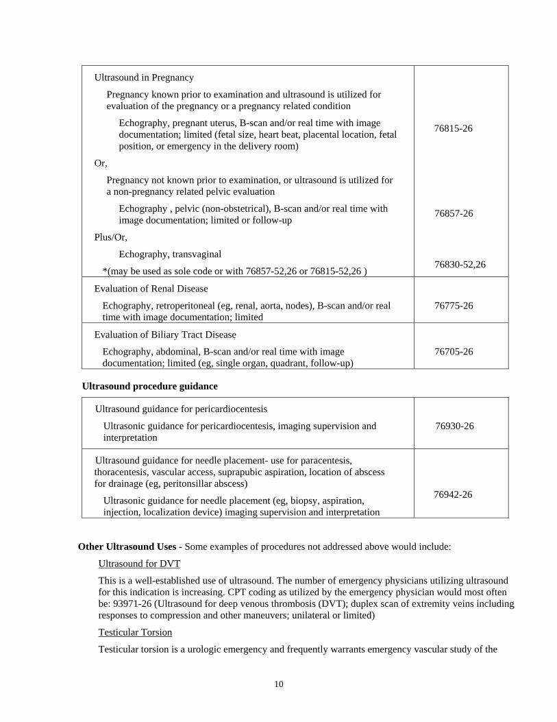
Primary Indications:
Trauma Ultrasound – Thoracoabdominal Trauma (FAST Exam)
Echography, abdominal, B-scan and/or real time with image documentation limited (eg, single organ, quadrant, follow-up)
Plus,
Echocardiography, transthoracic, real-time with image documentation (2D) or without M-Mode recording; follow-up or limited
*(for non-traumatic hemoperitoneum or ascites use 76705-26)
76705-26
+
93308-26
Abdominal Aortic Aneurysm
Echography, retroperitoneal (eg, renal, aorta, nodes), B-scan and/or real time with image documentation; limited
76775-26
Pericardial Fluid
Echocardiography, transthoracic, real-time with image documentation (2D) with or without M-Mode recording; follow-up or limited
93308-26
10
Ultrasound in Pregnancy
Pregnancy known prior to examination and ultrasound is utilized for evaluation of the pregnancy or a pregnancy related condition
Echography, pregnant uterus, B-scan and/or real time with image documentation; limited (fetal size, heart beat, placental location, fetal position, or emergency in the delivery room)
Or,
Pregnancy not known prior to examination, or ultrasound is utilized for a non-pregnancy related pelvic evaluation
Echography , pelvic (non-obstetrical), B-scan and/or real time with image documentation; limited or follow-up
Plus/Or,
Echography, transvaginal
*(may be used as sole code or with 76857-52,26 or 76815-52,26 )
76815-26
76857-26
76830-52,26
Evaluation of Renal Disease
Echography, retroperitoneal (eg, renal, aorta, nodes), B-scan and/or real time with image documentation; limited
76775-26
Evaluation of Biliary Tract Disease
Echography, abdominal, B-scan and/or real time with image documentation; limited (eg, single organ, quadrant, follow-up)
76705-26
Ultrasound procedure guidance
Ultrasound guidance for pericardiocentesis
Ultrasonic guidance for pericardiocentesis, imaging supervision and interpretation
76930-26
Ultrasound guidance for needle placement- use for paracentesis, thoracentesis, vascular access, suprapubic aspiration, location of abscess for drainage (eg, peritonsillar abscess)
Ultrasonic guidance for needle placement (eg, biopsy, aspiration, injection, localization device) imaging supervision and interpretation
76942-26
Other Ultrasound Uses - Some examples of procedures not addressed above would include:
Ultrasound for DVT
This is a well-established use of ultrasound. The number of emergency physicians utilizing ultrasound for this indication is increasing. CPT coding as utilized by the emergency physician would most often be: 93971-26 (Ultrasound for deep venous thrombosis (DVT); duplex scan of extremity veins including responses to compression and other maneuvers; unilateral or limited)
Testicular Torsion
Testicular torsion is a urologic emergency and frequently warrants emergency vascular study of the
11
scrotum. This study, done in a limited fashion to describe vascular flow to the testicles would be: 93976-26 (Duplex scan of arterial inflow and venous outflow of abdominal, pelvic, scrotal contents and/or retroperitoneal organs; limited study)
Urinary Bladder Sonography
Ultrasound may be utilized as a non-invasive method to check for urinary retention, post-void residual urine or the presence of urine in bladder prior to catheterization, for example in a dehydrated infant. Appropriate coding would be 76857-26; Echography, pelvic (non-obstetrical), B-scan and/or real time with image documentation; limited or follow-up. Note that if a suprapubic tap is done under ultrasound guidance one may substitute 76942-26 (ultrasound guidance for needle placement) for the echography code.
Musculoskeletal Sonography
The use of ultrasonic musculoskeletal examination by emergency physicians is infrequent. One example would be the limited examination of an infant’s hips, eg, for effusion, and is coded; 76886-26 (Echography of infant hips, real time with imaging documentation, limited, static eg, not requiring manipulation)
Foreign Body Localization
The localization of foreign bodies, particularly those that are radiolucent (eg, organic), is an attractive application for emergency department ultrasound. At this time the routine use of ultrasound for the identification of foreign bodies is considered investigational. If ultrasound were utilized to place a needle as a localization device for excision guidance, then 76942-26 (Ultrasonic guidance for needle placement) would accurately describe this ultrasound guidance procedure.
Peritonsillar Abscess Identification
A number of centers are currently utilizing ultrasound to evaluate the swollen tonsil, soft palate or retropharynx to identify abscess and guide medical treatment versus surgical treatment. At this time the routine use of ultrasound for the identification of peritonsillar abscess is considered investigational. If ultrasound were used for needle aspiration guidance then the 76942 code (ultrasound guidance for needle placement) would accurately describe this ultrasound guidance procedure.
Unlisted Ultrasound Procedures
The coding of a procedure not defined by CPT can be accomplished on a case-by-case basis utilizing the unlisted ultrasound procedure code 76999. As could be expected for a non-CPT-defined service, obtaining reimbursement for such a procedure is not the usual straightforward process. Submission of an unlisted procedure code almost universally requires supplemental documentation including a description of the procedure and rationale for medical necessity. In addition, because no work units can be assigned to an unlisted procedure code, as it would vary from use to use, reimbursement (if any) can vary significantly.
IV ICD-9 Coding and Medical Necessity
A. ICD-9 Coding
ICD-9, the International Classification of Diseases, 9th Revision, is a cataloging tool developed by the World Health Organization for the international comparison of morbidity and mortality data. ICD-9 codes were subsequently modified in the United States by the addition of clinical information and utilized in evaluating quality of care as well as for payment purposes. The ICD-9 system is analogous to the Dewey Decimal System, used for cataloging books, in that there are up to three digits to the left, and up to two digits to the right, of the decimal point (eg, 379.17), with the positions to the right of the
12
decimal indicating greater and greater specificity. ICD-9 codes may be driven by diagnosis, symptoms, signs, abnormal diagnostic tests, by external causes of injury (E codes) or factors influencing health status (V codes). Symptom codes, such as 789.01 abdominal pain, right upper quadrant, are often used for coding emergency visits, as an exact cause of symptoms, such as cholecystitis 575.10, may not be known at the time of discharge from the emergency department. E-codes identify the cause of injury, eg, motor vehicle accident, poisonings, bites, drowning, chemical exposures, etc. While mechanism of injury codes add valuable information about your practice and have great appeal to emergency physicians, most carriers will not recognize these codes as the primary diagnosis. Use of E codes, as a primary diagnosis will often result in denial of payment. Finally, multiple ICD-9 codes are frequently utilized to describe a single visit.
ICD-9 codes are often used by payors to determine why you performed a procedure as opposed to CPT codes that explain what procedure(s) were performed. Carriers often look first at these ICD-9 codes as an indicator of medical necessity by developing, as a first-pass edit, lists of ICD-9 codes that support certain CPT codes (see IV. B. Medical Necessity; below). Frequently associated with such lists of compatible ICD-9 codes is a statement that any code not listed would not be covered. However, even the denial of a claim, based upon such first-pass medical necessity edits, can usually be appealed. Proper use of ICD-9 codes will prevent many processing problems and help physicians report more intensive services when these services are required by a patient's condition. Complete and accurate ICD-9 coding in your current practice should support the medical necessity for most clinically indicated emergency department ultrasounds.
Medicare carriers are required to publish bulletins that clarify their view of proper coding, including coding for ultrasound procedures. These bulletins often include lists of indications and limitations of coverage as well as lists of covered ICD-9 codes (see VI.B. Covered Services and Non-covered Services below). A resource for searching a variety of regional policies can be found at the Center for Medicare and Medicaid Services (CMS or the agency formerly known as HCFA) sponsored Local Medical Review Policies web site (http://www.lmrp.net). The reader is advised to search such sites by carrier and for specific ultrasound examination policies, eg, abdominal or pelvic sonography.
B. Medical Necessity
Medical necessity or medically necessary services are described as those that are safe, effective and consistent with the symptoms or diagnosis of the illness or injury being treated. Obviously those services deemed medically unnecessary would lack the above characteristics. Services that are regarded as experimental or investigational are frequently deemed to be medically unnecessary. As the determination of medical necessity is often based on a payor’s concept of accepted professional medical standards, this determination may at times be subjective or may not reflect the most current application of medical technology. The designation of medically necessary services is dynamic and may change from time to time.
A covered service may be denied payment if the physician fails to demonstrate the medical necessity for that service. Indication(s) for an ultrasound study should be noted along with a procedure description and study findings in a procedure note placed in the patient’s medical record. Indications may include a symptom, diagnosis, physical finding or laboratory finding. In addition, appropriate ICD-9 codes describing those symptoms, findings or diagnoses should be applied at the time of medical record coding to assist in establishing the medical necessity for ultrasound procedures.
As medical necessity is determined by an individual patient’s symptoms, diagnoses or accepted professional medical standards of care, it is not clear that the protocol-driven use of ultrasound for a class of patients, eg, all those in motor vehicle accidents or all “trauma activations,” would be deemed medically necessary. One needs to distinguish between “screening studies” provided to patients without symptoms or finding, and “diagnostic studies” provided patients who have symptoms or findings. Screening examinations are not covered services by Medicare. For example,
13
a thoracoabdominal trauma ultrasound examination for an auto accident patient without abdominal pain, tenderness, intoxication, distracting injury or head trauma may be denied as a medically unnecessary study. An ultrasound for thoracoabdominal trauma examination in the same patient with abdominal pain or tenderness would likely be judged as medically necessary. Likewise, the routine use of ultrasound guidance for all central line placements may not be viewed as medically necessary (despite an evolving literature to the contrary; “Making Health Care Safer: A Critical Analysis of Patient Safety Practices” http://www.ahcpr.gov/clinic/ptsafety/chap21.htm). The placement of a central line under ultrasound guidance after a failed attempt or with patients who have poorly defined anatomic landmarks may be viewed as medically necessary by the same payor.
V Ultrasound Procedure Documentation
A. Ultrasound Documentation Background
Ultrasound documentation serves many purposes such as communication and continuity between healthcare providers, utilization and quality of care review, collection of data for research and education as well as for the management of risk. In addition, documentation is a key component in the proper coding of and reimbursement for emergency medicine ultrasound procedures. In this context documentation serves to identify specific procedures, as defined by CPT, and supports claims accuracy for the reimbursement of procedures. While most facilities offering emergency medicine ultrasound will define their own program-specific documentation methods, the following discussion addresses the issue of current and known requirements for the coding and claims submission of emergency medicine ultrasound procedures.
CPT states “A written report signed by the interpreting physician should be considered an integral part of a radiologic procedure or interpretation.” Medicare documentation requirements for ultrasound are not enumerated in statute or the Medicare Carrier Manual, but are found in Local Medical Review Policies. Such documentation requirements vary from region to region. Although these carrier requirements vary, there are sufficient requirements common among published local carrier policies to offer the documentation recommendations enumerated below. Contracts with private payors may have specific documentation requirements or may be silent in regard to diagnostic test documentation requirements. Absent any specific contractual requirements, a reasonable approach would be to follow local Medicare general documentation principles and adhere to the concept that the report should be similar to that usually prepared by a specialist in the field and consistent with the service furnished (HCFA memo regarding X-Ray/EKG Interpretations in the ED; http://www.acep.org/2,290,0.html). Again, a resource for searching a variety of regional policies regarding the use of ultrasonography, including documentation requirements, can be found at the CMS sponsored Local Medical Review Policies web site (http://www.lmrp.net).
B. Documentation and Image Archiving
Image archiving refers to the storage and retrieval of an ultrasound image(s). The retention of ultrasound images by emergency physicians is often undertaken during program startup to evaluate performance, for initial physician credentialling or as a component of a defined quality improvement or research project. The paradigm for emergency ultrasound use is that ultrasound performed by the emergency physician, is contemporaneous with patient care, and the diagnostic information obtained is used for immediate clinical decision-making or the guidance of high-risk procedures. Emergency physicians do not conduct consultative studies and generally do not utilize technicians to generate images, file rooms to store film or clerical personnel to track and retrieve film. In this context the archiving of images has received less attention than has the integration of clinical data into patient management. While some emergency providers affix a representative image(s) to the patient encounter form demonstrating relevant anatomy and/or pathology, many authorities in emergency ultrasound do not practice or advocate the archiving of emergency ultrasound images as an ongoing clinical
14
requirement.
Some ultrasound CPT codes, however, include the language “with image documentation”. This language indicates that when the unmodified CPT code is utilized it is expected that an image(s) be retained to meet criteria for the application of that unmodified, or complete, CPT code. The diagnostic ultrasound codes frequently used by emergency physicians (76506-76886) incorporate this language. As an example, the CPT code most often used by emergency physicians for evaluation of the abdominal aorta; echography, retroperitoneal (eg, renal, aorta, nodes), B-scan and/or real time with image documentation; limited (76775); contains this “image documentation” reference. The ultrasound guidance procedure codes (76930-76965) do not incorporate this language, but utilize “supervision and interpretation” language in its place. An example of an ultrasound guidance code utilized by emergency physicians would be: ultrasonic guidance for pericardiocentesis, imaging supervision and interpretation (76930). This “supervision and interpretation” language indicates that ultrasound imaging is utilized to supervise an invasive procedure eg, pericardiocentesis, and that the imaging supervision is separate from the procedure itself. Here, image retention is not required for the application of this guidance procedure CPT code. A written, separately identifiable, signed report remains a coding requirement.
The AMA publication, Principles of CPT Coding, states that “Physician interpretation of the results of diagnostic tests/studies may be reported separately with preparation of a separate distinctly identifiable signed written report. When separately reporting the interpretation of diagnostic tests/studies, modifier –26 is appended to the specific code for the diagnostic test/study reported.” Diagnostic image generation, storage, retrieval and image ownership is considered to be a facility responsibility, supported by payment of the technical component to the facility. Interpretation of images and report generation is a physician responsibility and is reimbursed as the professional component of a global service. When an unmodified CPT is utilized, or the technical component alone is coded for the diagnostic CPT codes, then image archiving would be required. Image retention for coding of the professional interpretation of a diagnostic ultrasound is not a specified CPT requirement.
Although physicians may report ultrasound image interpretation without image retention, the hospital will need some image archiving if they charge the technical component for the diagnostic ultrasound codes. Emergency physicians working cooperatively with hospitals may want to establish a practical policy for ultrasound image retention that can be accomplished within the emergency ultrasound paradigm. One mechanism currently utilized by some emergency physicians for image retention is to affix a representative thermal printer image(s) to the ED patient encounter form or on a progress note placed in the medical record (CPT does not enumerate specific views required, the number of views, the image documentation medium, how or where images are retained). Patient identification of the image is supplied, such as a patient label applied to the image. At least one image per CPT procedure coded, representative of relevant anatomy and/or pathology, is placed in the medical record. For example an aortic study might require a minimum of one representative image (aorta) and a FAST examination may require a minimum of two representative images (heart and abdomen). Documenting images in this fashion should meet CPT requirement for coding global diagnostic ultrasound CPT codes or the technical component of these codes. More elaborate retention/archiving systems could be pursued, depending on program design, resources and department specific policies.
The above discussion is based on CPT theory and not payor policy. Medicare payor policy regarding image retention requirements vary significantly by regional carrier. Some carriers have no published policies; others require only that the study indication or the physician report appear in the medical record. Some carriers require that images be available for review upon request. Again, emergency physicians should consult relevant local payors’ policies, with which participating providers are obligated to comply.
15
C. Ultrasound Documentation Requirements
Below are suggested guidelines for the development of documentation policy for emergency ultrasound programs culled from Medicare Local Medical Review Policies, CPT and authors’ opinion. The first three documentation criteria below are sample language quoted from regional Medicare carrier policies, and it is recommended that they be followed for all emergency ultrasound procedure notes. The fourth documentation criterion is from CPT and refers directly to image documentation or archiving. The last two criteria listed have been added to assist in the correct and consistent coding of emergency ultrasound procedures and do not represent Medicare or CPT-specified requirements.
Procedure Specific Requirements:
1. “A written interpretation and report must be completed for each test performed and must be maintained in the patient’s medical record.”
This written interpretation and report may take the form of a separately identifiable procedure note within the emergency department medical record, such as typically used for procedures like lumbar puncture, or central line placement. Alternatively, the report may take the form of a separate note appended to the medical record, such as typically done for consultative radiology reports. Regardless of the form used, the responsible physician must sign the report. (see CMS memo regarding X-Ray/EKG Interpretations in the ED; www.acep.org/2,290,0.html)
2. “Medical record documentation must indicate the medical necessity [of the diagnostic test].”
Indicating the medical necessity is essential for reimbursement and must be included in the documentation (see IV. B. Medical Necessity)
3. “This report must describe the structures or organs studied and supply an interpretation of the findings.”
This is the purpose of performing the ultrasound and is required documentation.
Image Documentation (archiving of images)
4. Image retention is not a CPT requirement for the ultrasound guidance procedure codes (76930-76965). CPT definitions for the diagnostic procedure codes do not specify image archiving by the physician when only the professional component (-26) is coded. When the global (unmodified) code or the technical portion of the diagnostic ultrasound CPT codes (76506-76886) and the non-invasive vascular diagnostic codes (93875-93990) are utilized, then image retention is a CPT requirement.
In consulting the Local Medicare Coverage Database (www.cms.hhs.gov/mcd) there is considerable carrier variability in documentation requirements relating to image retention. Some local medical review policies require image availability for review. Again, providers should consult relevant Local Coverage Determinations (LCDs).
Each emergency medicine ultrasound program should define their program-specific documentation policies, including image retention in accord with hospital requirements and compliant with local payor policy. Such requirements should be codified in policy and adhered to by emergency physician providers.
Additional Recommendations - These should assist in correct coding, though not specified by Medicare carriers or CPT, and should include:
5. The report should identify who performed the procedure.
This will help avoid confusion as to whether the physician performed and interpreted the test, or simply reviewed the report of another provider.
16
6. To facilitate accurate coding, the scope of the study should be described.
The performing physician should indicate whether the study was a limited or complete examination. In addition, the physician should indicate if this is a repeat examination by the same physician, a repeat examination by a second physician, and/or a reduced level of service.
Thus, an example of an emergency medicine ultrasound report for a patient with first trimester pelvic pain and vaginal bleeding might be as follows:
“I performed a limited transvaginal ultrasound exam of the pelvis to evaluate for IUP vs. signs of ectopic pregnancy in a patient with a positive pregnancy test and vaginal bleeding. The uterus and pouch of Douglas were visualized. There was a small (physiological) amount of free fluid in the pelvis. The uterus had an endometrial stripe of 12 mm with no definite sign of IUP. Impression: no evidence of an intrauterine pregnancy.”
VI Payor Policy
A. Payor Policy Background
Every payor, including Medicare, has its own rule sets regarding what services are covered, what constitutes a medically necessary service and what coding combinations are permissible (see definition of terms below). As a provider, when a physician agrees to accept assignment for Medicare or agrees to contract with any payor, he or she is agreeing to adhere to the payor’s payment policies. Medicare payor policy can vary by region and other payors may have dramatically different approaches to seemingly the same issues. A physician may need to review a payor’s methodologies (payor policy manuals) and while many contracts and payor policy manuals may be silent on these issues, an informed member of the practice should be prepared to review contractual language regarding the handling of ultrasound procedures and claims submission. These issues may become especially important as emergency ultrasound technology expands and payors begin to receive claims from physicians who have historically not conducted these procedures or utilized these codes. Medicare carriers, for example, are instructed to identify targets for auditing based upon data aberrations, such as the increased utilization of a family of codes. Finally, a practice member may also need to discuss denials of payment for ultrasound procedures as payor determination of reimbursement for emergency ultrasound may become clear only after claims are received and payors make determinations on previously undocumented policy.
The concepts outlined below will familiarize the reader with several payor approaches to the difficult task of identifying the correct coding and submission of claims for services. The concepts introduced will include covered services, non-covered services and coding edits.
B. Covered and Non-Covered Services
“Covered services” are those medical services that a payor will reimburse a provider for performing, under predefined circumstances. “Non-covered services” are those always excluded from coverage, and reimbursement from the payor is never available for them. For example, an annual physical examination may be a good medical practice, but it is not a Medicare “covered service;” therefore, as a “non-covered service” it is never paid for by Medicare. In the arena of ultrasound, examples of Medicare covered services would include echocardiography, abdominal sonography, abdominal aorta sonography, pregnancy diagnosis sonography and venous flow studies. An example of a non-covered ultrasound study would be the monitoring of cardiac output by Doppler, as it is considered investigational. Covered versus non-covered ultrasound services for Medicare can be viewed in the Coverage Issues Manual, Diagnostic Services 50-7 (http://www.cms.hhs.gov/mcd/search.asp?). The designation of covered and non-covered services may change from time to time.
17
In order to qualify for Medicare reimbursement a service must not only be a covered service, but must also meet the determination of “medical necessity” for the individual patient under consideration. In addition, the service must be provided in a setting appropriate for the patient’s condition to justify reimbursement and “Physicians who perform and/or interpret the studies must be capable of demonstrating training and experience specific to the study performed or interpreted.” For emergency physicians this credentialing requirement is supported by the ACEP Emergency Ultrasound Guidelines – 2001 and the ACEP Policy Statement, Use of Ultrasound Imaging by Emergency Physicians (http://www.acep.org/1,684,0.html; http://www.acep.org/1,33811,0.html).
Finally, while the term “covered service” is most frequently prospectively delineated for governmental payors, the same concept may not be so clearly predefined for non-governmental payors. Indeed it is not unusual that a provider learns only retrospectively that a service is not covered, ie, only after receiving and pursuing a reimbursement denial.
C. Payment Edits
Edits are policy-driven computer programs or manual reviews that check information available on payment claim forms in an effort to identify the coding of services with a higher probability of being incorrect or medically unnecessary. These edits vary from payor to payor, change periodically and, in the case of Medicare, may vary from region to region.
1) Edit types may include:
• Procedure to diagnosis (diagnosis edits)
Here the CPT code is compared to a list of previously approved ICD-9 codes. The procedures that are previously approved as compatible with the ICD-9 are automatically paid. All other CPT codes are denied, but may later be approved with additional information. They are often termed diagnosis or payment edits.
Example: migraine headache ICD-9 code with a limited abdominal echography CPT code.
• Procedure to procedure
The CPT code is compared to a second CPT code. The edit is designed to assure reporting a group of procedures with the most appropriate comprehensive code.
Example: CPT code for Ultrasound guidance for pericardiocentesis and a CPT code for a limited echocardiography (US guidance for pericardiocentesis includes the echocardiography in most circumstances for the same patient encounter).
• Frequency to time
Example: multiple limited abdominal examinations on same date of service.
• Site of Service
The CPT code is compared to the site where the service was rendered.
Example: global ultrasound service coded by a hospital-based physician (hospital site of service).
• Procedure to provider
Example: vascular surgery billed by an internist.
• Procedure to sex
Example: echography, pregnant uterus on a male patient.
Edits may select codes having a higher probability of being incorrect but cannot identify all
18
circumstances that may warrant legitimate use of the code. Again, documentation of medical necessity for the study in the medical record will be essential in any effort to overcome a first-pass edit denial. One should review all denials for appropriateness of the denied claim. A well-supported and timely appeal of a denial for medical necessity often results in payment. However, if claims are being rejected for medical necessity, your billing and coding policies may need review or revision. Improved, appropriate coding may provide information in a way that the payor can more readily recognize as medically necessary. A good discussion of Medicare claims review, audits and appeals mechanisms as well as medical necessity denials can be found in: Healthcare Fraud and Abuse: A Physicians Guide to Compliance (AMA Press; www.ama-assn.org).
2) Medicare Correct Coding Initiative Edits
These edits are found in The National Correct Coding Policy Manual for Part B Medicare Carriers. This policy manual includes guidelines and coding policy as well as coding edits that Medicare carriers use to approve or deny payment when the same physician, or facility, bills two or more CPT codes for the same date of service. These edits are revised on a quarterly basis. The policies generally identify codes that are mutually exclusive and identify fragmentation, or the unbundling, of services.
Most ultrasound codes utilized by emergency physicians in facilities do not trigger correct coding initiative edits when these codes are reported as the professional component only (use of a –26 modifier). It should be noted that when reported as unmodified, or global codes, there are a number of edits triggered. An example would be an unmodified 93308 (echography, transthoracic) would trigger an edit when reported with a 93042 (rhythm ECG, one to three leads; interpretation and report only), however reporting of a 93308-26 (echography, transthoracic; professional component) with a rhythm interpretation would not trigger that edit. For those physicians contemplating the reporting of global codes, the National Correct Coding Policy Manual for Part B Medicare Carriers should be consulted for every ultrasound code that will be utilized.
VII Frequently Asked Questions
1. What code should I use for the thoracoabdominal trauma or FAST ultrasound examination?
The FAST examination is not a single ultrasound procedure, but a clinical approach to the trauma patient that utilizes two distinct limited examinations currently described by CPT. The components of this clinical approach have been outlined by the FAST consensus Conference Committee: Scalea TM, Rodriguez A, Chiu WC, et al. Focused assessment by sonography for trauma (FAST): results from an international consensus conference. J Trauma 1999; 46(3):466-471. The CPT codes are (1) a limited transthoracic echocardiogram (93308-26) and (2) a limited abdominal ultrasound (76705-26). (ref. III.D.1.a.)
2. If I perform a diagnostic ultrasound and a related ultrasound-guided procedure during a single patient encounter, how do I code?
Generally one codes either the diagnostic ultrasound or the guidance ultrasound, but not both, for the same patient on the same visit. For example, one would not code both a diagnostic transthoracic echocardiography (93308-26) and an ultrasound guidance for pericardiocentesis (76930-26) on the same encounter. The surgical procedure, however, is coded in addition to the ultrasound code eg, the surgical procedure itself (33010, pericardiocentesis; initial) in addition to the ultrasound guidance (76930-26, ultrasound guidance for pericardiocentesis). (ref. III.D.2.)
19
3. What documentation is necessary for the coding of emergency department ultrasound examinations?
Proper documentation of ultrasound studies performed in the emergency department should include or indicate the following: • A separately identifiable written report for each test performed. This may be an independent
report(s) or may be incorporated within the patient treatment record. The report must be signed by the physician.
• The medical necessity must be included for each study performed and in the interest of clarity this should appear as the study indication.
• A description of the structures or organs studied and an interpretation of the findings. • Additional recommendations: a statement of who performed the study and the scope of the
examination performed (eg, limited or complete). (ref. V.C.)
4. Do I have to include an ultrasound image in the chart to code for an ultrasound that I perform?
Image retention is not an explicit CPT requirement when only the professional component of ultrasound CPT codes are utilized (ie, interpretation and report). The AMA publication, Principles of CPT Coding, states that “Physician interpretation of the results of diagnostic tests/studies may be reported separately with preparation of a separate distinctly identifiable signed written report. When separately reporting the interpretation of diagnostic tests/studies, modifier –26 is appended to the specific code for the diagnostic test/study reported.” When ultrasound guidance procedures (76930-76965) are coded a written interpretation and report is required, however image retention is not a requirement. When the diagnostic ultrasound CPT codes (76506-76886) or the non-invasive vascular diagnostic codes (93875-93990) are utilized and the global code or the technical component is coded, image retention is required based on the CPT “with image documentation” language included in the code definition. Finally, Medicare image retention requirements vary significantly by local carrier. Some carriers have no published requirements and some carriers require that images be available for review upon request. Again, emergency physicians should consult current relevant local payers’ policies. (see V. B. Documentation and Archiving)
5. Do I have to meet American College of Radiology standards for documentation in order to code for an ultrasound interpretation?
No, there are no CMS (agency formerly known as HCFA) requirements that documentation adhere to any specialty society guidelines. CMS opinion has primarily addressed X-ray and EKG interpretation and can be reviewed in the “HCFA Final Rule on X-ray/EKG Interpretations” and “HCFA memo regarding X-ray/EKG Interpretations in the ED.” (both found at www.acep.org/1,201,0.html)While these documents do not directly address ultrasound interpretation and reports, our opinion is that general principles of X-ray interpretation by emergency physicians would be similar to ultrasound examination and interpretation by emergency physicians. (1) Reports must be complete and similar to that usually prepared by a specialist in the field. (2) Reports must be consistent with the services furnished. (3) Reports need not specifically follow the American College of Radiology guidelines. (4) Interpretations and reports may be recorded either separately or within the emergency physician’s treatment record. (5) When multiple bills are submitted for the same report, CMS will pay for the interpretation and report that directly contributed to the diagnosis and treatment of the individual patient. The interpretation and report paid will usually be that provided contemporaneous with the patient’s visit and without consideration of the reporting physician’s specialty.
20
6. If the EP and a trauma surgeon both perform a FAST exam, can both physicians code and bill for their respective examinations?
A second physician may code and bill a repeat examination for the same patient on the same date, however, the second physician should utilize the –77 CPT code modifier. Both physicians must document the medical necessity for each examination performed. The indications for a repeat FAST examination on the same patient encounter might include such factors as the development of hemodynamic instability, a falling hematocrit, or development of increased abdominal pain/tenderness in a patient who had a negative initial FAST examination. Repeating a FAST examination to simply confirm the finding of another provider may be clinically reasonable, but would not warrant the coding of a separate study.
Remember that physicians of the same specialty in the same group, or working for the same employer are considered to be the same physician from a billing perspective. If the “same physician” repeats an examination then the –76 modifier is utilized. (ref. III.C.4.)
7. Can an emergency physician code a limited examination and the radiologist code a complete examination on the same patient encounter?
Two different physicians can code a limited examination and a complete examination, of the same anatomic description, on the same date of service, if two separate procedures are performed and are medically necessary. On some occasions an initial limited examination by an emergency physician will be inconclusive or demonstrate an unexpected finding requiring a complete examination, or follow-up examination, by a consulting radiologist. What is required is that each examination, limited or complete, stand on its own merit as a medically necessary study. The planned sequencing of a limited examination by one provider (emergency physician) followed by complete examination by a second provider (radiologist), both coding for the procedures, would not constitute proper or compliant coding. A repeat examination of the exact same type by a second physician would require a –77 modifier. Inasmuch as a complete study (comprehensive service) contains any corresponding limited service within it, it would be prudent for the second physician providing the comprehensive procedure to consider use the –77 modifier. Use of the –77 modifier in this later circumstance would assume that the second physician was aware that his/her service was a repeat examination.
A single provider would not code a limited exam followed by a complete examination, of the same anatomic description, as the initial limited exam would be included in the more comprehensive complete code. (ref. III.C.).
8. If I use ultrasound to diagnose pregnancy, how do I code for the study?
If pregnancy status is unknown prior to ultrasound examination and ultrasound were utilized to evaluate amenorrhea, pelvic pain, vaginal bleeding or non-gynecologic pelvic pathology, then the non-obstetric pelvic CPT code (76857-26) would be utilized. This would be true even if the result of the examination were the diagnosis of an intrauterine or ectopic pregnancy. (ref. III. D.1.b.)
9. If I use ultrasound to evaluate a pregnancy or a possible complication of pregnancy, how do I code the study?
When the patient is (1) known to be pregnant, including knowledge of a positive pregnancy test, and (2) the physician is utilizing ultrasound to evaluate the pregnancy or a suspected complication of pregnancy, then the obstetric pelvic codes would be utilized (eg, 76815-26). When these two criteria are met, the obstetric codes are utilized regardless of the study result. Thus, the obstetric pelvic codes would apply to the “known to be pregnant patient” even in the absence of an identified intrauterine pregnancy and if the patient were found to have an ectopic pregnancy, spontaneous abortion, molar pregnancy, ovarian torsion or a non-pregnancy related condition.
21
(ref. III. D.1.b.)
10. How do I code for a study in which there is an unexpected ultrasound finding?
Code for the studies indicated and performed. In many circumstances the examination indicated and the corresponding CPT code will be unchanged even when an unexpected finding occurs, eg, gallstones seen when doing a FAST examination. The gallstones would be recorded as an incidental finding in the abdominal portion of the FAST exam and would not require that an additional code or even a more comprehensive code be applied. In other circumstances a finding during the initial examination may necessitate a second examination indicated by the unexpected finding. For example, a non-obstetric pelvic ultrasound performed for urinary retention may suggest an abdominal aortic aneurysm at the level of the aortic bifurcation and warrant a retroperitoneal ultrasound to more fully evaluate the abdominal aorta. In this latter circumstance, if a second examination were performed and documented, then coding for the retroperitoneal ultrasound would be 76775-26 in addition to the limited non-obstetric pelvic 76857-26.
11. Does the introduction of APCs (ambulatory patient classification groups) change the CPT coding of emergency department ultrasounds?
No. Currently, physician services are excluded from APCs and are billed separately. It is important that as more emergency department ultrasounds are performed, facilities and physicians learn to accurately identify ultrasound examinations and CPT code descriptions to ensure complete and compliant reporting of procedures. Also, APCs are in their infancy and may be subject to some rapid changes, so be sure to remain up to date with this issue.
12. What impact does ICD-9 coding have on claims submission for emergency ultrasound examinations?
Payors frequently develop lists of pre-approved ICD-9 codes they feel are compatible with, support or verify the ultrasound CPT procedures listed on claim forms. The ICD-9 codes that are previously approved as compatible with the CPT codes are automatically paid. Other claims are often denied, but may later be approved when supplemented with additional information. This screening of CPT procedures by comparison to ICD-9 codes is designed to detect claims with a higher probability of being incorrect or unnecessary and are termed diagnosis or payment edits. As this screening process is often based on a payor’s concept of accepted professional medical standards such determinations may at times be subjective or may not reflect the most current application of medical technology. (ref. VI. C.)
13. Can hospital based emergency physicians own the US machine and code a global CPT based on ownership of the equipment?
No. Hospital-based emergency physicians report the professional component of the CPT by the addition of the (-26) modifier to describe their services.
Ultrasound CPT codes are global or complete codes. A global code, in this context, is the combination of a technical component and a professional component. In the hospital setting, the technical component, designated by a –TC modifier, is charged by the facility (ie, hospital) and includes reimbursement for the cost of equipment, service and maintenance, supplies and technician salaries. The professional component of the ultrasound service, indicated by the –26 modifier, is reported by the physician for professional services that include interpretation and report of the study results. In a like fashion, hospitals may not charge a global fee when physicians are employed and salaried. When physicians are employed by the hospital the technical component may be submitted to the Medicare fiscal intermediary on a form UB 92 and the hospital may also bill for the physician service, reassigned to the hospital, on the HCFA form 1500 submitted to the Medicare carrier. Finally physicians, including emergency physicians, may
22
utilize global or unmodified ultrasound CPT codes in physician offices, clinics or free standing emergency facilities (not operated by a hospital), that provide professional services as well as own and maintain the equipment.
The following issues relate to compliance surrounding hospital-based physician ownership of equipment and utilization of global CPT codes. (1) The global fee, when billed by a physician, should reflect a non-hospital location as a site of service and not a hospital site of service such as the ED. One would expect a coding edit to result in a payment denial or possible payor inquiry when a global CPT is submitted with a hospital site of service. (2) If the patient is admitted to the hospital within 72 hours of performance of the emergency department ultrasound, the technical component of the ultrasound charge should be rolled into the hospital inpatient DRG in the case of Medicare and must be backed out of the outpatient bill (“72 hour rule”). Recognition of these necessary charge adjustments is technically difficult and could lead to non-compliance with reimbursement regulations and/or payment policies. (3) Equipment purchase is not typically a cost sustained by hospital-based physicians, and hospital emergency physician groups do not typically receive revenue from equipment ownership. Financial relationships between physicians who utilize hospital services that entail using the physician’s own equipment are addressed by multiple fraud and abuse statutes and regulations. Physicians contemplating such arrangements are well advised to seek competent specialized legal counsel. (ref. III.C.1.)
Recommended