BioMed CentralFilaria Journal
ss
Open AcceResearchEvidence against Wolbachia symbiosis in Loa loaHelen F McGarry†1, Ken Pfarr†2, Gill Egerton1, Achim Hoerauf2, Jean-Paul Akue3, Peter Enyong4, Samuel Wanji5, Sabine L Kläger6, Albert E Bianco7, Nick J Beeching8 and Mark J Taylor*1Address: 1Molecular and Biochemical Parasitology, Liverpool School of Tropical Medicine, Pembroke Place, Liverpool, UK, 2Department of Helminthology, Bernhard Nocht Institute for Tropical Medicine, 20359 Hamburg, Germany, 3Department of Medical Parasitology, International Center for Medical Research of Franceville, BP 769, Franceville, Gabon, 4Tropical Medicine Research Station, P.O. BOX 55, Kumba, Cameroon, 5Research Foundation in Tropical diseases and Environment, P.O. Box 474, Buea, Cameroon, 6Department of Haematology, Box 234, Addenbrookes NHS Trust, Hills Road, Cambridge CB2 2QQ, UK, 7Wellcome Trust, 183 Euston Road, London, UK and 8Clinical Research Group, Liverpool School of Tropical Medicine, Liverpool, L3 5QA, UK
Email: Helen F McGarry - [email protected]; Ken Pfarr - [email protected]; Gill Egerton - [email protected]; Achim Hoerauf - [email protected]; Jean-Paul Akue - [email protected]; Peter Enyong - [email protected]; Samuel Wanji - [email protected]; Sabine L Kläger - [email protected]; Albert E Bianco - [email protected]; Nick J Beeching - [email protected]; Mark J Taylor* - [email protected]
* Corresponding author †Equal contributors
AbstractBackground: The majority of filarial nematode species are host to Wolbachia bacterialendosymbionts, although a few including Acanthocheilonema viteae, Onchocerca flexuosa and Setariaequina have been shown to be free of infection. Comparisons of species with and without symbiontscan provide important information on the role of Wolbachia symbiosis in the biology of thenematode hosts and the contribution of the bacteria to the development of disease. Previousstudies by electron microscopy and PCR have failed to detect intracellular bacterial infection in Loaloa. Here we use molecular and immunohistological techniques to confirm this finding.
Methods: We have used a combination of PCR amplification of bacterial genes (16S ribosomalDNA [rDNA], ftsZ and Wolbachia surface protein [WSP]) on samples of L. loa adults, third-stagelarvae (L3) and microfilariae (mf) and immunohistology on L. loa adults and mf derived from humanvolunteers to determine the presence or absence of Wolbachia endosymbionts. Samples used in thePCR analysis included 5 adult female worms, 4 adult male worms, 5 mf samples and 2 samples ofL3. The quality and purity of nematode DNA was tested by PCR amplification of nematode 5SrDNA and with diagnostic primers from the target species and used to confirm the absence ofcontamination from Onchocerca sp., Mansonella perstans, M. streptocerca and Wuchereria bancrofti.Immunohistology was carried out by light and electron microscopy on L. loa adults and mf andsections were probed with rabbit antibodies raised to recombinant Brugia malayi Wolbachia WSP.Samples from nematodes known to be infected with Wolbachia (O. volvulus, O. ochengi, Litomosoidessigmodontis and B. malayi) were used as positive controls and A. viteae as a negative control.
Results: Single PCR analysis using primer sets for the bacterial genes 16S rDNA, ftsZ, and WSPwere negative for all DNA samples from L. loa. Positive PCR reactions were obtained from DNAsamples derived from species known to be infected with Wolbachia, which confirmed the suitabilityof the primers and PCR conditions. The quality and purity of nematode DNA samples was verifiedby PCR amplification of 5S rDNA and with nematode diagnostic primers. Additional analysis by
Published: 2 May 2003
Filaria Journal 2003, 2:9
Received: 13 March 2003Accepted: 2 May 2003
This article is available from: http://www.filariajournal.com/content/2/1/9
© 2003 McGarry et al; licensee BioMed Central Ltd. This is an Open Access article: verbatim copying and redistribution of this article are permitted in all media for any purpose, provided this notice is preserved along with the article's original URL.
Page 1 of 7(page number not for citation purposes)
Filaria Journal 2003, 2 http://www.filariajournal.com/content/2/1/9
'long PCR' failed to produce any further evidence for Wolbachia symbiosis. Immunohistology of L.loa adults and mf confirmed the results of the PCR with no evidence for Wolbachia symbiosis.
Conclusion: DNA analysis and immunohistology provided no evidence for Wolbachia symbiosis inL. loa.
BackgroundThe majority of filarial nematodes are infected with Wol-bachia endosymbionts, including the major pathogenicspecies in humans, Wuchereria bancrofti, Brugia malayi andOnchocerca volvulus [1,2]. Research on the symbiosis ofWolbachia bacteria and filarial nematodes has highlightedthe contribution of bacteria to inflammatory diseasepathogenesis and the use of antibiotic therapy as a novelmethod of treatment [3–5]. A few filarial nematode spe-cies, including Acanthocheilonema viteae, Onchocerca flexu-osa and Setaria equina, are free of Wolbachia infection [6–9]. Studies using these species have helped define the con-tribution of Wolbachia to inflammatory pathogenesis [10–13] and the effects of antibiotic depletion on develop-ment and fertility [14,15]. Determining the extent of Wol-bachia infection in filarial nematodes could also shed lighton the evolutionary history of the symbiosis and giveinsight into the nature of the mutualistic association.
The association of Wolbachia with severe inflammatoryreactions post-treatment of B. malayi and O. volvulus withivermectin or diethylcarbamazine [10,16,17] promptedus to examine whether L. loa was infected with Wolbachiaand thus could potentially contribute to the rare but seri-ous severe adverse neurological events (SAE) followingivermectin treatment [18]. Previous electron microscopystudies have failed to find intracellular bacteria in L. loamicrofilariae [6,19] and adults [20,21] and PCR analysisof microfilariae from two patients also failed to detectWolbachia [22]. Here we have used molecular and immu-nohistochemical analysis to confirm this finding in alarger number of samples derived from different endemicareas.
MethodsParasitesNematode samples from infected humans and animalswere obtained with the approval of the ethics committeesand regulatory authorities of all institutions and countriesinvolved in this study.
Loa loaMicrofilariaeMicrofilariae samples were obtained from venous bloodsamples from individuals diagnosed with Loa loa fromCameroon (3), Gabon (2) and Benin (1). Whole bloodsamples were either frozen directly or filtered to collect
microfilariae, which were either frozen, fixed in 80% eth-anol or used directly for the extraction of DNA.
Third-stage larvae (L3)L3 larvae were collected from Chrysops fed on human vol-unteers from Cameroon. Engorged flies were maintainedin insectaries for 12 days at 23–28°C and 77–80%humidity. Heads of infected flies were dissected in RPMImedium and the recovered L3s washed three times. Larvaewere either frozen in liquid N2 or used to inoculate a drill,Mandrillus leucophaeus, for the recovery of adult worms.
Adult wormsTwo adult female worms were obtained following surgicalremoval from infected individuals in Gabon and fixed in80% ethanol. Adult worms (three female and four maleworms) were recovered from subcutaneous tissues of atwo-year old drill born in captivity, seven months aftersubcutaneous inoculation with 200 L3 in the inguinalregion and fixed with 4% formaldehyde in phosphatebuffered saline.
PCRPCR analyses were conducted in two separate laborato-ries, in the Liverpool School of Tropical Medicine and theBernhard Nocht Institute for Tropical Medicine, Ham-burg, and are therefore described for each laboratory.
LiverpoolDNA was extracted from the parasites by the phenol/chlo-roform method, as follows. Worms were placed in 500 µlof TEN (20 mM Tris pH 7.5; 50 mM EDTA; 100 mM NaCl)with 0.5% SDS, 0.1 mg/ml proteinase K and 1 µl β-mer-captoethanol, and incubated in a 55°C water bath untilthe parasites were digested. Phenol: chloroform: isoamylalcohol (25:24:1, Sigma, UK) was added to the lysate, gen-tly mixed, and after a 2 minute centrifugation, the aque-ous phase was removed to a clean tube. The organic phasewas re-extracted with 200 µl TEN and the aqueous phasescombined. To precipitate the DNA, 1.2 ml of room tem-perature ethanol was added and the DNA pelleted by cen-trifugation, followed by washing with ice cold 70%ethanol, centrifugation, and drying of the pellet; the pelletwas then resuspended in 200 µl of sterile distilled water.DNA concentration was determined by absorbance at 260nm (Adult female, 226, 157 µg/ml; microfilariae 73, 102µg/ml; L3, 2 µg/ml). By PCR, L. loa samples were con-firmed to be positive for L. loa DNA [23] and negative for
Page 2 of 7(page number not for citation purposes)
Filaria Journal 2003, 2 http://www.filariajournal.com/content/2/1/9
Onchocerca species [24], M. perstans and M. streptocerca[25] and Wuchereria bancrofti [26].
16s rDNAFor amplification of bacterial 16s rDNA, 5 µl of DNA wasamplified with the eubacterial primers 27f (5'-GAG AGTTTG ATC CTG GCT CAG-3') and 1495r (5'-CTA CGG CTACCT TGT TAC GA-3') as previously described [10].
ftsZTo increase the sensitivity of the reaction [27], ftsZ prim-ers (ftsZ UNIF 5'-GG [CT] AA [AG] GGT GC [AG] GCAGAA GA-3' and ftsZ UNIFR 5'-ATC [AG]AT [AG]CC AGTTGC AAG-3') [28] were used with a proof-reading DNApolymerase enzyme (Bio-X-Act, Bioline, U.K.). Onemicrolitre of DNA was amplified with 0.4 µM of eachprimer, 1 X buffer, 350 µM dNTPs, 2.5 U DNA polymeraseand between 1.5 mM and 2.5 mM MgCl2. After an initialdenaturation at 95°C for 2 minutes, samples were heatedat 94°C for 10 seconds, 65°C for 30 seconds, and 68°Cfor 1.5 mins for a total of ten cycles, after which the sam-ples were amplified for an additional 20 cycles with anannealing temperature of 55°C and an extension time of68°C for 1.5 mins plus an extra 20 seconds each cycle.
WSPWSP primers were based on the sequence of Brugia malayiWolbachia WSP (WSP-FILF 5'-CGC TTG CAG TAC AATAGT GAG-3' and WSP-FILR 5'-GCT TCT GCA CCA ATAGTG CT-3'). One microlitre of adult or 5 µl of microfilar-ial/L3 DNA was amplified with 0.2 µM of each primer, 1Xbuffer that contained 1.5 mM MgCl2, 0.1 mM of eachdNTP, 2.5 U of HotStarTaq DNA polymerase and water to50 µl. Following activation of the DNA polymerase at95°C for 15 minutes, the mixes were heated at 94°C for45 seconds, 60°C for 45 seconds with a decrease of 1°Cper cycle for 5 cycles, then at 55°C for 35 cycles, with anextension step at 72°C for 90 seconds and a final exten-sion step of 8 minutes.
PCR products were visualised on an agarose gel stainedwith ethidium bromide.
HamburgIndividual L. loa worms (4 male, 3 female) or microfilar-iae were homogenised in lysis buffer (50 mM Tris-HCl,pH 8; 20 mM EDTA; 2% SDS), then incubated for 30 min-utes at 37°C with 0.1 volume of 10 mg/ml Proteinase K(Qiagen, Hilden, Germany). The DNA was extracted twicein phenol:chloroform, ethanol precipitated, and the pelletwas resuspended in 200 µl water. The DNA concentrationas determined by absorbance at 260 nm had a range of15–145 µg/ml with an average of 53 µg/ml. PCR of thenematode 5S rDNA was performed as previouslydescribed [25] to confirm the quality of the DNA.
The following primer sets and annealing temperatureswere used to amplify the eubacterial 16S rDNA and ftsZsequences: 16S rDNA forward: AGA GTT TGA TCC TGGCTC AG, reverse: AAG AGG TGA TCC AGC C [14]; ftsZforward: CTT GGT GCT GGT GCT TTG CCT, reverse: TACCAA TCA TTG CTT TAC CCA. PCR was performed on 2 µlof genomic DNA in a 50 µl reaction in 1X Hotstar Taq®
buffer (Qiagen, Hilden, Germany) with 1.5 mM MgCl2,0.2 µM dNTPs, and 20 µM of each primer. The cycle con-ditions were an initial step of 95°C for 15 minutes, fol-lowed by 35 cycles of 94°C for 30 seconds, 55°C for 2minutes, 72°C for 1 minute, and a final extension at 72°Cfor 10 minutes. Products were separated on agarose gels in1X TBE and visualised with ethidium bromide. FtsZ prim-ers were also used with the Elongase® taq polymerase mix(Invitrogen, Paisley, United Kingdom) with 2 mM Mg2+ asper the manufacturer's protocol.
ImmunohistologyAntisera to recombinant Brugia malayi Wolbachia WSPA rabbit was immunised and boosted with purifiedrecombinant Brugia malayi Wolbachia WSP protein andthe serum tested in a Western blot. A single band of 28kDa was detected in B. malayi protein extract, whereasthere was no recognition of a Wolbachia-free A. viteaeextract or when pre-immunisation serum was used (notshown). Likewise, when used in immunohistology, thisantibody specifically labelled Wolbachia from 14 speciesof filarial nematodes tested but did not cross react withany nematode tissue (D. W. Büttner, pers. comm.; ourunpublished observation).
Immuno-electron microscopyL. loa microfilariae were fixed and embedded for immu-noelectron microscopy as described previously [29]. Sec-tions cut at 90 nm and mounted on nickle grids wereblocked with 1% bovine serum albumin in PBS with0.01% Tween 20 and then reacted with rabbit anti-WSPserum (dilutions of 1 in 20 to 1 in 100), washed and incu-bated with goat anti-rabbit colloidal gold conjugate (20nm diameter, British Biocell, UK). Sections were counter-stained with 2% aqueous uranyl acetate solution andexamined on a Phillips CM10 transmission electronmicroscope.
Light immunohistologyL. loa adult worms fixed with 4% formaldehyde in phos-phate buffered saline were embedded in paraffin. Sectionswere probed with rabbit anti-WSP serum (1:250) and vis-ualised using the alkaline phosphatase anti-alkaline phos-phatase (APAAP) method according to the manufacturer'srecommendations (Dako Diagnostika, Hamburg, Ger-many). Anti-rabbit mouse immunoglobulin was used as asecondary antibody (clone MR12/53, Dako Diagnostika)and Fast Red TR salt (Sigma) as the chromogen with
Page 3 of 7(page number not for citation purposes)
Filaria Journal 2003, 2 http://www.filariajournal.com/content/2/1/9
haematoxylin (Merck) as the counterstain. Brugia malayiadult female worms were used as a positive control.
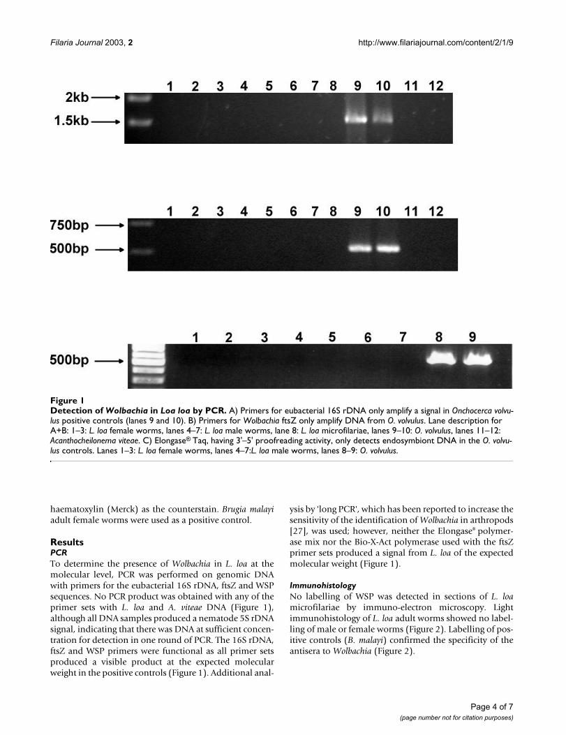
ResultsPCRTo determine the presence of Wolbachia in L. loa at themolecular level, PCR was performed on genomic DNAwith primers for the eubacterial 16S rDNA, ftsZ and WSPsequences. No PCR product was obtained with any of theprimer sets with L. loa and A. viteae DNA (Figure 1),although all DNA samples produced a nematode 5S rDNAsignal, indicating that there was DNA at sufficient concen-tration for detection in one round of PCR. The 16S rDNA,ftsZ and WSP primers were functional as all primer setsproduced a visible product at the expected molecularweight in the positive controls (Figure 1). Additional anal-
ysis by 'long PCR', which has been reported to increase thesensitivity of the identification of Wolbachia in arthropods[27], was used; however, neither the Elongase® polymer-ase mix nor the Bio-X-Act polymerase used with the ftsZprimer sets produced a signal from L. loa of the expectedmolecular weight (Figure 1).
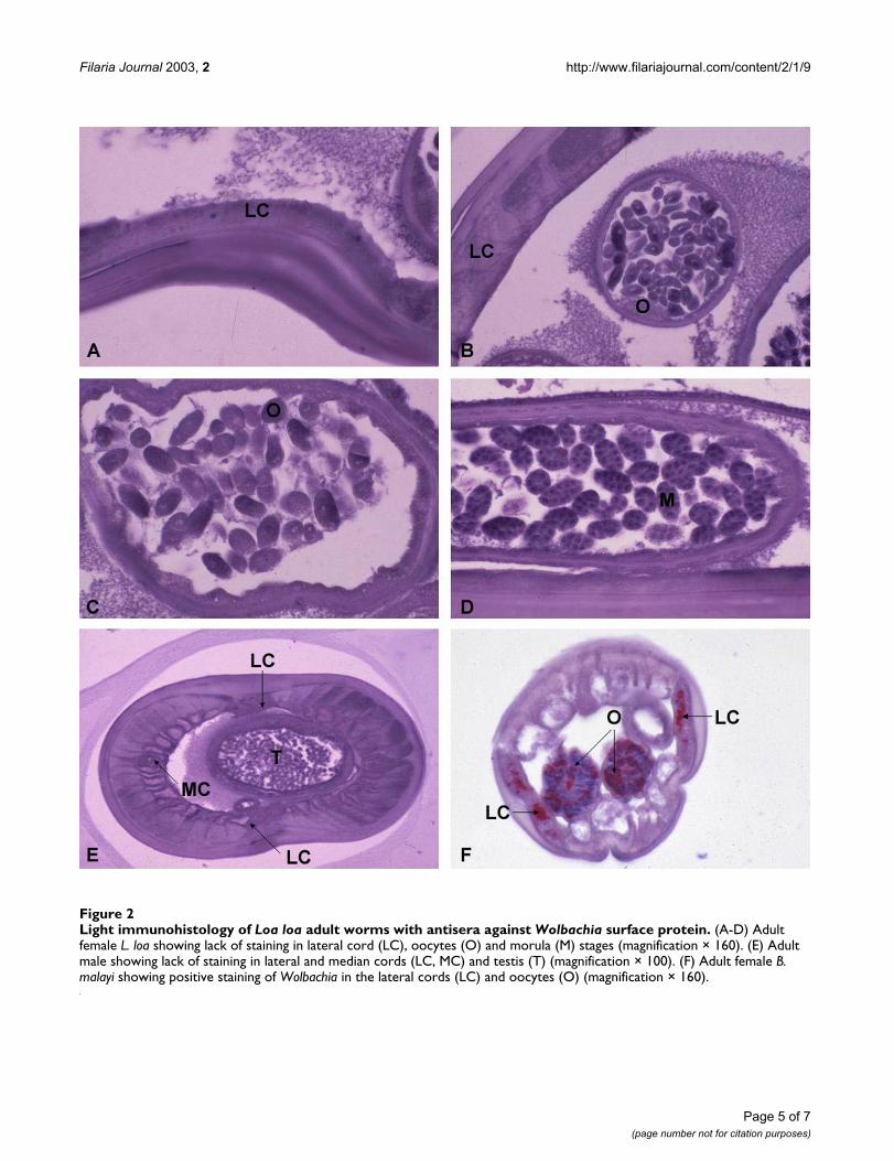
ImmunohistologyNo labelling of WSP was detected in sections of L. loamicrofilariae by immuno-electron microscopy. Lightimmunohistology of L. loa adult worms showed no label-ling of male or female worms (Figure 2). Labelling of pos-itive controls (B. malayi) confirmed the specificity of theantisera to Wolbachia (Figure 2).
Figure 1Detection of Wolbachia in Loa loa by PCR. A) Primers for eubacterial 16S rDNA only amplify a signal in Onchocerca volvu-lus positive controls (lanes 9 and 10). B) Primers for Wolbachia ftsZ only amplify DNA from O. volvulus. Lane description for A+B: 1–3: L. loa female worms, lanes 4–7: L. loa male worms, lane 8: L. loa microfilariae, lanes 9–10: O. volvulus, lanes 11–12: Acanthocheilonema viteae. C) Elongase® Taq, having 3'–5' proofreading activity, only detects endosymbiont DNA in the O. volvu-lus controls. Lanes 1–3: L. loa female worms, lanes 4–7:L. loa male worms, lanes 8–9: O. volvulus.
Page 4 of 7(page number not for citation purposes)
Filaria Journal 2003, 2 http://www.filariajournal.com/content/2/1/9
Figure 2Light immunohistology of Loa loa adult worms with antisera against Wolbachia surface protein. (A-D) Adult female L. loa showing lack of staining in lateral cord (LC), oocytes (O) and morula (M) stages (magnification × 160). (E) Adult male showing lack of staining in lateral and median cords (LC, MC) and testis (T) (magnification × 100). (F) Adult female B. malayi showing positive staining of Wolbachia in the lateral cords (LC) and oocytes (O) (magnification × 160).
Page 5 of 7(page number not for citation purposes)
Filaria Journal 2003, 2 http://www.filariajournal.com/content/2/1/9
DiscussionHere we present data of molecular and immunohistolog-ical analyses that failed to provide evidence for Wolbachiasymbiosis in L. loa. This confirms previous findings on L.loa by electron microscopy and PCR [6,19–22] andextends these observations to a larger sample of adultworms, infective larvae and isolates of microfilariae fromthree different endemic areas.
The release of Wolbachia into the blood following anti-filarial chemotherapy has been shown to be associatedwith severe systemic inflammatory reactions in individu-als infected with O. volvulus or B. malayi [16,17]. One ofthe objectives of this study was to determine whether Wol-bachia might contribute to the rare but severe neurologicaladverse events following ivermectin treatment of L. loa[18]. We conclude that the neurological consequences ofSAE following ivermectin treatment of individuals with L.loa are not associated with Wolbachia. In people co-infected with L. loa and O. volvulus or W. bancrofti, adverseevents induced by Wolbachia derived from the latter twospecies may nevertheless contribute to post-treatmentreactions. Double blind placebo-controlled trials to eval-uate the effect of doxycycline depletion of Wolbachia onthe development of post-treatment reactions to filarialchemotherapy are currently underway in individualsinfected with B. malayi, W. bancrofti, O. volvulus, and co-infection with O. volvulus and L. loa. In two patients withL. loa treated with doxycycline for six weeks (200 mg/day),microfilaraemia was still detected at 120 days of follow up[22].
Studies on species of filarial nematodes infected with Wol-bachia suggest that the symbiosis exists throughout allsamples of populations and individual parasites [1,2]. Theubiquity of infection, congruence with host phylogenyand deleterious effects of antibiotic clearance on embryo-genesis, development and viability suggest a mutualisticdependency [1,2]. It is clear, however, that some species offilariae, including L. loa, can cause widespread infectionwithout the need for bacterial symbionts. Although fur-ther studies are needed it has been suggested that theabsence of Wolbachia in A. viteae and Setaria sp. is a con-sequence of their divergence from the lineage prior to theacquisition of Wolbachia infection. Conversely, theabsence of Wolbachia from O. flexuosa and L. loa is morelikely to be due to the loss of bacterial symbionts [2]. Fur-ther analysis incorporating the results of the present studycould provide additional insights into the evolutionarybiology of the filarial nematode-Wolbachia symbiosis.
Although data collected so far support the conclusion thatfilarial nematode species with evidence of symbiosis areubiquitously infected, sampling of these species is inevita-bly limited and we cannot rule out the possibility that
populations or individual nematodes exist without infec-tion. Similarly with species shown to be aposymbiotic,populations may exist that contain symbionts, particu-larly if the absence of bacteria is due to a secondary loss ofWolbachia. In this regard it would be worthwhile to ana-lyse samples of monkey strains of L. loa, which may beancestrally 'primitive' compared to the strain parasitisinghumans. Additional studies on the extent of Wolbachiasymbiosis in infected species and the infection status ofthe human filariae M. perstans and M. streptocera areimportant areas for future research.
ConclusionsWe conclude that this study provides no evidence for Wol-bachia symbiosis in L. loa. It is therefore highly improba-ble that Wolbachia contributes to the neurologicalconsequences of SAE following ivermectin treatment inindividuals with infections of L. loa unaccompanied byother filarial species.
Competing interestsNone.
Authors' contributionsHelen McGarry – PCR analysis, preparation of draftmanuscript
Ken Pfarr – PCR analysis, preparation of draft manuscript
Gill Egerton – immunohistology
Achim Hoerauf – Interpretation of PCR data
Jean-Paul Akue – Collection, identification and process-ing of L. loa
Peter Enyong – Collection, identification and processingof L. loa
Samuel Wanji – Collection, identification and processingof L. loa
Sabine Kläger – Collection, identification and processingof L. loa
Ted Bianco – Collection, identification and processing ofL. loa
Nick Beeching – Collection, identification and processingof L. loa
Mark Taylor – Interpretation of data and preparation offinal manuscript
Page 6 of 7(page number not for citation purposes)
Filaria Journal 2003, 2 http://www.filariajournal.com/content/2/1/9
Publish with BioMed Central and every scientist can read your work free of charge
"BioMed Central will be the most significant development for disseminating the results of biomedical research in our lifetime."
Sir Paul Nurse, Cancer Research UK
Your research papers will be:
available free of charge to the entire biomedical community
peer reviewed and published immediately upon acceptance
cited in PubMed and archived on PubMed Central
yours — you keep the copyright
Submit your manuscript here:http://www.biomedcentral.com/info/publishing_adv.asp
BioMedcentral
AcknowledgementsWe thank all the people who provided samples of parasites. We thank Prof. Dietrich W. Büttner for the light immunohistochemistry and images of adult L. loa. We thank Dr. Tom Nutman and Dr. Amy Klion (NIH/NIAID, USA) for samples of microfilariae and Prof. Richard Lucius (Humboldt Uni-versity, Germany) for the supply of A. viteae. MJT thanks the Wellcome Trust for Senior Fellowship support.
References1. Taylor MJ and Hoerauf A: Wolbachia bacteria of filarial
nematodes Parasitol Today 1999, 15:437-442.2. Bandi C, Trees AJ and Brattig NW: Wolbachia in filarial nema-
todes: evolutionary aspects and implications for the patho-genesis and treatment of filarial diseases Vet Parasitol 2001,98:215-238.
3. Taylor MJ: A new insight into the pathogenesis of filarialdisease Curr Mol Med 2002, 2:299-302.
4. Taylor MJ and Hoerauf A: A new approach to the treatment offilariasis Curr Opin Infect Dis 2001, 14:727-731.
5. Hoerauf A, Adjei O and Buttner DW: Antibiotics for the treat-ment of onchocerciasis and other filarial infections Curr OpinInvestig Drugs 2002, 3:533-537.
6. McLaren DJ, Worms MJ, Laurence BR and Simpson MG: Micro-organisms in filarial larvae (Nematoda) Trans R Soc Trop MedHyg 1975, 69:509-514.
7. Bandi C, Anderson TJ, Genchi C and Blaxter ML: Phylogeny ofWolbachia in filarial nematodes Proc R Soc Lond B Biol Sci 1998,265:2407-2413.
8. Plenge-Bonig A, Kromer M and Buttner DW: Light and electronmicroscopy studies on Onchocerca jakutensis and O. flexu-osa of red deer show different host-parasite interactions Par-asitol Res 1995, 81:66-73.
9. Chirgwin SR, Porthouse KH, Nowling JM and Klei TR: The filarialendosymbiont Wolbachia sp. is absent from Setaria equina JParasitol 2002, 88:1248-1250.
10. Taylor MJ, Cross HF and Bilo K: Inflammatory responsesinduced by the filarial nematode Brugia malayi are mediatedby lipopolysaccharide-like activity from endosymbiotic Wol-bachia bacteria J Exp Med 2000, 191:1429-1436.
11. Brattig NW, Rathjens U, Ernst M, Geisinger F, Renz A and Tischen-dorf FW: Lipopolysaccharide-like molecules derived fromWolbachia endobacteria of the filaria Onchocerca volvulusare candidate mediators in the sequence of inflammatoryand antiinflammatory responses of human monocytesMicrobes Infect 2000, 2:1147-1157.
12. Brattig NW, Buttner DW and Hoerauf A: Neutrophil accumula-tion around Onchocerca worms and chemotaxis of neu-trophils are dependent on Wolbachia endobacteria MicrobesInfect 2001, 3:439-446.
13. Saint Andre A, Blackwell NM, Hall LR, Hoerauf A, Brattig NW, Volk-mann L, Taylor MJ, Ford L, Hise AG, Lass JH, Diaconu E and PearlmanE: The role of endosymbiotic Wolbachia bacteria in thepathogenesis of river blindness Science 2002, 295:1892-1895.
14. Hoerauf A, Nissen-Pahle K, Schmetz C, Henkle-Duhrsen K, BlaxterML, Buttner DW, Gallin MY, Al-Qaoud KM, Lucius R and Fleischer B:Tetracycline therapy targets intracellular bacteria in thefilarial nematode Litomosoides sigmodontis and results infilarial infertility J Clin Invest 1999, 103:11-18.
15. McCall J.W, Jun J.J. Bandi, C.: Wolbachia and the antifilarial prop-erties of tetracycline. An untold story Intalian Journal of Zoology1999, 66:7-10.
16. Cross HF, Haarbrink M, Egerton G, Yazdanbakhsh M and Taylor MJ:Severe reactions to filarial chemotherapy and release ofWolbachia endosymbionts into blood Lancet 2001, 358:1873-1875.
17. Keiser PB, Reynolds SM, Awadzi K, Ottesen EA, Taylor MJ and Nut-man TB: Bacterial endosymbionts of Onchocerca volvulus inthe pathogenesis of posttreatment reactions J Infect Dis 2002,185:805-811.
18. Boussinesq M, Gardon J, Gardon-Wendel N and Chippaux J-P: Clin-ical picture, epidemiology and outcome of Loa-associatedserious adverse events related to mass ivermectin treat-ment of onchocerciasis in Cameroon. Filaria J 2003.
19. Kozek WJ and Orihel TC: Ultrastructure of Loa loa microfilariaInt J Parasitol 1983, 13:19-43.
20. Franz M, Melles J and Buttner DW: Electron microscope study ofthe body wall and the gut of adult Loa loa Z Parasitenkd 1984,70:525-536.
21. Weber P: The fine structure of the female reproductive tractof adult Loa loa Int J Parasitol 1987, 17:927-934.
22. Brouqui P, Fournier P and Raoult D.: Doxycycline and eradicationof microfilaremia in patients with loiasis Emerg Infect Dis 2001,7:603-604.
23. Toure FS, Bain O, Nerrienet E, Millet P, Wahl G, Toure Y, DoumboO, Nicolas L, Georges AJ, McReynolds LA and Egwang TG: Detec-tion of Loa loa-specific DNA in blood from occult-infectedindividuals Exp Parasitol 1997, 86:163-170.
24. Zimmerman PA, Guderian RH, Aruajo E, Elson L, Phadke P, KubofcikJ and Nutman TB: Polymerase chain reaction-based diagnosisof Onchocerca volvulus infection: improved detection ofpatients with onchocerciasis J Infect Dis 1994, 169:686-689.
25. Fischer P, Buttner DW, Bamuhiiga J and Williams SA: Detection ofthe filarial parasite Mansonella streptocerca in skin biopsiesby a nested polymerase chain reaction-based assay Am J TropMed Hyg 1998, 58:816-820.
26. Zhong M, McCarthy J, Bierwert L, Lizotte-Waniewski M, Chanteau S,Nutman TB, Ottesen EA and Williams SA: A polymerase chainreaction assay for detection of the parasite Wuchereria ban-crofti in human blood samples Am J Trop Med Hyg 1996, 54:357-363.
27. Jeyaprakash A and Hoy MA: Long PCR improves WolbachiaDNA amplification: wsp sequences found in 76% of sixty-three arthropod species Insect Mol Biol 2000, 9:393-405.
28. Casiraghi M, Anderson TJ, Bandi C, Bazzocchi C and Genchi C: Aphylogenetic analysis of filarial nematodes: comparison withthe phylogeny of Wolbachia endosymbionts Parasitology 2001,122 Pt 1:93-103.
29. Jenkins RE, Taylor MJ, Gilvary N and Bianco AE: Characterizationof a secreted antigen of Onchocerca volvulus with host-pro-tective potential Parasite Immunol 1996, 18:29-42.
Page 7 of 7(page number not for citation purposes)
Recommended