Exotic Animal Ocular PathologyLaboratory Animal Ocular Pathology
Dick Dubielzig
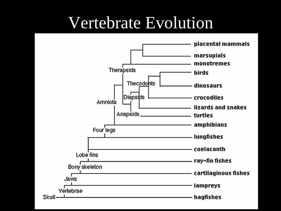
Vertebrate Evolution
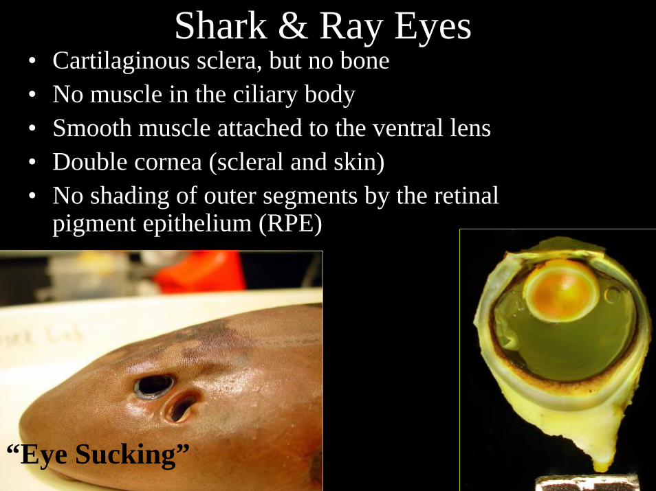
Shark & Ray Eyes• Cartilaginous sclera, but no bone• No muscle in the ciliary body• Smooth muscle attached to the ventral lens• Double cornea (scleral and skin) • No shading of outer segments by the retinal
pigment epithelium (RPE)
“Eye Sucking”
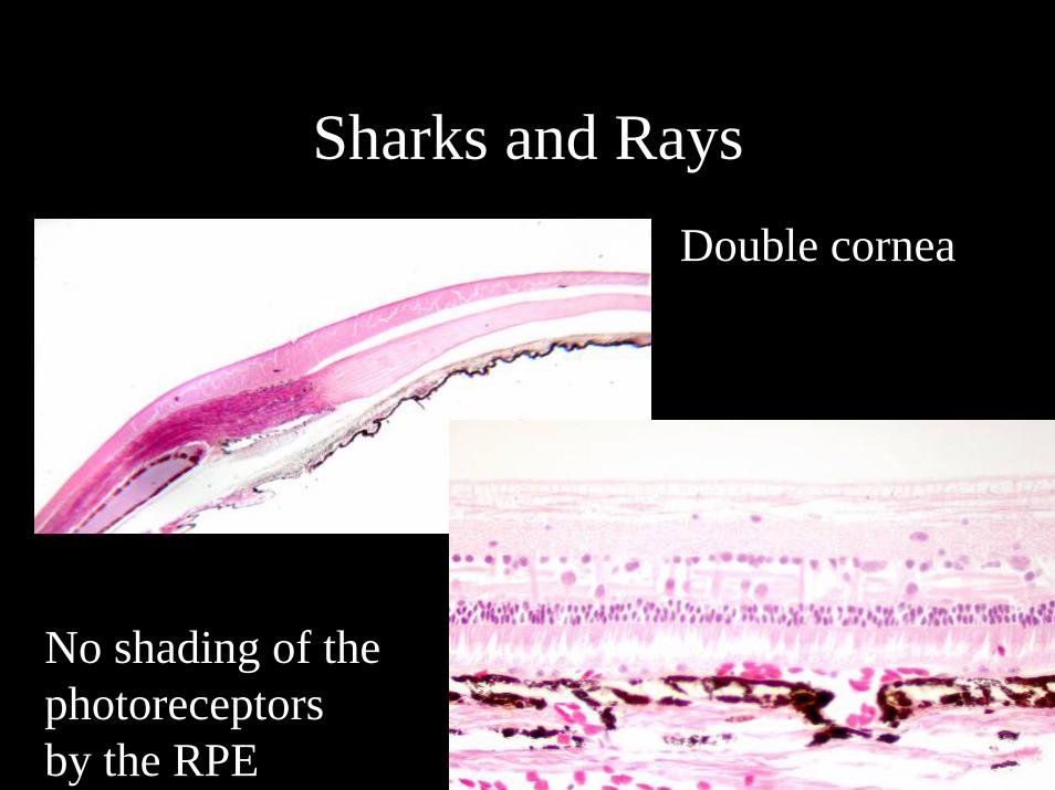
Sharks and RaysDouble cornea
No shading of the photoreceptorsby the RPE
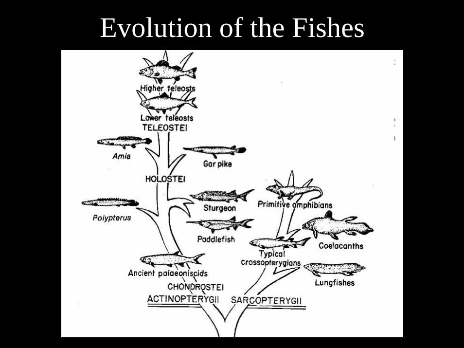
Evolution of the Fishes
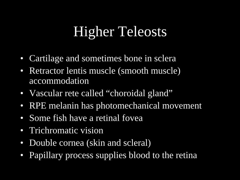
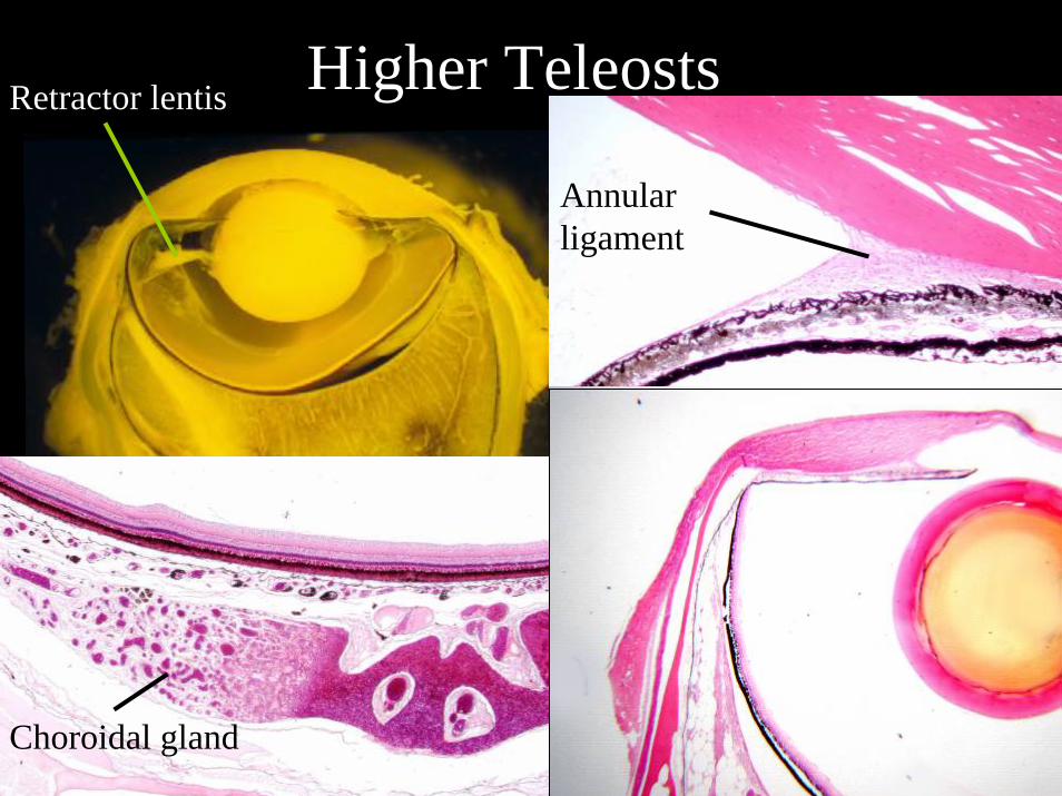
Higher Teleosts
• Cartilage and sometimes bone in sclera• Retractor lentis muscle (smooth muscle)
accommodation• Vascular rete called “choroidal gland”• RPE melanin has photomechanical movement• Some fish have a retinal fovea• Trichromatic vision• Double cornea (skin and scleral)• Papillary process supplies blood to the retina
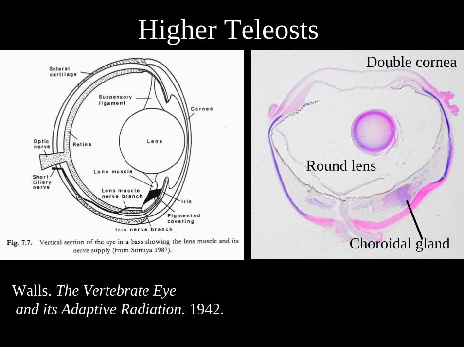
Higher Teleosts
Walls. The Vertebrate Eyeand its Adaptive Radiation. 1942.
Double cornea
Choroidal gland
Round lens
Higher Teleosts
Annular ligament
Choroidal gland
Retractor lentis
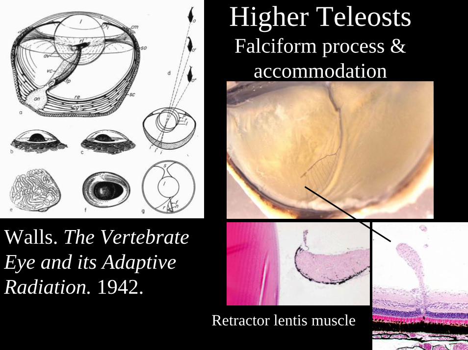
Higher TeleostsFalciform process &
accommodation
Retractor lentis muscle
Walls. The Vertebrate Eye and its Adaptive Radiation. 1942.
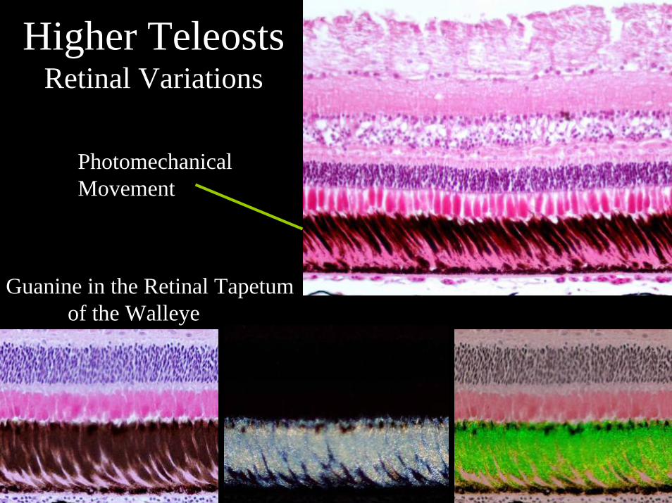
Higher TeleostsRetinal Variations
Photomechanical Movement
Guanine in the Retinal Tapetumof the Walleye
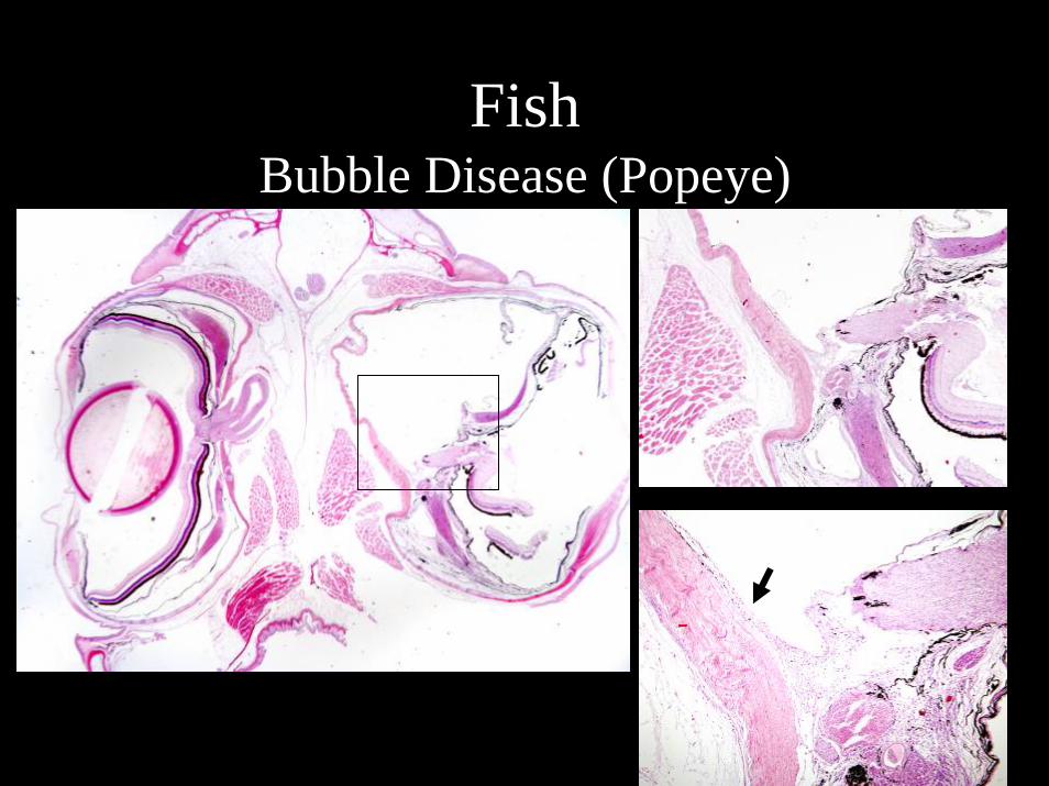
Fish Bubble Disease (Popeye)
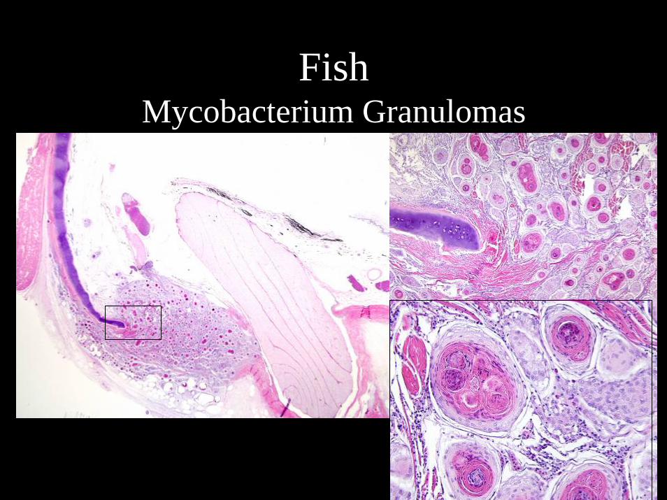
FishMycobacterium Granulomas
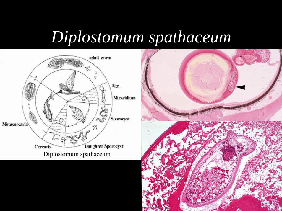
Diplostomum spathaceum

Teleost Primitive Neuroectodermal Tumor (PNET)
Fairy Basslet
Thanks to:Marie Pinkerton
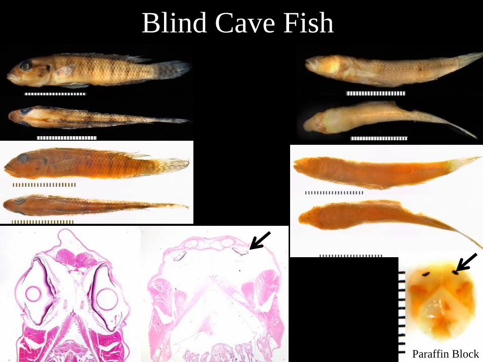
Blind Cave Fish
Paraffin Block
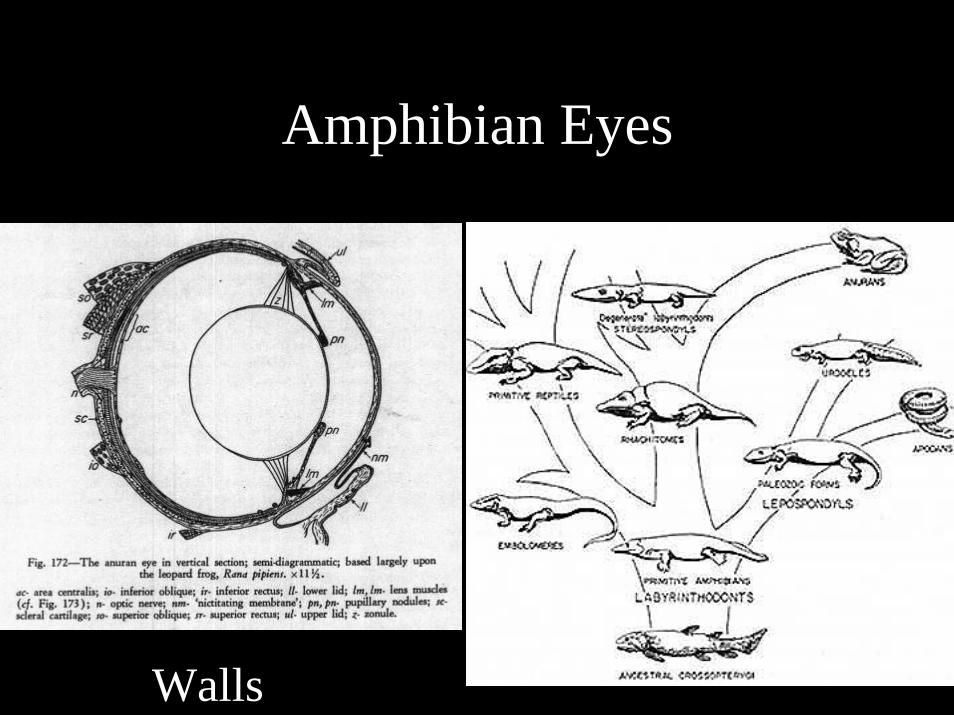
Amphibian Eyes
Walls
Features of Amphibian Eyes
• Cartilaginous sclera, but no bone• Trichromatic vision• Photomechanical motion in the RPE• Minimal amount of accommodation with smooth
muscle• Double cornea only in the tadpole• No annular pad in lens• Retractor bulbi muscle and eyelids
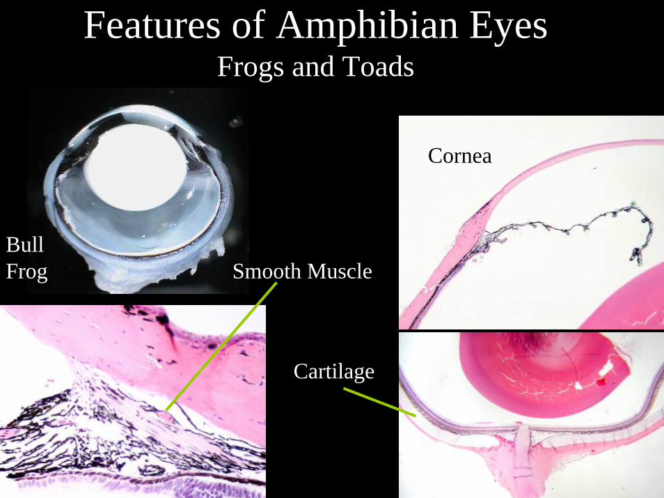
Features of Amphibian EyesFrogs and Toads
Smooth Muscle
Cartilage
Cornea
BullFrog
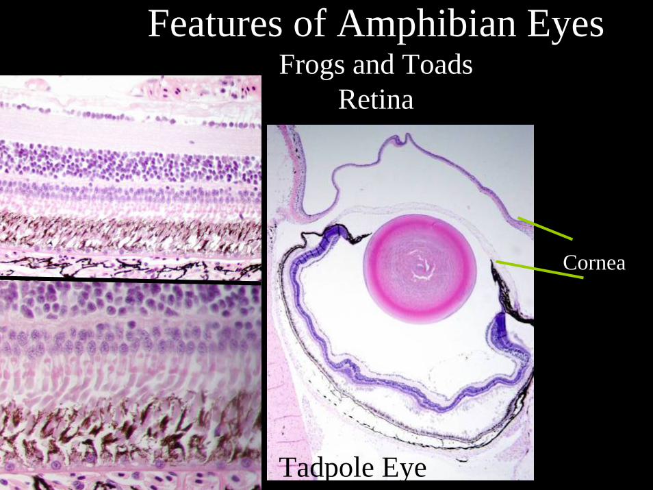
Features of Amphibian EyesFrogs and Toads
Retina
Tadpole Eye
Cornea
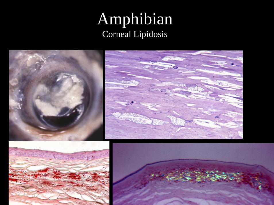
AmphibianCorneal Lipidosis
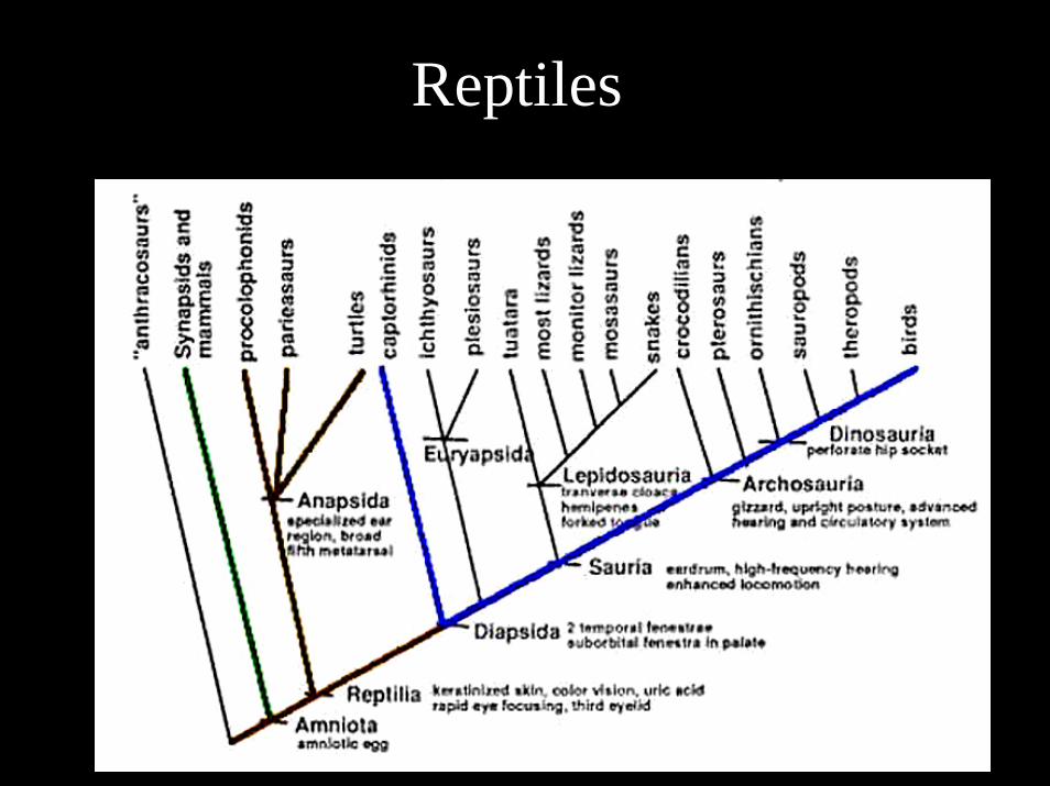
Reptiles
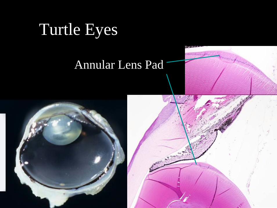
Turtle Eyes
Annular Lens Pad
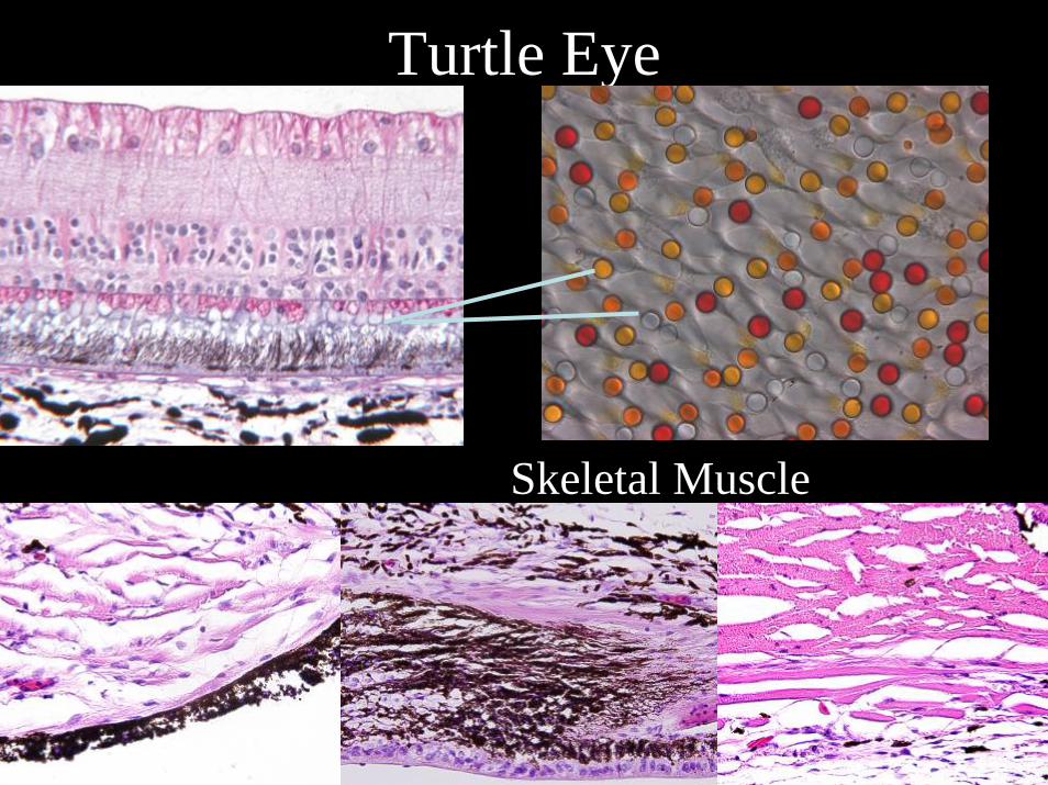
Turtle Eye
Skeletal Muscle
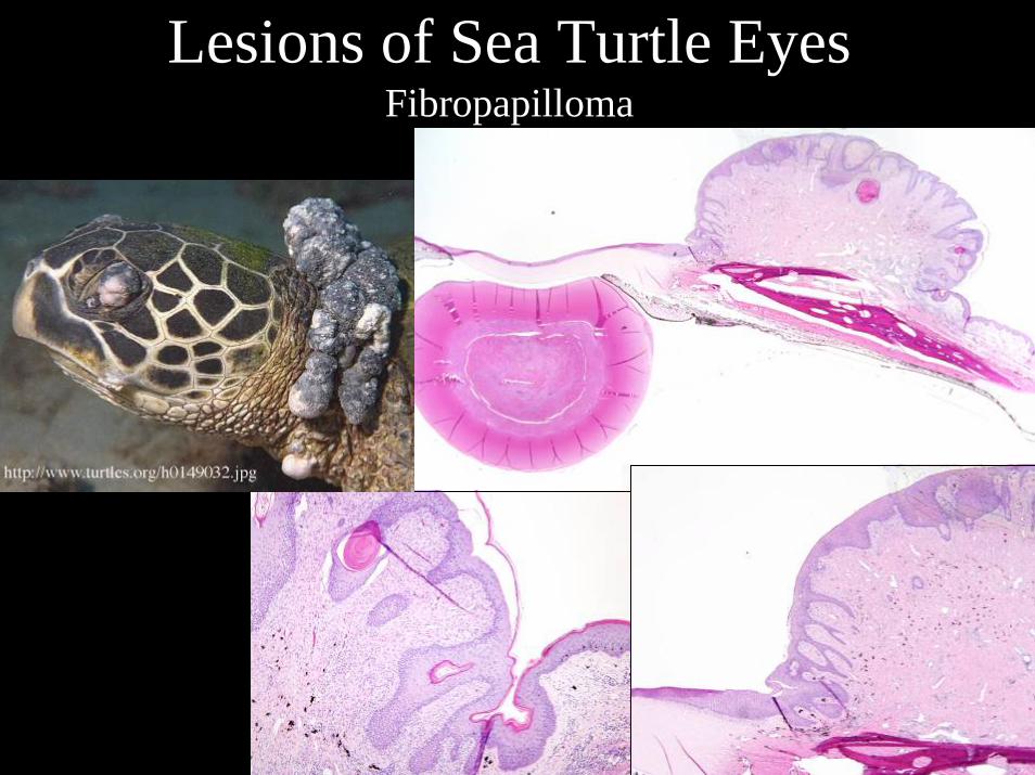
Lesions of Sea Turtle EyesFibropapilloma
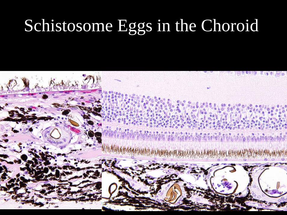
Schistosome Eggs in the Choroid
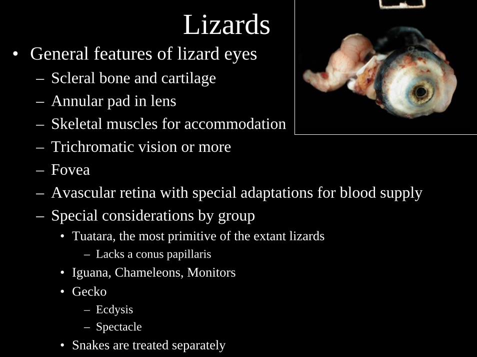
Lizards• General features of lizard eyes
– Scleral bone and cartilage– Annular pad in lens– Skeletal muscles for accommodation– Trichromatic vision or more– Fovea– Avascular retina with special adaptations for blood supply – Special considerations by group
• Tuatara, the most primitive of the extant lizards– Lacks a conus papillaris
• Iguana, Chameleons, Monitors• Gecko
– Ecdysis– Spectacle
• Snakes are treated separately
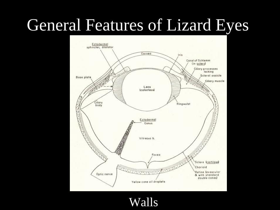
General Features of Lizard Eyes
Walls
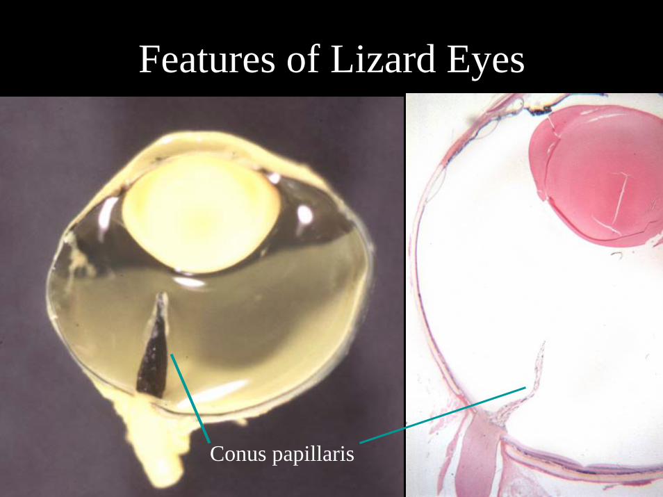
Features of Lizard Eyes
Conus papillaris
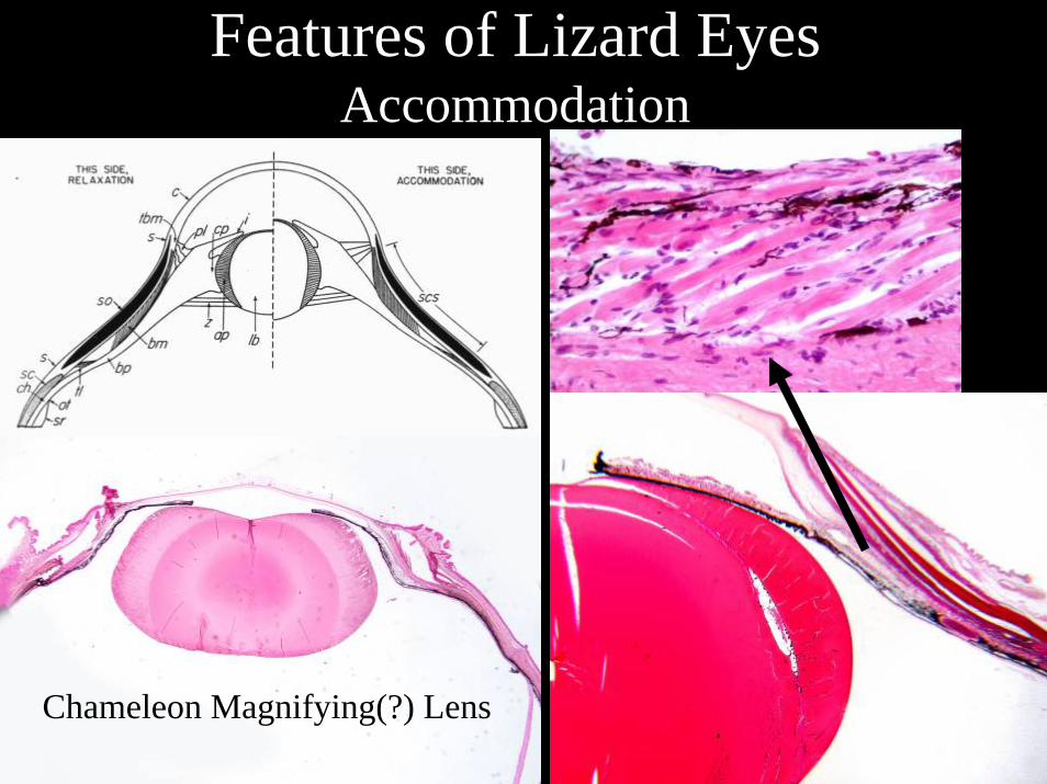
Features of Lizard EyesAccommodation
Chameleon Magnifying(?) Lens
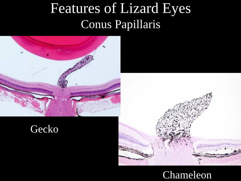
Features of Lizard EyesConus Papillaris
Gecko
Ch Chameleon
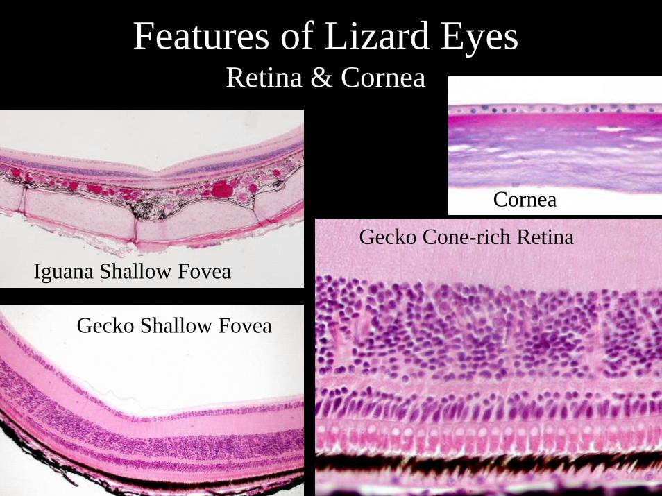
Features of Lizard EyesRetina & Cornea
Iguana Shallow Fovea
Gecko Shallow Fovea
Gecko Cone-rich Retina
Cornea
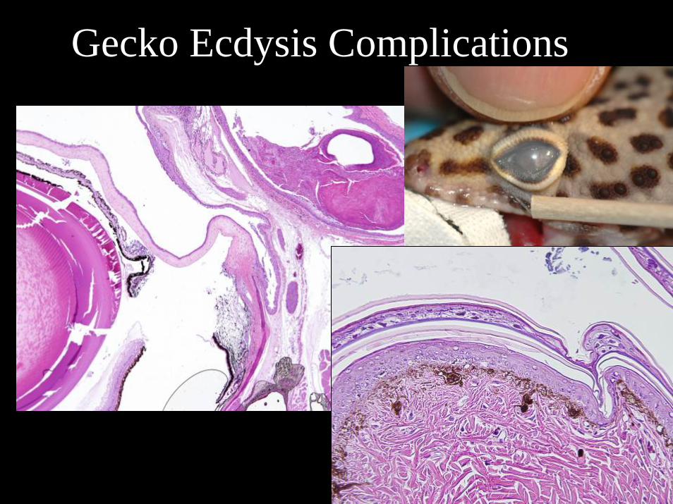
Gecko Ecdysis Complications
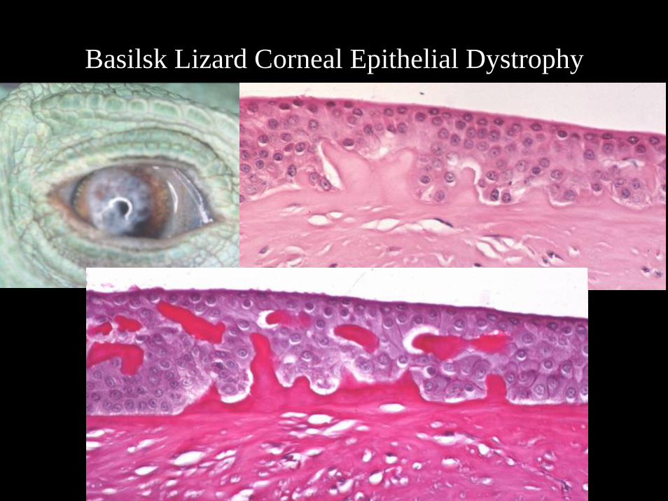
Basilsk Lizard Corneal Epithelial Dystrophy
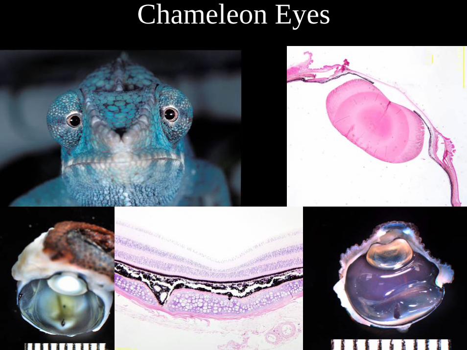
Chameleon Eyes
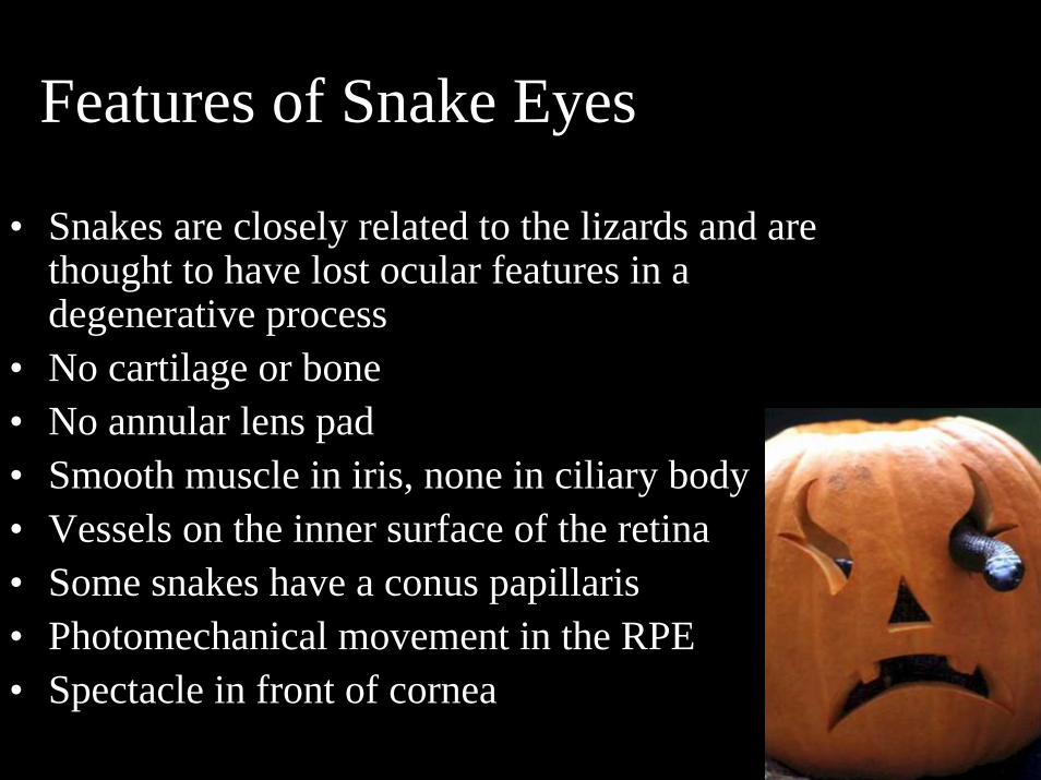
Features of Snake Eyes
• Snakes are closely related to the lizards and are thought to have lost ocular features in a degenerative process
• No cartilage or bone• No annular lens pad• Smooth muscle in iris, none in ciliary body• Vessels on the inner surface of the retina• Some snakes have a conus papillaris• Photomechanical movement in the RPE• Spectacle in front of cornea
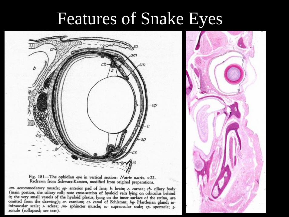
Features of Snake Eyes
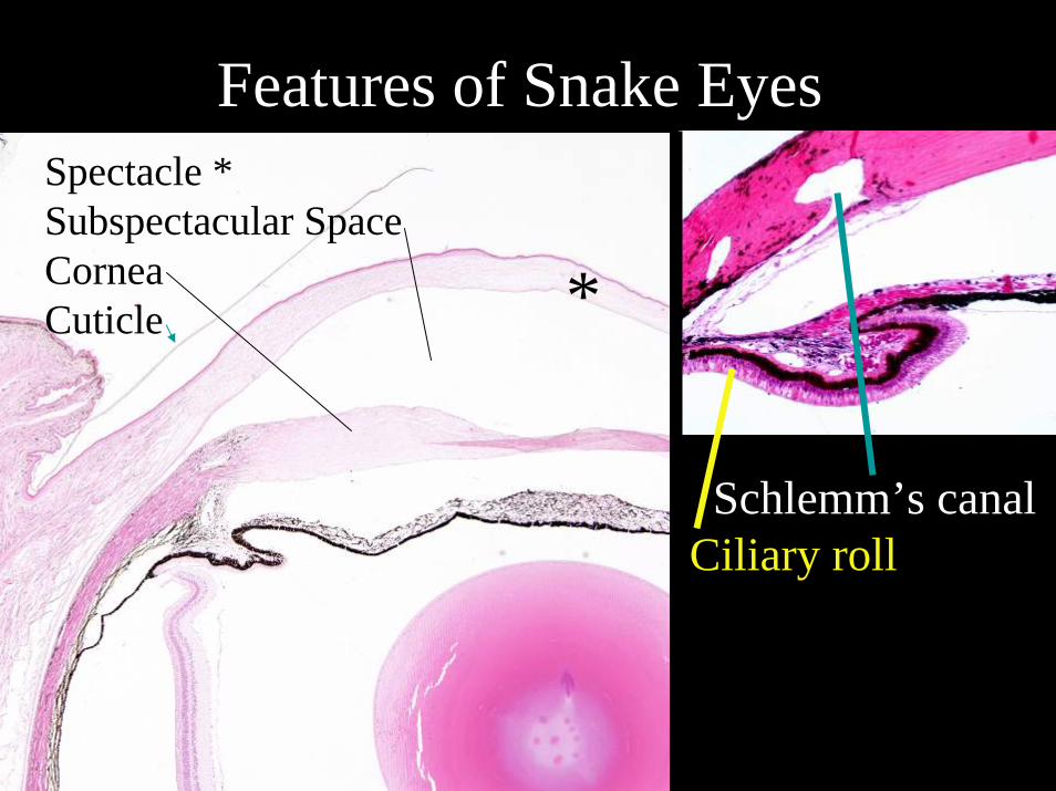
Features of Snake EyesSpectacle *Subspectacular SpaceCorneaCuticle *
Schlemm’s canalCiliary roll
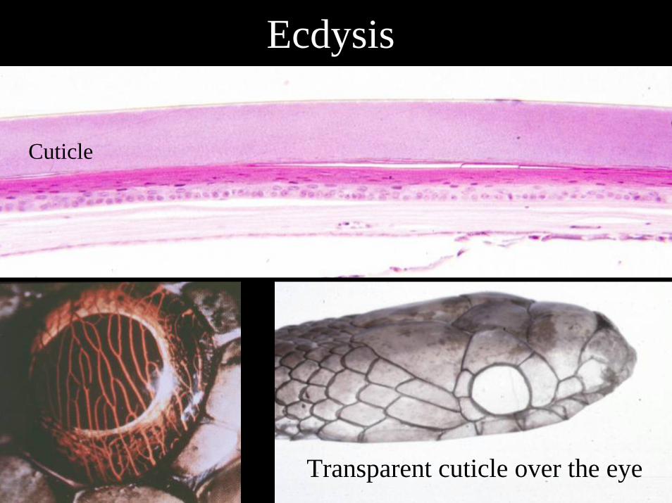
Ecdysis
Transparent cuticle over the eye
Cuticle
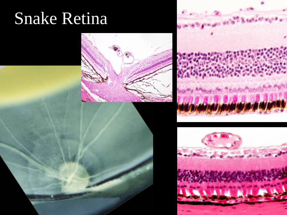
Snake Retina
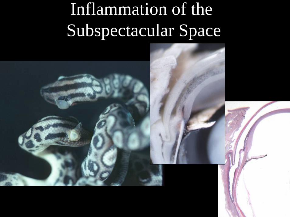
Inflammation of theSubspectacular Space
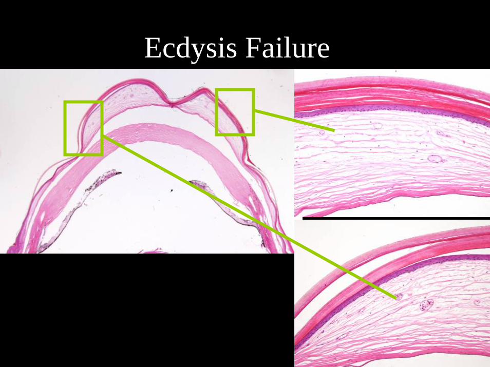
Ecdysis Failure

Surgical Drainage of the Subspectacular Space

Surgical Drainage of theSubspectacular Space
Damaged Cornea

Surgical Drainage of theSubspectacular Space
1
2
3
4
Day 1 Day 7
Day 7 Day 21
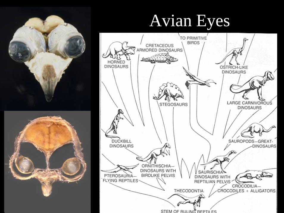
Avian Eyes
Features of Bird Eyes• Cartilage and well-developed ossicle
– Some birds have a tubular eye shape• Skeletal muscle in iris and ciliary body• Annular lens pad• Photomechanical movement in the RPE• Pecten oculi• Fovea common - some birds have two fovea• Corneal accommodation• Trichromatic vision or more
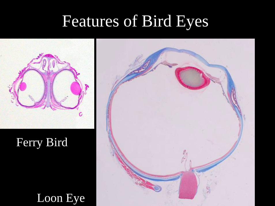
Features of Bird Eyes
Loon Eye
Ferry Bird
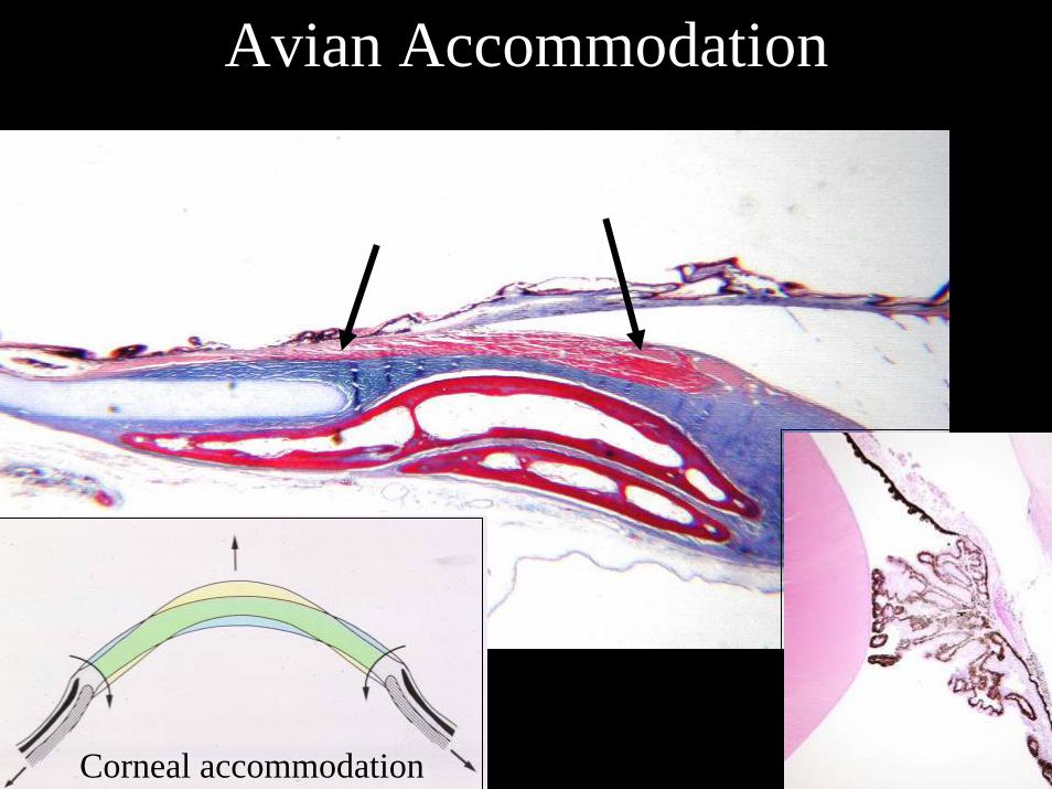
Avian Accommodation
Corneal accommodation
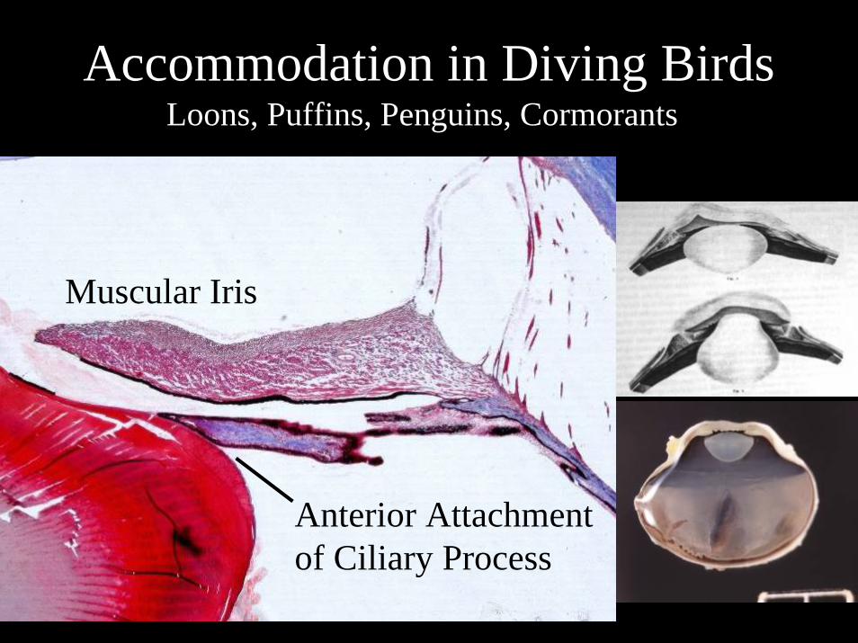
Accommodation in Diving BirdsLoons, Puffins, Penguins, Cormorants
Muscular Iris
Anterior Attachmentof Ciliary Process
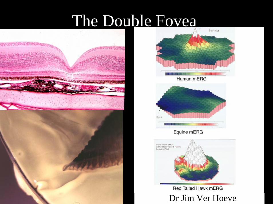
The Double Fovea
Dr Jim Ver Hoeve
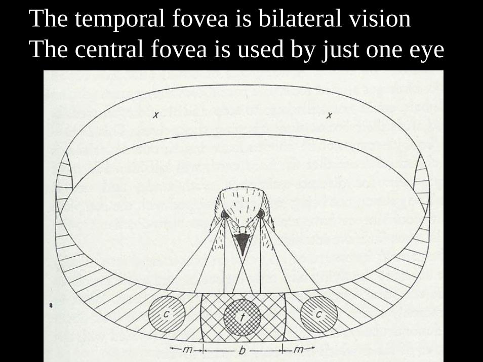
The temporal fovea is bilateral visionThe central fovea is used by just one eye
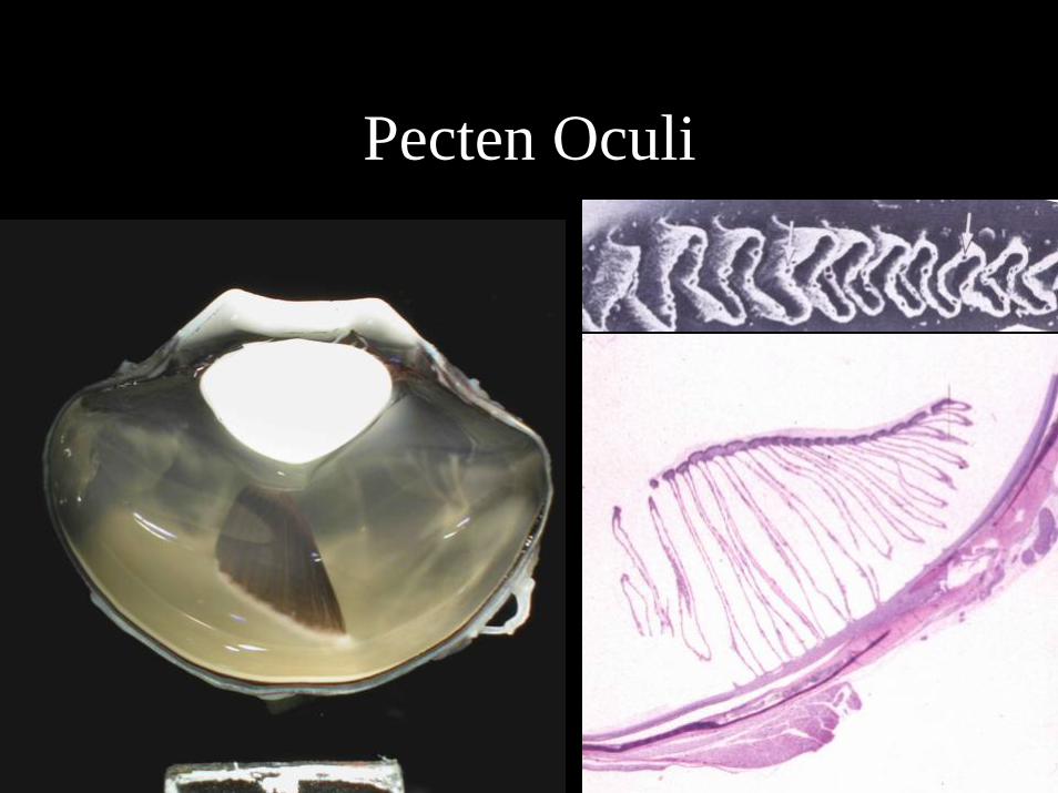
Pecten Oculi

Blunt Trauma
Cyclodialysis in Owl Retinal Avulsion in Hawk
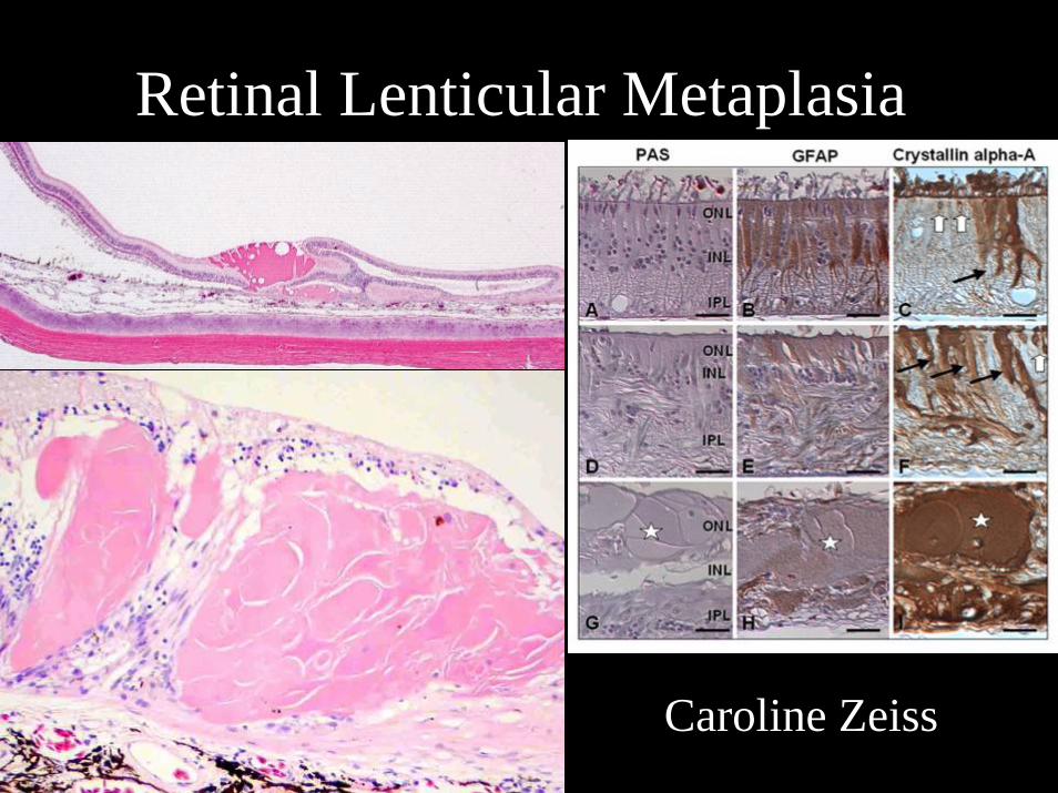
Retinal Lenticular Metaplasia
Caroline Zeiss
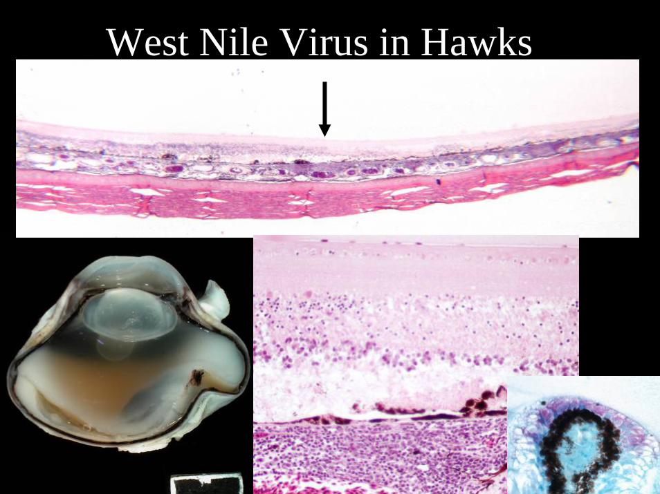
West Nile Virus in Hawks
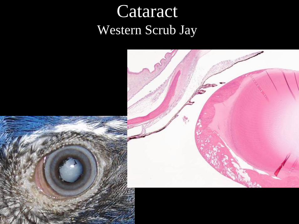
CataractWestern Scrub Jay
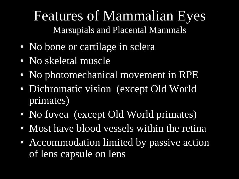
Features of Mammalian EyesMarsupials and Placental Mammals
• No bone or cartilage in sclera• No skeletal muscle• No photomechanical movement in RPE• Dichromatic vision (except Old World
primates)• No fovea (except Old World primates)• Most have blood vessels within the retina• Accommodation limited by passive action
of lens capsule on lens
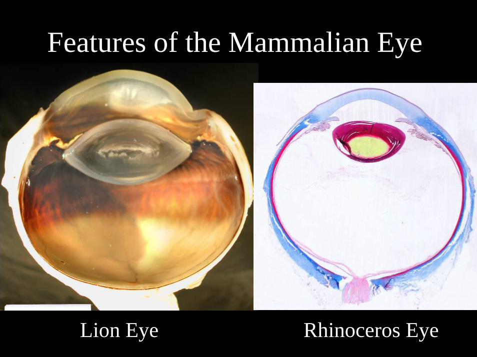
Features of the Mammalian Eye
Lion Eye Rhinoceros Eye
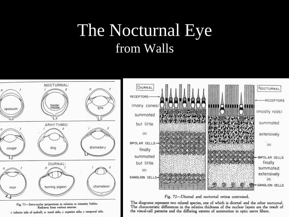
The Nocturnal Eyefrom Walls
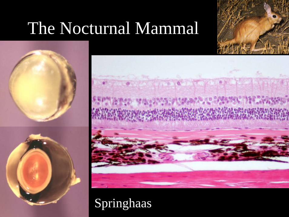
The Nocturnal Mammal
Springhaas

Retinal Atrophy in Albino Laboratory Rodents
Phototoxic RetinopathyNormal

Lipofuscinosis in Rescued Asiatic Black Bears
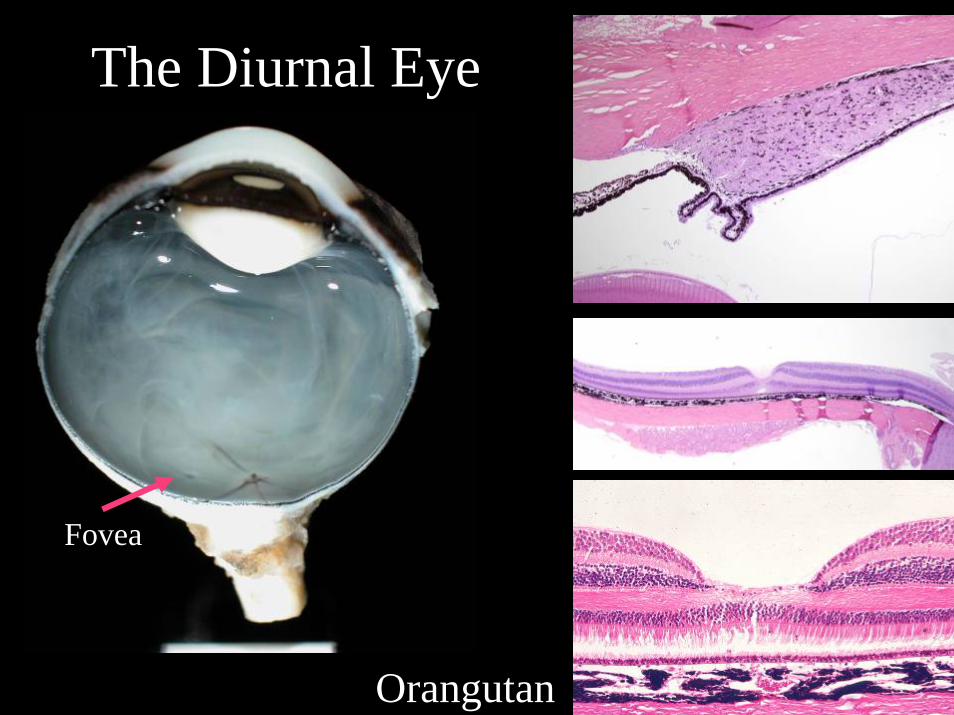
The Diurnal EyePrimate
Orangutan
Fovea
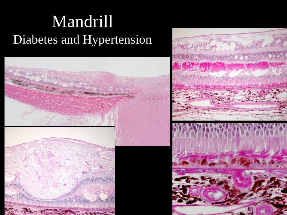
Mandrill Diabetes and Hypertension
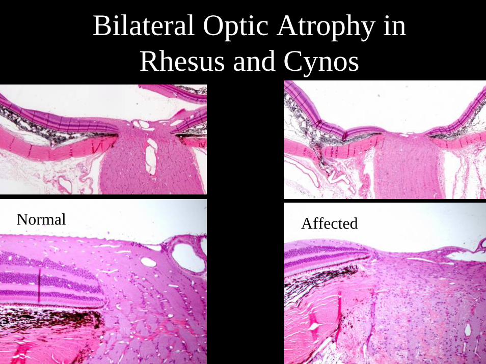
Bilateral Optic Atrophy in Rhesus and Cynos
Normal Affected

Diurnal EyeGround Squirrel
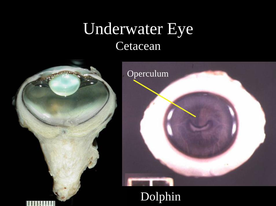
Underwater EyeCetacean
Dolphin
Operculum
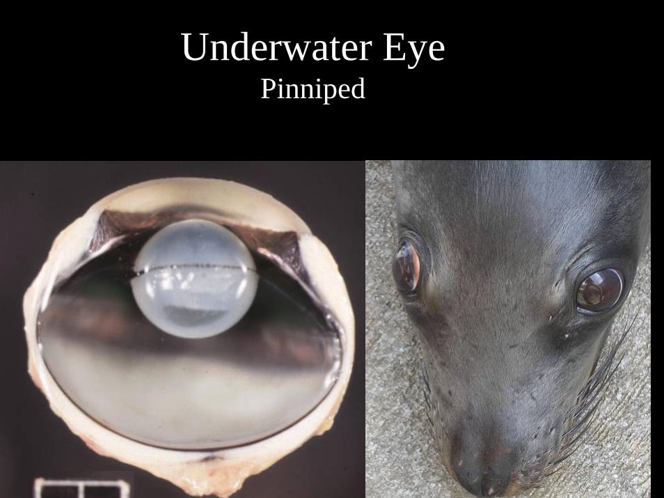
Underwater EyePinniped
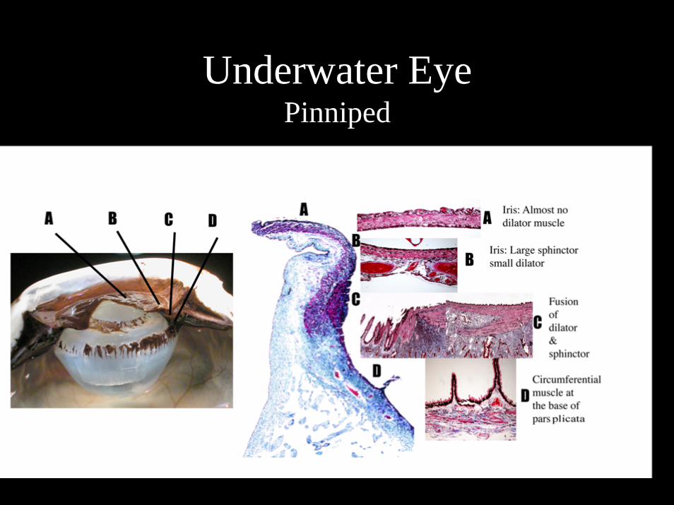
Underwater EyePinniped
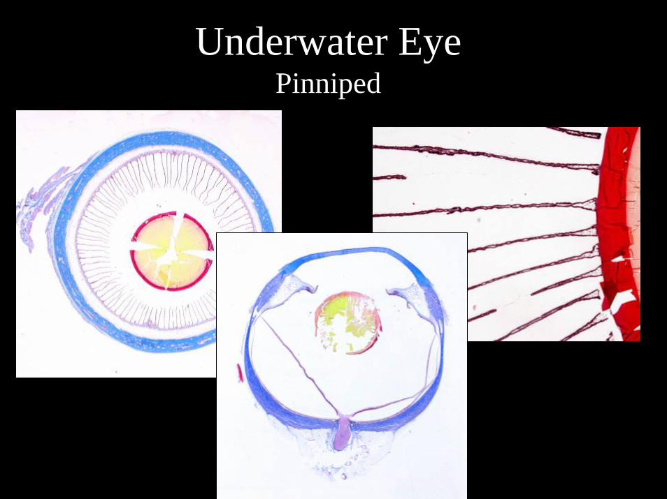
Underwater EyePinniped
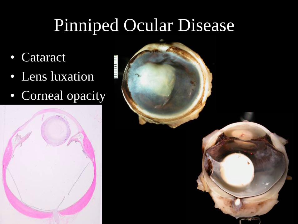
Pinniped Ocular Disease
• Cataract• Lens luxation• Corneal opacity
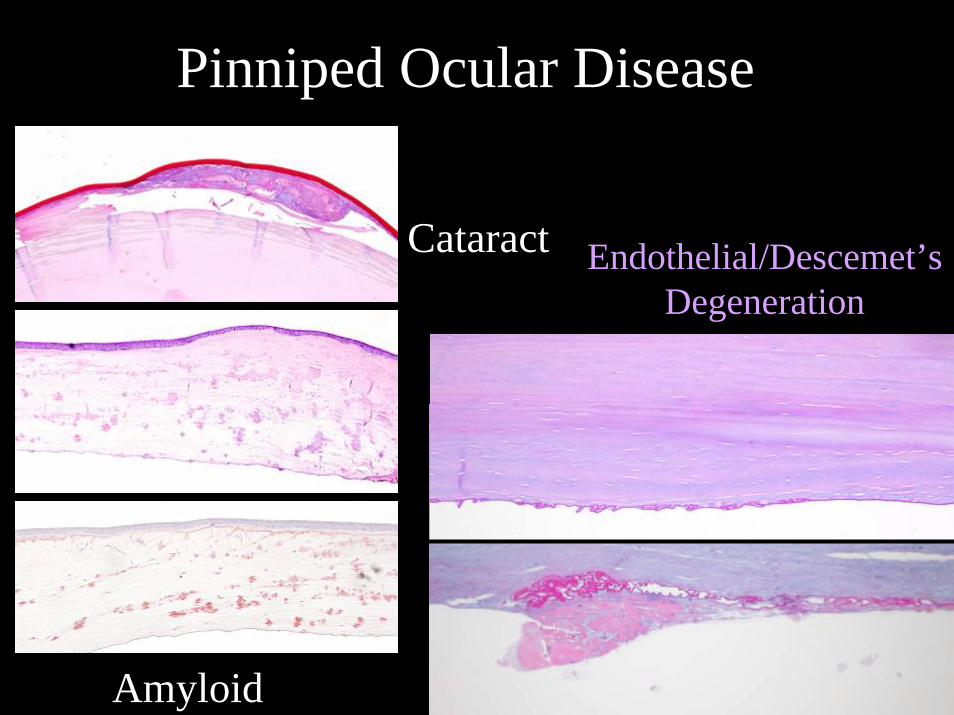
Pinniped Ocular Disease
Cataract
Amyloid
Endothelial/Descemet’sDegeneration
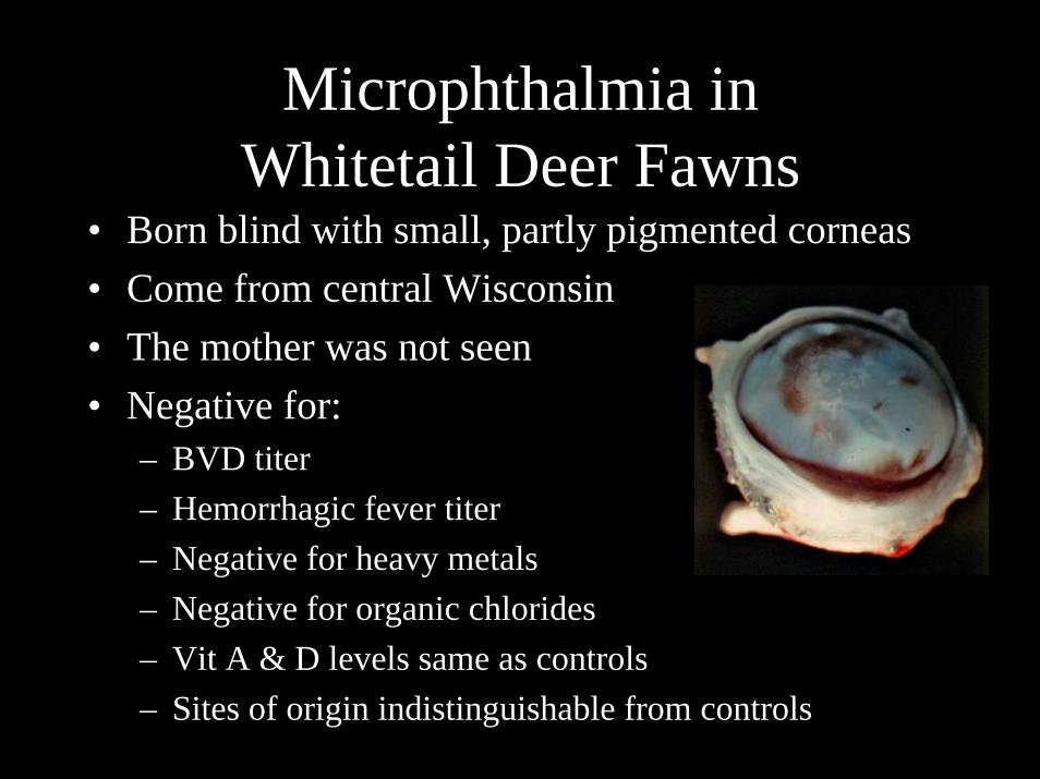
Microphthalmia in Whitetail Deer Fawns
• Born blind with small, partly pigmented corneas• Come from central Wisconsin• The mother was not seen• Negative for:
– BVD titer– Hemorrhagic fever titer– Negative for heavy metals– Negative for organic chlorides– Vit A & D levels same as controls– Sites of origin indistinguishable from controls
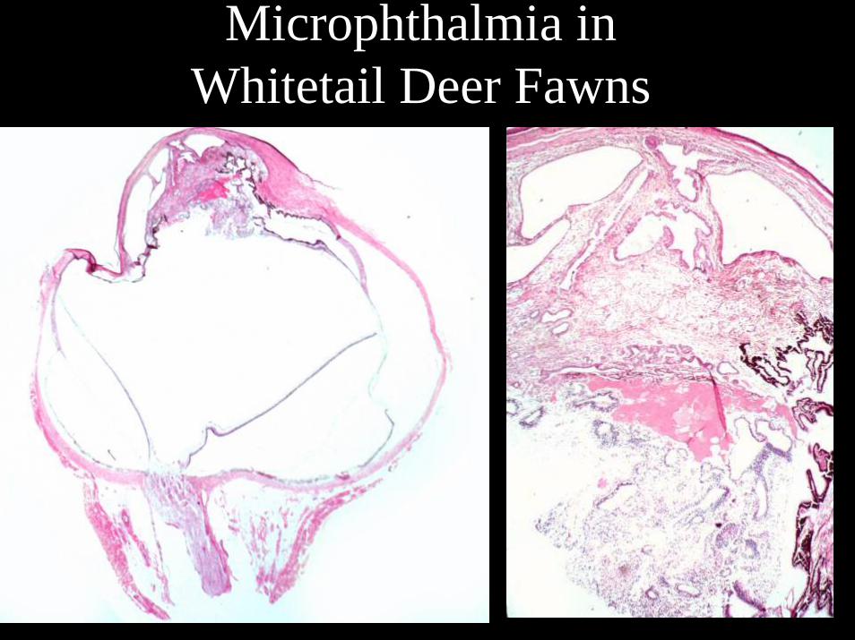
Microphthalmia in Whitetail Deer Fawns
Microphthalmia in Whitetail Deer Fawns

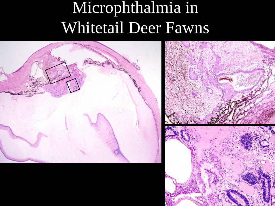
Microphthalmia in Whitetail Deer Fawns
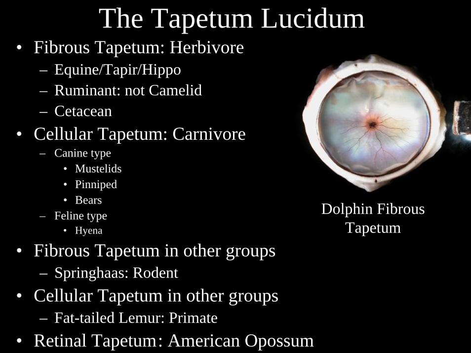
The Tapetum Lucidum• Fibrous Tapetum: Herbivore
– Equine/Tapir/Hippo– Ruminant: not Camelid– Cetacean
• Cellular Tapetum: Carnivore– Canine type
• Mustelids• Pinniped• Bears
– Feline type• Hyena
• Fibrous Tapetum in other groups– Springhaas: Rodent
• Cellular Tapetum in other groups– Fat-tailed Lemur: Primate
• Retinal Tapetum: American Opossum
Dolphin Fibrous Tapetum
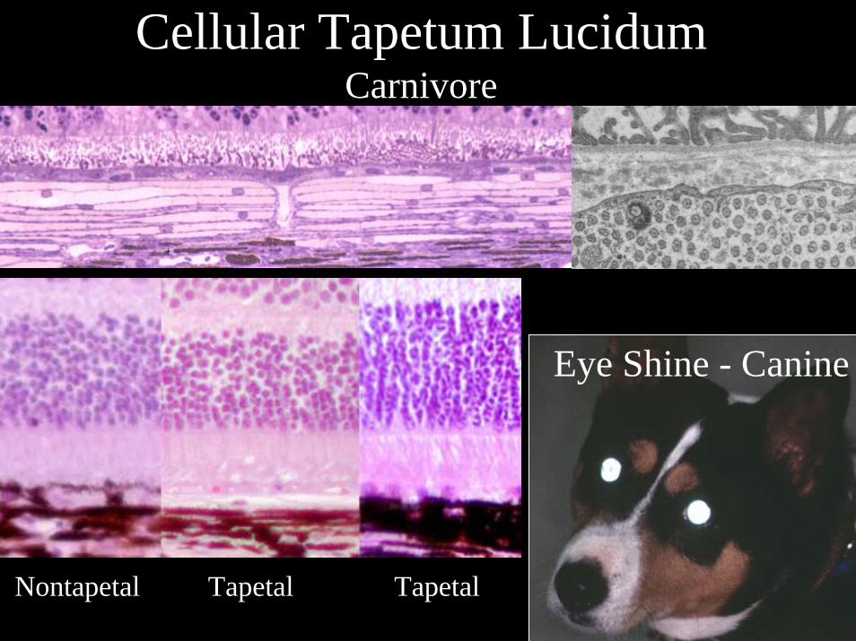
Cellular Tapetum LucidumCarnivore
Nontapetal Tapetal Tapetal
Eye Shine - Canine
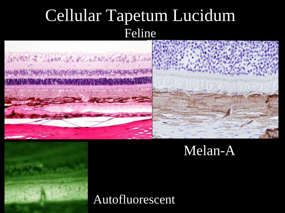
Cellular Tapetum LucidumFeline
Melan-A
Autofluorescent
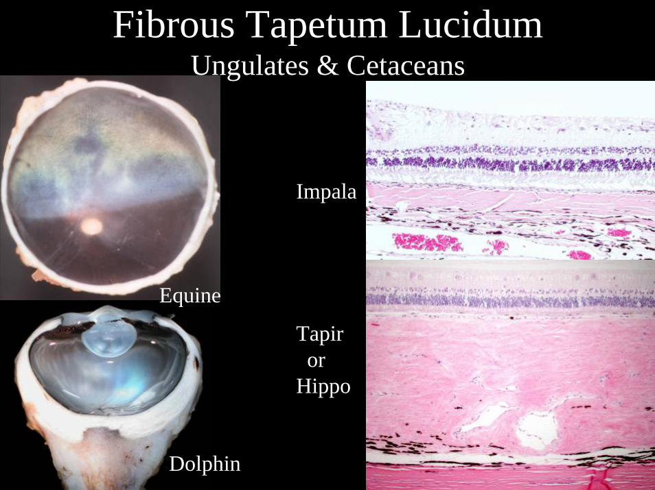
Fibrous Tapetum LucidumUngulates & Cetaceans
Equine
Dolphin
Impala
Tapiror
Hippo
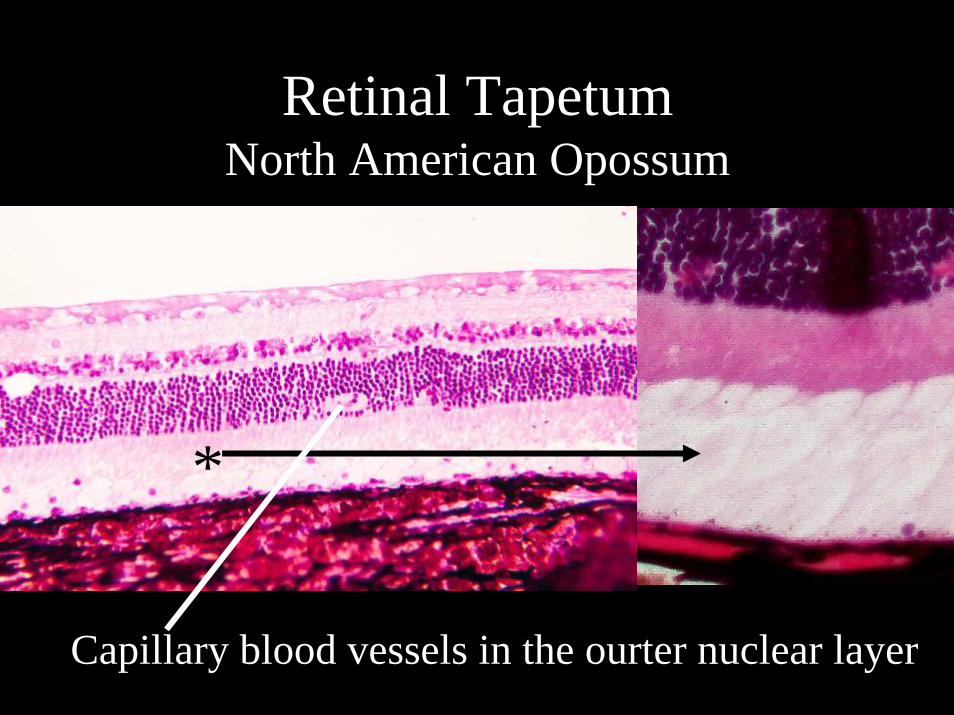
Retinal TapetumNorth American Opossum
*
Capillary blood vessels in the ourter nuclear layer
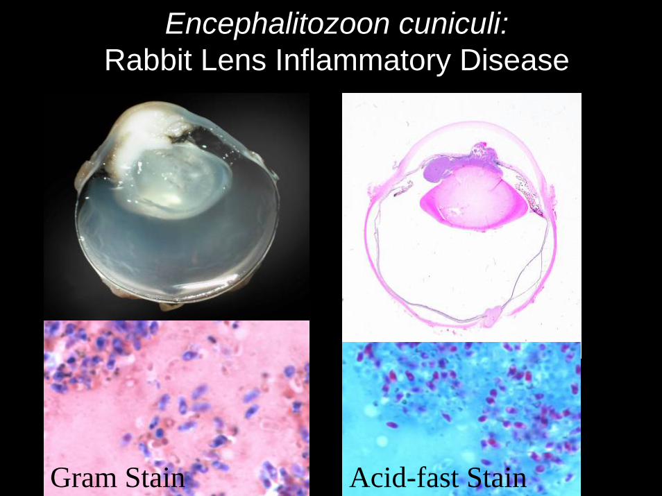
Encephalitozoon cuniculi: Rabbit Lens Inflammatory Disease
Gram Stain Acid-fast Stain
Recommended