Fetal Anemia 02/13/13
Anjulika Chawla, M.D.
Assistant Professor
Division of Pediatric Hematology/Oncology
Objectives
Definition of anemia
Diagnosis of fetal anemia
Normal developmental hematopoiesis
Etiology of fetal anemia
Decreased production
• Congenital, acquired
Malfunction of hemoglobin production
• Alpha thalassemia
Increased destruction
• Blood loss, hemolytic anemia
Treatment options
What does blood do?
Transports gasses, nutrients ,wastes,
hormones, heat
Regulates water balance, pH
Protection from infection, and
other alien invaders
What is blood?
Red blood cells : flexible sacks of
hemoglobin to carry gasses
White blood cells: cells with different
mechanisms to kill organisms
Platelets: make temporary walls to
keep from bleeding
Plasma : salt water that carries
everything else!
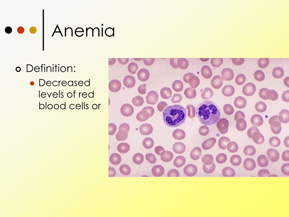
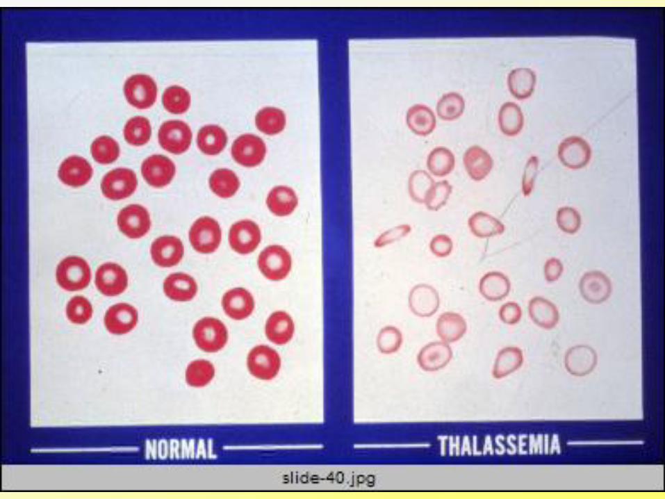
Anemia
Definition:
Decreased
levels of red
blood cells or
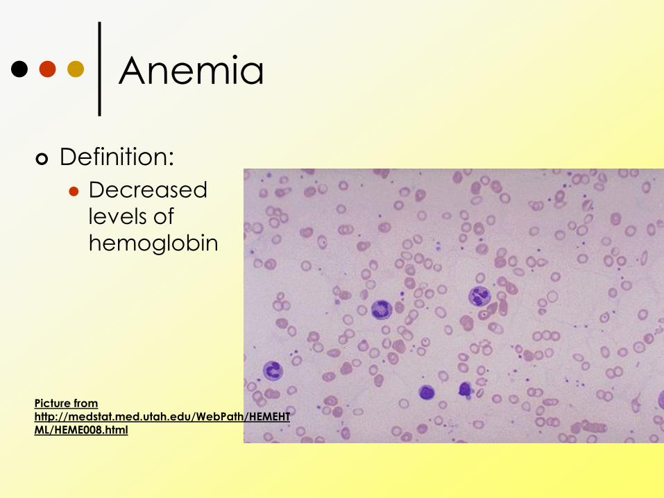
Anemia
Definition:
Decreased
levels of
hemoglobin
Picture from
http://medstat.med.utah.edu/WebPath/HEMEHT
ML/HEME008.html
Anemia
The fetus uses red blood cells to
carry oxygen in its circulation just
as children do.
When anemia is severe
(hemoglobin levels at 40-70% of
normal), the fetus can experience
heart failure and death.
Diagnosis of fetal anemia
Spectral analysis of amniotic fluid
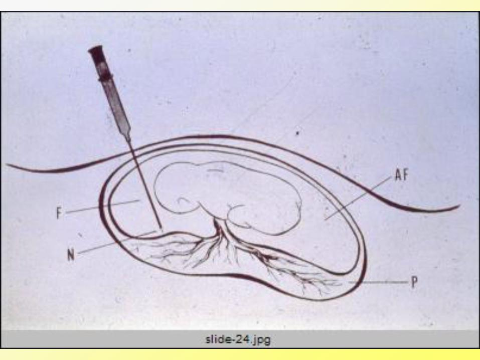
Cordocentesis
Doppler ultrasound – check for velocity of blood flow in the brain
Ultrasound of the heart can show signs
of strain
Ultrasound can also show signs of tissue
edema in severe anemia (hydrops
fetalis)
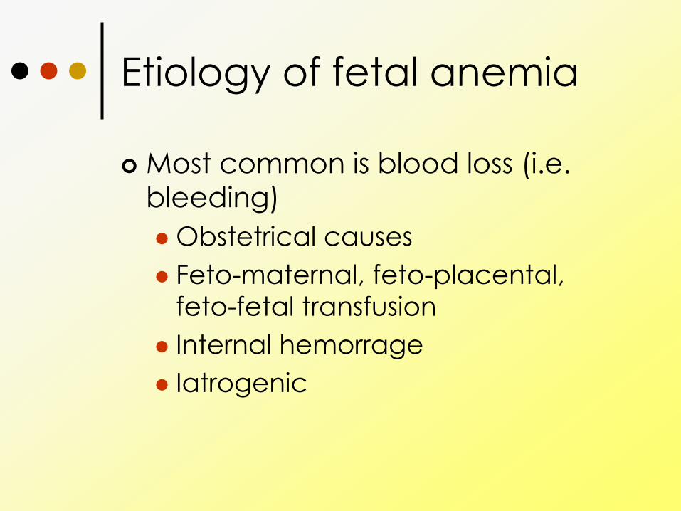
Etiology of fetal anemia
Most common is blood loss (i.e.
bleeding)
Obstetrical causes
Feto-maternal, feto-placental,
feto-fetal transfusion
Internal hemorrage
Iatrogenic
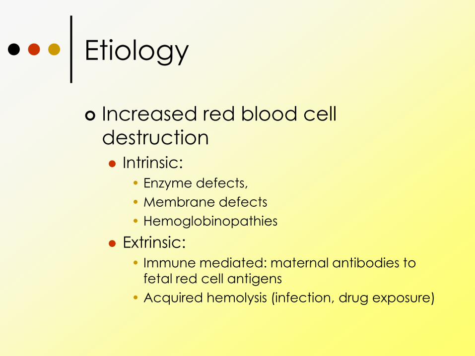
Etiology
Increased red blood cell
destruction Intrinsic:
• Enzyme defects,
• Membrane defects
• Hemoglobinopathies
Extrinsic:
• Immune mediated: maternal antibodies to
fetal red cell antigens
• Acquired hemolysis (infection, drug exposure)
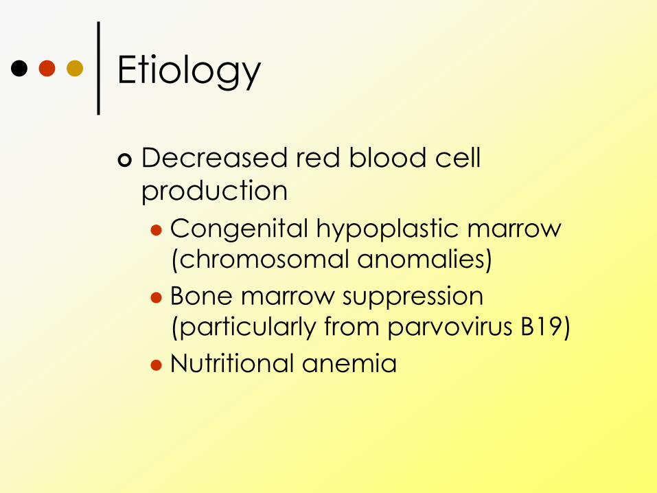
Etiology
Decreased red blood cell
production
Congenital hypoplastic marrow
(chromosomal anomalies)
Bone marrow suppression
(particularly from parvovirus B19)
Nutritional anemia
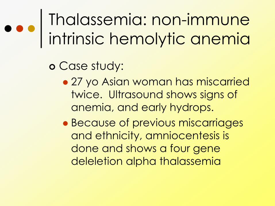
Thalassemia: non-immune
intrinsic hemolytic anemia
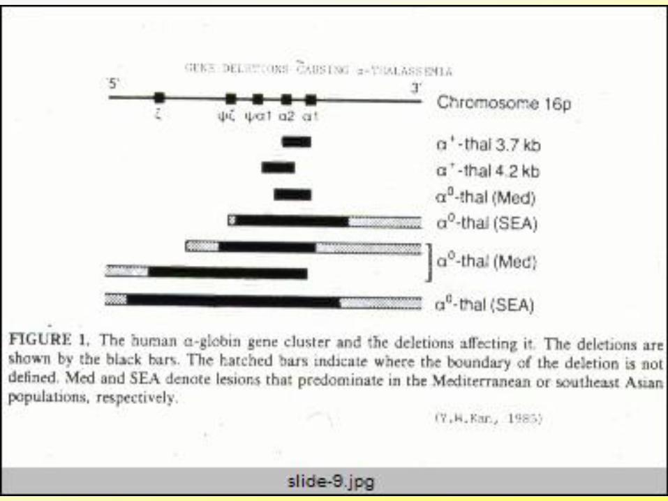
Case study:
27 yo Asian woman has miscarried
twice. Ultrasound shows signs of
anemia, and early hydrops.
Because of previous miscarriages
and ethnicity, amniocentesis is
done and shows a four gene
deleletion alpha thalassemia
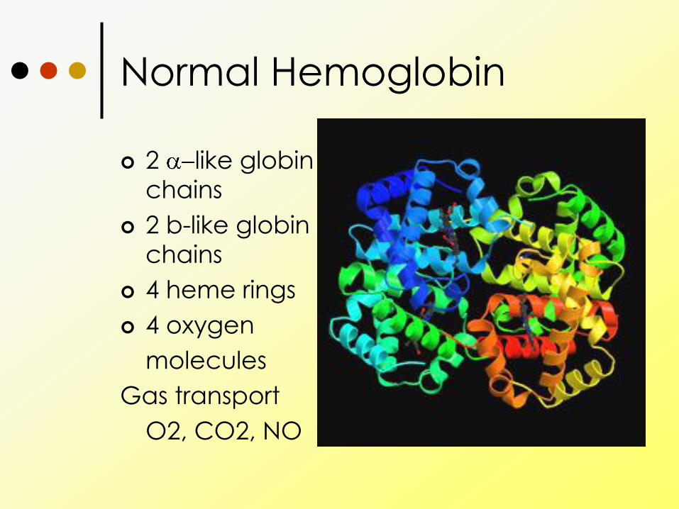
Normal Hemoglobin
2 like globin
chains
2 b-like globin
chains
4 heme rings
4 oxygen
molecules
Gas transport
O2, CO2, NO
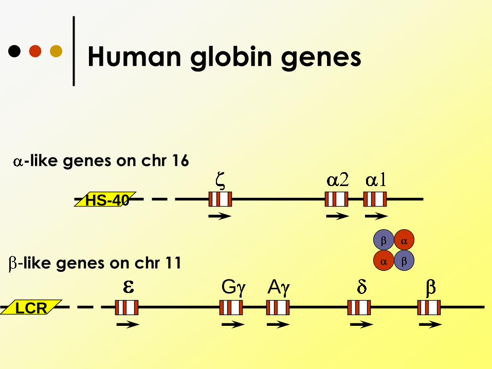
Human globin genes
-like genes on chr 16
-like genes on chr 11
G ALCR
HS-40
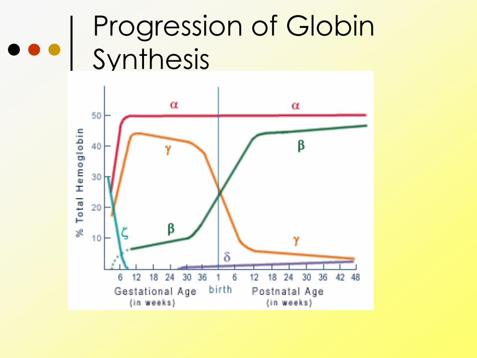
Progression of Globin
Synthesis
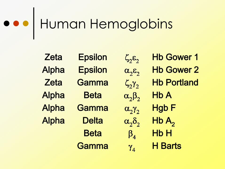
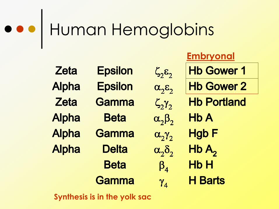
Human Hemoglobins
H Barts Gamma
Hb H Beta
Hb A2 Delta Alpha
Hgb F Gamma Alpha
Hb A Beta Alpha
Hb Portland Gamma Zeta
Hb Gower 2 Epsilon Alpha
Hb Gower 1 Epsilon Zeta
Human Hemoglobins
H Barts Gamma
Hb H Beta
Hb A2 Delta Alpha
Hgb F Gamma Alpha
Hb A Beta Alpha
Hb Portland Gamma Zeta
Hb Gower 2 Epsilon Alpha
Hb Gower 1 Epsilon Zeta
H Barts Gamma
Hb H Beta
Hb A2 Delta Alpha
Hgb F Gamma Alpha
Hb A Beta Alpha
Hb Portland Gamma Zeta
Hb Gower 2 Epsilon Alpha
Hb Gower 1 Epsilon Zeta
H Barts Gamma
Hb H Beta
Hb A2 Delta Alpha
Hgb F Gamma Alpha
Hb A Beta Alpha
Hb Portland Gamma Zeta
Hb Gower 2 Epsilon Alpha
Hb Gower 1 Epsilon Zeta
Embryonal
Synthesis is in the yolk sac
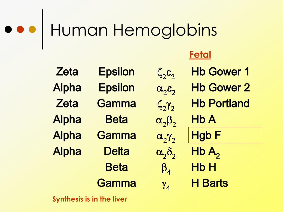
Human Hemoglobins
H Barts Gamma
Hb H Beta
Hb A2 Delta Alpha
Hgb F Gamma Alpha
Hb A Beta Alpha
Hb Portland Gamma Zeta
Hb Gower 2 Epsilon Alpha
Hb Gower 1 Epsilon Zeta
Fetal
Synthesis is in the liver
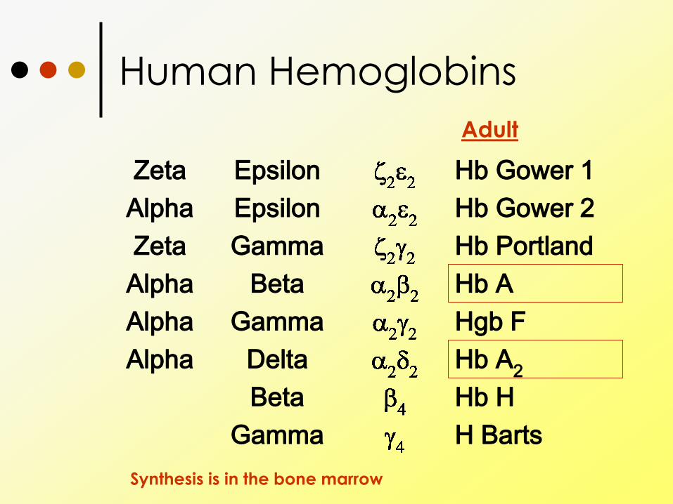
Human Hemoglobins
H Barts Gamma
Hb H Beta
Hb A2 Delta Alpha
Hgb F Gamma Alpha
Hb A Beta Alpha
Hb Portland Gamma Zeta
Hb Gower 2 Epsilon Alpha
Hb Gower 1 Epsilon Zeta
Adult
Synthesis is in the bone marrow
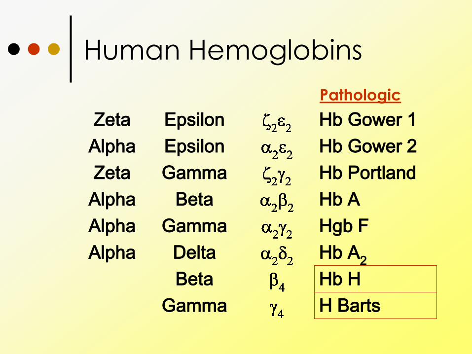
Human Hemoglobins
H Barts Gamma
Hb H Beta
Hb A2 Delta Alpha
Hgb F Gamma Alpha
Hb A Beta Alpha
Hb Portland Gamma Zeta
Hb Gower 2 Epsilon Alpha
Hb Gower 1 Epsilon Zeta
Pathologic
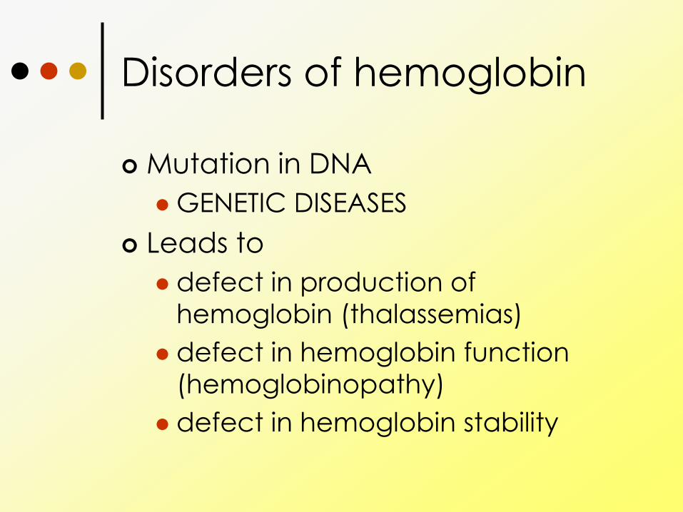
Disorders of hemoglobin
Mutation in DNA
GENETIC DISEASES
Leads to
defect in production of
hemoglobin (thalassemias)
defect in hemoglobin function
(hemoglobinopathy)
defect in hemoglobin stability
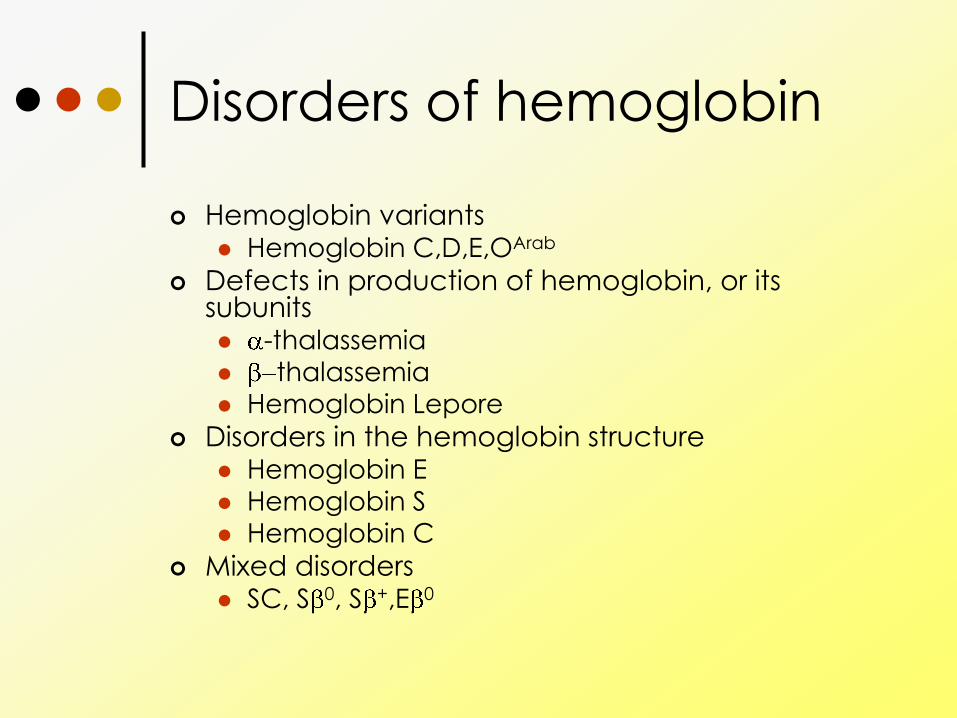
Disorders of hemoglobin
Hemoglobin variants Hemoglobin C,D,E,OArab
Defects in production of hemoglobin, or its subunits -thalassemia
thalassemia
Hemoglobin Lepore
Disorders in the hemoglobin structure Hemoglobin E
Hemoglobin S
Hemoglobin C
Mixed disorders SC, S 0, S +,E 0
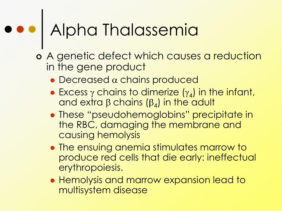
Alpha Thalassemia
A genetic defect which causes a reduction in the gene product
Decreased chains produced
Excess chains to dimerize ( 4) in the infant, and extra chains ( 4) in the adult
These “pseudohemoglobins” precipitate in the RBC, damaging the membrane and causing hemolysis
The ensuing anemia stimulates marrow to produce red cells that die early: ineffectual erythropoiesis.
Hemolysis and marrow expansion lead to multisystem disease
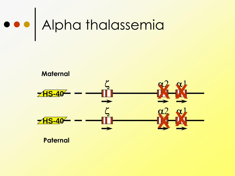
Alpha thalassemia
HS-40
HS-40
Maternal
Paternal
X
X
X
X
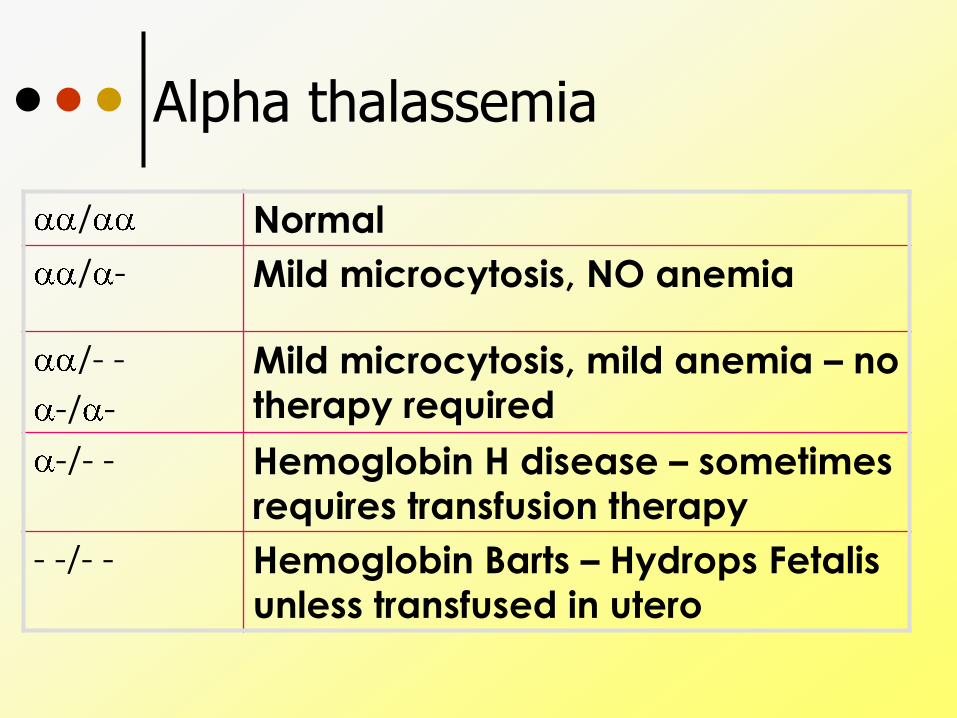
Alpha thalassemia
/ Normal
/ - Mild microcytosis, NO anemia
/- -
-/ -
Mild microcytosis, mild anemia – no
therapy required
-/- - Hemoglobin H disease – sometimes
requires transfusion therapy
- -/- - Hemoglobin Barts – Hydrops Fetalis
unless transfused in utero

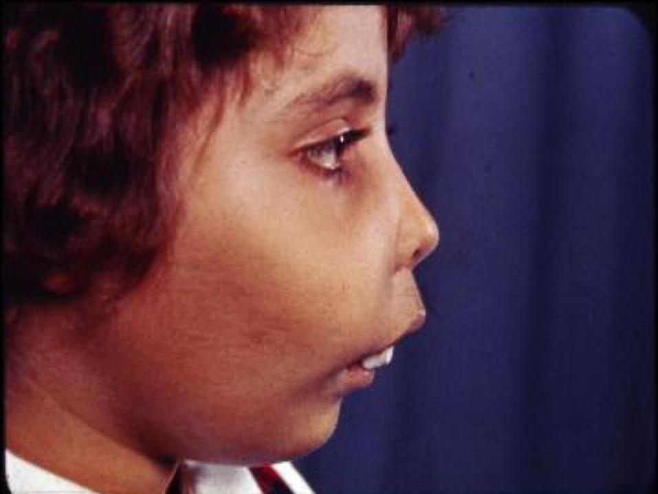
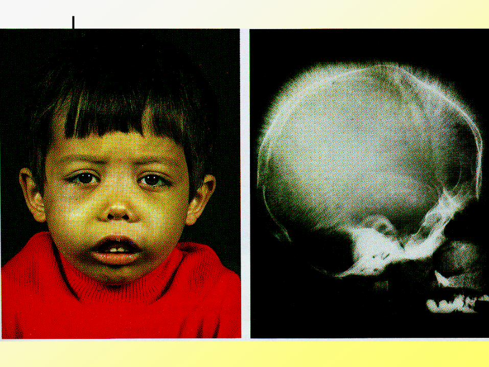
Natural History
Growth retardation
Delayed puberty
Pallor
Varying icterus
Skin Bronzing: gray-brown pigmentation
Features of hypermetabolic state
Hepatosplenomegaly
Skull changes:
frontal bossing
maxillary hyperplasia
Radiating striations
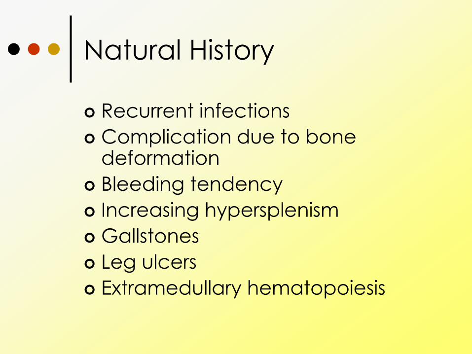
Natural History
Recurrent infections
Complication due to bone deformation
Bleeding tendency
Increasing hypersplenism
Gallstones
Leg ulcers
Extramedullary hematopoiesis
Treatment
Genetic counseling
Transfusion therapy
Iron overload treatment
Bone marrow transplant
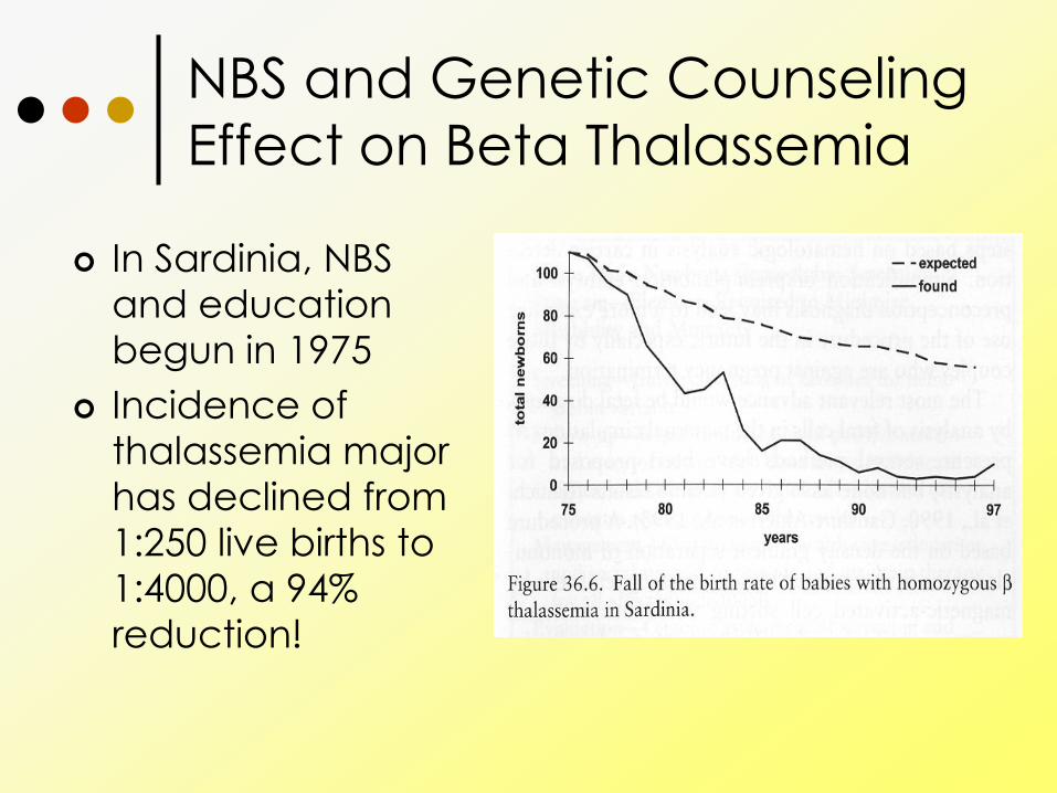
NBS and Genetic Counseling
Effect on Beta Thalassemia
In Sardinia, NBS
and education
begun in 1975
Incidence of
thalassemia major
has declined from 1:250 live births to
1:4000, a 94%
reduction!
Transfusion therapy
Corrects anemia and ineffective erythropoiesis
Consequences: Risk of fetal loss with each invasive
transfusion
Lifelong transfusions after birth
Time/effort/money
Risks of reaction, alloimmunization, infection
Iron overload • Liver deposition leads to cirrhosis • Endocrine
• Cardiac deposition leads to failure
• Iron chelation therapy
Natural History with Txfn
Endocrine disturbances – panhypopituitarism Impaired gonadotropins
Hypogonadism
IDDM
Adrenal insufficiency
Hypothyroidism
Hypoparathyroidism
Cirrhotic liver failure
Cardiac failure due to myocardial iron overload
Iron chelation
Desferroxamine
Chelates iron from the blood and tissues and excretes it in the urine and feces
Goal ferritin <2500 and liver iron stores <15mg/gm
Many drawbacks • Side efffects: Hearing loss, retinal damage, growth failure,
local skin reaction hypersenstivity
• Must be given continuous subcutaneously
• Expensive
Deferasirox Oral iron chelator, similar profile otherwise to desferroxamine Have to remember to take daily Side effects include skin rashes, risk of renal failure,
hearing loss Still expensive!
Avoid Iron Overload
Chelation
Exchange transfusion: remove
“bad blood” replace with “good
blood”
Erythracytapheresis: remove “bad
blood” replace with “good blood”
really, really fast with a machine
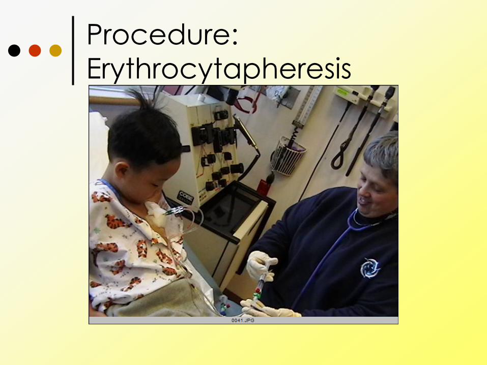
Procedure:
Erythrocytapheresis
Causes of death
Congestive heart failure
Arrythmia
Sepsis (postsplenectomy)
Multiple organ failure due to
hemochromocytosis
Thrombosis
Bone Marrow Transplant
Only curative option
Upfront mortality about 5% with matched sibling donor
Upfront mortality about 15% with unrelated matched donor
Morbidity from immunosuppression, toxicity of chemotherapy/radation, graft vs host disease
Recommended