GENETIC DIVERSITY OF WHITE TIGERS AND GENETIC
FACTORS RELATED TO COAT COLOR
Approved by: Research Advisor: Dr. Jan Janecka
Major: Biomedical Sciences Wildlife and Fisheries Sciences
May 2013
Submitted to Honors and Undergraduate Research Texas A&M University
in partial fulfillment of the requirements for the designation as
UNDERGRADUATE RESEARCH SCHOLAR
An Undergraduate Research Scholars Thesis
by
SARA ELIZABETH CARNEY
1
TABLE OF CONTENTS
Page
TABLE OF CONTENTS .....................................................................................................1
ABSTRACT .........................................................................................................................2
ACKNOWLEDGEMENTS .................................................................................................4
CHAPTER
I INTRODUCTION .......................................................................................5
Candidate genes for the white coat phenotype ................................8 Use of microsatellites to determine genetic diversity ......................9 II METHODS ................................................................................................11
Samples ..........................................................................................11 PCR methodology ..........................................................................11 Microsatellite analysis ...................................................................12 MC1R analysis ...............................................................................13
III RESULTS ..................................................................................................14
IV DISCUSSION ............................................................................................19
V CONCLUSION ..........................................................................................22
REFERENCES ..................................................................................................................24
2
ABSTRACT
Genetic Diversity of White Tigers and Genetic Factors Related to Coat Color. (May 2013)
Sara Elizabeth Carney Department of Veterinary Medicine and Biomedical Sciences
Texas A&M University
Research Advisor: Dr. Jan Janecka Department of Veterinary Medicine and Biomedical Sciences
White tigers are greatly cherished by the public, making them valuable to zoos and breeders.
Unfortunately, a number of health issues have occasionally surfaced within some of the white
tiger population such as neurological and facial defects. There is interest amongst private tiger
breeders to determine if these maladies are associated with the coat color or breeding practices,
and to find ways to prevent these health issues. The genes involved in producing the white
phenotype and the disease phenotype are currently unknown. Furthermore, the relationship
between the genes associated with coat color and levels of inbreeding also remain unknown.
Microsatellites are a tool frequently used within by geneticists and ecologists alike. These
segments of highly repeatable DNA mutate frequently and are variable in length. Thus
microsatellites can be used to determine heterozygosity within a population by detecting the
alleles present at the loci of interest. The amount of heterozygosity within a population can be
indicative of the amount of inbreeding present and overall levels of genetic diversity. A panel of
twelve microsatellites was used to analyze heterozygosity, thus inferring the levels of genetic
3
diversity present. Among the tigers sampled, estimated heterozygosity was determined to be
0.761 in white tigers and 0.772 in orange tigers.
The genes Melanocortin-1-Receptor (MC1R) and Agouti Signaling Protein (ASIP) have been
found to affect coat color phenotypes in other species similar to that of the white tiger, making
them ideal candidates for this project. These genes work antagonistically to each other in
production of melanin. MC1R is responsible for the production of α-melanocyte stimulating
hormone (α-MSH) while ASIP silences this activity. Thus, a loss-of-function associated with
MC1R or a gain-of-function associated with ASIP could lead to reduced pigment production.
This study continues the initial investigation by focusing on sequencing MC1R. Differences in
the nucleotides and amino acids of the sequences were compared though alignment in
Sequencher. At this time a causal mutation has not been found within exons 1 and 2 of ASIP or
MC1R.
4
Acknowledgements
First, I would like to thank everyone in the Texas A&M Molecular Cytogenetics and Genomics
Lab for providing a helpful and friendly environment for me to begin exploring the scientific
process. I would also like to thank Dr. Jan Janecka, Dr. Bhanu Chowdhary and Dr. Samantha
Steelman for allowing me the opportunity to embark on such an exciting project and for
providing ample guidance and encouragement. I am also grateful to my friend Emilee Larkin,
who sparked my interest in the subject and was always available to answer questions and provide
support. I would like to thank my friends and family for their enthusiasm and support.
I would also like to express my gratitude to those who provided samples for this project
including the San Francisco Zoo, In-sync Exotics (Vicky Keahey), Big Cat C.A.R.E, (Heidi
Riggs Berry), Sierra Endangered Cat Haven (Dale Anderson), Big Cat Rescue, Tiger Creek, the
Exotic Feline Rescue Center, Ferdinand and Antonin Fercos, T.I.G.E.R.S. (Doc Antle) and
REXANO (Zuzana Kukol). Your generosity has not only helped us begin to understand the
genetics of white tigers, but it has also provided me with the opportunity explore the world of
research and for that, I thank you.
5
CHAPTER I
INTRODUCTION
To many the white tiger, Panthera tigris, has been a source of awe, combining the power and
grace exhibited by the standard orange tiger with the rare beauty from its unusual coat color.
Though many find the white tiger to be inspiring, this is not a universally held opinion. Critics
contend that the white tiger is a detriment to tiger conservation, claiming that the tigers must be
inbred in order for the white coat to be present. Furthermore, they attribute the ailments faced by
some white tigers (eg. crossed-eyes and cleft palates) (Roychoudhury and Sankhala 1978) to the
white coat trait, believing it to be inseparable from inbreeding.
In light of this controversy, it is important to determine the white tiger’s role in conservation of
the species. Though some do not place priority on the preservation of the white tiger, it is evident
that the species as a whole is facing the threat of extinction. Three of the original eight tiger
subspecies, Bali (Panthera tigris balica), Caspian, (Panthera tigris virgata), and Javan
(Panthera tigris sondaica), have recently become extinct (Luo et al. 2004). The tiger population
has faced recent rapid decline. Within the last 100 years the wild tiger’s habitat has been reduced
to only 7% of the land in once roamed (Dinerstein et al. 2007). Poaching as well as habitat loss
and fragmentation poses the greatest threat to the wild tiger population. Deforestation has
significantly impacted the wildlife present in these areas particularly the tiger and its prey
(Kinnaird et al. 2003). The tiger faces additional risks associated with its dwindling population,
primarily decreased genetic diversity. Frequently, populations facing significant decline may
resort to inbreeding, potentially leading to inbreeding depression (Hedrick and Kalinowski
2000). Consequently, deleterious homozygotic traits that were once masked in a healthy
6
population of heterozygotes may become rampant in a genetically isolated population. Thus, this
genetically compromised population becomes increasingly vulnerable to disease (Lynch 1977).
While the wild tiger population faces steady decline, the captive population has successfully
propagated. Tigers have relatively few complications associated with reproduction, which often
plagues captive breeding programs. Additionally, captive-bred populations are protected from
many of the threats that face their wild counterparts, such as habitat degradation, disease and
poaching. Though the captive tiger has escaped many of these issues, loss of genetic diversity is
still a present concern within segments of the population (Lacy 1987). The white tiger is
particularly vulnerable to increased homozygosity due to selection for this phenotype. In many
ways the captive environment has allowed rare coat color polymorphisms such as that of the
white tiger to persist.
Though there are early reports of white tigers in India, the first lineage of captive white tigers
originated in what was known at the time as Rewa, (which is now Madhya Pradesh), from a
single male known as Mohan who was captured in 1951 (Thorton et al. 1966). The first breeding
of Mohan to Belgum, a wild orange female, was unsuccessful in producing a white offspring.
Mohan was subsequently bred to his daughter, Radha, produced from the previous cross. This
resulted in four litters, all producing white offspring (Thorton et al. 1966). It can be inferred that
Rewa, an F1, was heterozygous for the white coat allele. Thus the Rewa-Mohan cross gave
offspring of the union a 50% chance of being homozygous for and therefore expressing the white
coat allele. The white coat polymorphism is an autosomal characteristic inherited in a
Mendelian-recessive fashion (Thorton et al. 1966). Although inbreeding was prevalent in early
7
breeding of white tigers, it is not essential to produce a white tiger. Because the trait follows a
Mendelian inheritance pattern, the coat can be propagated given that both parents are carriers of
the allele.
Although the white coat polymorphism can be obtained without inbreeding, it can be challenging
to manage inbreeding levels while also selecting for the white phenotype. Because of this
breeders often resort to inbreeding to ensure that the trait is maintained. Mismanaged breeding
practices have reportedly led to an increase in health problems in some white tigers, such as
strabismus, facial deformities and neurological defects (Roychoudhury and Sankhala 1978).
However, it remains unclear to what extent these abnormalities are due to inbreeding. Some have
suggested that some of these health concerns may be linked to the white phenotype itself. For
example, strabismus, which is caused by retinal nerve fibers connecting at the opposite side of
the brain rather that the same side, is found in carnivores that are homozygous for an allele
within the albino series such as Siamese cats (Gulliery and Kaas 1973). Examination of a white
tiger’s lateral geniculate nucleus of the brain, (a region involved in processing visual information
gathered by the retina), revealed a defect of the A1 layer similar to, though less severe than that
of the Siamese (Gulliery and Kaas 1973). Therefore, determination of the degree of involvement
of the white phenotype versus inbreeding is essential in order to develop a scientifically based
breeding strategy for white tigers.
Though pigmentation and neurological development may seem unrelated, they are both derived
from the neural crest during the embryonic development of vertebrates (Rawles 1947).
Melanocyte precursors develop from the neural crest and spread to the hair and skin and
8
synthesize melanin (Rawles 1947). There are 2 forms of melanin: pheomelanin which produces
red or yellow pigment and eumelanin responsible for producing black or brown pigment
(Pawelek et al. 1982). These 2 types of melanin are structurally distinct; melanocytes producing
eumelanin tend to be more rounded than those producing eumelanin (Pawelek et al. 1982). White
tigers lack function in melanocytes producing pheomelanin, causing them to lack pigment where
other tigers would be orange. They carry pigment in their stripes which are gray or chocolate and
their eyes are blue. Therefore, white tigers are not albinos, though the coat of the white tiger is
due to an autosomal recessive mutation of the chinchilla allele, cch, and that locus is near the
albino locus (Robinson 1968).
Candidate genes for the white coat phenotype
Melanocortin-1-receptor (MC1R) and Agouti Signaling Protein (ASIP)
MC1R is responsible for regulating the hormone α-melanocyte stimulating hormone, (α-MSH),
which is involved in pigment production (Barsh 1996). MC1R has known effects on pigments in
many animals. In jaguars, Panthera onca, and jaguarundis, Puma yagouaroundi, a dominant
mutation of MC1R is responsible for melanism, the overproduction of eumelanin (Eizrik et al.
2003). However, melanism due to MC1R in domestic cats, Felis catus, follows a recessive
inheritance pattern (Eizrik et al. 2003). By contrast, repression of MC1R can lead to lack of
pigment, as in the case of the Kermode bear, Ursus americanus kermodei, which is a white color
morph of the black bear (Ritland et al. 2001). The lack of eumelanin is caused by a recessive
mutation at codon 298, replacing tyrosine with cytosine (Ritland et al. 2001). As with the case of
the white tiger, the Kermode bear is not an albino.
9
ASIP was also selected as a candidate gene due to antagonistic relationship to MC1R, silencing
the effects of α-MSH (Rieder, et al. 2001). Like MC1R, mutations in ASIP also affect some felid
species. Leopards, Panthera pardus, and Asian golden cats, Pardofelis temminckii, appear
melanistic due to single nucleotide polymorphism, (SNP), mutations in ASIP that cause ASIP to
loose function (Schneider et al. 2012). In this case, a loss-of-function has led to melanistic
individual. In contrast, a gain of function in ASIP could lead to an individual with reduced
pigment production. The ASIP gene contains three coding exons, the first two of which were
sequenced by former undergraduate research scholar, Emilee Larkin in an earlier phase of this
project (Larkin 2012).
Use of microsatellites to determine genetic diversity
Short tandem repeats of DNA known as microsatellites have shown to be invaluable in the
assessment of genetic diversity within a population or species. The high mutation rate of
microsatellites makes them ideal for individual identification and tracing evolution within a
population. Microsatellites are present within the non-coding regions of DNA and the mutations
that affect them influence the length of the microsatellite (Ellegren 2004). Because many
microsatellites are found within the non-coding region of DNA, they do not experience the same
evolutionary pressures found in genes within the coding segments of DNA (Ellegren 2004).
Additionally, the microsatellites are positioned next to a highly conserved region, known as the
flanking region. This region is identical among members of a species and sometimes between
closely related species, (Selkoe et al. 2006), thus allowing these regions to be compared more
effectively.
10
By assessing these candidate genes along with the levels of genetic diversity, we will gain insight
into the causes of the ailments faced by some white tigers. We will then use this information to
contribute to the understanding of the white tiger’s role in conservation and ideally resolve
controversy surrounding the breeding of white tigers. Additionally, the results of this study will
aid in the development of a responsible, genetically-based breeding program for white tigers.
11
CHAPTER II
METHODS
Samples
This study includes samples from 20 tigers (12 orange, 4 white, 3 golden tabby and 1 snow
white), 2 leopards (Panthera pardus) and 1 lion (Panthera leo). The DNA samples used in this
study were extracted from the blood via the PureGene DNA isolation protocol in agreement with
the College of Veterinary Medicine Clinical Research Review Committee (CRRC permit #10-44
J. Janecka). These samples were provided by zoos, sanctuaries and private owners including
T.I.G.E.R.S., Big Cat Rescue, Tiger Creek, REXANO, In-sync Exotics and Big Cat C.A.R.E,
among others.
PCR Methodology
The Multiplex Qiagen Typ-it Kit with Q was used for the PCRs for both microsatellites and
MC1R. The kit consists of sterile water, PCR Buffer Master Mix and Q Solution. These reagents
along with 20 µM of the respective forward and reverse primers were pooled to create a master
mix. To produce 10 µL reactions, 8.5 µL of the master mix was added to 1.5 µL of DNA and
placed in the thermal cycler. Samples were then incubated at 4ºC. The effectiveness of the PCR
was tested through visualization of the DNA by running a 1% agarose gel using 4 µL of each
sample using 0.1% mg/uL of ethidium bromide under ultraviolet light.
PCR for Microsatellites
12
For the microsatellites, the profile used for amplification is as follows: 5 minutes at 95ºC, 30
seconds at 95ºC, 1 minute and 30 seconds at 52 ºC, 30 seconds at 72ºC, steps 2 through 4 are
repeated 50 times, then 30 seconds at 60ºC. The samples were finally stored at 4ºC.
PCR for MC1R
Two set of forward and reverse primers were used for the MC1R PCR: MC1R4 forward, 5’-
GTTGTACAAGGGAGCTT-GG-3’, MC1R4 reverse, 5’-CATTGTCCTGAGCTGAC-AT-3’,
MC1R5 forward, 5’-CATTGTCCTTGAGCTGCAT-3’ , and MC1R5 reverse 5’-
GCCATAGGATATCCCCACCT-3. MC1R analysis required a touchdown PCR profile that is as
follows: 95 ºC for 5 minutes, 95ºC for 30 seconds, 65ºC for 1 minute and 30 seconds followed by
a 1ºC decrease per cycle, 1 minute at 72ºC, cycle to step 2 9 times, 30 seconds at 95ºC, 1 minute
and 30 seconds at 55ºC, 1 minute at 72ºC, cycle to step 6 for 20 times.
Microsatellite Analysis
A panel of 8 microsatellites (FCA 005, FCA 161, FCA 091, FCA 310, FCA032, FCA176,
FCA069 and FCA391) was used to measure genetic variation among the samples. These samples
were first diluted to 10ng/µL. This concentration was confirmed using the Nanodrop
spectrometer. Then the microsatellites were amplified using PCR via the Multiplex Qiagen Typ-
it Kit. This method uses 0.08 µL of each forward primer per reaction along with PCR Buffer
Master Mix, Q solution, and sterile water. After PCR amplification, the samples were run on a
1% agarose gel to confirm PCR success. The samples were subsequently genotyped using the
3730 DNA Analyzer in the Chowdhary lab. Genotyping data was then analyzed using the
13
GeneMapper 4.1 software. Genetic variation was assessed based on calculated heterozygosity at
each locus and among orange tigers and white tigers.
MC1R Analysis
Sequencing
The post-PCR samples were first cleaned up using the ExoSAP-IT treatment. Using this method,
5 µL of post-PCR product was added to 2 µL of ExoSAP-IT, then incubated in the thermal
cycler at 37ºC for 15 minutes then at 80ºC for another 15 minutes. The purpose of this step is to
eliminate excess primers and DTPs. The cleaned up product was then run through the Big Dye
terminator reaction, which involves adding 2µL of Big Dye v 1.1, 1 µL of 2 µM primer and 1 µ
of sterile water to 1 µL of the PCR product. The mixture was placed in the thermal cycler for the
cycles: 96ºC for 1 minute, 25 cycles of 96ºC for 10 seconds then 50ºC for 5 seconds and 60ºC for
4 minutes. The samples were then stored at 4ºC. Excess labeled DNTPs were cleaned up using a
Sephadex column. Dry G50 Sephadex was loaded into the wells of the plate and subsequently
hydrated with 300 µL of MilliQ water then left standing for approximately 30 minutes. The plate
was centrifuged at 2200 rmp for 10 minutes, then 20 µL from the Big Dye reactions was added
onto the column and centrifuged again at 2200 rmp for 10 minutes. The plates were then placed
in the spin-vac and afterwards rehydrated using 10 µL of formamide per sample. The samples
were then sequenced in the via the Applied Bio Systems 3730 DNA analyzer. Bases of low
quality were cut and aligned using Sequencher 4.7 (Gene Codes) software.
14
CHAPTER III
RESULTS
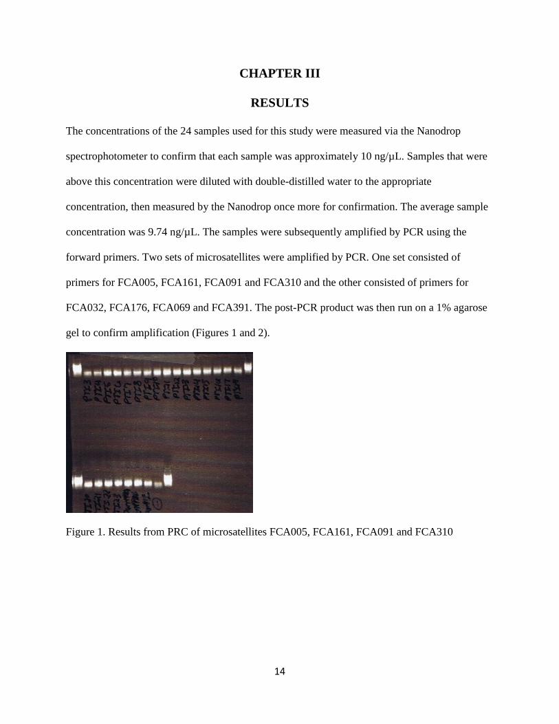
The concentrations of the 24 samples used for this study were measured via the Nanodrop
spectrophotometer to confirm that each sample was approximately 10 ng/µL. Samples that were
above this concentration were diluted with double-distilled water to the appropriate
concentration, then measured by the Nanodrop once more for confirmation. The average sample
concentration was 9.74 ng/µL. The samples were subsequently amplified by PCR using the
forward primers. Two sets of microsatellites were amplified by PCR. One set consisted of
primers for FCA005, FCA161, FCA091 and FCA310 and the other consisted of primers for
FCA032, FCA176, FCA069 and FCA391. The post-PCR product was then run on a 1% agarose
gel to confirm amplification (Figures 1 and 2).
Figure 1. Results from PRC of microsatellites FCA005, FCA161, FCA091 and FCA310
15
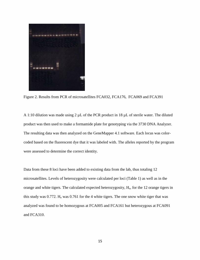
Figure 2. Results from PCR of microsatellites FCA032, FCA176, FCA069 and FCA391
A 1:10 dilution was made using 2 µL of the PCR product in 18 µL of sterile water. The diluted
product was then used to make a formamide plate for genotyping via the 3730 DNA Analyzer.
The resulting data was then analyzed on the GeneMapper 4.1 software. Each locus was color-
coded based on the fluorescent dye that it was labeled with. The alleles reported by the program
were assessed to determine the correct identity.
Data from these 8 loci have been added to existing data from the lab, thus totaling 12
microsatellites. Levels of heterozygosity were calculated per loci (Table 1) as well as in the
orange and white tigers. The calculated expected heterozygosity, He, for the 12 orange tigers in
this study was 0.772. He was 0.761 for the 4 white tigers. The one snow white tiger that was
analyzed was found to be homozygous at FCA005 and FCA161 but heterozygous at FCA091
and FCA310.
16
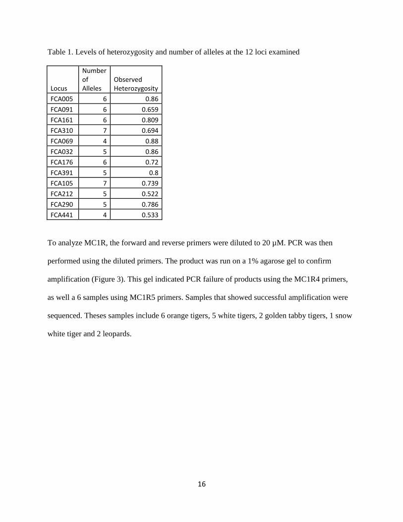
Table 1. Levels of heterozygosity and number of alleles at the 12 loci examined
Locus
Number of Alleles
Observed Heterozygosity
FCA005 6 0.86 FCA091 6 0.659 FCA161 6 0.809 FCA310 7 0.694 FCA069 4 0.88 FCA032 5 0.86 FCA176 6 0.72 FCA391 5 0.8 FCA105 7 0.739 FCA212 5 0.522 FCA290 5 0.786 FCA441 4 0.533
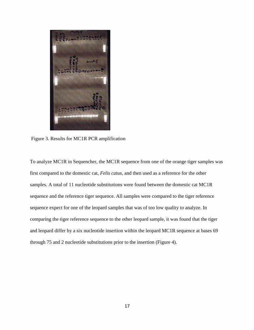
To analyze MC1R, the forward and reverse primers were diluted to 20 µM. PCR was then
performed using the diluted primers. The product was run on a 1% agarose gel to confirm
amplification (Figure 3). This gel indicated PCR failure of products using the MC1R4 primers,
as well a 6 samples using MC1R5 primers. Samples that showed successful amplification were
sequenced. Theses samples include 6 orange tigers, 5 white tigers, 2 golden tabby tigers, 1 snow
white tiger and 2 leopards.
17
Figure 3. Results for MC1R PCR amplification
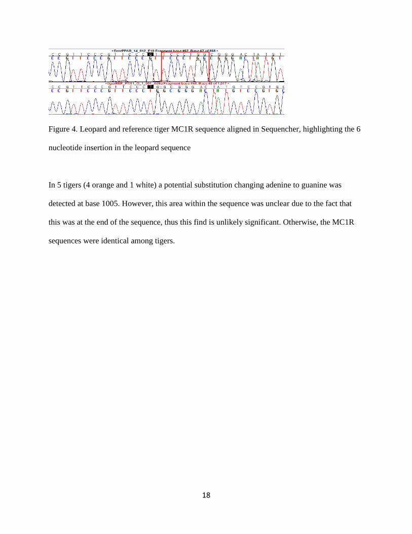
To analyze MC1R in Sequencher, the MC1R sequence from one of the orange tiger samples was
first compared to the domestic cat, Felis catus, and then used as a reference for the other
samples. A total of 11 nucleotide substitutions were found between the domestic cat MC1R
sequence and the reference tiger sequence. All samples were compared to the tiger reference
sequence expect for one of the leopard samples that was of too low quality to analyze. In
comparing the tiger reference sequence to the other leopard sample, it was found that the tiger
and leopard differ by a six nucleotide insertion within the leopard MC1R sequence at bases 69
through 75 and 2 nucleotide substitutions prior to the insertion (Figure 4).
18
Figure 4. Leopard and reference tiger MC1R sequence aligned in Sequencher, highlighting the 6
nucleotide insertion in the leopard sequence
In 5 tigers (4 orange and 1 white) a potential substitution changing adenine to guanine was
detected at base 1005. However, this area within the sequence was unclear due to the fact that
this was at the end of the sequence, thus this find is unlikely significant. Otherwise, the MC1R
sequences were identical among tigers.
19
CHAPTER IV
DISCUSSION
The need for adequate levels of genetic diversity is a particular concern for endangered
populations, primarily due to magnified effects of genetic drift and deleterious alleles as
compared to larger populations (Hedrick and Kalinowski 2000). In a natural environment
species that suffers from severe inbreeding faces an increased likelihood for extinction.
However, in a captive environment these alleles are able to persist for much longer due to
protection from outside threats (Lacy 1987). Therefore, it is equally important to maintain high
genetic diversity in both captive and wild populations, not only for the salvation of a species, but
also for the health of individuals.
There is a well-established correlation between heterozygosity and traits determining fitness,
such as weight, fecundity and developmental stability (Milton and Grant 1984). Subsequently,
the overall health of a population can be inferred by examining the heterozygosity of the
population in question. Populations with lower heterozygosity are also at greater risk for disease
acquisition. A classic example of the effects of decreased genetic diversity is the cheetah,
Acinonyx jubatus . which occurred as a result of a historic bottleneck. This lack of genetic
diversity has led to difficulties in captive breeding due to abnormalities of the spermatozoa
(O’brien et al. 1985). Furthermore, the major histocompatibility complex, (MHC), is identical in
cheetahs making the population susceptible to pathogens (O’brien et al. 1985).
20
Based on the data gained from our microsatellite analysis, it is apparent that among the white
tigers and orange tigers sampled, there is no statistically significant difference in heterozygosity.
This indicates that the white tigers included in this study were likely outbred to orange tigers,
maintaining higher heterozygosity. Though it is known that early captive white tiger populations
originated through inbreeding, (Thorton et al. 1966), it is clear from our results that not all white
tigers presently in captivity are significantly inbred. In an effort to broaden our understanding of
genetic diversity among white tigers and orange tigers, we will continue to incorporate additional
individuals and microsatellites, adding more power to our data.
The analysis of MC1R showed no causal mutation for the white coat phenotype. Though it
appeared that in some sequences a guanine was substituted for adenine, this result does not
provide clear results due to the fact that the sequences degrade toward the end and become
difficult to accurately assess in Sequencher. To better understand the significance of this finding
the samples would need to be sequenced again using the reverse primer. However, had this
anomaly been clear enough to consider a SNP, it still would not be considered a causal mutation
because of the fact that it was found in both orange and white tigers.
Because the cause of the white coat phenotype has not yet been found, we will continue
examining candidate genes by first sequencing of exon 3 of ASIP then looking at additional
candidate genes including the gene which codes for the enzyme tyrosinase, (TYR). This gene is
composed of 5 exons and 4 introns; mutations have known effects of pigment in humans and
mice, often leading to Oculocutaneous albinism type 1 (OCA 1) (Slominski et al. 2004). TYR
affects the melanogenesis pathway at an earlier stage than MC1R and ASIP, (Cieslak et al.

21
2011), by producing tyrosinase which functions as a catalyst in the reactions converting tyrosine
into melanin (Korner et al. 1982), making it essential in the production of eumelanin (Cieslak et
al. 2011).
22
CHAPTER V
CONCLUSION
Based on the analysis of 12 microsatellites, we have determined that there is not a significant
difference between white tigers and orange tigers in terms of heterozygosity. As this study
progresses, more microsatellites will be added in order to understand the levels of heterozygosity
at other loci. More individuals will also be incorporated into this study to broaden our
understanding of the heterozygosity within captive-bred tigers. Evidence suggesting that white
tigers are not inbred to a significantly greater degree than orange tigers could potentially alleviate
some of the controversy surrounding the breeding of white tigers. More importantly, it will
provide breeder with data that is necessary in order to make critical management decisions that
affect both species and individual health.
A causal mutation has not been discovered in ASIP or MC1R, but their assessment has
nonetheless been important in the search for the genetic origin of the white coat phenotype. We
will continue our analysis by sequencing exon 3 of ASIP and TYR and possibly other candidate
genes. Finding the gene responsible for this phenotype will provide new insight into the diversity
and well-being of tigers. This information will be used to if there is a link between the phenotype
itself and the health concerns sometimes appearing in white tigers.
As the tiger population continuously decreases in the wild, it becomes increasing apparent that
we must ensure the welfare of the tigers in captivity. As a flagship species, the tiger serves as an
ambassador for tigers in the wild, as well as conservation in general. Through increased research

23
we can gain the knowledge necessary to protect the beloved white tiger and ensure that white
tigers are carefully bred using the a management strategy that is genetically based.
24
REFERENCES
Barsh, G. S., 1996. The genetics of pigmentation: from fancy genes to complex traits. Trends in Genetics. 12, 299-305.
Cieslak, M., Reissmann, M., Hofreiter, M., Ludwig, A., 2011,. Colours of domestication.
Biological Reviews. 86, 885-899. Dinerstein, E., Loucks, C., Wikramanayake, E., Ginsberg, J., Sanderson, E., Seidensticker, J.,
Forrest, J., Bryja, G., Heydlauff, A., Klenzendorf, S., 2007. The Fate of Wild Tigers. Bioscience. 57, 508–514.
Eizirik, E., Yuhki, N., Johnson, W., Menotti-Raymond, M., Hannah, S., O’Brien, S., 2003.
Molecular Genetics and Evolution of Melanism in the Cat Family. Current Biology. 13, 448-453.
Ellegren, H., 2004. Microsatellites: simple sequences with complex evolution. Nature Reviews
Genetics. 5, 435-445. Guillery, R. W., Kaas, J. H., 1973. Genetic abnormality of the visual pathways in a “white” tiger. Science. 180, 1287-1289. Hedrick, P. W., Kalinowski, S. T., 2000. Inbreeding depression in conservation biology.
Annual Review of Ecology and Systematics. 139-162. Kinnaird, M. F., Sanderson, E. W., O'Brien, T. G., Wibisono, H. T., Woolmer, G., 2003.
Deforestation trends in a tropical landscape and implications for endangered large mammals. Conservation Biology. 17, 245-257.
Korner, A., Pawelek, J., 1982. Mammalian tyrosinase catalyzes three reactions in the biosynthesis of melanin. Science. 217, 1163-1165.
Lacy, R. C., 2005. Loss of genetic diversity from managed populations: interacting effects of
drift, mutation, immigration, selection, and population subdivision. Conservation Biology. 1,143-158.
Larkin, E. A., 2012. Investigation of genes associated with the white coat color in tigers.
(Undergraduate Research Thesis). Texas A&M University, College Station, Texas. Luo, S. J., Kim, J. H., Johnson, W. E., Walt, J. V. D., Martenson, J., Karanth, U. K., 2004. Phylogeography and genetic ancestry of tigers (Panthera tigris). PLoS Biology. 2, 2275-
2293.
25
Lynch, C. B., 1977. Inbreeding effects upon animals derived from a wild population of Mus musculus. Evolution. 31, 526-537.
Mitton, J. B., Grant, M. C., 1984. Associations among protein heterozygosity, growth rate, and developmental homeostasis. Annual review of ecology and systematics. 15, 479-499.
O'brien, S. J., Roelke, M. E., Marker, L., Newman, A., Winkler, C. A., Meltzer, D., Colly, L.,
Evermann, J. F., Bush, M., Wildt, D. E., 1985. Genetic basis for species vulnerability in the cheetah. Science. 227, 1428-1434.
Pawelek, J. M., Körner, A. M., 1982. The Biosynthesis of Mammalian Melanin: The
regulation of pigment formation, the key to disorders such as albinism and piebaldism, may also offer some clues for the treatment of melanoma. American scientist. 70, 136-145.
Rawles, M. E., 1947. Origin of pigment cells from the neural crest in the mouse embryo. Physiological zoology. 20, 248-266. Rieder, S., Taourit, S., Mariat, D., Langlois B., Guerin, G., 2001. Mutations in the agouti (ASIP),
the extension (MC1R), and the brown (TYRP1) loci and their association to coat color phenotype in horse (Equus cabaluus). Mammalian Genome. 12, 450-455.
Robinson, R., 1969. The white tigers of Rewa and gene homology in the Felidae. Genetica. 40, 198-200. Roychoudhury, A. K., Sankhala, K. S., 1979. Inbreeding in white tigers. Proceedings: Animal Sciences, 88, 311-323. Schneider, A., David, V. A., Johnson, W. E., O'Brien, S. J., Barsh, G. S., Menotti-Raymond, M.,
Eizirik, E., 2012. How the Leopard Hides Its Spots: ASIP Mutations and Melanism in Wild Cats. PloS one. 7, e50386.
Selkoe, K. A., Toonen, R. J., 2006. Microsatellites for ecologists: a practical guide to using
and evaluating microsatellite markers. Ecology letters, 9, 615-629.
Slominski, A., Tobin, D. J., Shibahara, S., Wortsman, J., 2004. Melanin pigmentation in mammalian skin and its hormonal regulation. Physiological reviews. 84, 1155-1228.
Thornton, I. W., Yeung, K. K., Sankhala, K. S., 1967. The genetics of the white tigers of Rewa. Journal of Zoology. 152, 127-135.
Recommended