Vol. 114 No. 1 July 2012
Intraoral molluscum contagiosum in a young immunocompetentpatientCyntia Helena Pereira de Carvalho, DDS,a Ana Luiza Dias Leite de Andrade,b
Denise Hélen Imaculada Pereira de Oliveira,b Emeline das Neves de Araújo Lima,c
Éricka Janine Dantas da Silveira, DDS, PhD,d and Ana Myriam Costa de Medeiros, DDS, PhDe FederalUniversity of Campina Grande, Patos, Paraíba, Brazil; and Federal University of Rio Grande do Norte, Natal,Brazil
Molluscum contagiosum (MC) is a contagious disease caused by a virus of the poxvirus family. In children, thedisease commonly manifests as a variable number of discrete umbilicated papules on the face and trunk. In healthy andimmunosuppressed adults, the disease appears on or near the genital organs and is often sexually transmitted. MC involvingthe intraoral mucosa has been documented but is rare. We report a case of MC involving the oral mucosa exclusively anddiscuss the main clinical, histopathologic, and therapeutic characteristics, comparing the findings with cases of this rare oral
presentation described in the literature. (Oral Surg Oral Med Oral Pathol Oral Radiol 2012;114:e57-e60)Molluscum contagiosum (MC) is a self-limited viralinfection that commonly affects the skin. Involvementof mucous membranes is rare. The disease can affectindividuals of any age, but its prevalence is higheramong children, sexually active adults, and immuno-compromised individuals (e.g., patients with acquiredimmunodeficiency syndrome).1-4
MC can be transmitted by direct contact with aninfected person, fomites, and self-inoculation. In chil-dren, lesions are commonly found on the face, trunk,and extremities, whereas in adults the genital regionand adjacent areas are the most affected.5 In addition,nonsexual transmission has been reported in adults andgenerally occurs at sites of trauma or other skin le-sions.6-8
Clinically, the infection manifests as single or mul-tiple asymptomatic, small (2-6 mm), flesh-colored orpink papules with a central depression. The infection isgenerally self-limiting and may resolve spontaneouslywithin �18 months of its emergence. Some lesionsmay not regress and require auxiliary treatment, such as
aProfessor, Oral Microbiology Department, Federal University ofCampina Grande.bMSc Student, Oral Pathology Department, Federal University of RioGrande do Norte.cPhD Student, Oral Pathology Department, Federal University of RioGrande do Norte.dProfessor, Oral Pathology Department, Federal University of RioGrande do Norte.eProfessor of Stomatology, Oral Pathology Department, Federal Uni-versity of Rio Grande do Norte.Received for publication Jul 12, 2011; returned for revision Oct 20,2011; accepted for publication Oct 25, 2011.© 2012 Elsevier Inc. All rights reserved.2212-4403/$ - see front matter
http://dx.doi.org/10.1016/j.oooo.2011.10.009curettage, cryotherapy with liquid nitrogen, electrocau-tery, and incision of the papules.9
Although involvement of mucous membranes hasbeen reported in the literature, cases in which the dis-ease affects oral soft tissues are extremely rare. Wereport a case of intraoral MC in an immunocompetentyoung patient and discuss the clinical features andtreatment of this rare presentation, comparing the pres-ent case with those described in the international liter-ature.
CASE REPORTA 13-year-old mulatto girl was seen at the Oral Diagnosis
Service of the Department of Dentistry, Federal University ofRio Grande do Norte. The patient was referred by anotherprofessional owing to the presence of multiple lesions on thelower labial mucosa whose diagnosis was not defined. Ex-traoral physical examination revealed no anomalies. Intraoral
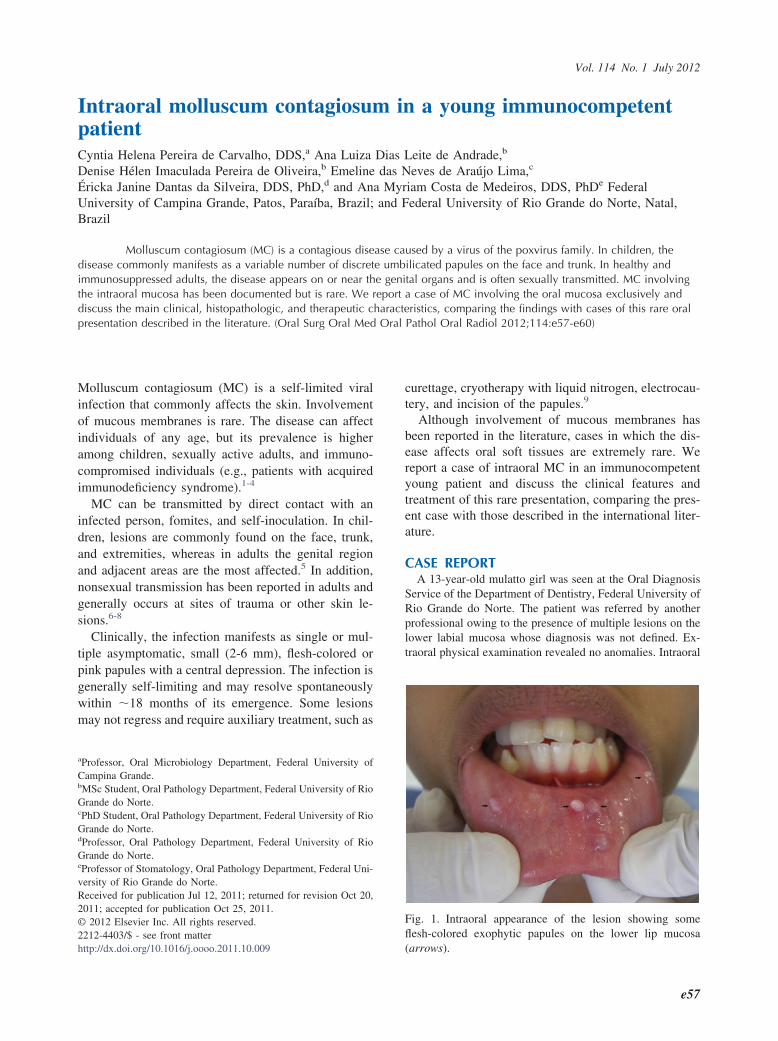
Fig. 1. Intraoral appearance of the lesion showing someflesh-colored exophytic papules on the lower lip mucosa
(arrows).e57
ORAL AND MAXILLOFACIAL PATHOLOGY OOOOe58 Carvalho et al. July 2012
clinical examination showed the presence of 4 asymptomaticsessile papules varying in size from 0.3 to 0.5 cm in greatest
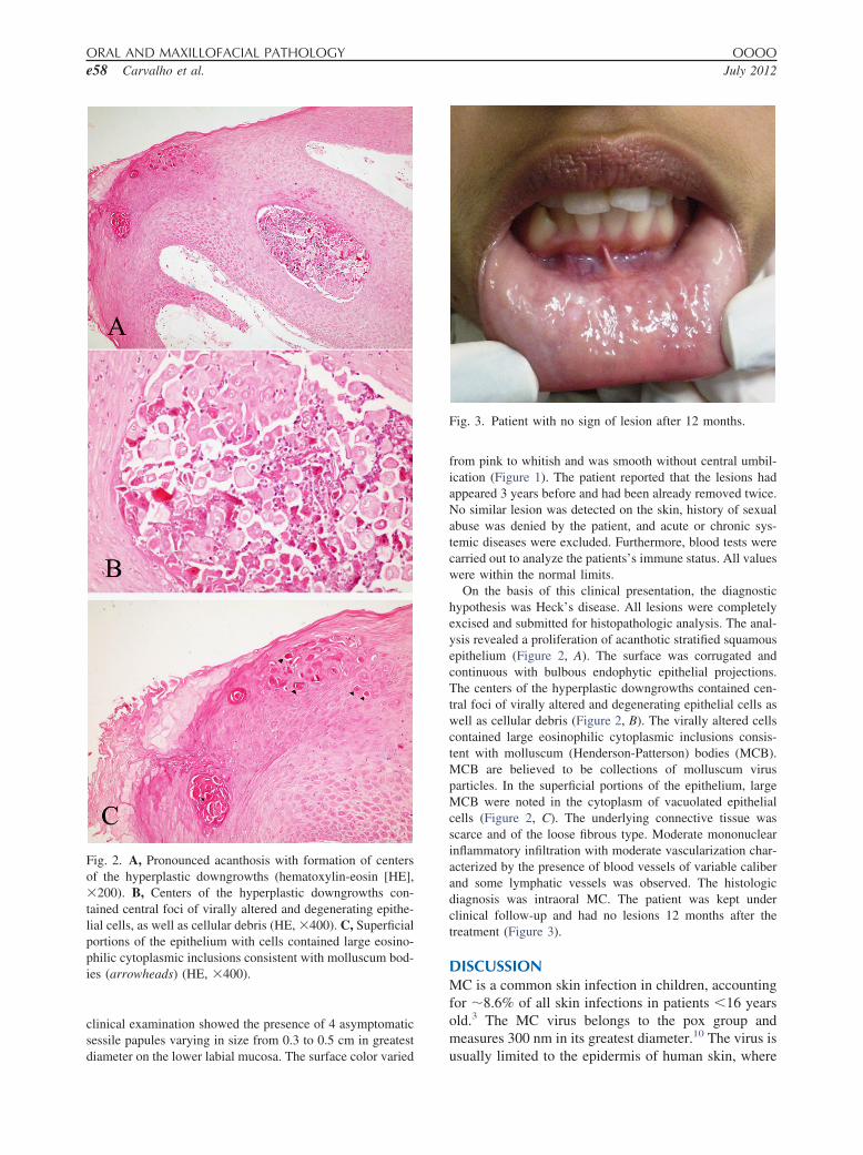
Fig. 2. A, Pronounced acanthosis with formation of centersof the hyperplastic downgrowths (hematoxylin-eosin [HE],�200). B, Centers of the hyperplastic downgrowths con-tained central foci of virally altered and degenerating epithe-lial cells, as well as cellular debris (HE, �400). C, Superficialportions of the epithelium with cells contained large eosino-philic cytoplasmic inclusions consistent with molluscum bod-ies (arrowheads) (HE, �400).
diameter on the lower labial mucosa. The surface color varied
from pink to whitish and was smooth without central umbil-ication (Figure 1). The patient reported that the lesions hadappeared 3 years before and had been already removed twice.No similar lesion was detected on the skin, history of sexualabuse was denied by the patient, and acute or chronic sys-temic diseases were excluded. Furthermore, blood tests werecarried out to analyze the patients’s immune status. All valueswere within the normal limits.
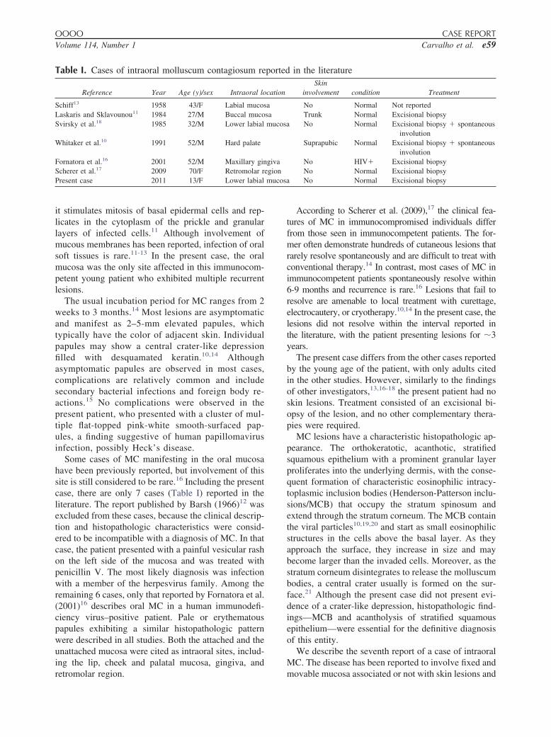
On the basis of this clinical presentation, the diagnostichypothesis was Heck’s disease. All lesions were completelyexcised and submitted for histopathologic analysis. The anal-ysis revealed a proliferation of acanthotic stratified squamousepithelium (Figure 2, A). The surface was corrugated andcontinuous with bulbous endophytic epithelial projections.The centers of the hyperplastic downgrowths contained cen-tral foci of virally altered and degenerating epithelial cells aswell as cellular debris (Figure 2, B). The virally altered cellscontained large eosinophilic cytoplasmic inclusions consis-tent with molluscum (Henderson-Patterson) bodies (MCB).MCB are believed to be collections of molluscum virusparticles. In the superficial portions of the epithelium, largeMCB were noted in the cytoplasm of vacuolated epithelialcells (Figure 2, C). The underlying connective tissue wasscarce and of the loose fibrous type. Moderate mononuclearinflammatory infiltration with moderate vascularization char-acterized by the presence of blood vessels of variable caliberand some lymphatic vessels was observed. The histologicdiagnosis was intraoral MC. The patient was kept underclinical follow-up and had no lesions 12 months after thetreatment (Figure 3).
DISCUSSIONMC is a common skin infection in children, accountingfor �8.6% of all skin infections in patients �16 yearsold.3 The MC virus belongs to the pox group andmeasures 300 nm in its greatest diameter.10 The virus is
Fig. 3. Patient with no sign of lesion after 12 months.
usually limited to the epidermis of human skin, where
mucosa
OOOO CASE REPORTVolume 114, Number 1 Carvalho et al. e59
it stimulates mitosis of basal epidermal cells and rep-licates in the cytoplasm of the prickle and granularlayers of infected cells.11 Although involvement ofmucous membranes has been reported, infection of oralsoft tissues is rare.11-13 In the present case, the oralmucosa was the only site affected in this immunocom-petent young patient who exhibited multiple recurrentlesions.
The usual incubation period for MC ranges from 2weeks to 3 months.14 Most lesions are asymptomaticand manifest as 2–5-mm elevated papules, whichtypically have the color of adjacent skin. Individualpapules may show a central crater-like depressionfilled with desquamated keratin.10,14 Althoughasymptomatic papules are observed in most cases,complications are relatively common and includesecondary bacterial infections and foreign body re-actions.15 No complications were observed in thepresent patient, who presented with a cluster of mul-tiple flat-topped pink-white smooth-surfaced pap-ules, a finding suggestive of human papillomavirusinfection, possibly Heck’s disease.
Some cases of MC manifesting in the oral mucosahave been previously reported, but involvement of thissite is still considered to be rare.16 Including the presentcase, there are only 7 cases (Table I) reported in theliterature. The report published by Barsh (1966)12 wasexcluded from these cases, because the clinical descrip-tion and histopathologic characteristics were consid-ered to be incompatible with a diagnosis of MC. In thatcase, the patient presented with a painful vesicular rashon the left side of the mucosa and was treated withpenicillin V. The most likely diagnosis was infectionwith a member of the herpesvirus family. Among theremaining 6 cases, only that reported by Fornatora et al.(2001)16 describes oral MC in a human immunodefi-ciency virus–positive patient. Pale or erythematouspapules exhibiting a similar histopathologic patternwere described in all studies. Both the attached and theunattached mucosa were cited as intraoral sites, includ-ing the lip, cheek and palatal mucosa, gingiva, and
Table I. Cases of intraoral molluscum contagiosum re
Reference Year Age (y)/sex Intraoral lo
Schiff13 1958 43/F Labial mucosLaskaris and Sklavounou11 1984 27/M Buccal mucoSvirsky et al.18 1985 32/M Lower labial
Whitaker et al.10 1991 52/M Hard palate
Fornatora et al.16 2001 52/M Maxillary ginScherer et al.17 2009 70/F Retromolar rPresent case 2011 13/F Lower labial
retromolar region.
According to Scherer et al. (2009),17 the clinical fea-tures of MC in immunocompromised individuals differfrom those seen in immunocompetent patients. The for-mer often demonstrate hundreds of cutaneous lesions thatrarely resolve spontaneously and are difficult to treat withconventional therapy.14 In contrast, most cases of MC inimmunocompetent patients spontaneously resolve within6-9 months and recurrence is rare.16 Lesions that fail toresolve are amenable to local treatment with curettage,electrocautery, or cryotherapy.10,14 In the present case, thelesions did not resolve within the interval reported inthe literature, with the patient presenting lesions for �3years.
The present case differs from the other cases reportedby the young age of the patient, with only adults citedin the other studies. However, similarly to the findingsof other investigators,13,16-18 the present patient had noskin lesions. Treatment consisted of an excisional bi-opsy of the lesion, and no other complementary thera-pies were required.
MC lesions have a characteristic histopathologic ap-pearance. The orthokeratotic, acanthotic, stratifiedsquamous epithelium with a prominent granular layerproliferates into the underlying dermis, with the conse-quent formation of characteristic eosinophilic intracy-toplasmic inclusion bodies (Henderson-Patterson inclu-sions/MCB) that occupy the stratum spinosum andextend through the stratum corneum. The MCB containthe viral particles10,19,20 and start as small eosinophilicstructures in the cells above the basal layer. As theyapproach the surface, they increase in size and maybecome larger than the invaded cells. Moreover, as thestratum corneum disintegrates to release the molluscumbodies, a central crater usually is formed on the sur-face.21 Although the present case did not present evi-dence of a crater-like depression, histopathologic find-ings—MCB and acantholysis of stratified squamousepithelium—were essential for the definitive diagnosisof this entity.
We describe the seventh report of a case of intraoralMC. The disease has been reported to involve fixed and
in the literatureSkin
involvement condition Treatment
No Normal Not reportedTrunk Normal Excisional biopsyNo Normal Excisional biopsy � spontaneous
involutionSuprapubic Normal Excisional biopsy � spontaneous
involutionNo HIV� Excisional biopsyNo Normal Excisional biopsyNo Normal Excisional biopsy
ported
cation
asamucosa
givaegion
movable mucosa associated or not with skin lesions and
ORAL AND MAXILLOFACIAL PATHOLOGY OOOOe60 Carvalho et al. July 2012
to affect healthy and HIV-seropositive adults. The pres-ent case differs from those reported so far because thepatient was only 13 years old and presented recurrentlesions that had not regressed over the preceding 3years despite her immunocompetent status.
REFERENCES1. Dave S, Thappa DM, Karthikeyan K. Disseminated and disfig-
uring molluscum contagiosum in a child. Pediatr Dermatol2003;20:436-9.
2. Madan V, August PJ. Facial molluscum contagiosum in a patientwith rheumatoid arthritis taking methotrexate. Clin Exp Derma-tol 2007;33:347.
3. Monteagudo B, Cabanillas M, Suárez-Amor O, Vázquez-BlancoM, López-Mouriño VM. El molusco contagioso como infecciónde transmisión sexual. Cad Aten Primaria 2009;16:176-9.
4. Vanhooteghem O, Henrijean A, de la Brassine M. Epidemiology,clinical picture and treatment of molluscum contagiosum: liter-ature review. Ann Dermatol Venereol 2008;135:326-32.
5. Bikowski JB Jr. Molluscum contagiosum: the need for physicianintervention and new treatment options. Cutis 2004;73:202-6.
6. Hendricks WM, Myers DE, Hu CH. Molluscum contagiosumoccurring in an epidermal inclusion cyst. Cutis 1980;26:180-4.
7. Isaac F. Molluscum contagiosum limited to a scar. Dermato-logica 1980;160:351-3.
8. Mobacken H, Nordin P. Molluscum contagiosum among cross-country runners. J Am Acad Dermatol 1987;17:519-20.
9. Sanfilippo AM, Barrio V, Kulp-Shorten C, Callen JP. Commonpediatric and adolescent skin conditions. J Pediatr Adolesc Gy-necol 2003;16:269-83.
10. Whitaker SB, Wiegand SE, Budnick SD. Intraoral molluscumcontagiosum. Oral Surg Oral Med Oral Pathol 1991;72:334-6.
11. Laskaris G, Sklavounou A. Molluscum contagiosum of the oralmucosa. Oral Surg Oral Med Oral Pathol 1984;58:688-91.
12. Barsh LI. Molluscum contagiosum of the oral mucosa. Report of
a case. Oral Surg Oral Med Oral Pathol 1966;22:42-6.13. Schiff B. Molluscum contagiosum of the buccal mucosa. ArchDermatol 1958;78:90.
14. Jones AC, McGuff HS, Alderson GL. Oral and maxillofacialpathology case of the month. Molluscum contagiosum. Tex DentJ 2005;122:1158-62.
15. Plotnick RD, Brown MD. Molluscum contagiosum and papillo-mas. In: Mannis MJ, Macsai MS, Huntley AC, editors. Eye andskin disease. Philadelphia: Lippincott-Raven; 1996. p. 489-95.
16. Fornatora ML, Reich RF, Gray RG, Freedman PD. Intraoralmolluscum contagiosum: a report of a case and a review of theliterature. Oral Surg Oral Med Oral Pathol Oral Radiol Endod2001;92:318-20.
17. Scherer P, Fries J, Mischkowski RA, Neugebauer J, Scheer M,Zöller JE. Intraoral molluscum contagiosum imitating a squa-mous-cell carcinoma in an immunocompetent person—case re-port and review of the literature. Int J Oral Maxillofac Surg2009;38:802-5.
18. Svirsky JA, Sawyer DR, Page DG. Molluscum contagiosum ofthe lower lip. Int J Dermatol 1985;24:668-9.
19. Epstein WL. Molluscum contagiosum. Semin Dermatol 1992;11:184-9.
20. Ficarra G, Cortis S, Rubino I, Romagnoli P. Facial and perioralmolluscum contagiosum in patients with HIV infection. Oralsurg oral Medicine J Oral Pathol 1994;78:621-6.
21. Neville BW, Damm DD, Allen CM, Bouquot JE. Epithelialpathology and viral infections. In: Neville BW, Damm DD, AllenCM, Bouquot JE, editors. Oral and maxillofacial pathology. 3rded. Philadelphia: Saunders; 2008.
Reprint requests:
Cyntia Helena Pereira de CarvaloDepartamento de Odontologia—UFRNPrograma de Pós-Graduação em Patologia Oral.Av. Salgado Filho, 1787Lagoa. Nova. CEP: 59.056-000Natal, RNBrazil
[email protected]Recommended