Membranous Conjunctivitis
Tarun Prudvi B.MBBS 3rd Professional Part 1
What is all about this.
In children some cases present with conjunctivitis of either unilateral or both eyes.
There may be typical conjunctivitis signs showing presence of follicles, papillae.
The conjunctiva may be reddened (congested) w/ itching or watering of eye. The child may cry in pain.
By ocular observation we might find presence of discharge in the lower fornix or at the canthus of the eye.
Though membranous conjunctivitis occur in children it might also present in adults.
Membranous conjunctivitis
It is characterized by unilateral or bilateral conjunctivitis of eyes with presence of either pseudo membrane or fibrinous membrane either on palpebral or bulbar conjunctiva which is made-up of fibrinous exudate that may or may not be firmly adherent to the epithelium of conjunctiva.
Causes of membranous conjunctivitis
▪ Bacterial causes
a) Corynyebacteruim diphtheriae
b) Beta Hemolytic streptococci
c) Streptococcus pneumoniae
d) Neisseria gonorrhea
e) Staphylococcus aureus
f) Escherichia coli
Chemical and thermal burns
Viral.
These things need emphasis.
▪ Diphtheritic infection chiefly occurs in children who are not immunized.
▪ Streptococcal conjunctivitis occurs in children associated with measles, scarlet fever, whooping cough, influenza.
It might occur in elderly individuals with erysipelas.
Membrane
Clinical Presentation
▪ Mild cases– Swelling of the lids with
mucopurulent or sanguineous discharge
– On everting the lid palpebral conjunctiva is seen to be covered by white membrane that is easily peelable. This form is referred as Pseudo membranous conjunctivitis.
– Associated with signs of conjunctivitis.
▪ Severe cases
– Lids are brawny (hefty)
– Conjunctiva shows semi solid exudates; which impair mobility and prevent formation of free discharge. And it compresses the vessels and tend to necrotize the cornea and underlying conjunctiva
– Here, membrane separates less easily and may tend to bleed.
– This form is referred as membranous conjunctivitis
– Associated with signs of conjunctivitis.
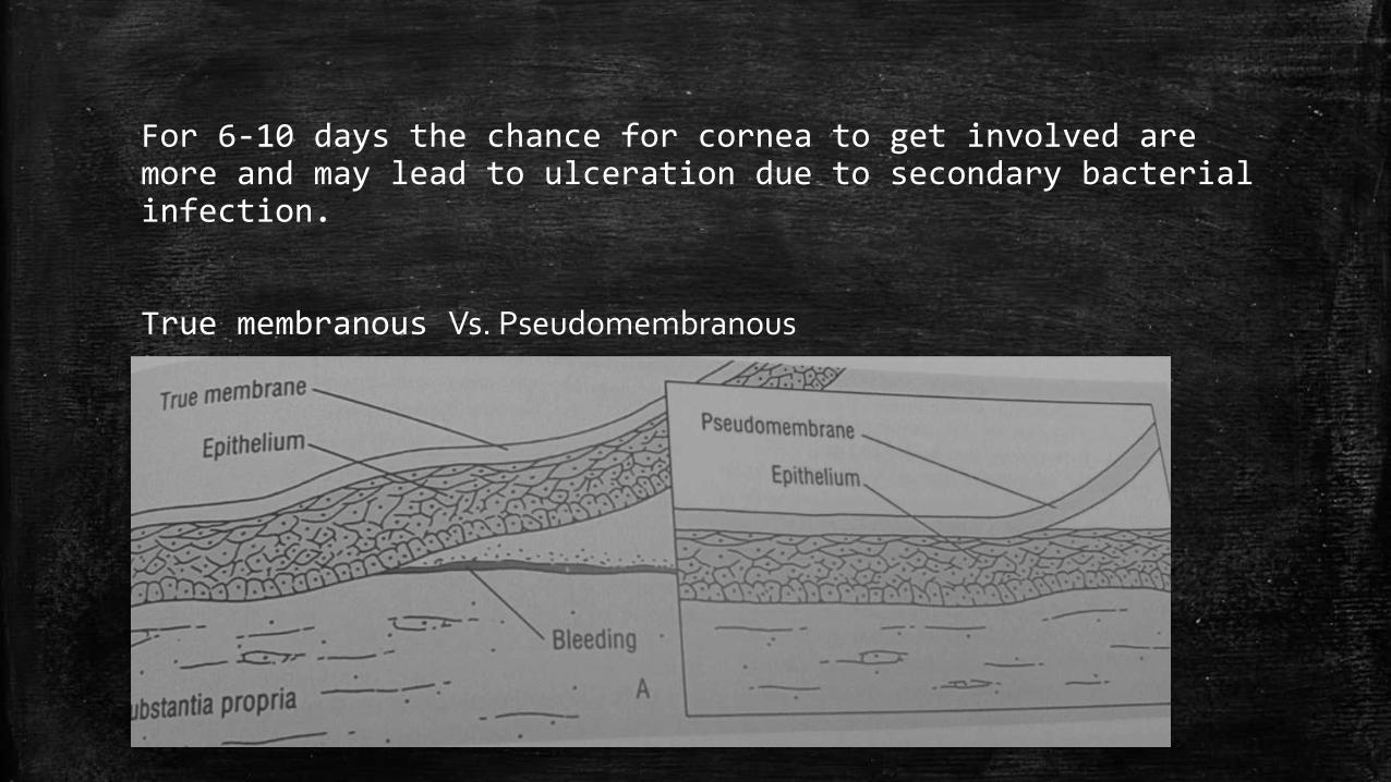
For 6-10 days the chance for cornea to get involved are more and may lead to ulceration due to secondary bacterial infection.
True membranous Vs. Pseudomembranous
Membrane is made-up of fibrinous exudate that may or may not be firmly adherent to the epithelium of conjunctiva.
If adhered tightly it is membranous and vice-versa.
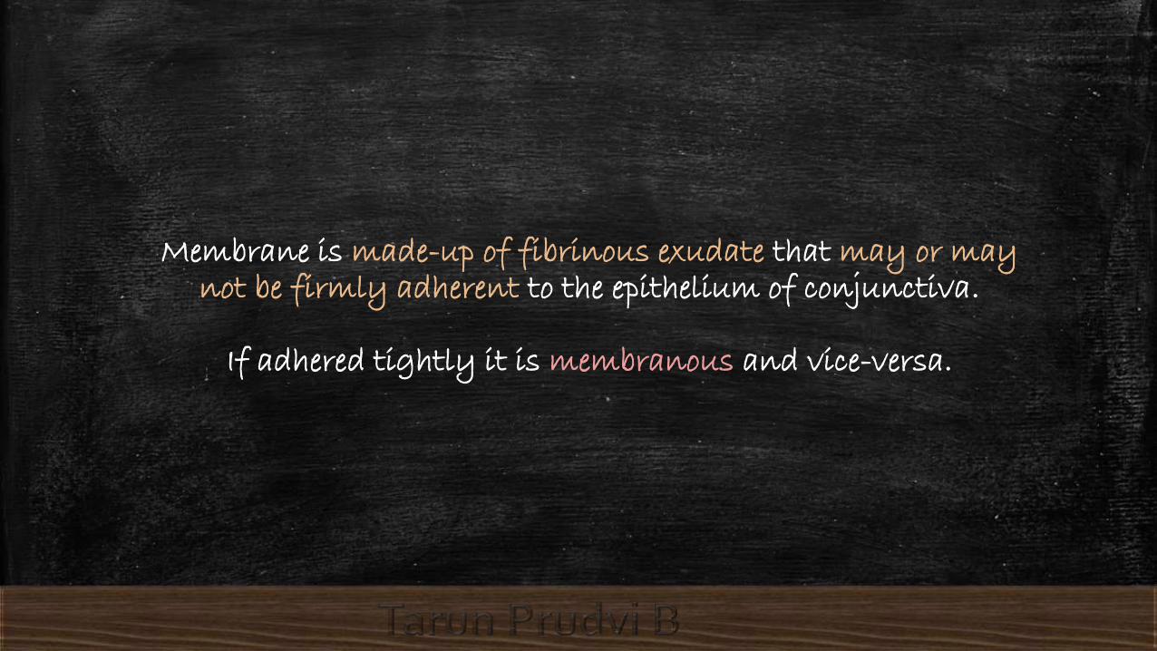
Pseudomembranous
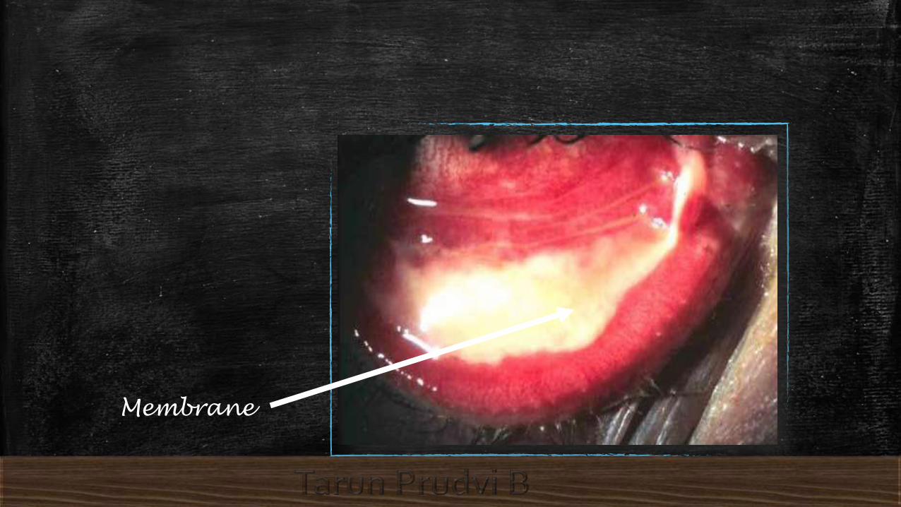
This is a picture of eye showing inflammatory conjunctivitis .
Conjunctiva shows congestion.
In the lower palpebral conjunctiva you see easily peelable membrane due to inflammation.
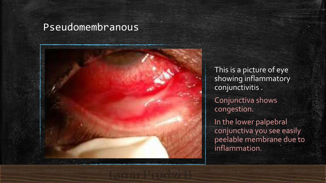
Membranous conjunctivitis
It is picture of eye with adenoviral keratoconjunctivitis
Note the presence of membrane in lower palpebral conjunctiva.
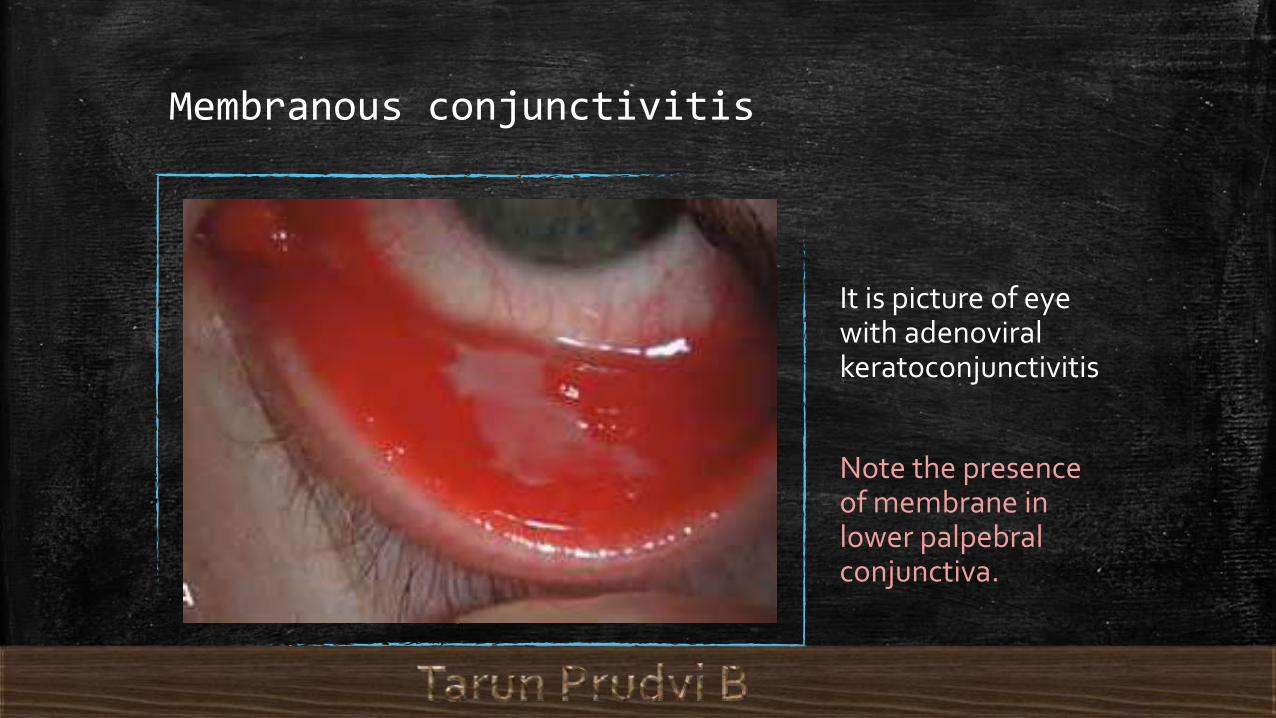
DifferentialLigneous conjunctivitis is a rare form of chronic conjunctivitischaracterized by recurrent, fibrin-rich pseudomembranous lesions of wood-like consistency that develop mainly on the underside of the eyelid (tarsal conjunctiva).It is generally a systemic disease which may involve the periodontal tissue, the upper and lower respiratory tract, kidneys, middle ear, and female genitalia.

Diagnosis:
Take a part of membrane and send for microbiological examination.
And start with antibiotics.

Treatment
In children who are not immunized every case is treated as diphtheritic infection unless films and cultures give negative evidence.
Administration of freshly made topical 10,000 units/ml drops from injectable solution. And systemic administration of penicillin along with antidiphtheritic serum (4000-10000 units repeated 12 hourly)
In other bacterial causes antibiotic drops are prescribed. If cornea involved the cyclopegics are given.

Complications:
If membrane is removed inadvertently it may precipitate symblepharon. So removal of membrane is not required.

Thank You
Recommended