University of Nebraska - LincolnDigitalCommons@University of Nebraska - Lincoln
Karl Reinhard Papers/Publications Natural Resources, School of
12-2013
On head lice and social interaction in archaicAndean coastal populationsBernardo ArriazaUniversidad de Tarapacá, Arica, Chile, [email protected]
Vivien StandenUniversidad de Tarapacá, Arica, Chile, [email protected]
Karl ReinhardUniversity of Nebraska-Lincoln, [email protected]
Aduto AraújoEscola Nacional de Saúde Pública, Fundação Oswaldo Cruz, Rio de Janeiro, [email protected]
Jörg HeukelbachFederal University of Ceará, Fortaleza, Brazil, [email protected]
See next page for additional authors
Follow this and additional works at: http://digitalcommons.unl.edu/natresreinhard
Part of the Disorders of Environmental Origin Commons, Environmental Public HealthCommons, International Public Health Commons, Medical Pathology Commons, OtherImmunology and Infectious Disease Commons, Parasitic Diseases Commons, and the ParasitologyCommons
This Article is brought to you for free and open access by the Natural Resources, School of at DigitalCommons@University of Nebraska - Lincoln. Ithas been accepted for inclusion in Karl Reinhard Papers/Publications by an authorized administrator of DigitalCommons@University of Nebraska -Lincoln.
Arriaza, Bernardo; Standen, Vivien; Reinhard, Karl; Araújo, Aduto; Heukelbach, Jörg; and Dittmar, Katharina, "On head lice andsocial interaction in archaic Andean coastal populations" (2013). Karl Reinhard Papers/Publications. Paper 1.http://digitalcommons.unl.edu/natresreinhard/1

AuthorsBernardo Arriaza, Vivien Standen, Karl Reinhard, Aduto Araújo, Jörg Heukelbach, and Katharina Dittmar
This article is available at DigitalCommons@University of Nebraska - Lincoln: http://digitalcommons.unl.edu/natresreinhard/1
1. Introduction
Head lice, Pediculus humanus capitis De Geer (1767), have been a part of mummy studies for decades. Zinsser (1935) presented the importance of lice in mummy studies in his classic publica-tion Rats, Lice and History (republished in 2007). Previously, Ewing (1924) had published the discovery of lice from Peruvian mum-mies. Both authors pointed out the comparative value of mummy louse studies in documenting the intra-population variation of lice as they adapted to different hair morphology in diverse human populations. Although Zinsser and Ewing highlighted the poten-tial of population-based studies, such potential went unexplored for decades. Recently, a series of case reports have been published, drawing on small, or single mummy samples (Araújo et al., 2000; Arriaza et al., 2012a, 2012b; Raoult et al., 2008; Rivera et al., 2008). Most studies were not quantitative, but were successful in painting a general picture of the arrival of head lice in the New World. Araújo et al. (2000) reported head lice from hair associated with an archaic human skeleton in northeastern Brazil. The find-ing was radiocarbon dated to more than 10,000 years ago indicat-ing that the introduction of lice into the New World probably oc-curred with the earliest migrants. Based on molecular analysis of
Chiribaya head lice (960 B.P. Peru) from two individuals, Raoult et al. (2008) showed that pre-conquest head lice populations likely had haplotype links to the Old World, pointing to ancestral mi-grations of host and parasite into the New World. Rivera et al. (2008) found louse nits/eggs on six of seven 4000-year-old mum-mies from Camarones, on the coast of northern Chile, however they did not report nit/egg density. This small sample hinted that high levels of head lice infestation were reached in archaic coastal Andean populations.
Levels of infestation of 44% were documented in mummies from Canyon de Chelly, Arizona, dating to 800–900 years ago (El-Najjar et al., 1998). In general, louse infestation was relatively rare in the Southwestern United States in prehistory. These case stud-ies have been important in documenting louse distribution through time and space.
The first ancient population-based study on head lice infesta-tion was performed by Reinhard and Buikstra (2003), who ana-lyzed 146 Chiribaya culture mummies from the Moquegua valley of southern Peru. They found that 92 of the mummies were suffi-ciently preserved for analysis. They then assessed the distribution of parasites in host populations, searching for a pattern of over-dis-persal—which means that a very small percentage of hosts harbor
Published in International Journal of Paleopathology 3:4 (December 2013), pp. 257–268; doi: 10.1016/j.ijpp.2013.10.001Copyright © 2013 Elsevier Inc. Used by permission.Submitted February 8, 2013; revised September 3, 2013; accepted October 7, 2013; published online November 8, 2013.
On head lice and social interaction in archaic Andean coastal populations
Bernardo Arriaza,1 Vivien Standen,2 Karl Reinhard,3
Adauto Araújo,4 Jörg Heukelbach,5 and Katharina Dittmar 6
1. Instituto de Alta Investigación, Universidad de Tarapacá, Arica, Chile2. Departamento de Antropología, Universidad de Tarapacá, Arica, Chile
3. School of Natural Resources, University of Nebraska–Lincoln, USA4. Escola Nacional de Saúde Pública, Fundação Oswaldo Cruz, Rio de Janeiro RJ 21041–210, Brazil
5. Department of Community Health, School of Medicine, Federal University of Ceará, Fortaleza, Brazil6. Department of Biological Sciences, University at Buffalo, NY, USA
Corresponding author — B. Arriaza, tel 56 58 2255371; email [email protected]
AbstractArchaic mummies from northern Chile were examined for the presence of Pediculus humanus capitis. The excellent preser-vation of mummies and louse nits/eggs permitted a study of the degree of head lice infestation. We studied 63 Chinchorro mummies (ca. 5000–3000 years B.P.) from the Arica-Camarones coast. An area of 2 cm × 2 cm on each mummy’s head was systematically inspected for louse nits/eggs. Hairs with nits/eggs and lice were collected and analyzed using optic and scan-ning electronic microscopy. About 79% (50/63) of the mummies resulted positive for pediculosis, with an average of 2.1 nits/eggs/cm2 per positive individual. Microscopic analyses revealed the micromorphology of all developmental stages, includ-ing eggs/nits, nymphal instars and adults. Chinchorro people lived in small huts increasing the transmission of ectoparasites. Considering that head lice thrive in crowded conditions, their prevalence could be used as an bioindicator to assess and de-bate cultural behavior (e.g., degree of crowdedness and sedentism) and to study paleoepidemiology in prehistoric populations.
Keywords: Mummies, Pediculosis, Bioarchaeology, Atacama Desert
257
digitalcommons.unl.edu
258 A r r i A z A e t A l . i n I n t e r n at I o n a l J o u r n a l o f P a l e o Pat h o l o g y 3 (2013)
the majority of parasites. In parasitological terms, this phenome-non is best described by a negative binomial distribution (Ander-son, 1993), or “wormy hosts”, in the case of intestinal parasites, when few hosts carry a great number of parasites who contrib-ute to continuing transmission dynamics in endemic communi-ties (Croll and Ghadirian, 1981). This phenomenon has been ob-served with other ectoparasitic diseases such as tungiasis, where a small number of individuals carried most of the parasites in a community (Heukelbach et al., 2007). In order to validate the pa-leoepidemiological value of parasite data, this characteristic distri-bution had to be demonstrated with archaeological remains. Re-inhard and Buikstra (2003) were able to quantify infestations on an individual basis by calculating the maximum number of eggs and nits cemented to hair shafts on the scalps of the 92 Chiribaya mummies. The infestation of nits and eggs on Chiribaya hosts re-flected the negative binomial distribution. This supports the statis-tical value of louse parasitological data when large numbers of hu-man remains can be evaluated.
Following this population-based approach, we have studied sev-eral hundred mummies from all cultural phases of the Arica region in northern Chile. In this paper, we focus on the presence of Pedic-ulus humanus capitis (P. humanus capitis) in Chinchorro populations (5000–3000 years B.P.; n = 63 individuals) that inhabited the arid coastal region of the Atacama Desert. The Chinchorro were fish-ers and hunter-gatherers who lived along the dry but fertile coast at the fringes of the Atacama Desert in northern Chile and south-ern Peru (Arriaza, 1995; Aufderheide et al., 1993; Santoro et al., 2012). They are known for their elaborated mortuary practices, including artificial mummification (Arriaza, 1995; Arriaza et al., 2012c; Marquet et al., 2012; Rivera, 1991; Standen, 1997). The Chinchorro were expert morticians who transformed the corpses of the dead into polychromous preserved bodies (mummies). They used various techniques and mummification styles that changed through time to preserve individuals of both sexes and all ages, in-cluding fetuses. Even though there were regional and chronolog-ical variations, two types of artificially prepared mummies stand out: the black and the red style (Arriaza, 1995). The black mum-mies were the earliest (ca. 7000–5000 years B.P.) and are basically clay modeled or reconstructed bodies. They have an inner struc-ture of bones, sticks, clay and reeds. Clay masks and short black wigs made of human hair adorn the heads. Externally, the bod-ies are completely painted black, including the face, using manga-nese pigments. The red mummies (ca. 5000–4000 years B.P.), in contrast, have incisions for organ removal, stuffing of cavities and externally are fully painted with red ocher, except the face mask which is painted with manganese. These red mummies have long wigs of black human hair. In addition to these types of mummies, there are also Chinchorro bodies with natural mummification, es-pecially after the red style period. Independent of mummification procedures, most bodies were wrapped in reed matts and buried ly-ing on their back with a few grave goods (fishhooks, harpoons and net bags) in the sands of the Atacama Desert. In general, mummi-fication techniques faced cultural changes, but the maritime sub-sistence and technology for fishing and gathering remained the same. Also, later Chinchorro populations (ca. 3500 years B.P.) no longer practiced complex artificial mummification, annular skull deformation increased, heads were adorned with colored threads (headbands), and fine basket weaving and horticultural products appeared (Rivera, 1991; Standen, 1997).
Scholars have studied many aspects of Chinchorro ways of life and endoparasitism, but ectoparasite studies and their cultural rel-evance have been minimally addressed so far. Rivera et al. (2008), at the Camarones-15 site, found 98 operculated Pediculus capitis eggs and 265 non-operculated eggs. Arriaza et al. (2008, 2012a, 2012b) also reported three positive cases of pediculosis in ten pre-
Columbian mummies of different periods from Arica and Iquique (northern Chile) and a heavily infested late agropastoral Chiribaya mummy (ca. 1280–960 years B.P.). Likewise, Reinhard and Buiks-tra (2003) reported pediculosis in three Chiribayan sites from Peru, with prevalence ranging from 20% to 70%.
The general archaeological evidence posits that Chinchorro pop-ulations were composed of small bands of hunter-gatherers and col-lectors who lived in semi-permanent camps along the coast (Núñez, 1983; Muñoz and Chacama, 1982; Rivera, 1991). Based on this in-formation, it is thought that the Chinchorros were highly mobile groups. Using pediculosis as a bioindicator, we provide evidence in this paper that the Chinchorro daily life was more sedentary and that social bonding was likely strong and enduring. Yet, there are still several questions regarding archaic lice infestations: Why is Ri-vera’s reported frequency so high for this Chinchorro site? Is this value unusual, or did all Chinchorro sites have similar infestation rates? What is the social significance of pediculosis in these early populations? To answer these questions we systematically expanded the sample size and chrono-geographic framework to describe the extent that archaic coastal populations were affected by pediculosis, in order to discuss its bioarchaeological significance.
2. Materials and methods
The samples studied came from eleven coastal archaeological sites in the city of Arica in northern Chile, ranging from one to twenty-two individuals per site. In total we studied 63 mummies, 41 natu-rally mummified and 22 artificially prepared bodies. All are housed at the Museum of Archaeology of the Universidad de Tarapacá, Arica, Chile. The latter included the addition of a hair wig as part of the artificial mummification procedures (Arriaza, 1995; Stan-den, 1997). In artificially prepared bodies of newborns and infants, clearly the wigs were not made from their own hair, but probably from the hair of older relatives (Arriaza, 1995; Standen, 1997). Two broad, main age categories were considered: adults and sub-adults. The latter includes ages around 15 and younger. Thus, the demographic profile of the total sample can be subdivided into 42 adults and 21 subadults (26 males, 25 females and 12 individuals whose sex it was not possible to determine). A summary of the study sample demographs per site is presented in Table 1.
2.1. Macroscopic analysis
At the Museum of Archaeology, all mummy heads were exam-ined for the presence of head lice (nits or empty egg-shells, em-bryonated eggs and lice). If heads were detached by previous au-topsies, they were carefully deposited on a 60 cm × 60 cm sheet of acid free paper and inspected visually with the aid of a 10× magni-fication glass. Complete bodies were studied in their storage trays.
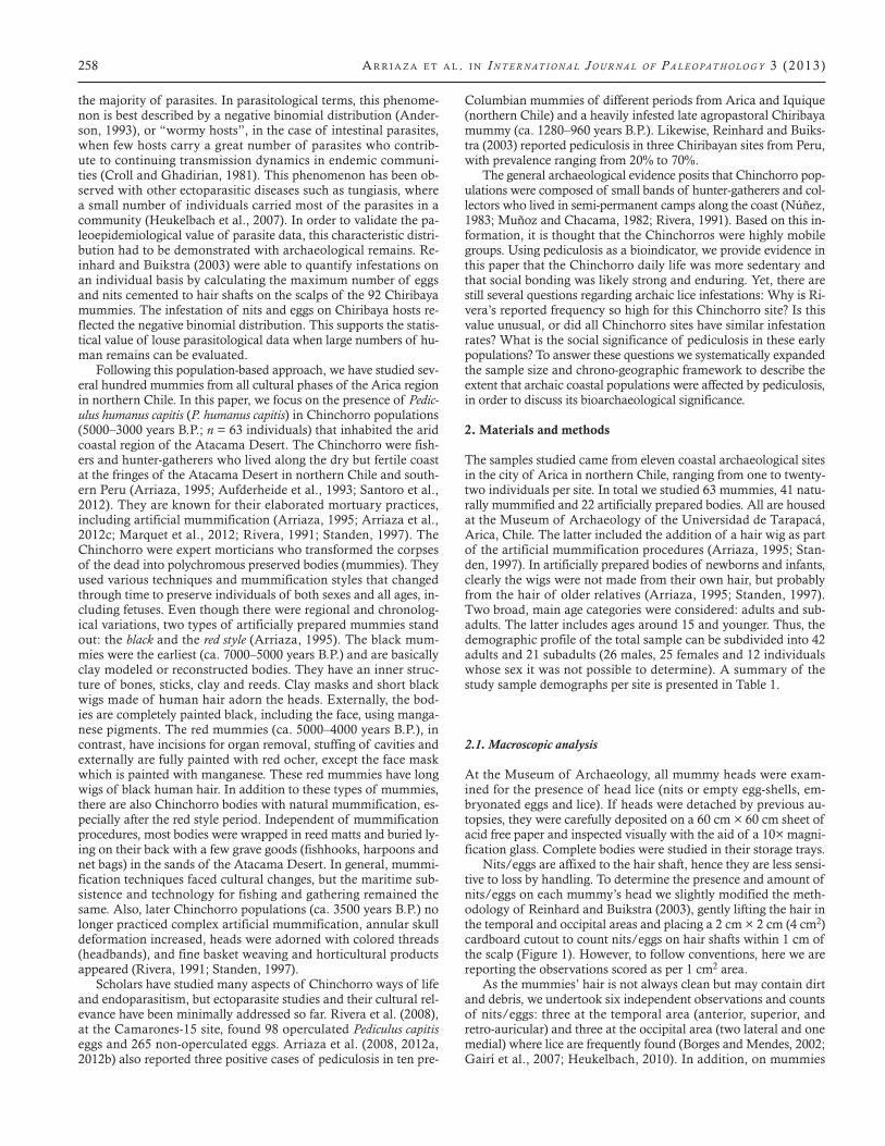
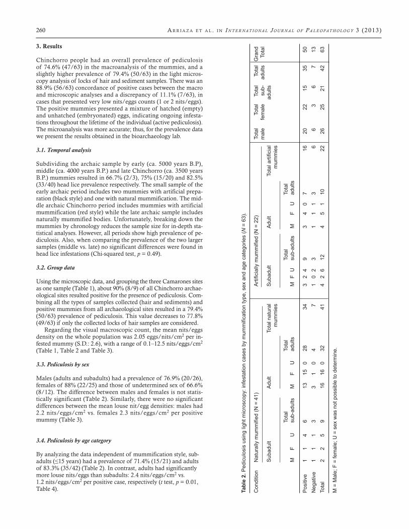
Nits/eggs are affixed to the hair shaft, hence they are less sensi-tive to loss by handling. To determine the presence and amount of nits/eggs on each mummy’s head we slightly modified the meth-odology of Reinhard and Buikstra (2003), gently lifting the hair in the temporal and occipital areas and placing a 2 cm × 2 cm (4 cm2) cardboard cutout to count nits/eggs on hair shafts within 1 cm of the scalp (Figure 1). However, to follow conventions, here we are reporting the observations scored as per 1 cm2 area.
As the mummies’ hair is not always clean but may contain dirt and debris, we undertook six independent observations and counts of nits/eggs: three at the temporal area (anterior, superior, and retro-auricular) and three at the occipital area (two lateral and one medial) where lice are frequently found (Borges and Mendes, 2002; Gairí et al., 2007; Heukelbach, 2010). In addition, on mummies
H e A d l i c e A n d s o c i A l i n t e r A c t i o n i n A rc H A i c A n d e A n c oA s tA l p o p u l At i o n s 259
with detached hair (due to previous autopsies), three counts were undertaken in similar areas, whenever possible, and recorded sep-arately. Hair from archaeological settings (including mummies) is often brittle and crumbles easily as the head is inspected; therefore, we collected all sediments (hair fragments, soil debris and ectopar-asitic developmental stages) that fell on the acid-free paper during examination for an in-depth analysis. We also measured the length of the hair per individual (from the scalp to maximum length). We considered a mummy to be positive for pediculosis if any stage of
the ectoparasite was found, even if only hatched eggs or cementing residues were present. In contrast, we considered a mummy to be symptomatic for active pediculosis if nymphs, adults, or unhatched eggs were found on an individual.
2.2. Microscopic analysis
We cut and collected small locks of hair where nits/eggs were observed for further microscopy studies at the Bioarchaeology Laboratory of the Instituto de Alta Investigación of the Univer-sidad de Tarapacá, Arica. We recorded the number of nits/eggs, louse nymphal instars and adult lice found in the hair samples collected. All specimens were stored in acid-free tissue and sterile tubes. We first used stereomicroscopy (Olympus SZX-7) to sep-arate all hair and parasites from other debris. Then all samples were observed using another microscope with a higher magnifi-cation (Olympus BX-41) to verify sex and to determine whether unhatched eggs contained embryos. Sex of lice was determined when possible according to morphological characteristics (Heu-kelbach, 2010; Nuttall, 1917).
To complement this study and yet preserve precious material for subsequent molecular analyses, a subset of selected samples (9 lice and 37 nits) were further analyzed using environmental scan-ning electron microscopy (Zeiss, EVO-LS 10) to pursue ultrastruc-tural analysis of the micromorphology of nits/eggs and adults of P. humanus capitis (Arriaza et al., 2012a). The samples were mounted on aluminum stubs without gold/palladium alloy coat-ing and viewed under variable pressure mode (VP). The chamber pressure was 150 Pascals, the working distance 4–8 mm and the acceleration voltage 15 kV. Undesired charging of dried, uncoated specimens was controlled by careful choice of incident beam en-ergy and nitrogen gas levels.
Figure 1. Illustration of nit/egg counting technique with a 2 cm × 2 cm square on a mummy head.
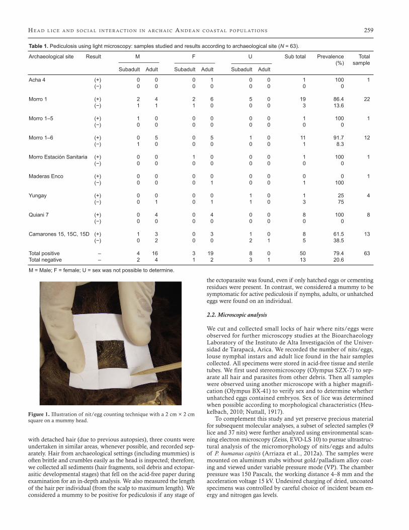
Table 1. Pediculosis using light microscopy: samples studied and results according to archaeological site (N = 63).
Archaeological site Result M F U Sub total Prevalence Total (%) sample Subadult Adult Subadult Adult Subadult Adult
Acha 4 (+) 0 0 0 1 0 0 1 100 1 (−) 0 0 0 0 0 0 0 0
Morro 1 (+) 2 4 2 6 5 0 19 86.4 22 (−) 1 1 1 0 0 0 3 13.6
Morro 1–5 (+) 1 0 0 0 0 0 1 100 1 (−) 0 0 0 0 0 0 0 0
Morro 1–6 (+) 0 5 0 5 1 0 11 91.7 12 (−) 1 0 0 0 0 0 1 8.3
Morro Estación Sanitaria (+) 0 0 1 0 0 0 1 100 1 (−) 0 0 0 0 0 0 0 0
Maderas Enco (+) 0 0 0 0 0 0 0 0 1 (−) 0 0 0 1 0 0 1 100
Yungay (+) 0 0 0 0 1 0 1 25 4 (−) 0 1 0 1 1 0 3 75
Quiani 7 (+) 0 4 0 4 0 0 8 100 8 (−) 0 0 0 0 0 0 0 0
Camarones 15, 15C, 15D (+) 1 3 0 3 1 0 8 61.5 13 (−) 0 2 0 0 2 1 5 38.5
Total positive – 4 16 3 19 8 0 50 79.4 63Total negative – 2 4 1 2 3 1 13 20.6
M = Male; F = female; U = sex was not possible to determine.
260 A r r i A z A e t A l . i n I n t e r n at I o n a l J o u r n a l o f P a l e o Pat h o l o g y 3 (2013)
3. Results
Chinchorro people had an overall prevalence of pediculosis of 74.6% (47/63) in the macroanalysis of the mummies, and a slightly higher prevalence of 79.4% (50/63) in the light micros-copy analysis of locks of hair and sediment samples. There was an 88.9% (56/63) concordance of positive cases between the macro and microscopic analyses and a discrepancy of 11.1% (7/63), in cases that presented very low nits/eggs counts (1 or 2 nits/eggs). The positive mummies presented a mixture of hatched (empty) and unhatched (embryonated) eggs, indicating ongoing infesta-tions throughout the lifetime of the individual (active pediculosis). The microanalysis was more accurate; thus, for the prevalence data we present the results obtained in the bioarchaeology lab.
3.1. Temporal analysis
Subdividing the archaic sample by early (ca. 5000 years B.P), middle (ca. 4000 years B.P.) and late Chinchorro (ca. 3500 years B.P.) mummies resulted in 66.7% (2/3), 75% (15/20) and 82.5% (33/40) head lice prevalence respectively. The small sample of the early archaic period includes two mummies with artificial prepa-ration (black style) and one with natural mummification. The mid-dle archaic Chinchorro period includes mummies with artificial mummification (red style) while the late archaic sample includes naturally mummified bodies. Unfortunately, breaking down the mummies by chronology reduces the sample size for in-depth sta-tistical analyses. However, all periods show high prevalence of pe-diculosis. Also, when comparing the prevalence of the two larger samples (middle vs. late) no significant differences were found in head lice infestations (Chi-squared test, p = 0.49).
3.2. Group data
Using the microscopic data, and grouping the three Camarones sites as one sample (Table 1), about 90% (8/9) of all Chinchorro archae-ological sites resulted positive for the presence of pediculosis. Com-bining all the types of samples collected (hair and sediments) and positive mummies from all archaeological sites resulted in a 79.4% (50/63) prevalence of pediculosis. This value decreases to 77.8% (49/63) if only the collected locks of hair samples are considered.
Regarding the visual macroscopic count, the mean nits/eggs density on the whole population was 2.05 eggs/nits/cm2 per in-fested mummy (S.D.: 2.6), with a range of 0.1–12.5 nits/eggs/cm2 (Table 1, Table 2 and Table 3).
3.3. Pediculosis by sex
Males (adults and subadults) had a prevalence of 76.9% (20/26), females of 88% (22/25) and those of undetermined sex of 66.6% (8/12). The difference between males and females is not statis-tically significant (Table 2). Similarly, there were no significant differences between the mean louse nit/egg densities: males had 2.2 nits/eggs/cm2 vs. females 2.3 nits/eggs/cm2 per positive mummy (Table 3).
3.4. Pediculosis by age category
By analyzing the data independent of mummification style, sub-adults (≤15 years) had a prevalence of 71.4% (15/21) and adults of 83.3% (35/42) (Table 2). In contrast, adults had significantly more louse nits/eggs than subadults: 2.4 nits/eggs/cm2 vs. 1.2 nits/eggs/cm2 per positive case, respectively (t test, p = 0.01, Table 4). Ta
ble
2.Pediculosisusinglightm
icroscopy:infestationcasesbymum
mificationtype,sexandagecategories(N
= 6
3).
Condition
Naturallymum
mified(N
=41)
Artificiallymum
mified(N
=22)
Total
Total
Total
TotalGrand
mal
e fe
mal
e su
b-
adul
ts
Tota
l
Subadult
Adult
Totalnatural
Subadult
Adult
Totalartificial
adults
m
umm
ies
mum
mie
s
Tota
l
Tota
l
To
tal
To
tal
M
F
U
sub-
adul
ts
M
F U
ad
ults
M
F U
su
b-ad
ults
M
F
U
adul
ts
Pos
itive
1
1 4
6 13
15
0
28
34
3 2
4 9
3 4
0 7
16
20
22
15
35
50N
egat
ive
1 1
1 3
3 1
0 4
7 1
0 2
3 1
1 1
3 6
6 3
6 7
13To
tal
2 2
5 9
16
16
0 32
41
4
2 6
12
4 5
1 10
22
26
25
21
42
63
M =
Mal
e; F
= fe
mal
e; U
= s
ex w
as n
ot p
ossi
ble
to d
eter
min
e.
H e A d l i c e A n d s o c i A l i n t e r A c t i o n i n A rc H A i c A n d e A n c oA s tA l p o p u l At i o n s 261
3.5. Pediculosis by mummification type
Separating the positive samples by natural vs. artificial mummifi-cation resulted in similar prevalences of 82.9% (34/41) and 72.7% (16/22) respectively (Table 2). However, the mean nit/egg den-sity was significantly higher in natural mummies compared to ar-tificially prepared bodies: 2.6 nits/eggs/cm2 vs. 1.0 nits/eggs/cm2 per infested mummy (t test, p = 0.004, Table 6).
Naturally mummified subadults and adults had prevalences of pediculosis of 66.7% (6/9) and 87.5% (28/32) respectively (Table 2). However, this difference was not statistically significant. Sub-adults had a significantly lower mean of 1.6 nits/eggs/cm2 per positive case, as compared to the adult mean of 2.9 nits/eggs/cm2 per positive case (t test, p = 0.04, Table 5). Artificially mummified subadults had 75% (9/12) head lice prevalence and adults 70% (7/10), with a similar mean nits/eggs/cm2 density: 0.8 vs. 1.1 re-spectively (Table 5).
After controlling for sex, naturally mummified bodies of both sexes had similar mean values around 2.8 nits/eggs/cm2 per pos-itive mummy. Artificially mummified females had nearly double the nit/egg density than artificially prepared males: 1.5 nits/eggs/cm2 vs. 0.7 nits/eggs/cm2 per positive case, respectively (t test, p = 0.016, Table 6). Naturally mummified males and females had similar prevalence of pediculosis [77.7% (14/18) vs. 88% (16/18)]. Likewise artificially mummified bodies of both sexes showed simi-lar prevalence values [75% (6/8) vs. 85.7% (6/7)].
4. Pediculosis by lice only
Microscopically, of the 63 mummies analyzed 21 of them pre-sented lice (33.3%) in various regions of the head (e.g. temporal and occipital). Fifty microscopically examined mummies were positive for pediculosis (nits/eggs or lice) compared to 47 posi-tive cases macroscopically analyzed. From these 21 positive Chin-chorro mummies we collected 232 head lice (94 adults and 138 nymphs). Of the 232 lice, 228 were found on 20 naturally mummi-fied bodies and one of these mummies (Quiani 7 T9, adult male) accounted for nearly half of the total (110 lice). On these positive natural mummies, 18 were adult individuals (221 lice) and 2 were
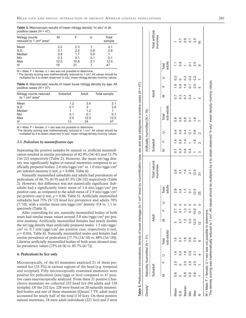
Table 3. Macroscopic results of mean nit/egg density “in situ” in all positive cases (N = 47).
Nit/egg counts M F U Total reduced to 1 cm2 area* sample
Mean 2.2 2.3 1 2.1S.D. 3.1 2.5 0.8 2.6Median 0.8 1.7 0.9 1Min 0.3 0.1 0.1 0.1Max 12.5 10.8 2.1 12.5N 19 21 7 47
M = Male; F = female; U = sex was not possible to determine.* The density scoring was mathematically reduced to 1 cm2. All values should be
multiplied by 4 to obtain observed “in situ” mean nit/egg density mummy values.
Table 4. Macroscopic results of mean louse nit/egg density by age. All positive cases (N = 47).
Nit/egg counts reduced Subadult Adult Total sample to 1 cm2 area*
Mean 1.2 2.4 2.1S.D. 0.7 3 2.6Median 1 1.2 1Min 0.1 0.1 0.1Max 2.5 12.5 12.5N 13 34 47
M = Male; F = female; U = sex was not possible to determine.*The density scoring was mathematically reduced to 1 cm2. All values should be
multiplied by 4 to obtain observed “in situ” mean nit/egg density mummy values.
Tabl
e 5.
Ped
icul
osis
mic
roan
alys
is: d
ensi
ty o
f lou
se n
its/e
ggs
(in 1
cm
2 area)bymum
mificationtype,sexandagecategoriesinpositivecases(N
= 4
7).
Den
sity
* Naturallymum
mified(N
=31)
Artificiallymum
mified(N
=16)
Subadult
Adult
Totalnatural
Subadult
Adult
Totalartificial
m
umm
ies
mum
mie
s
M
F
U
Tota
l M
F
U
Tota
l
M
F U
To
tal
M
F U
To
tal
sub-
adul
ts
ad
ults
su
b-ad
ults
adul
ts
Mea
n 0.
8 2.
5 1.
6 1.
6 3
2.7
0 2.
9 2.
6 0.
7 1.
4 0.
4 0.
8 0.
7 1.
6 0.
2 1.
1 1
S.D
. 0
0 0.
5 0.
7 3.
7 3.
1 0
3.4
3.1
0.3
0.5
0.5
0.5
0.3
0.9
0 0.
9 0.
7M
edia
n 0.
8 2.
5 1.
6 1.
6 1.
2 1.
3 0
1.3
1.4
0.8
1.4
0.4
0.8
0.7
1.8
0.2
1 0.
9M
in
0.8
2.5
0.9
0.8
0.3
0.1
0 0.
1 0.
1 0.
4 1
0.1
0.1
0.4
0.1
0.2
0.1
0.1
Max
0.
8 2.
5 2.
1 2.
5 12
.5
10.6
0
12.5
12
.5
1 1.
8 0.
8 1.
8 1
2.5
0.2
2.5
2.5
N
1 1
4 6
12
13
0 25
31
3
2 2
7 3
5 1
9 16
M =
Mal
e; F
= fe
mal
e; U
= s
ex w
as n
ot p
ossi
ble
to d
eter
min
e.* T
he d
ensi
ty s
corin
g w
as m
athe
mat
ical
ly re
duce
d to
1 c
m2 .
All
valu
es s
houl
d be
mul
tiplie
d by
4 to
obt
ain
obse
rved
“in
situ
” mea
n de
nsity
mum
my
valu
es.
262 A r r i A z A e t A l . i n I n t e r n at I o n a l J o u r n a l o f P a l e o Pat h o l o g y 3 (2013)
subadult individuals (7 lice). In contrast, only 4 lice were found in an artificially mummified child (Camarones 15D T16C1).The adult head lice subsample had the following sex distribution: 34 female, 56 male and 4 indeterminate specimens. Considering the 21 positive mummies (or with head lice), each Chinchorro individual hosted on average 11 lice (S.D.= 23.8; Median = 4; Min = 1; Max = 110).
5. Discussion
Nearly all Chinchorro sites investigated presented evidence of P. humanus capitis. Prevalence was high and endemic. On average, four out of five Chinchorro individuals presented louse nits/eggs and adult specimens. Density, however, was variable with a mean of about 2.05 nits/eggs/cm2nits/eggs/cm2 per individual. The presence of a living louse in the hair is considered an active infes-tation (Heukelbach, 2010:48). While this is not possible to score in the mummies, unhatched eggs are good proxies for potential ac-tive infestations.
Although the average number of nits/eggs/cm2 is relatively low, this is not the case if we consider the complete head surface. Using a rough estimate that an adult individual has an area of 500–600 cm2 of scalp with hair, and each Chinchorro had on aver-age 2.05 nits/eggs/cm2, then each Chinchorro had the potential to host at least 1025–1230 nits/eggs. Also, considering that we found 26.6% (119/448) embryonated eggs and 73.4% (329/448) hatched eggs (Table 7), then each Chinchorro had between 748 and 898 eggs that finished the life cycle of the ectoparasite and 267–320 eggs that were likely to convert to infestation.
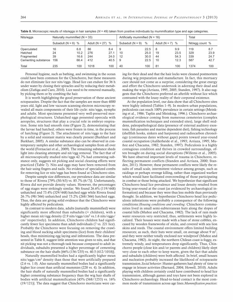
Also it is interesting to note that despite the low average density of 2.05 nits/eggs/cm2, unhatched eggs or adult lice were present in all positive cases, indicating an active infestation. This contrasts with current studies by Williams et al. (2001) on recent popula-tions, which found that only seven percent of children with fewer than 5 nits/eggs per 0.6 cm actually developed an active infes-tation. The calculated average number of 11 lice per mummy is likely an underestimation, because lice are prone to falling off the head while handling the mummy. Still, some individuals were car-rying extremely high parasite loads, following the negative bino-mial distribution (over-dispersal) typical for parasite epidemiology in endemic communities (Figure 4), and confirming previous re-sults on mummy headlice (Reinhard and Buikstra, 2003). Three mummies presented 7.5 nits/eggs/cm2, accounting for 32.2% of all the nits/eggs observed in the hair (Figure 5).
Even though cultural attitudes toward head lice vary, symptoms depend on the duration of the infestation and the number of lice and unhatched eggs present on the scalp (Heukelbach, 2010:44). Head lice feed several times per day on their host, which may pro-voke mood changes and sleeping disturbances. Repeated expo-sure to antigenic compounds in louse saliva can cause sensitiza-tion, with intense itching and subsequent scratching. Although this is known to facilitate secondary bacterial infection (Feldmeier and Heukelbach, 2009), no obvious evidence of scalp infectious
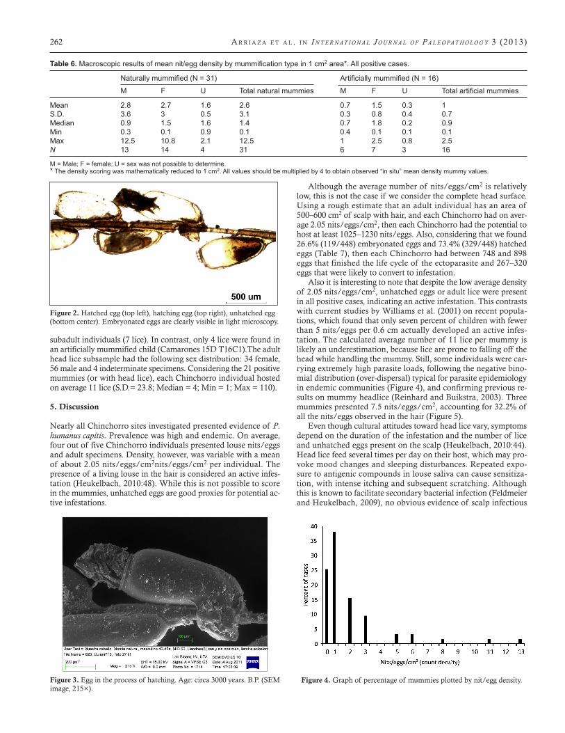
Figure 2. Hatched egg (top left), hatching egg (top right), unhatched egg (bottom center). Embryonated eggs are clearly visible in light microscopy.
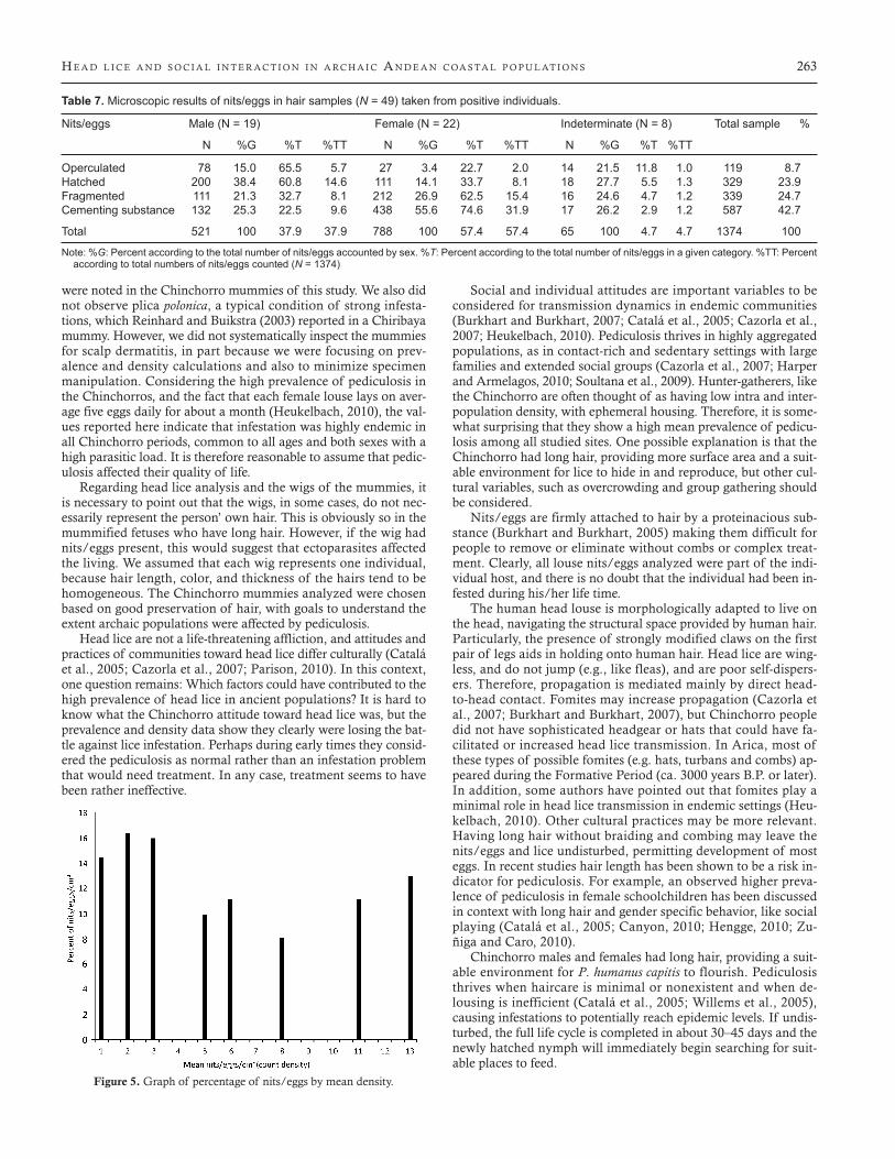
Figure 3. Egg in the process of hatching. Age: circa 3000 years. B.P. (SEM image, 215×).
Figure 4. Graph of percentage of mummies plotted by nit/egg density.
Table 6. Macroscopicresultsofmeannit/eggdensitybymummificationtypein1cm2 area*. All positive cases.
Naturallymummified(N=31) Artificiallymummified(N=16)
M F U Totalnaturalmummies M F U Totalartificialmummies
Mean 2.8 2.7 1.6 2.6 0.7 1.5 0.3 1S.D. 3.6 3 0.5 3.1 0.3 0.8 0.4 0.7Median 0.9 1.5 1.6 1.4 0.7 1.8 0.2 0.9Min 0.3 0.1 0.9 0.1 0.4 0.1 0.1 0.1Max 12.5 10.8 2.1 12.5 1 2.5 0.8 2.5N 13 14 4 31 6 7 3 16
M = Male; F = female; U = sex was not possible to determine.* The density scoring was mathematically reduced to 1 cm2. All values should be multiplied by 4 to obtain observed “in situ” mean density mummy values.
H e A d l i c e A n d s o c i A l i n t e r A c t i o n i n A rc H A i c A n d e A n c oA s tA l p o p u l At i o n s 263
were noted in the Chinchorro mummies of this study. We also did not observe plica polonica, a typical condition of strong infesta-tions, which Reinhard and Buikstra (2003) reported in a Chiribaya mummy. However, we did not systematically inspect the mummies for scalp dermatitis, in part because we were focusing on prev-alence and density calculations and also to minimize specimen manipulation. Considering the high prevalence of pediculosis in the Chinchorros, and the fact that each female louse lays on aver-age five eggs daily for about a month (Heukelbach, 2010), the val-ues reported here indicate that infestation was highly endemic in all Chinchorro periods, common to all ages and both sexes with a high parasitic load. It is therefore reasonable to assume that pedic-ulosis affected their quality of life.
Regarding head lice analysis and the wigs of the mummies, it is necessary to point out that the wigs, in some cases, do not nec-essarily represent the person’ own hair. This is obviously so in the mummified fetuses who have long hair. However, if the wig had nits/eggs present, this would suggest that ectoparasites affected the living. We assumed that each wig represents one individual, because hair length, color, and thickness of the hairs tend to be homogeneous. The Chinchorro mummies analyzed were chosen based on good preservation of hair, with goals to understand the extent archaic populations were affected by pediculosis.
Head lice are not a life-threatening affliction, and attitudes and practices of communities toward head lice differ culturally (Catalá et al., 2005; Cazorla et al., 2007; Parison, 2010). In this context, one question remains: Which factors could have contributed to the high prevalence of head lice in ancient populations? It is hard to know what the Chinchorro attitude toward head lice was, but the prevalence and density data show they clearly were losing the bat-tle against lice infestation. Perhaps during early times they consid-ered the pediculosis as normal rather than an infestation problem that would need treatment. In any case, treatment seems to have been rather ineffective.
Social and individual attitudes are important variables to be considered for transmission dynamics in endemic communities (Burkhart and Burkhart, 2007; Catalá et al., 2005; Cazorla et al., 2007; Heukelbach, 2010). Pediculosis thrives in highly aggregated populations, as in contact-rich and sedentary settings with large families and extended social groups (Cazorla et al., 2007; Harper and Armelagos, 2010; Soultana et al., 2009). Hunter-gatherers, like the Chinchorro are often thought of as having low intra and inter-population density, with ephemeral housing. Therefore, it is some-what surprising that they show a high mean prevalence of pedicu-losis among all studied sites. One possible explanation is that the Chinchorro had long hair, providing more surface area and a suit-able environment for lice to hide in and reproduce, but other cul-tural variables, such as overcrowding and group gathering should be considered.
Nits/eggs are firmly attached to hair by a proteinacious sub-stance (Burkhart and Burkhart, 2005) making them difficult for people to remove or eliminate without combs or complex treat-ment. Clearly, all louse nits/eggs analyzed were part of the indi-vidual host, and there is no doubt that the individual had been in-fested during his/her life time.
The human head louse is morphologically adapted to live on the head, navigating the structural space provided by human hair. Particularly, the presence of strongly modified claws on the first pair of legs aids in holding onto human hair. Head lice are wing-less, and do not jump (e.g., like fleas), and are poor self-dispers-ers. Therefore, propagation is mediated mainly by direct head-to-head contact. Fomites may increase propagation (Cazorla et al., 2007; Burkhart and Burkhart, 2007), but Chinchorro people did not have sophisticated headgear or hats that could have fa-cilitated or increased head lice transmission. In Arica, most of these types of possible fomites (e.g. hats, turbans and combs) ap-peared during the Formative Period (ca. 3000 years B.P. or later). In addition, some authors have pointed out that fomites play a minimal role in head lice transmission in endemic settings (Heu-kelbach, 2010). Other cultural practices may be more relevant. Having long hair without braiding and combing may leave the nits/eggs and lice undisturbed, permitting development of most eggs. In recent studies hair length has been shown to be a risk in-dicator for pediculosis. For example, an observed higher preva-lence of pediculosis in female schoolchildren has been discussed in context with long hair and gender specific behavior, like social playing (Catalá et al., 2005; Canyon, 2010; Hengge, 2010; Zu-ñiga and Caro, 2010).
Chinchorro males and females had long hair, providing a suit-able environment for P. humanus capitis to flourish. Pediculosis thrives when haircare is minimal or nonexistent and when de-lousing is inefficient (Catalá et al., 2005; Willems et al., 2005), causing infestations to potentially reach epidemic levels. If undis-turbed, the full life cycle is completed in about 30–45 days and the newly hatched nymph will immediately begin searching for suit-able places to feed.
Figure 5. Graph of percentage of nits/eggs by mean density.
Table 7. Microscopic results of nits/eggs in hair samples (N = 49) taken from positive individuals.
Nits/eggs Male (N = 19) Female (N = 22) Indeterminate (N = 8) Total sample %
N %G %T %TT N %G %T %TT N %G %T %TT
Operculated 78 15.0 65.5 5.7 27 3.4 22.7 2.0 14 21.5 11.8 1.0 119 8.7Hatched 200 38.4 60.8 14.6 111 14.1 33.7 8.1 18 27.7 5.5 1.3 329 23.9Fragmented 111 21.3 32.7 8.1 212 26.9 62.5 15.4 16 24.6 4.7 1.2 339 24.7Cementing substance 132 25.3 22.5 9.6 438 55.6 74.6 31.9 17 26.2 2.9 1.2 587 42.7
Total 521 100 37.9 37.9 788 100 57.4 57.4 65 100 4.7 4.7 1374 100
Note: %G: Percent according to the total number of nits/eggs accounted by sex. %T: Percent according to the total number of nits/eggs in a given category. %TT: Percent according to total numbers of nits/eggs counted (N = 1374)
264 A r r i A z A e t A l . i n I n t e r n at I o n a l J o u r n a l o f P a l e o Pat h o l o g y 3 (2013)
Personal hygiene, such as bathing, and swimming in the ocean could have been common for the Chinchorro, but these measures do not eliminate lice nor nits/eggs. Head lice can endure for 36 h under water by closing their spiracles and by reducing their metab-olism (Zuñiga and Caro, 2010). Lice need to be removed manually, by picking them or by combing the hair.
It is worth highlighting the good preservation of these ancient ectoparasites. Despite the fact that the samples are more than 4000 years old, light and low vacuum scanning electron microscopy re-vealed all main components of their life cycle: nits/eggs, nymphs and adults. Most specimens presented very well-preserved mor-phological structures. Unhatched eggs presented opercula with aeropyles, structures that play a crucial role in embryo respira-tion. Some nits had smooth rims (Figure 2), demonstrating that the larvae had hatched; others were frozen in time, in the process of hatching (Figure 3). The attachment of nits/eggs to the hair by a solid and resistant matrix was clearly present (Burkhart and Burkhart, 2005). A similar substance also can be observed in con-temporary samples and other archaeological samples from all over the world (Fornaciari et al., 2009). The remaining substance sheds light into cleaning attempts and nit/egg removal. The fact that of all microscopically studied nits/eggs 42.7% had cementing sub-stance only, suggests nit picking and social cleaning efforts were practiced (Table 7). Nits and eggs may have been removed from the hair using fingers and nails. No evidence of specialized combs for removing lice or nits/eggs has been found at Chinchorro sites.
Despite sample size differences, our prevalence data are similar to those of Rivera [79% (50/63) vs. 85.7% (6/7)]. Unfortunately, Rivera did not provide density values. However, the percentages of egg stages were strikingly similar. We found 26.6% (119/448) unhatched and 73.4% (329/448) hatched eggs while Rivera found 26.7% (98/366) unhatched and 73.2% (268/366) hatched eggs. Thus, the data are giving solid evidence that the Chinchorro were highly affected by pediculosis.
In contrast to modern data, adults (naturally mummified) were significantly more affected than subadults (= children), with a higher mean nit/egg density (2.9 nits/eggs/cm2 vs.1.6 nits/eggs/cm2 respectively). In addition, Chinchorro children had signifi-cantly fewer lice specimens than adult individuals (7 vs. 221 lice). Probably the Chinchorro were focusing on removing the crawl-ing and blood sucking adult specimens (lice) from their children’ heads, thus minimizing egg laying and infestations. The data pre-sented in Table 8, suggest little attention was given to nits, and that nit picking was not a thorough task because compared to adult in-dividuals, subadults presented a higher percentage of cementing substance on the hair shafts [66% (156/235) vs. 40.5 (412/1018)].
Naturally mummified bodies had a significantly higher mean nits/eggs/cm2 density than those that were artificially prepared (2.6 vs. 1.0). Also naturally mummified bodies had a significantly higher lice count than artificial mummies (228 vs. 4). In addition, the hair shafts of naturally mummified bodies had a significantly higher cementing substance frequency than the wig hair shafts of bodies with artificial mummification [45% (568/1253) vs. 16% (19/121)]. The data suggest that Chinchorro morticians were car-
ing for their dead and that the hair locks were cleaned postmortem during wig preparation and manufacture. In fact, this mortuary care should not come as a surprise, considering the great energy and effort the Chinchorro undertook in adorning their dead and making the wigs (Arriaza, 1995, 2005; Standen, 1997). It also sug-gests that the Chinchorro preferred an afterlife without lice which contrasted with the lousy reality of their corporeal existence.
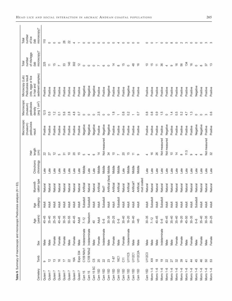
At the population level, our data show that all Chinchorro sites were highly infested (Tables 1–9). In modern urban populations, pediculosis can reach 100% prevalence in certain settings (Meink-ing et al., 1986; Taplin and Meinking, 1996.). Chinchorro archae-ological evidence coming from numerous cemeteries (complex mummification techniques and extended sites), large shell mid-dens, paleopathological data (presence of external auditory exos-tosis, fish parasites and marine dependent diet), fishing technology (shellfish hooks, sinkers and harpoons) and radiocarbon chronol-ogy (continuous dates without gaps) points toward year-round coastal occupation (Aufderheide et al., 1993; Arriaza, 1995; Mu-ñoz and Chacama, 1982; Standen, 1997). Pediculosis is a highly contagious condition and thrives in crowded surroundings, of-ten brought on during social disruptions (Willems et al., 2005). We have observed important levels of trauma in Chinchorro, re-flecting permanent conflicts (Standen and Arriaza, 2000; Stan-den, 2011). However, these permanent conflicts were likely the re-sults of small scale, intermittent violence among groups, such as raidings or perhaps revenge killing, rather than organized warfare which would have facilitated overcrowding of those participating in combat or those being attacked. We hypothesize that significant Chinchorro head lice prevalence and louse density resulted from living year-round at the coast (as evidenced by archaeological in-formation) and because they were thus less mobile than previously thought. In addition, we hypothesize Chinchorro head lice pedic-ulosis infestations were probably a consequence of the following conditions:Housing conditions and crowding: Chinchorro commu-nities lived in small semi-subterranean huts along the slopes of coastal hills (Muñoz and Chacama, 1982). The lack of rain made water resources very restricted; thus, settlements were highly lo-calized. Their houses were made with a base of stone cobbles, en-cased by wooden frameworks of sticks, and covered with sea lion skins and reeds. The coastal environment offers limited building resources; as such, their huts were small, on average about 9 m2, and they were neither totally enclosed nor windproof (Muñoz and Chacama, 1982). At night, the northern Chilean coast is foggy, ex-tremely windy, and temperatures drop significantly. Thus, Chin-chorro people (close kin and/or parents and children) likely slept very close to each other to keep warm, given the fact that adults and subadults (children) were both affected. In brief, small houses and nucleation probably increased the likelihood of ectoparasite transmission.Social behavior: Hunter-gatherers had plenty of time to engage in leisure activities (Barnard, 2004; Howell, 2010). Adults playing with children certainly could have contributed to head lice transmission, although games and toys have not been explored in Chinchorro archaeology. Head-to-head contact is the most com-mon mode of transmission across age lines.Mummification practices:
Table 8. Microscopic results of nits/eggs in hair samples (N=49)takenfrompositiveindividualsbymummificationtypeandagecategories.
Nits/eggs Naturallymummified(N=33) Artificiallymummified(N=16) Total
Subadult (N = 6) % Adult (N = 27) % Subadult (N = 9) % Adult (N = 7) % Nit/egg count %
Operculated 16 6.8 86 8.4 9 22.5 8 9.9 119 8.7Hatched 24 10.2 276 27.1 10 25.0 19 23.5 329 23.9Fragmented 39 16.6 244 24.0 12 30.0 44 54.3 339 24.7Cementing substance 156 66.4 412 40.5 9 22.5 10 12.3 587 42.7
Total 235 100 1018 100 40 100 81 100 1374 100
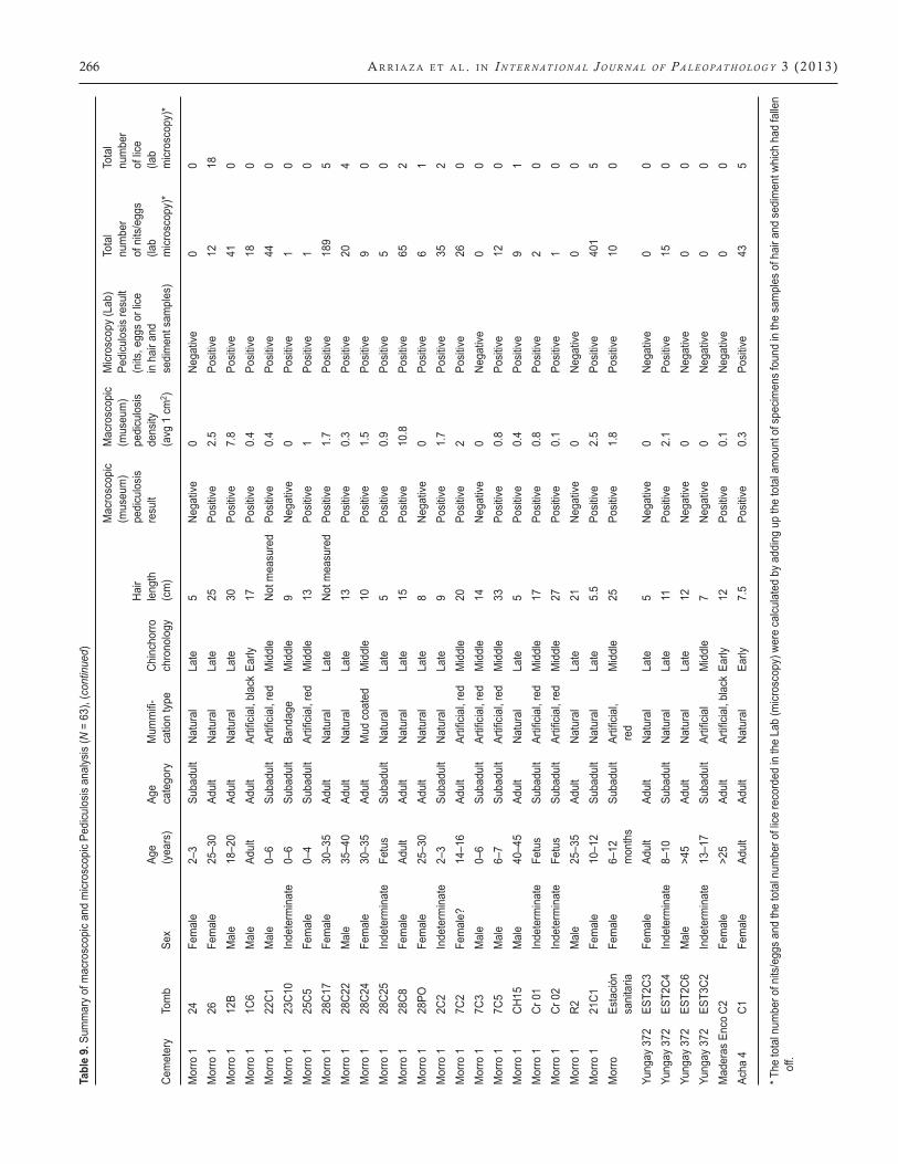
H e A d l i c e A n d s o c i A l i n t e r A c t i o n i n A rc H A i c A n d e A n c oA s tA l p o p u l At i o n s 265Ta
ble
9. S
umm
ary
of m
acro
scop
ic an
d m
icros
copi
c Pe
dicu
losis
ana
lysis
(N =
63)
.
Mac
rosc
opic
Mac
rosc
opic
Micr
osco
py (L
ab)
Tota
l To
tal
(mus
eum
) (m
useu
m)
Pedi
culo
sis re
sult
num
ber
num
ber
Ha
ir pe
dicu
losis
pe
dicu
losis
(n
its, e
ggs
or lic
e of
nits
/egg
s of
lice
Ag
eAg
eMum
mifi-
Chinchorro
length
result
density
inhairand
(lab
(lab
Cem
eter
y To
mb
Sex
(yea
rs)
cate
gory
ca
tion
type
ch
rono
logy
(c
m)
(a
vg 1
cm
2 ) se
dim
ent s
ampl
es)
micr
osco
py)*
micr
osco
py)*
Qui
ani 7
9
Mal
e 40
–45
Adul
t Na
tura
l La
te
22
Posit
ive
12.5
Po
sitive
22
5 11
0Q
uian
i 7
12
Mal
e 35
–40
Adul
t Na
tura
l La
te
27
Posit
ive
0.5
Posit
ive
11
0Q
uian
i 7
13
Fem
ale
25–3
5 Ad
ult
Natu
ral
Late
12
Po
sitive
1.
2 Po
sitive
15
0
Qui
ani 7
16
Fe
mal
e 40
–45
Adul
t Na
tura
l La
te
5 Po
sitive
0.
1 Po
sitive
7
0Q
uian
i 7
17
Fem
ale
30–3
5 Ad
ult
Natu
ral
Late
31
Po
sitive
5.
6 Po
sitive
33
28
Qui
ani 7
22
M
ale
50–5
5 Ad
ult
Natu
ral
Late
10
Po
sitive
2.
8 Po
sitive
14
0 22
Qui
ani 7
16
A Fe
mal
e 40
–45
Adul
t Na
tura
l La
te
20
Posit
ive
4.8
Posit
ive
302
4Q
uian
i 7
Expo
334
M
ale
Adul
t Ad
ult
Natu
ral
Late
15
Po
sitive
0.
7 Po
sitive
12
1
Cam15
C109Niño1Indeterminate
7–12
Subadult
Artificia
lMiddle
5Ne
gative
0Ne
gative
00
Cam
15
C109
Niñ
o2
Inde
term
inat
e Ne
wbor
n Su
badu
lt Na
tura
l La
te
4 Ne
gativ
e 0
Nega
tive
0 0
Cam
15
SC
C5
Mal
e 40
Ad
ult
Natu
ral
Late
6
Nega
tive
0 Ne
gativ
e 0
0Ca
m15D
20
Female
Adult
Adult
Artificia
lMiddle
14
Positive
2.5
Positive
7
0Ca
m 1
5D
22
Inde
term
inat
e 0–
6 Su
badu
lt Re
d?
Mid
dle
Not m
easu
red
Nega
tive
0 Po
sitive
6
0Ca
m15D
23
Male
30–35
Adult
Artificia
l(face)Middle
34
Negative
0Ne
gative
00
Cam15D
7–87
Female
20–25
Adult
Artificia
lMiddle
10
Positive
1.8
Positive
14
0Ca
m15D
16C1
Male
0–6
Subadult
Artificia
lMiddle
7Po
sitive
1
Positive
8
4Ca
m 1
5D
C11
Fem
ale
30–4
0 Ad
ult
Natu
ral
Late
20
Po
sitive
0.
8 Po
sitive
15
0
Cam15D
U1
11C5
Indeterminate
18–20
Adult
Artificia
lMiddle
15
Positive
0.2
Negative
00
Cam15D
U1
12C1
Male
30–40
Adult
Artificia
l?
Middle
11
Positive
1
Positive
48
0Ca
m15D
U1
12C2
AMale
>45
Adult
Artificia
l,
Middle
9Po
sitive
0.7
Positive
16
0
mud
coa
ted
Cam
15D
U1
12C3
M
ale
30–3
5 Ad
ult
Natu
ral
Late
4
Posit
ive
0.8
Posit
ive
10
0M
orro
1–5
XI
II M
ale
7–12
Su
badu
lt Na
tura
l La
te
16
Posit
ive
0.8
Posit
ive
15
0M
orro
1–6
18
M
ale
40–4
5 Ad
ult
Natu
ral
Late
26
Po
sitive
0.
9 Po
sitive
11
1
Mor
ro 1
–6
19
Inde
term
inat
e 0–
6 Su
badu
lt Na
tura
l La
te
Not m
easu
red
Posit
ive
1.5
Posit
ive
36
0M
orro
1–6
22
M
ale
40–4
5 Ad
ult
Natu
ral
Late
No
t mea
sure
d Ne
gativ
e 0
Posit
ive
4 0
Mor
ro 1
–6
27
Mal
e 35
–40
Adul
t Na
tura
l La
te
10
Posit
ive
1.4
Posit
ive
9 8
Mor
ro 1
–6
32
Fem
ale
35–4
0 Ad
ult
Natu
ral
Late
14
Po
sitive
0.
5 Po
sitive
16
0
Mor
ro 1
–6
39
Fem
ale
18–2
0 Ad
ult
Natu
ral
Late
8
Posit
ive
5.2
Posit
ive
56
2M
orro
1–6
41
M
ale
45–5
0 Ad
ult
Natu
ral
Late
11
.5
Posit
ive
4.7
Posit
ive
134
3M
orro
1–6
44
Fe
mal
e 30
–35
Adul
t Na
tura
l La
te
9 Po
sitive
1.
3 Po
sitive
16
0
Mor
ro 1
–6
45
Mal
e 0–
6 Su
badu
lt Na
tura
l La
te
4 Ne
gativ
e 0
Nega
tive
0 0
Mor
ro 1
–6
46
Fem
ale
35–4
0 Ad
ult
Natu
ral
Late
8
Nega
tive
0 Po
sitive
8
0M
orro
1–6
50
M
ale
30–3
5 Ad
ult
Natu
ral
Late
No
t mea
sure
d Po
sitive
3
Posit
ive
37
3M
orro
1–6
53
Fe
mal
e 20
–25
Adul
t Na
tura
l La
te
32
Posit
ive
0.6
Posit
ive
13
3
266 A r r i A z A e t A l . i n I n t e r n at I o n a l J o u r n a l o f P a l e o Pat h o l o g y 3 (2013) Ta
ble
9. S
umm
ary
of m
acro
scop
ic an
d m
icros
copi
c Pe
dicu
losis
ana
lysis
(N =
63)
, (co
ntin
ued)
Mac
rosc
opic
Mac
rosc
opic
Micr
osco
py (L
ab)
Tota
l To
tal
(mus
eum
) (m
useu
m)
Pedi
culo
sis re
sult
num
ber
num
ber
Ha
ir pe
dicu
losis
pe
dicu
losis
(n
its, e
ggs
or lic
e of
nits
/egg
s of
lice
Ag
eAg
eMum
mifi-
Chinchorro
length
result
density
inhairand
(lab
(lab
Cem
eter
y To
mb
Sex
(yea
rs)
cate
gory
ca
tion
type
ch
rono
logy
(c
m)
(a
vg 1
cm
2 ) se
dim
ent s
ampl
es)
micr
osco
py)*
micr
osco
py)*
Mor
ro 1
24
Fe
mal
e 2–
3 Su
badu
lt Na
tura
l La
te
5 Ne
gativ
e 0
Nega
tive
0 0
Mor
ro 1
26
Fe
mal
e 25
–30
Adul
t Na
tura
l La
te
25
Posit
ive
2.5
Posit
ive
12
18M
orro
1
12B
Mal
e 18
–20
Adul
t Na
tura
l La
te
30
Posit
ive
7.8
Posit
ive
41
0Morro1
1C6
Male
Adult
Adult
Artificia
l,blackEa
rly
17
Positive
0.4
Positive
18
0Morro1
22C1
Male
0–6
Subadult
Artificia
l,red
Middle
Notm
easured
Positive
0.4
Positive
44
0M
orro
1
23C1
0 In
dete
rmin
ate
0–6
Suba
dult
Band
age
Mid
dle
9 Ne
gativ
e 0
Posit
ive
1 0
Morro1
25C5
Female
0–4
Subadult
Artificia
l,red
Middle
13
Positive
1
Positive
1
0M
orro
1
28C1
7 Fe
mal
e 30
–35
Adul
t Na
tura
l La
te
Not m
easu
red
Posit
ive
1.7
Posit
ive
189
5M
orro
1
28C2
2 M
ale
35–4
0 Ad
ult
Natu
ral
Late
13
Po
sitive
0.
3 Po
sitive
20
4
Mor
ro 1
28
C24
Fem
ale
30–3
5 Ad
ult
Mud
coa
ted
Mid
dle
10
Posit
ive
1.5
Posit
ive
9 0
Mor
ro 1
28
C25
Inde
term
inat
e Fe
tus
Suba
dult
Natu
ral
Late
5
Posit
ive
0.9
Posit
ive
5 0
Mor
ro 1
28
C8
Fem
ale
Adul
t Ad
ult
Natu
ral
Late
15
Po
sitive
10
.8
Posit
ive
65
2M
orro
1
28PO
Fe
mal
e 25
–30
Adul
t Na
tura
l La
te
8 Ne
gativ
e 0
Posit
ive
6 1
Mor
ro 1
2C
2 In
dete
rmin
ate
2–3
Suba
dult
Natu
ral
Late
9
Posit
ive
1.7
Posit
ive
35
2Morro1
7C2
Female?
14–16
Adult
Artificia
l,red
Middle
20
Positive
2
Positive
26
0Morro1
7C3
Male
0–6
Subadult
Artificia
l,red
Middle
14
Negative
0Ne
gative
00
Morro1
7C5
Male
6–7
Subadult
Artificia
l,red
Middle
33
Positive
0.8
Positive
12
0M
orro
1
CH15
M
ale
40–4
5 Ad
ult
Natu
ral
Late
5
Posit
ive
0.4
Posit
ive
9 1
Morro1
Cr01
Indeterminate
Fetus
Subadult
Artificia
l,red
Middle
17
Positive
0.8
Positive
2
0Morro1
Cr02
Indeterminate
Fetus
Subadult
Artificia
l,red
Middle
27
Positive
0.1
Positive
1
0M
orro
1
R2
Mal
e 25
–35
Adul
t Na
tura
l La
te
21
Nega
tive
0 Ne
gativ
e 0
0M
orro
1
21C1
Fe
mal
e 10
–12
Suba
dult
Natu
ral
Late
5.
5 Po
sitive
2.
5 Po
sitive
40
1 5
Morro
Estació
n
Female
6–12
Subadult
Artificia
l,Middle
25
Positive
1.8
Positive
10
0
sani
taria
mon
ths
re
d Yu
ngay
372
ES
T2C3
Fe
mal
e Ad
ult
Adul
t Na
tura
l La
te
5 Ne
gativ
e 0
Nega
tive
0 0
Yung
ay 3
72
EST2
C4
Inde
term
inat
e 8–
10
Suba
dult
Natu
ral
Late
11
Po
sitive
2.
1 Po
sitive
15
0
Yung
ay 3
72
EST2
C6
Mal
e >4
5 Ad
ult
Natu
ral
Late
12
Ne
gativ
e 0
Nega
tive
0 0
Yungay372
EST3C2
Indeterminate
13–17
Subadult
Artificia
lMiddle
7Ne
gative
0Ne
gative
00
MaderasEncoC2
Female
>25
Adult
Artificia
l,blackEa
rly
12
Positive
0.1
Negative
00
Acha
4
C1
Fem
ale
Adul
t Ad
ult
Natu
ral
Early
7.
5 Po
sitive
0.
3 Po
sitive
43
5
* The
tota
l num
ber o
f nits
/egg
s an
d th
e to
tal n
umbe
r of l
ice re
cord
ed in
the
Lab
(micr
osco
py) w
ere
calcu
late
d by
add
ing
up th
e to
tal a
mou
nt o
f spe
cimen
s fo
und
in th
e sa
mpl
es o
f hai
r and
sed
imen
t whi
ch h
ad fa
llen
off.
H e A d l i c e A n d s o c i A l i n t e r A c t i o n i n A rc H A i c A n d e A n c oA s tA l p o p u l At i o n s 267
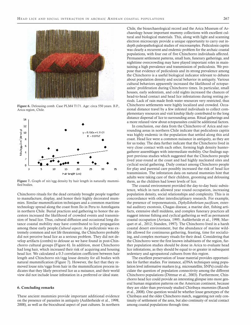
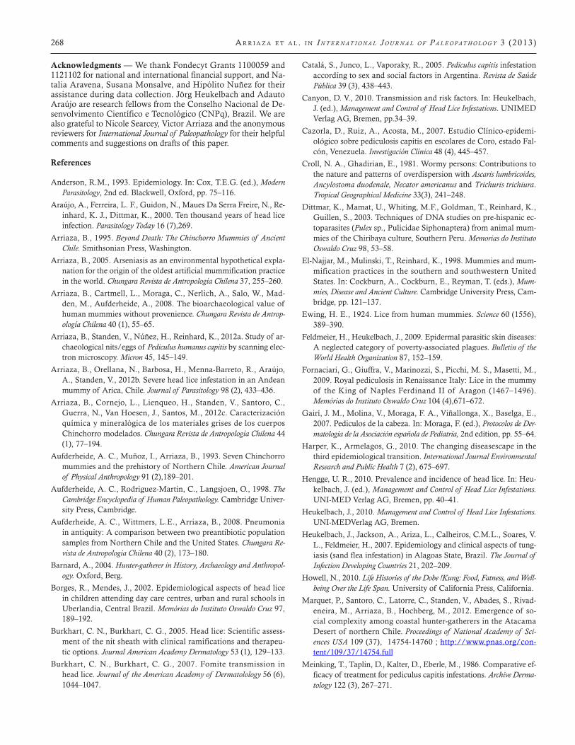
Chinchorro rituals for the dead certainly brought people together to manufacture, display, and honor their highly decorated mum-mies. Similar mummification techniques and a common maritime technology spread along the coast from Ilo in Peru to Antofagasta in northern Chile. Burial practices and gathering to honor the an-cestors increased the likelihood of crowded events and transmis-sion of head lice. Thus, cultural diffusion and occasional long dis-tance coastal mobility may have contributed to lice propagation among these early people.Cultural aspects: As pediculosis was ex-tremely common and not life threatening, the Chinchorro probably did not perceive head lice as a serious problem. They did not de-velop artifacts (combs) to delouse as we have found in post-Chin-chorro cultural groups (Figure 6). In addition, most Chinchorro had long hair, which increased their risk of acquiring and housing head lice. We calculated a 0.3 correlation coefficient between hair length and Chinchorro nit/egg louse density for all bodies with natural mummification (Figure 7). However, the fact that they re-moved louse nits/eggs from hair in the mummification process in-dicates that they likely perceived lice as a nuisance, and their world view did not include louse infestation in a preferred or ideal state.
6. Concluding remarks
These ancient mummies provide important additional evidence on the presence of parasites in antiquity (Aufderheide et al., 1998, 2008), as well as the biocultural aspect of past cultures. In northern
Chile, the bioarchaeological record and the Arica Museum of Ar-chaeology house important mummy collections with excellent cul-tural and biological materials. This, along with light and scanning electron microscopy provide a unique opportunity to carry out in-depth paleopathological studies of microsamples. Pediculosis capitis was clearly a recurrent and endemic problem for the archaic coastal populations, with four out of five Chinchorro individuals affected. Permanent settlement patterns, small huts, funerary gatherings, and nighttime overcrowding may have played important roles in main-taining a high prevalence and transmission of pediculosis. We pro-pose that evidence of pediculosis and its strong prevalence among the Chinchorro is a useful biological indicator relevant to debates about population density and social behavior in antiquity. Various cultural behaviors apparently increased the likelihood of ectopar-asites’ proliferation during Chinchorro times. In particular, small houses, early sedentism, and cold nights increased the chances of head-to-head contact and head lice infestations during resting pe-riods. Lack of rain made fresh water resources very restricted, thus Chinchorro settlements were highly localized and crowded. Occa-sional distance travel by a few infested individuals to collect com-plementary resources and visit kinship likely contributed to the long distance dispersal of lice to surrounding areas. Ritual gatherings and a more relaxed view about ectoparasites could be additional factors.
In conclusion, our data from the Chinchorro of Arica and sur-rounding areas in northern Chile indicate that pediculosis capitis was highly endemic in the population that settled along this arid coast. Head lice were a common nuisance in antiquity, as they are for us today. The data further indicate that the Chinchorro lived in very close contact with each other, forming high density hunter-gatherer assemblages with intermediate mobility. Our findings sup-port previous studies which suggested that the Chinchorro people lived year-round at the coast and had highly nucleated sites and cyclical social gathering. Daily contact among Chinchorro people for play and parental care possibly increased the risk of head lice transmission. The infestation data on natural mummies hint that adults were taking care of their children, grooming and delousing them, as the children had lower levels of lice.
The coastal environment provided the day-to-day basic subsis-tence, which in turn allowed year round occupation, increasing population density, social relationships and complexity. This is in concordance with other interdisciplinary research. For example, the presence of treponematosis, Diphyllobothrium pacificum, exter-nal auditory exostosis, Chagas disease, a marine diet, fishing arti-facts, extensive shell middens, and complex funerary practices all suggest intense fishing and cyclical gathering as well as permanent coastal occupation (Arriaza, 1995; Aufderheide et al., 1998; Mar-quet et al., 2012; Standen, 1997). The Chinchorro lived in a harsh coastal desert environment, but the abundance of marine wild-life allowed for continuous gathering, feasting, time for socializ-ing, and complex mortuary rituals for their dead. Considering that the Chinchorro were the first known inhabitants of the region, fur-ther population studies should be done in Arica to evaluate head lice infestations, as they should be equal to or greater in subsequent sedentary and agropastoral cultures from this region.
The excellent preservation of louse material provides opportuni-ties for further studies. For instance, aDNA techniques using popu-lation level informative markers (e.g. microsatellite, SNPs) could elu-cidate the question of population connectivity among the different Chinchorro populations (Dittmar et al., 2003). Furthermore, Chin-chorro head lice could provide an interesting glimpse into more gen-eral human migration patterns on the American continent, because they are older than previously studied Chiribaya mummies (Raoult et al., 2008). One question would be whether louse genotypes of the Chiribaya and the older Chinchorro match, suggesting not only con-tinuity of settlement of the area, but also continuity of social contact among coastal populations through time.
Figure 6. Delousing comb. Case PLM4 T171. Age: circa 550 years. B.P., Arica region, Chile.
Figure 7. Graph of nit/egg density by hair length in naturally mummi-fied bodies.
268 A r r i A z A e t A l . i n I n t e r n at I o n a l J o u r n a l o f P a l e o Pat h o l o g y 3 (2013)
Acknowledgments — We thank Fondecyt Grants 1100059 and 1121102 for national and international financial support, and Na-talia Aravena, Susana Monsalve, and Hipólito Nuñez for their assistance during data collection. Jörg Heukelbach and Adauto Araújo are research fellows from the Conselho Nacional de De-senvolvimento Científico e Tecnológico (CNPq), Brazil. We are also grateful to Nicole Searcey, Victor Arriaza and the anonymous reviewers for International Journal of Paleopathology for their helpful comments and suggestions on drafts of this paper.
References
Anderson, R.M., 1993. Epidemiology. In: Cox, T.E.G. (ed.), Modern Parasitology, 2nd ed. Blackwell, Oxford, pp. 75–116.
Araújo, A., Ferreira, L. F., Guidon, N., Maues Da Serra Freire, N., Re-inhard, K. J., Dittmar, K., 2000. Ten thousand years of head lice infection. Parasitology Today 16 (7),269.
Arriaza, B., 1995. Beyond Death: The Chinchorro Mummies of Ancient Chile. Smithsonian Press, Washington.
Arriaza, B., 2005. Arseniasis as an environmental hypothetical expla-nation for the origin of the oldest artificial mummification practice in the world. Chungara Revista de Antropología Chilena 37, 255–260.
Arriaza, B., Cartmell, L., Moraga, C., Nerlich, A., Salo, W., Mad-den, M., Aufderheide, A., 2008. The bioarchaeological value of human mummies without provenience. Chungara Revista de Antrop-ología Chilena 40 (1), 55–65.
Arriaza, B., Standen, V., Núñez, H., Reinhard, K., 2012a. Study of ar-chaeological nits/eggs of Pediculus humanus capitis by scanning elec-tron microscopy. Micron 45, 145–149.
Arriaza, B., Orellana, N., Barbosa, H., Menna-Barreto, R., Araújo, A., Standen, V., 2012b. Severe head lice infestation in an Andean mummy of Arica, Chile. Journal of Parasitology 98 (2), 433–436.
Arriaza, B., Cornejo, L., Lienqueo, H., Standen, V., Santoro, C., Guerra, N., Van Hoesen, J., Santos, M., 2012c. Caracterización química y mineralógica de los materiales grises de los cuerpos Chinchorro modelados. Chungara Revista de Antropología Chilena 44 (1), 77–194.
Aufderheide, A. C., Muñoz, I., Arriaza, B., 1993. Seven Chinchorro mummies and the prehistory of Northern Chile. American Journal of Physical Anthropology 91 (2),189–201.
Aufderheide, A. C., Rodriguez-Martin, C., Langsjoen, O., 1998. The Cambridge Encyclopedia of Human Paleopathology. Cambridge Univer-sity Press, Cambridge.
Aufderheide, A. C., Wittmers, L.E., Arriaza, B., 2008. Pneumonia in antiquity: A comparison between two preantibiotic population samples from Northern Chile and the United States. Chungara Re-vista de Antropologia Chilena 40 (2), 173–180.
Barnard, A., 2004. Hunter-gatherer in History, Archaeology and Anthropol-ogy. Oxford, Berg.
Borges, R., Mendes, J., 2002. Epidemiological aspects of head lice in children attending day care centres, urban and rural schools in Uberlandia, Central Brazil. Memórias do Instituto Oswaldo Cruz 97, 189–192.
Burkhart, C. N., Burkhart, C. G., 2005. Head lice: Scientific assess-ment of the nit sheath with clinical ramifications and therapeu-tic options. Journal American Academy Dermatology 53 (1), 129–133.
Burkhart, C. N., Burkhart, C. G., 2007. Fomite transmission in head lice. Journal of the American Academy of Dermatolology 56 (6), 1044–1047.
Catalá, S., Junco, L., Vaporaky, R., 2005. Pediculus capitis infestation according to sex and social factors in Argentina. Revista de Saúde Pública 39 (3), 438–443.
Canyon, D. V., 2010. Transmission and risk factors. In: Heukelbach, J. (ed.), Management and Control of Head Lice Infestations. UNIMED Verlag AG, Bremen, pp.34–39.
Cazorla, D., Ruiz, A., Acosta, M., 2007. Estudio Clínico-epidemi-ológico sobre pediculosis capitis en escolares de Coro, estado Fal-cón, Venezuela. Investigación Clínica 48 (4), 445–457.
Croll, N. A., Ghadirian, E., 1981. Wormy persons: Contributions to the nature and patterns of overdispersion with Ascaris lumbricoides, Ancylostoma duodenale, Necator americanus and Trichuris trichiura. Tropical Geographical Medicine 33(3), 241–248.
Dittmar, K., Mamat, U., Whiting, M.F., Goldman, T., Reinhard, K., Guillen, S., 2003. Techniques of DNA studies on pre-hispanic ec-toparasites (Pulex sp., Pulicidae Siphonaptera) from animal mum-mies of the Chiribaya culture, Southern Peru. Memorias do Instituto Oswaldo Cruz 98, 53–58.
El-Najjar, M., Mulinski, T., Reinhard, K., 1998. Mummies and mum-mification practices in the southern and southwestern United States. In: Cockburn, A., Cockburn, E., Reyman, T. (eds.), Mum-mies, Disease and Ancient Culture. Cambridge University Press, Cam-bridge, pp. 121–137.
Ewing, H. E., 1924. Lice from human mummies. Science 60 (1556), 389–390.
Feldmeier, H., Heukelbach, J., 2009. Epidermal parasitic skin diseases: A neglected category of poverty-associated plagues. Bulletin of the World Health Organization 87, 152–159.
Fornaciari, G., Giuffra, V., Marinozzi, S., Picchi, M. S., Masetti, M., 2009. Royal pediculosis in Renaissance Italy: Lice in the mummy of the King of Naples Ferdinand II of Aragon (1467–1496). Memórias do Instituto Oswaldo Cruz 104 (4),671–672.
Gairí, J. M., Molina, V., Moraga, F. A., Viñallonga, X., Baselga, E., 2007. Pediculos de la cabeza. In: Moraga, F. (ed.), Protocolos de Der-matología de la Asociación española de Pediatría, 2nd edition, pp. 55–64.
Harper, K., Armelagos, G., 2010. The changing diseasescape in the third epidemiological transition. International Journal Environmental Research and Public Health 7 (2), 675–697.
Hengge, U. R., 2010. Prevalence and incidence of head lice. In: Heu-kelbach, J. (ed.), Management and Control of Head Lice Infestations. UNI-MED Verlag AG, Bremen, pp. 40–41.
Heukelbach, J., 2010. Management and Control of Head Lice Infestations. UNI-MEDVerlag AG, Bremen.
Heukelbach, J., Jackson, A., Ariza, L., Calheiros, C.M.L., Soares, V. L., Feldmeier, H., 2007. Epidemiology and clinical aspects of tung-iasis (sand flea infestation) in Alagoas State, Brazil. The Journal of Infection Developing Countries 21, 202–209.
Howell, N., 2010. Life Histories of the Dobe !Kung: Food, Fatness, and Well-being Over the Life Span. University of California Press, California.
Marquet, P., Santoro, C., Latorre, C., Standen, V., Abades, S., Rivad-eneira, M., Arriaza, B., Hochberg, M., 2012. Emergence of so-cial complexity among coastal hunter-gatherers in the Atacama Desert of northern Chile. Proceedings of National Academy of Sci-ences USA 109 (37), 14754-14760 ; http://www.pnas.org/con-tent/109/37/14754.full
Meinking, T., Taplin, D., Kalter, D., Eberle, M., 1986. Comparative ef-ficacy of treatment for pediculus capitis infestations. Archive Derma-tology 122 (3), 267–271.
H e A d l i c e A n d s o c i A l i n t e r A c t i o n i n A rc H A i c A n d e A n c oA s tA l p o p u l At i o n s 268-A
Muñoz, I., Chacama, J., 1982. Investigaciones arqueológicas en las pobla-ciones pre-cerámicas de la costa de Arica. Documento de Trabajo, vol. 2. Universidad deTarapacá, Arica, pp. 3–97.
Núñez, L., 1983. Paleoindio y Arcaico en Chile: Diversidad, Secuencia y Proceso. In: Serie Monografías 3. Ediciones Cuicuilco, México.
Nuttall, G.H.F., 1917. The biology of Pediculus humanus. Parasitology 10 (01), 80–185.
Parison, J., 2010. Head lice and the impact of knowledge, attitudes and practices—A social science overview. In: Heukelbach, J. (ed.), Management and Control ofHead Lice Infestations. UNI-MED Verlag AG, Bremen, pp. 104–109.
Raoult, D., Reed, D., Dittmar, K., Kirchman, J., Rolain, J., Guillen, S., Light, J., 2008. Molecular identification of lice from Pre-Co-lumbian mummies. The Journal of Infectious Diseases 197, 535–543.
Reinhard, K., Buikstra, J., 2003. Louse infestation of the Chiribaya culture, southern Peru: Variation in prevalence by age and sex. Memórias do Instituto Oswaldo Cruz 98 (Supl 1), 173–179.
Rivera, M., 1991. The prehistory of northern Chile: A synthesis. Jour-nal of World Prehistory 5, 1–47.
Rivera, M., Mumcuoglu, K., Matheny, R., Matheny, D., 2008. Head lice eggs, anthropophthirus capitis, from mummies of the Chin-chorro tradition, Camarones 15-D, northern Chile. Chungara Re-vista de Antropología Chilena 40 (1), 31–39.
Santoro, C., Rivadeneira, M., Latorre, C., Rothhammer, R., Stan-den, V., 2012. Rise and decline of Chinchorro sacred landscapes along the hyperarid coast of theAtacama Desert. Chungara Revista de Antropología Chilena 44 (4), 637–653.
Soultana, V., Euthumia, P., Maniatis, A., Roussaki-Schultze, A., 2009. Prevalence of pediculosis capitis among school children in Greece and risk factors: A questionnaire survey. Pediatric Dermatology 26 (6), 701–705.
Standen, V. G., 1997. Early funerary patterns of the Chinchorro cul-ture (Northern Chile). Latin American Antiquity 8 (2), 134–156.
Standen, V. G., Arriaza, B., 2000. Trauma in the preceramic coastal populations of northern Chile: Violence or occupational hazards? American Journal of Physical Anthropology 112 (2), 239–249.
Standen, V. G., 2011. Violencia y cultura en cazadores, pescadores y recolec-tores deldesierto de Atacama (norte de Chile): 8.900-3.700 A.P. Ph.D. Dis-sertation. Universidad Nacional Autónoma de México, Mexico, DF.
Taplin, D., Meinking, T., 1996. Pediculosis infestations. Pediatric Der-matology 7, 1480–1515.
Williams, L. K., Reichert, M. A., MacKenzie, W. R., Hightower, A. W., Blake, P. A., 2001. Lice, nits and school policy. Pediatrics 107, 1011–1015.
Willems, S., Lapeere, H., Haedens, N., Pasteels, I., Naeyaert, J., De Maesenner, J., 2005. The importance of socioeconomic status and individual characteristics on the prevalence of head lice in school children. European Journal of Dermatology 15(5), 387–392.
Zinsser, H., 1935. Rats, Lice and History. Little, Brown & Company, Boston.
Zuñiga, I., Caro, J., 2010. Pediculosis: Una ectoparasitosis emergente en México. Revista de Enfermedades Infecciosas en Pediatría 24 (94), 56–63.
Recommended