CLINICAL PRACTICE GUIDELINE HN–002
version 1
Page 1 of 22
ORAL CAVITY CANCER
Effective Date: February, 2014
The recommendations contained in this guideline are a consensus of the Alberta Provincial Head and Neck Tumour
Team synthesis of currently accepted approaches to management, derived from a review of relevant scientific literature. Clinicians applying these guidelines should, in consultation with the patient, use independent medical
judgment in the context of individual clinical circumstances to direct care.
CLINICAL PRACTICE GUIDELINE HN–002 version 1
Page 2 of 22
BACKGROUND
The incidence of oral cavity cancer is not well documented because it is often grouped with other subsites rather than being described in accord with the anatomical definition. Cancer of the oral cavity comprises nearly 30 percent of all malignant tumours of the head and neck.1 Squamous cell carcinomas (SCCs) represent approximately 90 percent of oral cavity cancer cases. Minor salivary gland cancers, melanomas, lymphomas and sarcomas are less common. Sites of tumour origin of the oral cavity include the buccal mucosa, floor of mouth, anterior tongue, alveolar ridge, retromolar trigone and the hard palate. Lifestyle, habits, demographics, as well as genetic factors influence geographic variations in the incidence of disease. In North America, tobacco smoking and alcohol use are the principal risk factors for developing oral cavity cancer. Oral tobacco use, periodontal disease, radiation and immune deficiency have also been implicated.
Despite improved quality of life for patients with oral cavity cancer over the past 30 years, 5-year overall survival (OS) remains in the range of 50–60 percent.1 Treatment for oral cavity cancer is highly complex because of the variety of tumour subsites and the anatomical constraints of the head and neck. In patients who are successfully treated, they often face quality of life issues related to changes in appearance and organ function, such as talking, chewing and swallowing.
The literature about oral cavity cancer treatment is composed largely of nonrandomized, retrospective or monocentric studies, hence the importance of treatment being decided on a case-by-case basis by a multidisciplinary team. The ultimate goal of oral cavity cancer treatment is to eliminate the cancer, preserve or restore form and function, minimize treatment complications and to prevent any subsequent new primary cancers. To achieve these goals, current treatment modalities include surgery, radiotherapy (RT), chemotherapy and combined modality treatments. Influencing the choice of treatment are tumour, patient and treatment team factors.2 Tumour factors that affect the choice of initial treatment of oral cavity cancer are primary site, size and proximity to bone, status of regional cervical lymph nodes, previous treatment and histology. Staging for tumours of the oral cavity are presented in Appendix A. Patient factors include age, general health and tolerance to treatment, occupation, lifestyle and other socioeconomic considerations. Finally, treatment team factors depend on the technical capabilities and support services from various disciplines.
This guideline was developed to outline treatment recommendations for patients with early and advanced stage oral cavity cancer. These guidelines should be applied in the context of the recommendations outlined in Alberta Health Services, CancerControl Alberta guideline, The Organization and Delivery of Healthcare Services for Head and Neck Cancer Patients.
GUIDELINE QUESTIONS
1. What diagnostic and baseline investigations are recommended for patients with suspected orconfirmed oral cavity cancer?
2. What are the recommended treatment options for early-stage oral cavity cancer (T1–2, N0)?3. What are the recommended treatment options for advanced-stage oral cavity cancer (T3, N0; T4a,
Any N; T1–3, N1–3; T4b, any N or unresectable nodal disease or unfit for surgery)?4. What is the recommended follow-up after treatment for oral cavity cancer?
CLINICAL PRACTICE GUIDELINE HN–002
version 1
Page 3 of 22
DEVELOPMENT AND REVISION HISTORY This guideline was reviewed and endorsed by the Alberta Provincial Head and Neck Tumour Team. Members of the Alberta Provincial Head and Neck Tumour Team include medical oncologists, radiation oncologists, surgical oncologists, head and neck reconstructive surgeons, nurses, pathologists, pharmacists, dentists, dietitians, and other allied health professionals. Evidence was selected and reviewed by a working group comprised of members from the Alberta Provincial Head and Neck Tumour Team and a Knowledge Management Specialist from the Guideline Utilization Resource Unit. A detailed description of the methodology followed during the guideline development process can be found in the Guideline Utilization Resource Unit Handbook. This guideline was originally developed in February, 2014. SEARCH STRATEGY PubMED, MEDLINE and the Cochrane Database of Systematic Reviews were searched to May 6, 2013 for literature on the treatment of oral cavity cancer. The following search terms were used: mouth floor (MeSH [Medical Subject Heading]) OR mouth neoplasm (MeSH) OR oral cavity cancer OR buccal mucosa cancer OR tongue neoplasms (MeSH) OR anterior tongue cancer OR alveolar ridge cancer OR retromolar trigone cancer OR hard palate cancer; results were limited to human subjects (19+ years), published in English. Except in the case of frequent citation, only studies with equal to or greater than 50 patients and who were followed for more than 3 years were considered for the literature review. In addition, studies reporting patient populations with mixed tumour sites were only considered if greater than 50 percent of the patients had oral cavity cancer. Reference lists of included studies were also searched to identify further trials. The National Guidelines Clearinghouse and SAGE Directory of Cancer Guidelines were also searched from 2008 to 2013 for guidelines on oral cavity cancer. TARGET POPULATION The recommendations outlined in this guideline are intended for adults over the age of 18 years with oral squamous cell carcinoma. Different principles may apply to pediatric patients or patients with rarer histopathology. RECOMMENDATIONS The Alberta Provincial Head and Neck Tumour Team reviewed the recommendations of several different guidelines, including those from the Cancer Care Ontario Program in Evidence-based Care,3 the German Guideline Group4 and the National Comprehensive Cancer Network (NCCN)5. The Alberta Provincial Head and Neck Tumour Team have adopted the recommendations of the NCCN, with modifications to fit the Alberta context. 1. Initial work-up and supportive care evaluations. The following investigations are recommended at diagnosis for all patients with suspected or confirmed oral cavity cancer:
• Complete head and neck examination • Biopsy • Chest imaging
CLINICAL PRACTICE GUIDELINE HN–002
version 1
Page 4 of 22
• Nutrition, speech and swallowing evaluation/therapy should be conducted by a registered dietician and a speech-language/swallowing therapist and is recommended for patients who have significant weight loss (>10% ideal body weight), and/or difficulty with speech/swallowing, and/or for patients whose treatment is likely to affect speech/swallowing
• Computed tomography (CT) with contrast and/or magnetic resonance imaging (MRI) with contrast of primary site and neck, as indicated
• Positron emission tomography-computed tomography (PET-CT), as indicated • Chest CT scan, if not included with other imaging • Examination under anesthesia with endoscopy, as indicated • Preanesthesia studies • Dental/prosthodontic evaluation, including jaw imaging, as indicated
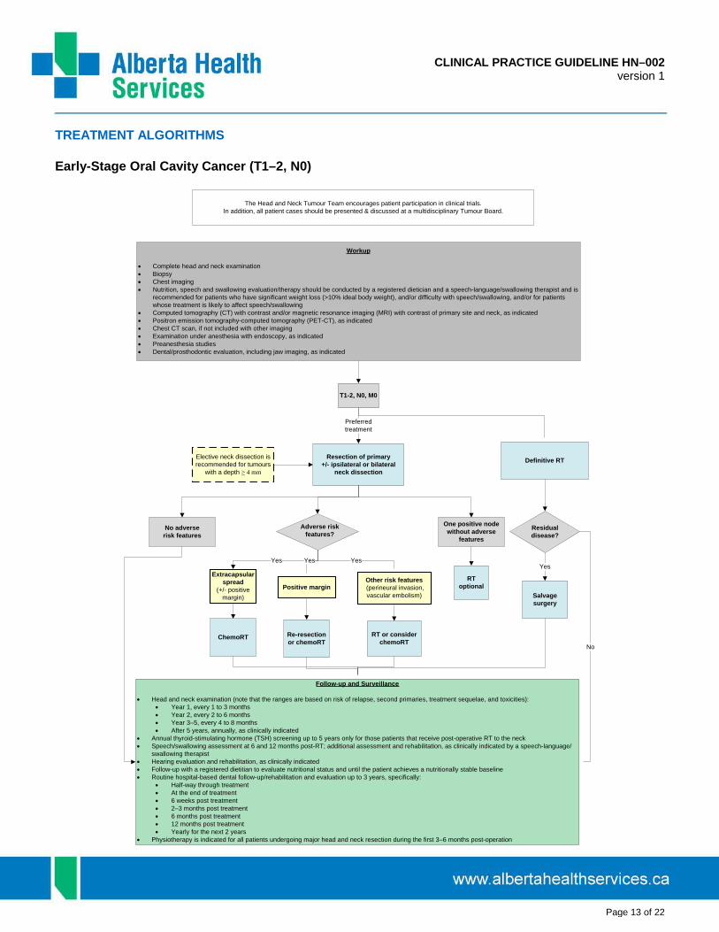
2. Treatment options for early-stage oral cavity cancer (T1–2, N0). Patient participation in clinical trials is recommended. For standard treatment, all cases should be presented and discussed at a multidisciplinary Tumour Board to decide the best treatment option for each patient.
• Preferred treatment: Patients with no contraindication to surgery and who are agreeable should undergo resection of primary site plus/minus ipsilateral or bilateral neck dissection (ND), which should be guided by tumour thickness.
o Elective ND is recommended for tumours with a depth ≥ 4 mm. For a depth of 2 to 4 mm, clinical judgment should be utilized to determine appropriateness of elective ND.
If a patient has one positive node without adverse risk features, RT is optional. If a patient has the following adverse risk features, treatment after resection includes:
o Extracapsular spread +/- positive margin: chemoRT o Positive margin: if feasible, consider re-resection to achieve negative margins; if re-
resection is not possible consider chemoRT o Perineural invasion and/or vascular embolism: RT alone; chemoRT may be considered, the
decision should be based on clinical judgment and discussion at the multidisciplinary Tumour Board
If a patient has none of the above adverse risk features, he or she should proceed with follow–up and surveillance.
• Alternative treatment: Patients with a contraindication to surgery should undergo definitive RT.
Patients who go on to develop recurrent or residual disease should be considered for salvage therapy.
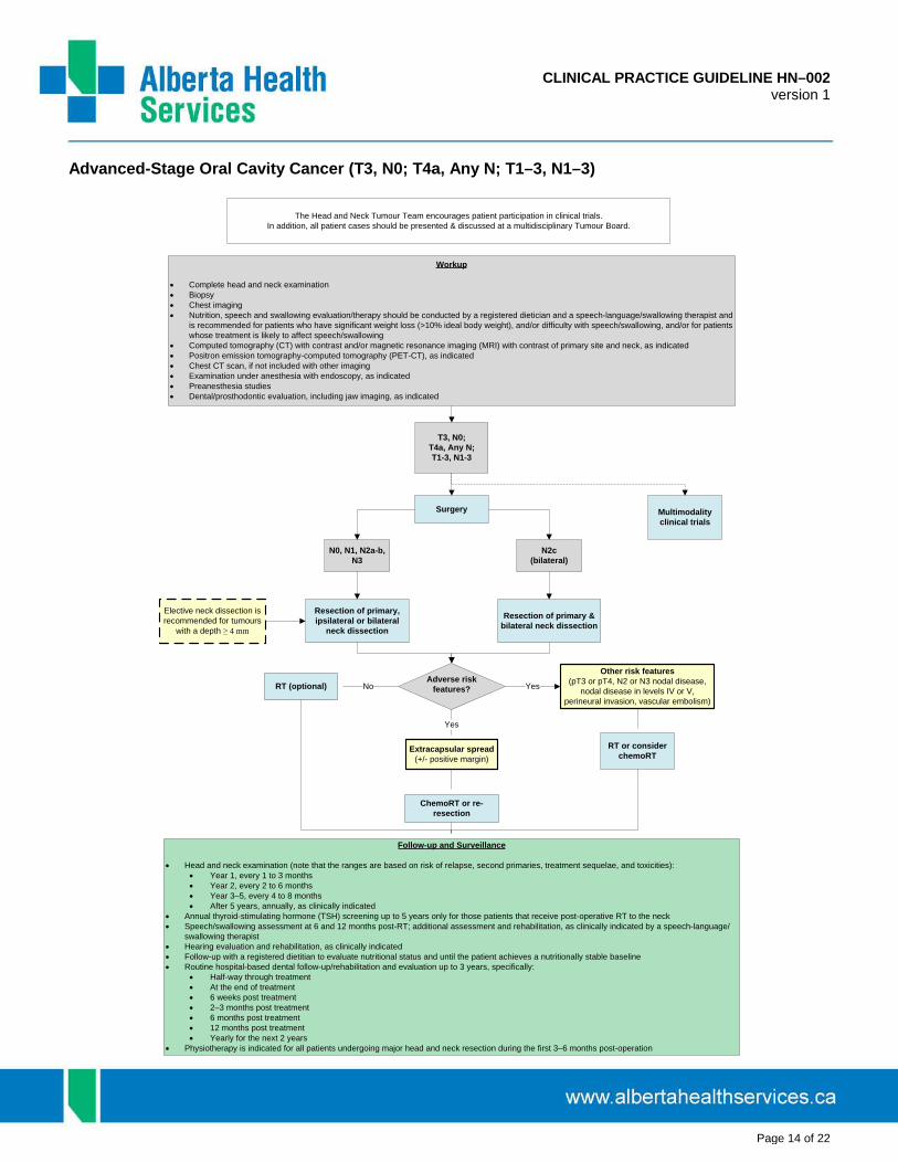
Please click here to view the early-stage treatment algorithm. 3a. Treatment options for advanced-stage oral cavity cancer (T3, N0; T1–3, N1–3; T4a, Any N ). Inclusion of patients in multimodality clinical trials is recommended. In lieu of a clinical trial, patients should undergo surgery. Patients with N2c neck disease should undergo primary tumour resection and bilateral ND, while all other patients (N0, N1, N2a–b and N3) should undergo primary tumour resection. Ipsilateral or bilateral dissection may be indicated for some patients, especially if the depth of invasion is ≥ 4 mm; the decision should be based on clinical judgment and discussion at the multidisciplinary Tumour Board.
CLINICAL PRACTICE GUIDELINE HN–002
version 1
Page 5 of 22
If a patient has the following adverse risk features, treatment after resection includes: • Extracapsular spread +/- positive margin: chemoRT or re-resection • pT3 or pT4, and/or N2 or N3 nodal disease, and/or nodal disease in levels IV or V, and/or
perineural invasion, and/or vascular embolism: RT alone; chemoRT may be considered, the decision should be based on clinical judgment and discussion at the multidisciplinary Tumour Board
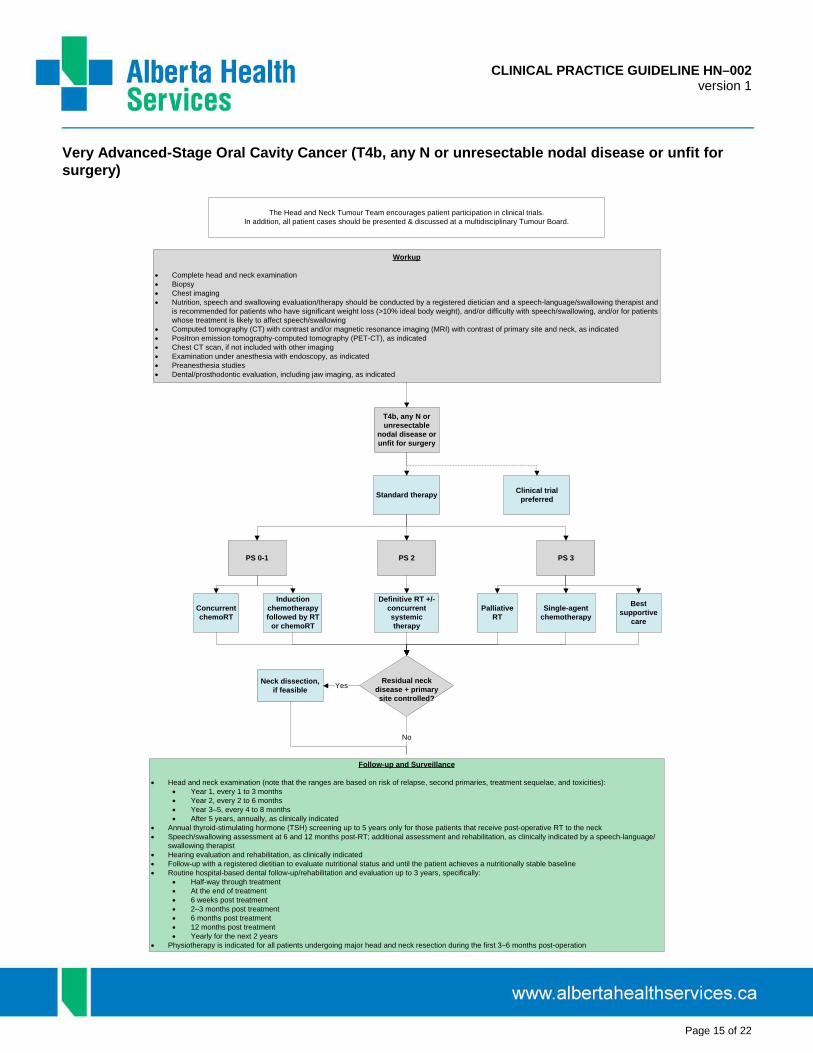
If a patient has none of the above adverse risk features RT is considered optional before proceeding with follow–up and surveillance. Please click here to view the advanced-stage treatment algorithm. 3b. Treatment options for very advanced-stage oral cavity cancer (T4b, Any N or unresectable nodal disease or unfit for surgery). Patient participation in clinical trials is recommended. In lieu of a clinical trial, patients should undergo therapy dependent on their Eastern Cooperative Oncology Group (ECOG) performance status (PS). ECOG PS levels are presented in Appendix B.
• PS 0–1 patients should undergo both concurrent systemic therapy and RT or induction chemotherapy followed by RT or chemoRT. Induction chemotherapy should only be done in a tertiary setting due to toxicity concerns.
• PS 2 patients should undergo RT with or without concurrent systemic therapy dependent on their treatment goals.
• PS 3 patients have the option of undergoing palliative RT, single-agent chemotherapy or opt for best supportive care.
In the instance of residual neck disease and if feasible, conduct ND if the primary tumour site is controlled following the above treatments. For oral cavity cancers, the risk of regional metastases and the need for adjuvant elective ND increases as thickness of the lesion increases. Please click here to view the very advanced-stage treatment algorithm. 4. Reconstruction. The Alberta Provincial Head and Neck Tumour Team agree that patients with oral cavity cancer should be assessed by an experienced Head and Neck Reconstructive Surgeon, as defined in The Organization and Delivery of Healthcare Services for Head and Neck Cancer Patients guideline, prior to surgical treatment. If reconstruction of surgical defects is required, it should be performed at the discretion of the Head and Neck Reconstructive Surgeon. 5. Follow-up and surveillance. The following schedule should be taken into account to manage complications related to treatment, to detect disease recurrence and/or the development of new disease:
• Head and neck examination (note that the ranges are based on risk of relapse, second primaries, treatment sequelae, and toxicities):
o Year 1, every 1 to 3 months o Year 2, every 2 to 6 months o Year 3–5, every 4 to 8 months o After 5 years, annually, as clinically indicated
• Annual thyroid-stimulating hormone (TSH) screening up to 5 years only for those patients that receive post-operative RT to the neck
CLINICAL PRACTICE GUIDELINE HN–002
version 1
Page 6 of 22
• Speech/swallowing assessment at 6 and 12 months post-RT; additional assessment and rehabilitation, as clinically indicated by a speech-language/swallowing therapist
• Hearing evaluation and rehabilitation, as clinically indicated • Follow-up with a registered dietitian to evaluate nutritional status and until the patient achieves a
nutritionally stable baseline • Routine hospital-based dental follow-up/rehabilitation and evaluation up to 3 years, specifically:
o Half-way through treatment o At the end of treatment o 6 weeks post treatment o 2–3 months post treatment o 6 months post treatment o 12 months post treatment o Yearly for the next 2 years
• Physiotherapy is indicated for all patients undergoing major head and neck resection during the first 3–6 months post-operation
DISCUSSION Diagnosis Commonly observed signs and symptoms in oral cavity cancer include, non-healing ulcer, pain, bleeding or ill-fitting dentures. More advanced lesions may present with speech difficulties, dysphasia, otalgia (ear pain), hypersalavation and neck mass(es). A complete head and neck examination is required to begin to diagnose oral cavity cancer. Attention should be paid to tongue mobility, extension to adjacent sites and involvement of the primary echelon nodal drainage levels I and II. In individual cases, endoscopic examination under anesthesia can be used for a more thorough examination, although the actual diagnosis of oral cavity cancer can only be confirmed with a tissue biopsy. Pre-anesthetic studies should precede any invasive procedures requiring general anesthesia in patients who have had previous treatment with surgery, RT or chemotherapy. Imaging studies of the head and neck areas using CT with contrast and/or MRI should be used to assess the extent of local and regional tumour spread, the depth of invasion and lymphadenopathy. Chest imaging, usually CT scan, will identify pulmonary metastasis. Dental/prosthodontic evaluation, including jaw imaging is required prior to commencement of treatment in patients at risk of oral complications. Finally, nutrition, speech and swallowing evaluation/therapy are critical to optimize quality of life during and after treatment. Early-Stage Disease Approximately 30–40 percent of patients with oral cavity cancer present with stage I and II disease.1 Surgery is considered the gold standard for treatment because it requires a single intervention and is usually performed with a minimum amount of morbidity. RT is recommended for patients with a contraindication to surgery and can achieve similar rates of local control and survival. However, multiple interventions are required over a period of several weeks and the incidence of long-term sequelae is high (e.g., xerostomia, dysphagia and osteoradionecrosis). Thus, the choice of treatment (surgery versus RT) should be based on an assessment of the patient’s ability to tolerate and accept surgery. Please see ‘Surgical management of the neck’ below regarding the surgical management of N0 neck oral cavity cancer patients.
CLINICAL PRACTICE GUIDELINE HN–002
version 1
Page 7 of 22
Advanced-Stage Disease Surgery. Patients with advanced-stage oral cavity cancer should be enrolled in multimodality clinical trials, if available, and if the patient is agreeable. The preferred management of advanced, operable head and neck cancers consists of surgical resection followed by RT or chemoRT. Patients treated by surgery and RT are known to experience locoregional recurrence rates of 30 percent, distant metastasis rates of 25 percent and 5-year survival rates of 40 percent.6 Patients treated in Alberta with post-operative adjuvant RT had a reported 5-year metastasis-free survival rate of 75 percent and this increased to 84 percent with the addition of adjuvant chemotherapy for selected indications.7 Surgical management of the neck. Lymph node metastasis occurs in approximately 45 percent of patients at presentation of oral cavity SCC.8 As such, ND has been advocated for the removal and control of metastatic disease in the cervical region.9 The specific indications for a modified radical versus a selective ND are still under debate. The current literature suggests that modified radical ND, where all five levels of lymphatic tissue are removed, is reserved for extensive disease cases (e.g., N3 disease, extensive extracapsular spread or identifiable disease in levels IV or V).9 Selective ND can be used to treat N1 or N2a disease; yielding outcomes similar to those treated with modified radical ND.9 There is still debate regarding the removal of level IV during selective ND. Elective ND for patients that do not have clinical or radiologic evidence of cervical metastatic disease can be therapeutic and diagnostic. Supraomohyoid ND for levels I, II and III is the recommended procedure for elective management of the neck.9,10 Elective removal of level IV is under debate due to the risk of injury to level IV structures such as the thoracic duct or phrenic nerve. Spiro et al. conducted a retrospective review of N0 floor of mouth and oral tongue SCC patients and found that measurement of tumour thickness is a good way to select those oral cancer patients who are most likely to benefit from elective ND; indicating ND for tongue and floor of mouth tumours, specifically, with a thickness of 2–4 mm depth of invasion.11 A prospective study of patients with SCC of the tongue found that tumour thickness had prognostic significance for nodal metastasis, local recurrence and survival. The authors found that tumours of up to 3 mm thickness had 10 percent nodal metastasis, 0 percent local recurrence and 100 percent 5-year actuarial DFS; tumour thickness between 3 mm and 9 mm had 50 percent nodal metastasis, 11 percent local recurrence and 77 percent 5-year actuarial DFS; tumours of more than 9 mm thickness had 65 percent nodal metastasis, 26 percent local recurrence and 60 percent 5-year actuarial DFS.12 Huang et al. conducted a meta-analysis of studies examining the relationship between tumour thickness and cervical lymph-node involvement in oral cavity SCC and concluded that the optimal tumour thickness cutoff point for prompting prophylactic neck management is 4 mm.13 A systematic review of randomized controlled trials (RCTs) comparing different surgical treatment modalities for oral cavity cancer patients found evidence that elective ND of N0 neck nodes reduces locoregional recurrence.10 However, the same study found that there is insufficient evidence to conclude that elective ND increases OS or disease-free survival (DFS) compared to therapeutic ND. The authors of the study also concluded that there is no evidence that elective radical ND increases OS compared to elective selective ND. The application of sentinel lymph node biopsy in the management of oral cavity cancer patients with a clinically negative neck has been reported;14,15 however, this procedure is not well-established.9
CLINICAL PRACTICE GUIDELINE HN–002
version 1
Page 8 of 22
Chemoradiotherapy. Data from two trials, phase III Radiotherapy Oncology Group (RTOG) 85-03 and phase II RTOG 88-24 was used to identify characteristics of tumours that predicted locoregional recurrence of disease following surgery and PORT.16 The presence of tumors in two or more lymph nodes and/or extracapsular spread of nodal disease and/or microscopic-size tumor involvement of the surgical margins of resection were found to impart a high risk of local-regional relapse. Study authors hypothesized that following surgery, the concurrent addition of chemotherapy to RT might increase the likelihood of local-regional control of disease for patients with high-risk characteristics. In 2004, evidence was established for the use of concurrent postoperative chemotherapy and RT in patients with locally advanced head and neck cancers, with the publication of trials conducted in Europe (European Organization for Research and Treatment of Cancer [EORTC] trial 22931)17 and the United States RTOG trial 9501.18 Adjuvant chemotherapy-enhanced RT was shown to be more efficacious than PORT for these tumours in terms of locoregional control and survival. However, additional studies were required to determine which patients were most suitable for such intense treatment.19 Risk levels. Data from a retrospective subgroup analysis of the selection criteria, clinical and pathologic risk factors and treatment outcomes from EORTC 22931 and RTOG 9501 showed that microscopically involved resection margins and extracapsular spread of tumour from neck nodes are the most significant prognostic factors for poor outcome. Adjuvant chemotherapy-enhanced RT improves outcome in patients with one or both of these risk factors who are medically fit to receive chemotherapy.19 While both EORTC 22931 and RTOG 9501 provided strong evidence on the efficacy of surgery and adjuvant concurrent chemotherapy and RT, they were not oral cavity site specific. In a retrospective study to assess survival outcomes of advanced oral cavity SCC treated by differing modalities, authors found that patients receiving postoperative adjuvant chemoRT had improved overall, disease-specific, disease-free and metastatic free survival compared to surgical resection with postoperative adjuvant RT, chemoRT only or RT only (p<0.05).7 Moreover, patients with extracapsular spread treated with postoperative chemoRT versus postoperative adjuvant RT had 55 percent survival advantage at 5 years (p<0.05). Reconstruction. Reconstructive surgery following resection is considered when there is functional or aesthetic loss of structures in the oral cavity.2 Strong evidence to support the use of reconstructive surgery does not exist; therefore, reconstruction of surgical defects should be performed at the discretion of the surgeon and aim to maintain as much mobility of structures in the oral cavity as possible.20 A cross-sectional, multicenter study of 1334 oral and oropharyngeal cancer patients found that 63.8 percent reported speech problems and 75.4 percent reported swallowing problems.20 Multivariate analysis found that reconstruction was not a predictive factor of functional outcome.20 Mucke and colleagues examined the effect of microvascular free flap reconstruction on survival among 274 patients with oral SCC and found that when they controlled for extent of tumour, only T3 and T4 tumours were significantly associated with survival (p<0.001, HR = 0.46, 95% CI, 0.31–0.69).21 Small cohort data specific to Alberta has shown that, with appropriate reconstruction and regular attendance at rehabilitation sessions, good functional outcomes can be predicted in patients with early or advanced stage oral cavity cancer.22-27 Site-Specific Treatment Anterior tongue. The incidence of tongue cancer exceeds all other sites in the oral cavity, accounting for almost 30 percent of oral cavity cancer patients.1 Surgery is recommended for small, anterior and well-lateralized lesions; however, some debate exists regarding the impairment of normal speech and/or swallowing following surgery of large T1 and T2 lesions. A longitudinal study of T2 and T3 patients
CLINICAL PRACTICE GUIDELINE HN–002
version 1
Page 9 of 22
undergoing surgical resection and radial forearm free flap reconstruction in Alberta found that swallowing and tongue mobility functional problems are not evident 12 months postoperatively.23 Elective ND is recommended for T2–4 tumours and a clinically negative neck, especially if the depth of invasion is ≥ 4 mm,5 because of the high incidence of occult cervical nodal disease28 and the staging information provided by the ND to help determine the necessity and type of postoperative treatment. The role of contralateral ND in patients with T1 disease and a clinically negative neck is difficult to define. Lim et al. conducted a retrospective comparative study of stage I or II oral tongue SCC patients who had undergone ipsilateral elective ND simultaneously with the primary lesion. The management of the contralateral N0 necks involved observation or elective ND. The 5-year actuarial DFS rates were 82 percent for the observation group and 68 percent for the elective ND group; this difference was not statistically significant (p=0.182). The authors of the study conclude that ipsilateral elective neck management is indicated for stage I and II SCC of the tongue; however, they also suggest that contralateral occult lymph node metastasis is unlikely in early-stage tongue cancer and that elective ND offers no survival benefit in comparison to observation.29 Conversely, more recent data suggests that early-stage tongue cancer patients have a greater than expected rate of neck failure, with contralateral recurrence accounting for close to 40 percent of recurrences.30 In this more recent study, neck failure occurred mainly in patients who had primary tumours > 4 mm thick; multivariate analysis indicated that tumour thickness was the only independent predictor of neck failure.30 Similarly, Bier-Laning et al. found that the risk for contralateral metastases was greatest for patients with tumours > 3.75 mm thick and < 9.5 mm thick.31 A more recent prospective randomized study of N0 neck stage I or II tongue cancer patients found that the 5-year disease-specific survival rate for the observation arm (87 percent) versus the elective ND arm (89 percent) was not significant; however, 37 percent of the patients in the observation arm ultimately required a second surgery, in the form of a ND, and adjuvant RT for recurrent disease.32 Buccal mucosa. In Southeast Asia, SCC of the buccal mucosa (buccal SCC) is the most common type of oral cavity cancer. In North America and Western Europe it accounts for only approximately 10 percent of all oral cavity cancers.33 This difference is due mainly to the habit of betel quid chewing in Southeast Asia, which exposes the buccal mucosa to high doses of carcinogens. Literature related to the treatment of buccal SCC is limited to case series, with small patient numbers and variable treatment modalities. In addition, much of the literature originates from Southeast Asia where the treatment approach and disease biology may differ from that in North America. Thus, treatment of these patients has been largely guided by case series from single institutions. In contrast to large studies from Southeast Asia that involve patients who present at an advanced-stage, most patients documented in studies that originate in Western countries present with early-stage local disease (T1–T2).34 In one of the largest North American case series (n=119), Diaz et al. reported that 71 percent of buccal SCC patients were treated with surgery exclusively, while 29 percent of patients received preoperative RT or PORT. Patients who received PORT had either extracapsular nodal spread or positive margins on final pathological evaluation. The 5-year survival rates for patients with T1, T2, T3 and T4 primary tumours were 79 percent, 65 percent, 56 percent and 69 percent, respectively. Five-year survival rates for patients with N0 and N+ neck disease were 70 percent and 49 percent, respectively. Rates for patients with and without extracapsular spread were 24 percent and 69 percent (p=0.0052). Elective treatment of the neck reduced the regional recurrence rate from 25 percent to 10 percent in patients with N0 neck disease, but local recurrence was still about 30 percent irrespective of neck treatment.35 Similarly, a 10-yr retrospective chart review of Canadian patients treated for buccal SCC at a single institution (n=70) showed surgery (transoral or transmentum cheek flap) was the primary treatment modality for the majority of patients (87 percent).34 The general management approach for patients with
CLINICAL PRACTICE GUIDELINE HN–002
version 1
Page 10 of 22
high-risk pathological features (e.g., extranodal extension, positive margins) was primary surgery with adjuvant RT and chemoRT. Primary RT, with or without chemotherapy, was offered to patients who were medically unfit or refused surgery. At this institution, elective NDs are usually performed in patients with T1 tumours and a depth of invasion greater than 4 or 5 mm and for T2N0 tumours. The overall 5-year survival rate for T1–T2-sized tumours was 73.6 percent and 64.0 percent for T3–T4 tumours. Rates for pN0 patients were 87.3 percent and 50.8 percent for patients with nodal metastases. Floor of mouth. Floor of mouth SCCs are aggressive oral cavity neoplasms that can quickly metastasize to cervical nodes due to the lack of any substantial fascial barriers. Patients with floor of mouth cancer typically present with painful infiltrative ulcerative lesions that may bleed.1 The preferred treatment approach in operable patients is usually surgery because of the risk of radiation-induced bone necrosis. Surgical treatment outcomes vary directly with tumour size and the status of the surgical margins; negative margins can be technically difficult to achieve without rim mandibulectomy due to the proximity of and/or occult invasion into the mandible.1 PORT with or without concomitant chemotherapy is indicated for patients who have positive margins, mandibular bone erosion or pathologically positive lymph nodes after elective ND. Rodgers et al. conducted a retrospective analysis of 194 patients managed with curative intent for previously untreated floor of mouth SCC. 117 patients received RT alone, 36 patients received surgery alone and 41 patients received surgery and RT together (10 patients were treated preoperatively and 31 patients were treated postoperatively). The authors of the study found that initial and ultimate control rates for RT alone or surgery alone for early-stage lesions did not differ significantly. On the other hand, combined therapy for T3 and T4 lesions was clearly associated with a significant improvement in local control rates compared to single modality therapy. However, 5-year cause-specific survival was not significantly different for advanced–stage lesions between combination (63–25 percent) and single modality groups (67–25 percent for surgery alone and 67–20 percent for RT alone), due to the effect of distant metastases. In conclusion, the authors recommend surgery for early–stage floor of mouth cancer and combination therapy for advanced-stages.36 Floor of mouth cancers have a high incidence of occult nodal disease and therefore, prophylactic ND is recommended in most cases. The Alberta Provincial Head and Neck Tumour Team members agree with the recommendations of the National Comprehensive Cancer Network, which state that for SCC, the depth of invasion is currently the best predictor of occult metastatic disease.5 The NCCN recommends that for tumours with a depth greater than or equal to 4 mm, elective ND should be strongly considered if RT is not already planned; conversely, for a depth less than 2 mm, elective ND is only indicated in highly selective situations.5 Clinical judgment should be used to determine the appropriateness of dissection for tumours with a depth between 2 to 4 mm. Retromolar trigone. The retromolar trigone is the firm area just behind the back molars in the lower jaw. SCC of the retromolar trigone is relatively uncommon; patients usually have a long history of tobacco abuse and heavy alcohol consumption.37 The presenting symptom is typically pain that is exacerbated by chewing. In comparison to other sites in the oral cavity, local recurrence rate is higher with these tumours due to microscopic extension to the mandible and maxilla; therefore, it is important to determine the true invasive margin. For tumours involving the retromolar trigone, the optimal extent of surgery is controversial;37 a resection margin of at least 1 cm in all directions is recommended.9 Treatment options for retromolar trigone cancer include surgery and RT. Patients with early-stage lesions are usually treated with single modality surgical management, whereas patients with advanced-stage
CLINICAL PRACTICE GUIDELINE HN–002
version 1
Page 11 of 22
disease are frequently treated with primary surgery and adjuvant RT or chemoRT. Mendenhall et al. compared treatment outcomes for retromolar trigone SCC patients treated with RT alone or RT combined with surgery. The 5-year cause-specific survival rates after definitive RT compared with surgery and RT were as follows: stages I–III, 56 percent and 83 percent; stage IV, 50 percent and 61 percent; and overall, 52 percent and 69 percent, respectively. Multivariate analyses found that the likelihood of cure was better with combined treatments in comparison to RT alone. The authors of the study conclude that the optimal treatment for patients with SCC of the retromolar trigone is surgery, particularly in comparison to RT alone, which has a high probability of severe complications such as osteoradionecrosis. That being said, the authors do recommend that PORT be employed for those with a significant risk of residual disease, particularly patients with positive margins and/or extracapsular extensions. High-risk patients should also be considered for adjuvant chemotherapy to be administered concomitantly with PORT.37 As with other oral cavity cancers, early lesions of the retromolar trigone are characterized by early invasion of the mandible and high rates of regional metastases. As such, elective ND in levels I–III is recommended. Hard palate and alveolar ridge. Malignant neoplasms of the hard palate and upper alveolar ridge comprise approximately 5 percent of oral cavity malignancies.1 Unlike other areas of the oral cavity where SCC makes up the majority of pathology, the palate is rich in minor salivary gland carcinomas and other rare malignancies.1 Few studies report the treatment of hard palate cancer due to the relatively rare incidence of disease; studies that do report outcomes are small (n=<50); thus, no strong evidence exists suggesting that any specific treatment is more effective than the other. The alveolar ridge is the area just behind the upper and lower front teeth. Early lesions of the alveolar ridges are often initially evaluated during a visit to the dentist for gingival bleeding. Like, hard palate SCC, the peer–reviewed literature specifically pertaining to the outcomes of therapy for alveolar ridge SCC is limited by small sample size and failure to separately analyze outcomes by site and treatment modality.38 Generally speaking, most lesions of the hard palate and alveolar ridge are treated with primary surgery. Combined modality therapy provides better locoregional disease control than single modality therapy.1 PORT with or without concomitant chemotherapy is indicated for patients with positive resection margins, bone erosion or pathologically positive lymph nodes after elective ND.1 Tumours of the hard palate rarely metastasize to the neck because the periosteum serves as an early barrier to spread;1 therefore, a ND is rarely warranted in the absence of demonstrable regional disease. If disease extends beyond the hard palate, however, elective treatment of the neck is indicated even in N0 neck patients. Follow-up There are no RCTs which have compared pre-defined follow-up strategies with no follow-up. Thus, there is no definitive evidence suggesting that any specific programme is better or more efficient at detecting recurrences or improving survival and quality of life. Oral cavity cancer behaves differently in each person and follow-up plans should take into consideration the individual’s situation. The Canadian Cancer Society suggests that the following symptoms should be reported to a doctor without waiting for the next scheduled appointment:39
CLINICAL PRACTICE GUIDELINE HN–002
version 1
Page 11 of 22
• Pain • Discharge from the wound • Trismus (difficulty opening the jaw) • Weight loss • Changes in vision, hearing or taste
• Difficulty in chewing, speech or swallowing
• Any new lump or swelling in the oral cavity or neck
Patients should be educated about symptomology and the need for additional visits if any of the above symptoms arise. The chance of oral cavity cancer recurring is greatest within the first three years, so close follow-up is needed during this time.40 Treatment for oral cavity cancer, in general, is highly complex and can result in substantial side effects experienced by the patient, such as self-esteem and body image issues, dry mouth, difficulty chewing, speech and swallowing problems, taste changes and dental problems, to name a few.41 Supportive care plans should be incorporated into follow-up strategies to help cope with these side effects.
CLINICAL PRACTICE GUIDELINE HN–002
version 1
Page 13 of 22
TREATMENT ALGORITHMS Early-Stage Oral Cavity Cancer (T1–2, N0)
Resection of primary +/- ipsilateral or bilateral
neck dissection
Definitive RT
Workup
• Complete head and neck examination• Biopsy• Chest imaging• Nutrition, speech and swallowing evaluation/therapy should be conducted by a registered dietician and a speech-language/swallowing therapist and is
recommended for patients who have significant weight loss (>10% ideal body weight), and/or difficulty with speech/swallowing, and/or for patients whose treatment is likely to affect speech/swallowing
• Computed tomography (CT) with contrast and/or magnetic resonance imaging (MRI) with contrast of primary site and neck, as indicated• Positron emission tomography-computed tomography (PET-CT), as indicated• Chest CT scan, if not included with other imaging• Examination under anesthesia with endoscopy, as indicated• Preanesthesia studies• Dental/prosthodontic evaluation, including jaw imaging, as indicated
The Head and Neck Tumour Team encourages patient participation in clinical trials. In addition, all patient cases should be presented & discussed at a multidisciplinary Tumour Board.
Follow-up and Surveillance
• Head and neck examination (note that the ranges are based on risk of relapse, second primaries, treatment sequelae, and toxicities):• Year 1, every 1 to 3 months• Year 2, every 2 to 6 months• Year 3–5, every 4 to 8 months• After 5 years, annually, as clinically indicated
• Annual thyroid-stimulating hormone (TSH) screening up to 5 years only for those patients that receive post-operative RT to the neck• Speech/swallowing assessment at 6 and 12 months post-RT; additional assessment and rehabilitation, as clinically indicated by a speech-language/
swallowing therapist • Hearing evaluation and rehabilitation, as clinically indicated• Follow-up with a registered dietitian to evaluate nutritional status and until the patient achieves a nutritionally stable baseline• Routine hospital-based dental follow-up/rehabilitation and evaluation up to 3 years, specifically:
• Half-way through treatment• At the end of treatment• 6 weeks post treatment• 2–3 months post treatment• 6 months post treatment• 12 months post treatment• Yearly for the next 2 years
• Physiotherapy is indicated for all patients undergoing major head and neck resection during the first 3–6 months post-operation
T1-2, N0, M0
Preferredtreatment
Residual disease?
Salvage surgery
Yes
Adverse risk features?
One positive node without adverse
features
ChemoRT RT or consider chemoRT
RT optional
No adverserisk features
Extracapsular spread
(+/- positive margin)
Other risk features (perineural invasion, vascular embolism)
Positive margin
Re-resection or chemoRT
Elective neck dissection is recommended for tumours
with a depth ≥ 4 mm
Yes Yes Yes
No
CLINICAL PRACTICE GUIDELINE HN–002
version 1
Page 14 of 22
Advanced-Stage Oral Cavity Cancer (T3, N0; T4a, Any N; T1–3, N1–3)
T3, N0;T4a, Any N;T1-3, N1-3
Surgery
N0, N1, N2a-b, N3
N2c (bilateral)
Resection of primary, ipsilateral or bilateral
neck dissection
Resection of primary & bilateral neck dissection
Multimodality clinical trials
Workup
• Complete head and neck examination• Biopsy• Chest imaging• Nutrition, speech and swallowing evaluation/therapy should be conducted by a registered dietician and a speech-language/swallowing therapist and
is recommended for patients who have significant weight loss (>10% ideal body weight), and/or difficulty with speech/swallowing, and/or for patients whose treatment is likely to affect speech/swallowing
• Computed tomography (CT) with contrast and/or magnetic resonance imaging (MRI) with contrast of primary site and neck, as indicated• Positron emission tomography-computed tomography (PET-CT), as indicated• Chest CT scan, if not included with other imaging• Examination under anesthesia with endoscopy, as indicated• Preanesthesia studies• Dental/prosthodontic evaluation, including jaw imaging, as indicated
Adverse risk features?RT (optional)
ChemoRT or re-resection
RT or consider chemoRT
The Head and Neck Tumour Team encourages patient participation in clinical trials. In addition, all patient cases should be presented & discussed at a multidisciplinary Tumour Board.
Extracapsular spread (+/- positive margin)
Other risk features (pT3 or pT4, N2 or N3 nodal disease,
nodal disease in levels IV or V, perineural invasion, vascular embolism)
Elective neck dissection is recommended for tumours
with a depth ≥ 4 mm
Follow-up and Surveillance
• Head and neck examination (note that the ranges are based on risk of relapse, second primaries, treatment sequelae, and toxicities):• Year 1, every 1 to 3 months• Year 2, every 2 to 6 months• Year 3–5, every 4 to 8 months• After 5 years, annually, as clinically indicated
• Annual thyroid-stimulating hormone (TSH) screening up to 5 years only for those patients that receive post-operative RT to the neck• Speech/swallowing assessment at 6 and 12 months post-RT; additional assessment and rehabilitation, as clinically indicated by a speech-language/
swallowing therapist • Hearing evaluation and rehabilitation, as clinically indicated• Follow-up with a registered dietitian to evaluate nutritional status and until the patient achieves a nutritionally stable baseline• Routine hospital-based dental follow-up/rehabilitation and evaluation up to 3 years, specifically:
• Half-way through treatment• At the end of treatment• 6 weeks post treatment• 2–3 months post treatment• 6 months post treatment• 12 months post treatment• Yearly for the next 2 years
• Physiotherapy is indicated for all patients undergoing major head and neck resection during the first 3–6 months post-operation
Yes
Yes
No
CLINICAL PRACTICE GUIDELINE HN–002
version 1
Page 15 of 22
Very Advanced-Stage Oral Cavity Cancer (T4b, any N or unresectable nodal disease or unfit for surgery)
T4b, any N or unresectable
nodal disease or unfit for surgery
Standard therapy
PS 0-1 PS 2
Concurrent chemoRT
Definitive RT +/- concurrent systemic therapy
Clinical trial preferred
Residual neck disease + primary site controlled?
Neck dissection, if feasible
The Head and Neck Tumour Team encourages patient participation in clinical trials. In addition, all patient cases should be presented & discussed at a multidisciplinary Tumour Board.
PS 3
Induction chemotherapy followed by RT
or chemoRT
Palliative RT
Single-agent chemotherapy
Best supportive
care
Workup
• Complete head and neck examination• Biopsy• Chest imaging• Nutrition, speech and swallowing evaluation/therapy should be conducted by a registered dietician and a speech-language/swallowing therapist and
is recommended for patients who have significant weight loss (>10% ideal body weight), and/or difficulty with speech/swallowing, and/or for patients whose treatment is likely to affect speech/swallowing
• Computed tomography (CT) with contrast and/or magnetic resonance imaging (MRI) with contrast of primary site and neck, as indicated• Positron emission tomography-computed tomography (PET-CT), as indicated• Chest CT scan, if not included with other imaging• Examination under anesthesia with endoscopy, as indicated• Preanesthesia studies• Dental/prosthodontic evaluation, including jaw imaging, as indicated
Follow-up and Surveillance
• Head and neck examination (note that the ranges are based on risk of relapse, second primaries, treatment sequelae, and toxicities):• Year 1, every 1 to 3 months• Year 2, every 2 to 6 months• Year 3–5, every 4 to 8 months• After 5 years, annually, as clinically indicated
• Annual thyroid-stimulating hormone (TSH) screening up to 5 years only for those patients that receive post-operative RT to the neck• Speech/swallowing assessment at 6 and 12 months post-RT; additional assessment and rehabilitation, as clinically indicated by a speech-language/
swallowing therapist • Hearing evaluation and rehabilitation, as clinically indicated• Follow-up with a registered dietitian to evaluate nutritional status and until the patient achieves a nutritionally stable baseline• Routine hospital-based dental follow-up/rehabilitation and evaluation up to 3 years, specifically:
• Half-way through treatment• At the end of treatment• 6 weeks post treatment• 2–3 months post treatment• 6 months post treatment• 12 months post treatment• Yearly for the next 2 years
• Physiotherapy is indicated for all patients undergoing major head and neck resection during the first 3–6 months post-operation
Yes
No
CLINICAL PRACTICE GUIDELINE HN–002
version 1
Page 16 of 22
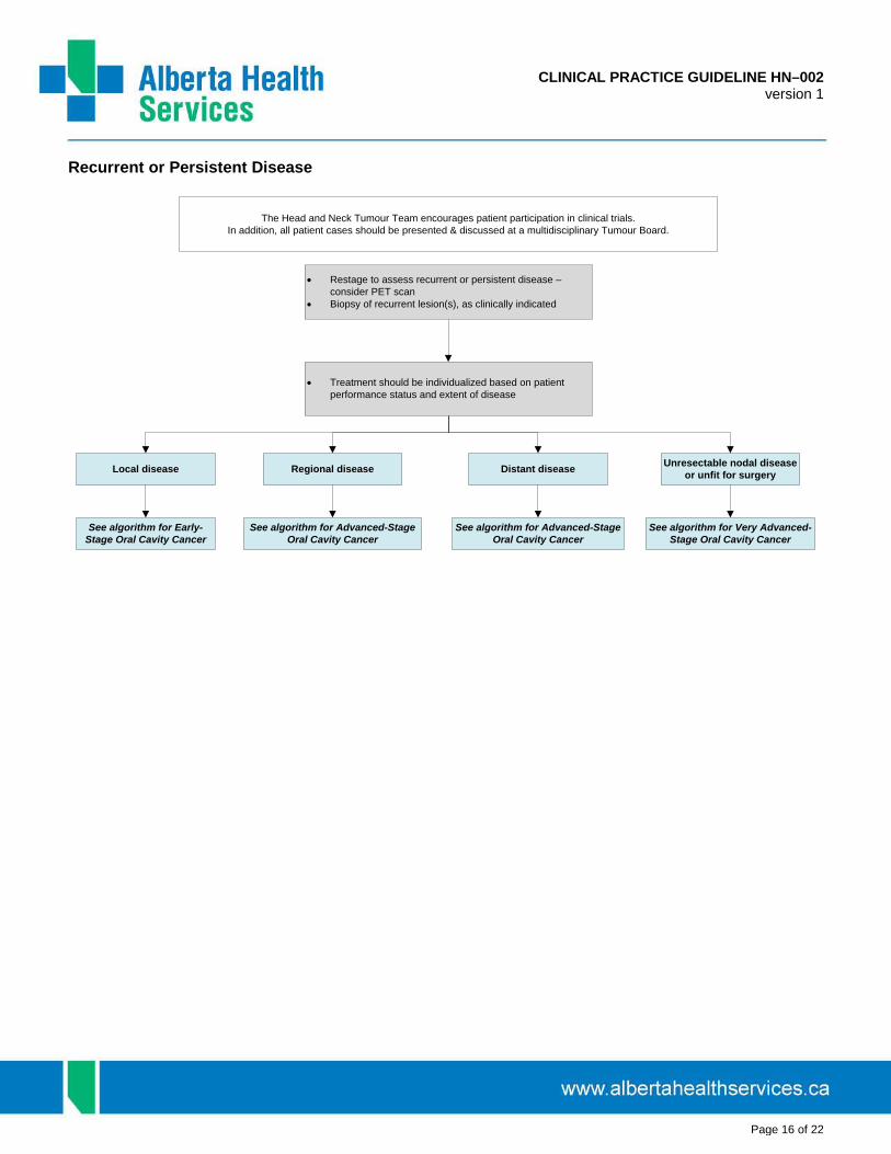
Recurrent or Persistent Disease
• Restage to assess recurrent or persistent disease – consider PET scan
• Biopsy of recurrent lesion(s), as clinically indicated
The Head and Neck Tumour Team encourages patient participation in clinical trials. In addition, all patient cases should be presented & discussed at a multidisciplinary Tumour Board.
Distant disease
• Treatment should be individualized based on patient performance status and extent of disease
Local disease Regional disease
See algorithm for Early-Stage Oral Cavity Cancer
See algorithm for Advanced-Stage Oral Cavity Cancer
Unresectable nodal disease or unfit for surgery
See algorithm for Advanced-Stage Oral Cavity Cancer
See algorithm for Very Advanced-Stage Oral Cavity Cancer
CLINICAL PRACTICE GUIDELINE HN–002
version 1
Page 17 of 22
GLOSSARY OF ABBREVIATIONS Acronym Description ChemoRT Chemoradiotherapy CT Computed tomography DFS Disease–free survival ECOG Eastern Cooperative Oncology Group EORTC European Organization for Research and Treatment of Cancer MRI Magnetic resonance imaging MeSH Medical Subject Heading NCCN National Comprehensive Cancer Network ND Neck dissection OS Overall survival PS Performance status PET–CT Positron emission tomography–computed tomography PORT Postoperative radiotherapy RCT Randomized controlled trial RT Radiotherapy RTOG Radiotherapy Oncology Group SCC Squamous cell carcinoma TNM Tumour, node, metastasis TSH Thyroid–stimulating hormone
DISSEMINATION • Present the guideline at the local and provincial tumour team meetings and weekly rounds. • Post the guideline on the Alberta Health Services website. • Send an electronic notification of the new guideline to all members of CancerControl Alberta. MAINTENANCE A formal review of the guideline will be conducted at the Annual Provincial Meeting in 2015. If critical new evidence is brought forward before that time, however, the guideline working group members will revise and update the document accordingly. CONFLICT OF INTEREST Participation of members of the Alberta Provincial Head and Neck Tumour Team in the development of this guideline has been voluntary and the authors have not been remunerated for their contributions. There was no direct industry involvement in the development or dissemination of this guideline. CancerControl Alberta recognizes that although industry support of research, education and other areas is necessary in order to advance patient care, such support may lead to potential conflicts of interest. Some members of the Alberta Provincial Head and Neck Tumour Team are involved in research funded by industry or have other such potential conflicts of interest. However the developers of this guideline are satisfied it was developed in an unbiased manner.
CLINICAL PRACTICE GUIDELINE HN–002
version 1
Page 18 of 22
REFERENCES 1. Rapidis A. Multidisciplinary management of oral cavity and maxillary sinus cancers. In: Bernier J, editor. Head
and neck cancer: multimodality management: Springer Science+Business Media, LLC; 2011. p. 363. 2. Shah JP, Gil Z. Current concepts in management of oral cancer--surgery. Oral Oncol 2009 Apr-May;45(4-5):394-
401. 3. Gilbert R, Devries-Aboud M, Winquist E, Waldron J, McQuestion M, the Head and Neck Disease Site Group.
The management of head and neck cancer in Ontario. Program in Evidence-Based Care, Cancer Care Ontario 2009;5-3.
4. Wolff KD, Follmann M, Nast A. The diagnosis and treatment of oral cavity cancer. Dtsch Arztebl Int 2012;109(48):829-835.
5. National Comprehensive Cancer Network. Head and Neck Cancers. Version I.2013. 2013; Available at: http://www.nccn.org/professionals/physician_gls/pdf/head-and-neck.pdf. Accessed 04/03, 2013.
6. Laramore GE, Scott CB, al-Sarraf M, Haselow RE, Ervin TJ, Wheeler R, et al. Adjuvant chemotherapy for resectable squamous cell carcinomas of the head and neck: report on Intergroup Study 0034. Int J Radiat Oncol Biol Phys 1992;23(4):705-713.
7. Zhang H, Dziegielewski PT, Biron VL, Szudek J, Al-Qahatani KH, O'Connell DA, et al. Survival outcomes of patients with advanced oral cavity squamous cell carcinoma treated with multimodal therapy: a multi-institutional analysis. J Otolaryngol Head Neck Surg 2013 Apr 19;42(1):30-0216-42-30.
8. Shah JP. Patterns of cervical lymph node metastasis from squamous carcinomas of the upper aerodigestive tract. Am J Surg 1990 Oct;160(4):405-409.
9. Ow TJ, Myers JN. Current management of advanced resectable oral cavity squamous cell carcinoma. Clin exp otorhinolaryngol 2011 Mar;4(1):1-10.
10. Bessell A, Glenny AM, Furness S, Clarkson JE, Oliver R, Conway DI, et al. Interventions for the treatment of oral and oropharyngeal cancers: surgical treatment. Cochrane Database Syst Rev 2011 Sep 7;(9):CD006205. doi(9):CD006205.
11. Spiro RH, Huvos AG, Wong GY, Spiro JD, Gnecco CA, Strong EW. Predictive value of tumor thickness in squamous carcinoma confined to the tongue and floor of the mouth. Am J Surg 1986 Oct;152(4):345-350.
12. Yuen AP, Lam KY, Wei WI, Lam KY, Ho CM, Chow TL, et al. A comparison of the prognostic significance of tumor diameter, length, width, thickness, area, volume, and clinicopathological features of oral tongue carcinoma. Am J Surg 2000 Aug;180(2):139-143.
13. Huang SH, Hwang D, Lockwood G, Goldstein DP, O'Sullivan B. Predictive value of tumor thickness for cervical lymph-node involvement in squamous cell carcinoma of the oral cavity: a meta-analysis of reported studies. Cancer 2009 Apr 1;115(7):1489-1497.
14. Ross GL, Soutar DS, Gordon MacDonald D, Shoaib T, Camilleri I, Roberton AG, et al. Sentinel node biopsy in head and neck cancer: preliminary results of a multicenter trial. Ann Surg Oncol 2004 Jul;11(7):690-696.
15. Civantos FJ, Zitsch RP, Schuller DE, Agrawal A, Smith RB, Nason R, et al. Sentinel lymph node biopsy accurately stages the regional lymph nodes for T1-T2 oral squamous cell carcinomas: results of a prospective multi-institutional trial. J Clin Oncol 2010 Mar 10;28(8):1395-1400.
16. Cooper JS, Pajak TF, Forastiere A, Jacobs J, Fu KK, Ang KK, et al. Precisely defining high-risk operable head and neck tumors based on RTOG #85-03 and #88-24: targets for postoperative radiochemotherapy?. Head Neck 1998 Oct;20(7):588-594.
17. Bernier J, Domenge C, Ozsahin M, Matuszewska K, Lefebvre JL, Greiner RH, et al. Postoperative irradiation with or without concomitant chemotherapy for locally advanced head and neck cancer. N Engl J Med 2004 May 6;350(19):1945-1952.
18. Cooper JS, Pajak TF, Forastiere AA, Jacobs J, Campbell BH, Saxman SB, et al. Postoperative concurrent radiotherapy and chemotherapy for high-risk squamous-cell carcinoma of the head and neck. N Engl J Med 2004 May 6;350(19):1937-1944.
19. Bernier J, Cooper JS, Pajak TF, van Glabbeke M, Bourhis J, Forastiere A, et al. Defining risk levels in locally advanced head and neck cancers: a comparative analysis of concurrent postoperative radiation plus chemotherapy trials of the EORTC (#22931) and RTOG (# 9501). Head Neck 2005 Oct;27(10):843-850.
20. Suarez-Cunqueiro M, Schramm A, Schoen R, Seoane-Leston J, Otero-Cepeda X, Bormann K, et al. Speech and swallowing impairment after treatment for oral and oropharyngeal cancer. 2008; . Accessed 12, 134.
CLINICAL PRACTICE GUIDELINE HN–002
version 1
Page 19 of 22
21. Mucke T, Wolff K, Wagenpfeil S, Mitchell DA, Holzle F. Immediate microsurgical reconstruction after tumor ablation predicts survival among patients with head and neck carcinoma. 2010; . Accessed 1, 17.
22. Dziegielewski PT, Ho ML, Rieger J, Singh P, Langille M, Harris JR, et al. Total glossectomy with laryngeal preservation and free flap reconstruction: objective functional outcomes and systematic review of the literature. Laryngoscope 2013 Jan;123(1):140-145.
23. Brown L, Rieger JM, Harris J, Seikaly H. A longitudinal study of functional outcomes after surgical resection and microvascular reconstruction for oral cancer: tongue mobility and swallowing function. J Oral Maxillofac Surg 2010 Nov;68(11):2690-2700.
24. Loewen IJ, Boliek CA, Harris J, Seikaly H, Rieger JM. Oral sensation and function: a comparison of patients with innervated radial forearm free flap reconstruction to healthy matched controls. Head Neck 2010 Jan;32(1):85-95.
25. Seikaly H, Rieger J, O'Connell D, Ansari K, Alqahtani K, Harris J. Beavertail modification of the radial forearm free flap in base of tongue reconstruction: technique and functional outcomes. Head Neck 2009 Feb;31(2):213-219.
26. O'Connell DA, Rieger J, Harris JR, Dziegielewski P, Zalmanowitz J, Sytsanko A, et al. Swallowing function in patients with base of tongue cancers treated with primary surgery and reconstructed with a modified radial forearm free flap. Arch Otolaryngol Head Neck Surg 2008 Aug;134(8):857-864.
27. Uwiera T, Seikaly H, Rieger J, Chau J, Harris JR. Functional outcomes after hemiglossectomy and reconstruction with a bilobed radial forearm free flap. J Otolaryngol 2004 Dec;33(6):356-359.
28. Yang TL, Wang CP, Ko JY, Lin CF, Lou PJ. Association of tumor satellite distance with prognosis and contralateral neck recurrence of tongue squamous cell carcinoma. Head Neck 2008 May;30(5):631-638.
29. Lim YC, Lee JS, Koo BS, Kim SH, Kim YH, Choi EC. Treatment of contralateral N0 neck in early squamous cell carcinoma of the oral tongue: elective neck dissection versus observation. Laryngoscope 2006 Mar;116(3):461-465.
30. Ganly I, Goldstein D, Carlson DL, Patel SG, O'Sullivan B, Lee N, et al. Long-term regional control and survival in patients with "low-risk," early stage oral tongue cancer managed by partial glossectomy and neck dissection without postoperative radiation: the importance of tumor thickness. Cancer 2013 Mar 15;119(6):1168-1176.
31. Bier-Laning CM, Durazo-Arvizu R, Muzaffar K, Petruzzelli GJ. Primary tumor thickness as a risk factor for contralateral cervical metastases in T1/T2 oral tongue squamous cell carcinoma. Laryngoscope 2009 May;119(5):883-888.
32. Yuen AP, Ho CM, Chow TL, Tang LC, Cheung WY, Ng RW, et al. Prospective randomized study of selective neck dissection versus observation for N0 neck of early tongue carcinoma. Head Neck 2009 Jun;31(6):765-772.
33. Parkin DM, Pisani P, Ferlay J. Estimates of the worldwide incidence of eighteen major cancers in 1985. Int J Cancer 1993 Jun 19;54(4):594-606.
34. Bachar G, Goldstein DP, Barker E, Lea J, O'Sullivan B, Brown DH, et al. Squamous cell carcinoma of the buccal mucosa: outcomes of treatment in the modern era. Laryngoscope 2012 Jul;122(7):1552-1557.
35. Diaz EM,Jr, Holsinger FC, Zuniga ER, Roberts DB, Sorensen DM. Squamous cell carcinoma of the buccal mucosa: one institution's experience with 119 previously untreated patients. Head Neck 2003 Apr;25(4):267-273.
36. Rodgers LW,Jr, Stringer SP, Mendenhall WM, Parsons JT, Cassisi NJ, Million RR. Management of squamous cell carcinoma of the floor of mouth. Head Neck 1993 Jan-Feb;15(1):16-19.
37. Mendenhall WM, Morris CG, Amdur RJ, Werning JW, Villaret DB. Retromolar trigone squamous cell carcinoma treated with radiotherapy alone or combined with surgery. Cancer 2005 Jun 1;103(11):2320-2325.
38. Werning JW, Mendenhall WM. Cancer of the hard palate and upper alveolar ridge. In: Werning JW, editor. Oral cancer: diagnosis, management, and rehabilitation: Thieme Medical Publishers, Inc.; 2007. p. 119.
39. Canadian Cancer Society. Follow-up after treatment for oral cavity cancer. 2013; Available at: http://www.cancer.ca/en/cancer-information/cancer-type/oral/treatment/follow-up/?region=ab. Accessed 10/03, 2013.
40. Manikantan K, Khode S, Dwivedi RC, Palav R, Nutting CM, Rhys-Evans P, et al. Making sense of post-treatment surveillance in head and neck cancer: when and what of follow-up. Cancer Treat Rev 2009 Dec;35(8):744-753.
41. Canadian Cancer Society. Supportive care for oral cavity cancer. 2013; Available at: http://www.cancer.ca/en/cancer-information/cancer-type/oral/supportive-care/?region=ab. Accessed 10/03, 2013.
42. Edge SB, Byrd DR, Compton CC, Fritz AG, Greene FL, Trotti A editors. AJCC Cancer Staging Manual. 7th ed. New York, NY: Springer; 2010.

CLINICAL PRACTICE GUIDELINE HN–002
version 1
Page 20 of 22
43. Oken MM, Creech RH, Tormey DC, Horton J, Davis TE, McFadden ET, et al. Toxicity and response criteria of the Eastern Cooperative Oncology Group. Am J Clin Oncol 1982 Dec;5(6):649-655.
CLINICAL PRACTICE GUIDELINE HN–002
version 1
Page 21 of 22
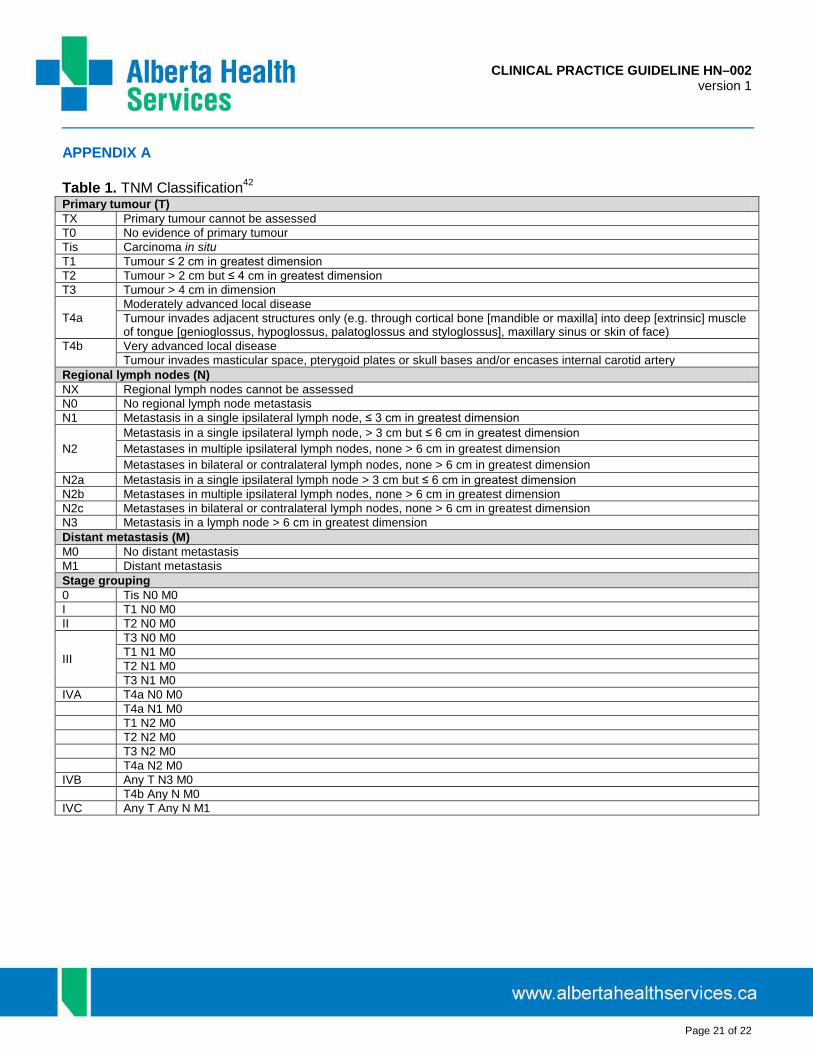
APPENDIX A Table 1. TNM Classification42 Primary tumour (T) TX Primary tumour cannot be assessed T0 No evidence of primary tumour Tis Carcinoma in situ T1 Tumour ≤ 2 cm in greatest dimension T2 Tumour > 2 cm but ≤ 4 cm in greatest dimension T3 Tumour > 4 cm in dimension
T4a Moderately advanced local disease Tumour invades adjacent structures only (e.g. through cortical bone [mandible or maxilla] into deep [extrinsic] muscle of tongue [genioglossus, hypoglossus, palatoglossus and styloglossus], maxillary sinus or skin of face)
T4b Very advanced local disease Tumour invades masticular space, pterygoid plates or skull bases and/or encases internal carotid artery
Regional lymph nodes (N) NX Regional lymph nodes cannot be assessed N0 No regional lymph node metastasis N1 Metastasis in a single ipsilateral lymph node, ≤ 3 cm in greatest dimension
N2 Metastasis in a single ipsilateral lymph node, > 3 cm but ≤ 6 cm in greatest dimension Metastases in multiple ipsilateral lymph nodes, none > 6 cm in greatest dimension Metastases in bilateral or contralateral lymph nodes, none > 6 cm in greatest dimension
N2a Metastasis in a single ipsilateral lymph node > 3 cm but ≤ 6 cm in greatest dimension N2b Metastases in multiple ipsilateral lymph nodes, none > 6 cm in greatest dimension N2c Metastases in bilateral or contralateral lymph nodes, none > 6 cm in greatest dimension N3 Metastasis in a lymph node > 6 cm in greatest dimension Distant metastasis (M) M0 No distant metastasis M1 Distant metastasis Stage grouping 0 Tis N0 M0 I T1 N0 M0 II T2 N0 M0
III
T3 N0 M0 T1 N1 M0 T2 N1 M0 T3 N1 M0
IVA T4a N0 M0 T4a N1 M0 T1 N2 M0 T2 N2 M0 T3 N2 M0 T4a N2 M0 IVB Any T N3 M0 T4b Any N M0 IVC Any T Any N M1
CLINICAL PRACTICE GUIDELINE HN–002
version 1
Page 22 of 22
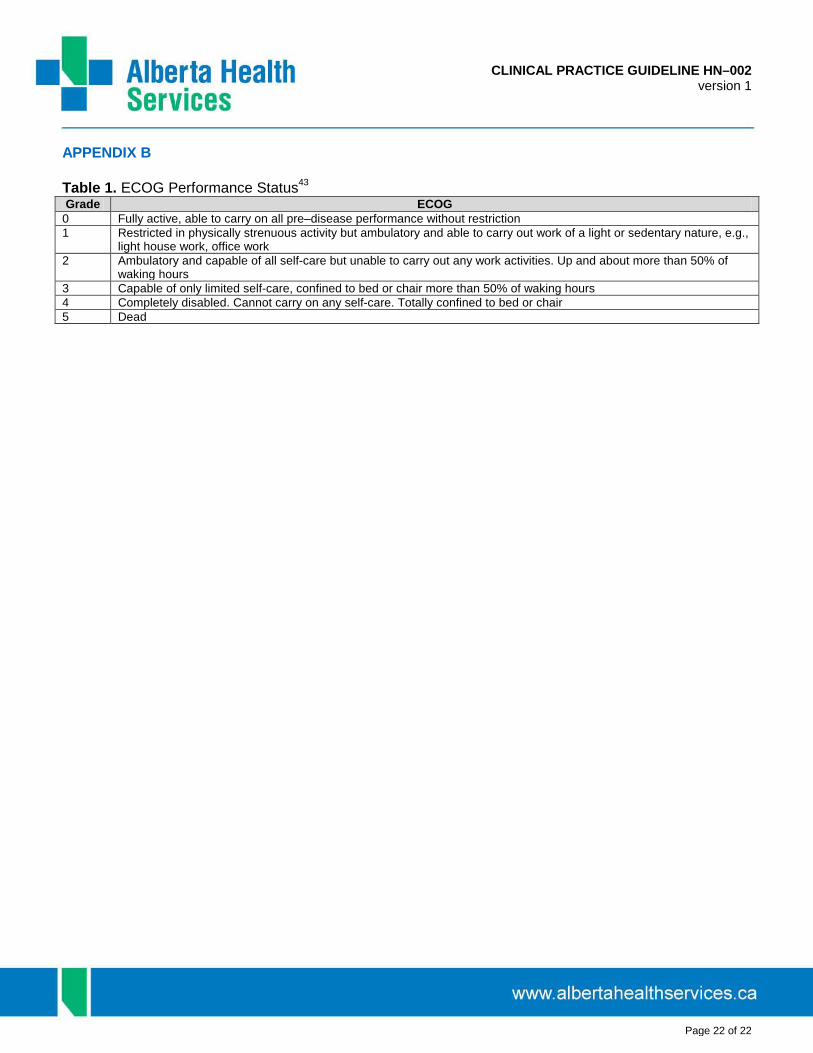
APPENDIX B Table 1. ECOG Performance Status43 Grade ECOG
0 Fully active, able to carry on all pre–disease performance without restriction 1 Restricted in physically strenuous activity but ambulatory and able to carry out work of a light or sedentary nature, e.g.,
light house work, office work 2 Ambulatory and capable of all self-care but unable to carry out any work activities. Up and about more than 50% of
waking hours 3 Capable of only limited self-care, confined to bed or chair more than 50% of waking hours 4 Completely disabled. Cannot carry on any self-care. Totally confined to bed or chair 5 Dead
Recommended