Am J Blood Res 2018;8(2):5-16www.AJBlood.us /ISSN:2160-1992/AJBR0078774
Original ArticleComparison of DNA demethylating and histone deacetylase inhibitors hydralazine-valproate versus vorinostat-decitabine incutaneous t-cell lymphoma in HUT78 cells
Alejandro Schcolnik-Cabrera1*, Guadalupe Domínguez-Gómez1*, Alfonso Dueñas-González2
1Division of Basic Research, Instituto Nacional De Cancerología, México; 2Unidad De Investigacion Biomédica En Cancer, Instituto De Investigaciones Biomédicas UNAM/Instituto Nacional De Cancerología, México. *Equal con-tributors.
Received April 30, 2018; Accepted May 31, 2018; Epub June 5, 2018; Published June 15, 2018
Abstract: Purpose: Cutaneous T-cell lymphoma (CTCL) is an uncommon extranodal non-Hodgkin T-cell lymphoma that originates from mature T lymphocytes homed at the skin. Epigenetic alterations observed in CTCL are not limited to overexpression of Histone Deacetylases but also to DNA hypermethylation. The known synergy between Histone deacetylase inhibitors (HDACi) and DNA methyltransferases inhibitors (DNMTi) suggests that combining these agent classes could be effective for CTCL. Methods: In this study, the combinations of the HDACi and DNMTi hydralazine/valproate (HV) and vorinostat/decitabine (VD) were compared in regard to viability inhibition, clonoge-nicity, pharmacological interaction and cell cycle effects in the CTCL cell line Hut78. In addition, the effect of these combinations was evaluated in normal peripheral blood mononuclear cells. Results: The results show that each of the DNMTi and HDACi exerts growth inhibition, mostly by inducing apoptosis as shown in the cell cycle distribution. However, in the combination of HV the interaction is more synergic and also it inhibits the clonogenic capacity of cells over time. Additionally, the HV combination seems to affect in a minor degree the viability of peripheral blood mononuclear cells. Conclusions: The results of this study and the preclinical and clinical evidence on the efficacy of combining HDACi with DNMTi strongly suggest that more studies are needed with this drug class combination in CTCL, particularly with the hydralazine-valproate scheme, which is safe, and these drugs are widely available and administered by oral route.
Keywords: CTCL, HDACi, DNMTi, hydralazine, valproate, vorinostat, decitabine, synergy
Introduction
Cutaneous T-cell lymphoma (CTCL) is an uncom-mon extranodal non-Hodgkin T-cell lymphoma that originates from mature T lymphocytes ho- med at the skin, and represents the majority of all primary cutaneous lymphomas. Among them, mycosis fungoides (MF) accounts for al- most 50% of all primary cutaneous lympho-mas. This lymphoma is characterized by hav- ing a chronic and relapsing course, and since there are no curative modalities with the pos-sible exception of patients with minimal patch and plaque lesions confined to the skin, the treatment, which includes external beam ra- diation, phototherapy, chemotherapy and bio-logical agents, is used sequentially for the pa- tients [1]. Most patients with limited disease burden, in general, show long-term survival and
are commonly treated with corticosteroids, top-ical nitrogen mustard, phototherapy and other forms of systemic treatments. Systemic appro- aches such as bexarotene, denileukin diftitox, alemtuzumab, lenalidomide, Toll-like receptor agonists, pralatrexate, bortezomib, vorinostat, romidepsin, as well as cytotoxic chemothera- py, are frequently employed in patients with advanced and erythrodermic MF [2, 3].
CTCL overexpresses several histone deacety-lases (HDACs), which include HDACs 1, 2, and 6, and this disease is highly sensitive to HDAC inhibitors (HDACi) [4, 5]. However, the epigene-tic alterations observed in CTCL are not limited to overexpression of HDACs but also to DNA hypermethylation [6]. On this regard, van Doorn et al. by using differential methylation hybridi- zation, reported on 28 CTCL samples, 35 CpG
In vitro comparison of hydralazine valproate versus vorinostat decitabine in CTCL
6 Am J Blood Res 2018;8(2):5-16
(cytosine-guanine) islands hypermethylated in at least 4 of these 28 samples, plus 6 of 8 genes hypermethylated and analyzed with a candidate-gene approach. Of note, treatment with the DNA methyltransferase inhibitor (DN- MTi) decitabine led to demethylation and re-expression of BCL7a and PTPRG, in the CTCL cell line MyLa [7].
This observation, and the known synergy be- tween HDACi and DNMTi [8, 9], suggests that combining these agent classes could be effec-tive for CTCL. In fact, preclinical studies com-bining these two agent classes have been un- dertaken, with promising results in lymphoma [10, 11]. However, in our knowledge, only the drug combination of romidepsin with azaciti- dine has been preclinically tested in CTCL [12]. The fact that the HDACi valproate in combina-tion with the DNMTi hydralazine has shown promising efficacy in patients with CTCL, and because they are well tolerated, led us to com-pare this pharmacological combination against the one HDACi vorinostat and the DNMTi de- citabinein the Hut78 CTCL cancer cell line. Our results demonstrate that hydralazine and val-proate exert more synergism than decitabine and vorinostat against CTCL cells with similar cell cycle effects, but a higher reduction in clo-nogenic capacity of treated cells over time.
Materials and methods
Cell line
The CTCL cell line Hut78 was obtained from the ATCC (Manassas, VA, USA). Hut78 cell line and healthy leukocytes recovered from buffy coats were cultured at 37°C in a humidified atmo-sphere containing 5% CO2 in complete medium, composed of RPMI-1640 medium supplement-ed with 10% fetal bovine serum and 1% antibi-otic-antimycotic solution (all from Invitrogen Life Technologies, Carlsbad, CA, USA).
Healthy leukocytes
Buffy coats were kindly obtained from the blood bank of the Instituto Nacional de Cancerología (Mexico) for leukocyte recovery. Briefly, buffy coats contained in heparinized Terumo bags (Terumo BCT, Lakewood, CO, USA) were treat- ed at 1:1 ratio with ACK lysing buffer (Life Technologies), mixed for 5 min in a platform mixer and then centrifuged at 250 G during 20 min to lyse the erythrocytic content. This pro-cess was repeated 3 times until a free-erythro-cyte white pellet-containing leukocyte was ob-
tained. Cells were then resuspended in RPMI-1640 complete medium and used for viability assay experiments.
Viability curves in the Hut78 cell line
Hut78 cells were seeded into 12-well microtiter plates (Corning) at a density of 5×10⁴ cells/well into 1 mL complete medium. The following day, cells were treated for a 72 hour-period with either hydralazine (Sigma, St. Louis, MO, USA), magnesium valproate (Psicofarma, México), vorinostat (Sigma, St. Louis, MO, USA), or de- citabine (Sigma, St. Louis, MO, USA) at escalat-ing doses. All the compounds but vorinostat were resuspended in distilled water, and the latter was resuspended in absolute ethanol (Sigma, St. Louis, MO, USA). Fresh complete medium containing each drug was changed every 24 hours by recovering all the content of each well, with posterior centrifugation (5 min at 120 G), decantation of the supernatant and resuspension of the cell pellet in the medi-um-containing drug. After 72 hours of treat-ment, pellets were recovered and resuspend- ed in 1 mL RPMI-1640 medium to assess cell viability by trypan blue exclusion assay. Briefly, resuspended cells were gently mixed at 1:1 ratio with trypan blue stain solution (Life Te- chnologies) and then cell viability was evaluat-ed with a TC10TM Automated Cell Counter (Bio- Rad). The cytotoxic effect of each treatment was expressed as a percentage of cell viabi- lity relative to control cells treated with the vehicle of each compound at the highest eva- luated dose. The data of the dose-response curve at different concentrations per drug was plotted in the SigmaPlot software 10.0. The percentage of growth inhibition was calculated, and IC20-IC50 values (the concentration of each drug that achieves 20%-50% growth inhibition) were graphically obtained from the survival curves.
Pharmacological interactions
Increasing doses of hydralazine (IC20, IC30, IC40, and IC50) were combined with their respective increasing doses of valproic acid (IC20, IC30, IC40, and IC50). At the same time, increasing doses of vorinostat (IC20, IC30, IC40, and IC50) were combined with their respective increasing doses of decitabine (IC20, IC30, IC40, and IC50). The resulting mixes (HV IC20, HV IC30, HV IC40, HV IC50 for hydralazine and valproate, and VD IC20, VD IC30, VD IC40, VD IC50 for vorinostat and decitabine) were employed for viability curve
In vitro comparison of hydralazine valproate versus vorinostat decitabine in CTCL
7 Am J Blood Res 2018;8(2):5-16
assays. Cells were seeded into 12-well microti-ter plates at 5×10⁴ cells/well with 1 mL of com-plete medium, and then treated for a 72 hour-period with the different combinations of the ICs of HV and VD. After 72 hours, pellets were recovered and resuspended in 1 mL RPMI-1640 medium to assess cell viability by trypan blue exclusion assay.
Synergism evaluation
HV and VD interactions were determined using the combination index (CI) method from the mathematical formula of Chou and Talalay (CalcuSyn software, Biosoft). The CI is a numer-ical representation of the pharmacological interaction, which takes into account the dose-response curve of each drug and the growth inhibition curve of the drug combination to determine synergism, addition or inhibition. This model uses the formula fa/fu = [D/Dm], where fa is the fraction of cells inhibited, fu = 1-fa, the unaffected fraction, D is the concen- tration of the drug, Dm is the potency of the drug, and m is the shape of the dose-effect curve. Table 1 shows the interpretation of CI values with the corresponding pharmacologi- cal interaction.
Cell cycle assays
After performing combinatory viability assays with the most synergistic doses, cells were stained with propidium iodide (Sigma) for 1 h and analyzed for DNA content on the flow cytometer BD FACSCantoTM II (BD Biosciences). Debris and aggregates were gated out during
data acquisition, and 20,000 gated events we- re collected for each sample. Cell cycle analy-ses were performed employing the ModFit LT software (Verity Software House). Results are expressed in percentage of cells for each cell phase.
Clonogenic capacity assays
After performing combinatory viability assays with the most synergistic doses, remaining cells were seeded into 25 cm2 cell culture fla- sks (Corning) at a density of 2×10³ cells/flask into 5 mL complete medium. The whole medi-um was changed every 48 hours with a fresh complete medium. Cells were let growing dur-ing 2 weeks and then viability was measured by trypan blue exclusion assay.
Viability evaluation in healthy leukocytes
The most synergistic doses of each drug combi-nation were employed during a 72 h-period in healthy leukocytes to evaluate viability. Healthy leukocytes were seeded into 12-well microtiter plates at a density of 5×10⁴ cells/well into 1 mL complete medium. The following day, cells were treated for a 72 hour-period with either HV or VD at the most synergic doses, and the medi-um with the respective drug mixture was ch- anged every 24 hours as with the Hut78 cell line. After 72 hours of treatment, cellular via- bility was measured by trypan blue exclusion assay.
Statistical analysis
Three independent experiments were perform- ed and data was expressed as means ± SD. Data were statistically analyzed by using Gra- phPad Prism V6 software (GraphPad Software Inc., La Jolla, CA, USA). Significant differences were determined using one-way analysis of variance (ANOVA) followed by Tukey correction to determine significant difference between each test group against its respective control. A P value of <0.05 was considered statistically significant.
Results
Individual DNMTi and HDACi agents diminish in a dose-dependent manner cellular viability in the Hut78 lymphoma cell line
To demonstrate that the epigenetic agents, either individual DNMTi or HDACi decrease the
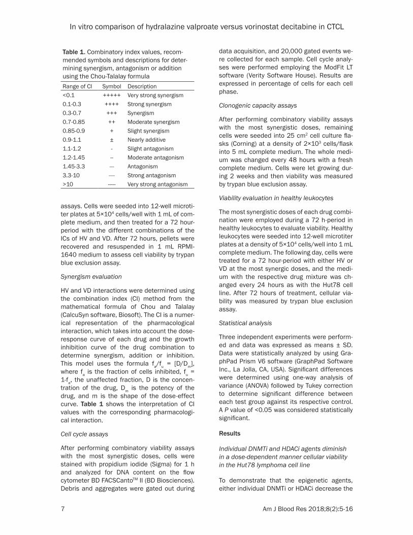
Table 1. Combinatory index values, recom-mended symbols and descriptions for deter-mining synergism, antagonism or addition using the Chou-Talalay formulaRange of CI Symbol Description<0.1 +++++ Very strong synergism0.1-0.3 ++++ Strong synergism0.3-0.7 +++ Synergism0.7-0.85 ++ Moderate synergism0.85-0.9 + Slight synergism0.9-1.1 ± Nearly additive1.1-1.2 - Slight antagonism1.2-1.45 -- Moderate antagonism1.45-3.3 --- Antagonism3.3-10 ---- Strong antagonism>10 ----- Very strong antagonism
In vitro comparison of hydralazine valproate versus vorinostat decitabine in CTCL
8 Am J Blood Res 2018;8(2):5-16
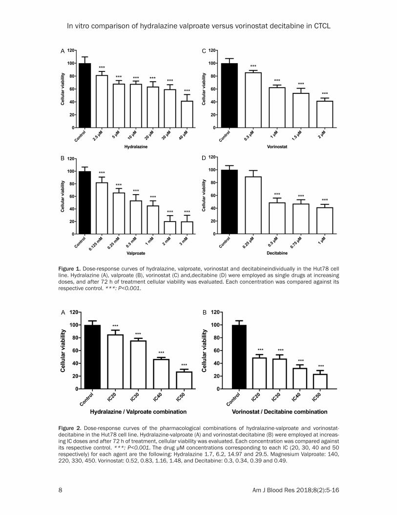
Figure 1. Dose-response curves of hydralazine, valproate, vorinostat and decitabineindividually in the Hut78 cell line. Hydralazine (A), valproate (B), vorinostat (C) and,decitabine (D) were employed as single drugs at increasing doses, and after 72 h of treatment cellular viability was evaluated. Each concentration was compared against its respective control. ***: P<0.001.
Figure 2. Dose-response curves of the pharmacological combinations of hydralazine-valproate and vorinostat-decitabine in the Hut78 cell line. Hydralazine-valproate (A) and vorinostat-decitabine (B) were employed at increas-ing IC doses and after 72 h of treatment, cellular viability was evaluated. Each concentration was compared against its respective control. ***: P<0.001. The drug µM concentrations corresponding to each IC (20, 30, 40 and 50 respectively) for each agent are the following: Hydralazine 1.7, 6.2, 14.97 and 29.5. Magnesium Valproate: 140, 220, 330, 450. Vorinostat: 0.52, 0.83, 1.16, 1.48, and Decitabine: 0.3, 0.34, 0.39 and 0.49.
In vitro comparison of hydralazine valproate versus vorinostat decitabine in CTCL
9 Am J Blood Res 2018;8(2):5-16
cellular viability of the CTCL cell line Hut78, cells were treated with increasing doses of hydralazine, valproate, vorinostat or decitabine. As shown in Figure 1, hydralazine shows its inhibitory effects starting at 2.5 µM in a dose-dependent manner. These effects were statisti-cally significant. The inhibition with valproate
was observed since 0.125 mM and was also dose-dependent. At doses higher than 3mM, viability was 0% (data not shown). Both vorino-stat and decitabine also inhibited viability in a dose-dependent manner. For vorinostat, signifi-cant inhibition started at 0.5 µM and the high-est effect was seen at 2 µM. The highest effect
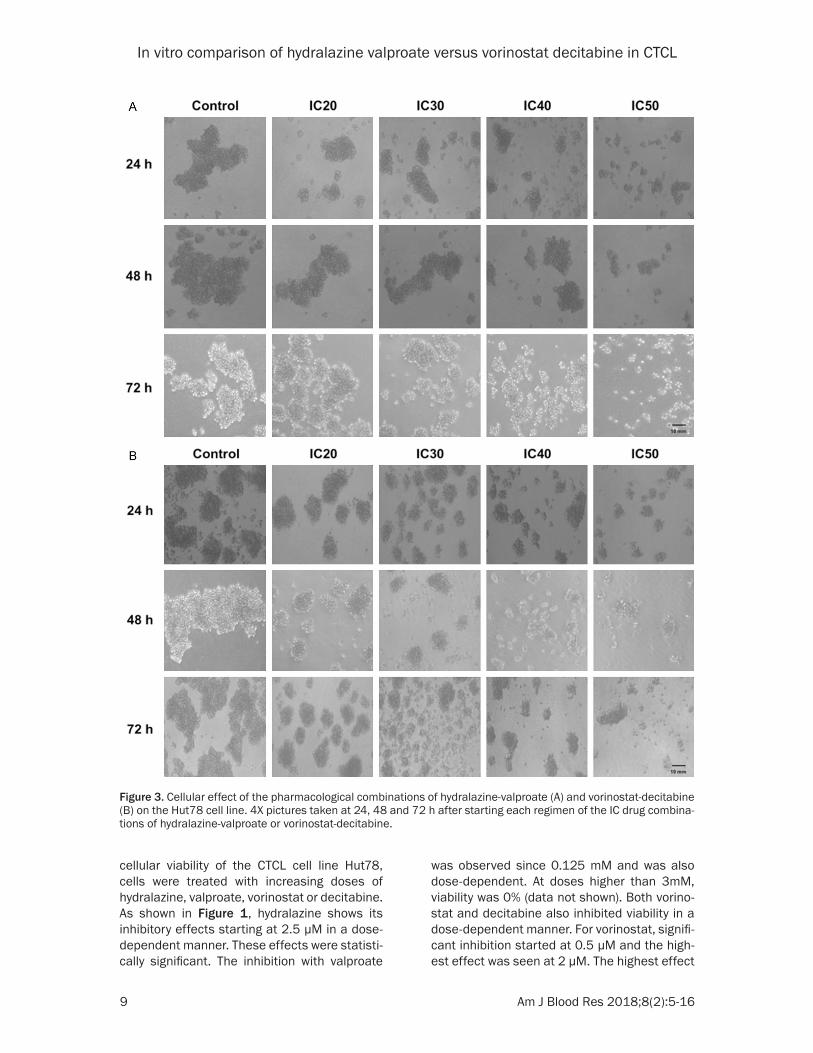
Figure 3. Cellular effect of the pharmacological combinations of hydralazine-valproate (A) and vorinostat-decitabine (B) on the Hut78 cell line. 4X pictures taken at 24, 48 and 72 h after starting each regimen of the IC drug combina-tions of hydralazine-valproate or vorinostat-decitabine.
In vitro comparison of hydralazine valproate versus vorinostat decitabine in CTCL
10 Am J Blood Res 2018;8(2):5-16
of decitabine was observed at 1 µM, but inhibi-tion started at 0.25 µM.
The combinations of hydralazine-valproate and of vorinostat-decitabine work in a synergistic way to reduce cellular viability
In order to determine the pharmacological in- teraction between these agents, the IC20, IC30, IC40 and IC50 doses for each of the agents we- re determined using the SigmaPlot software.
clearly above 1, indicative of an antagonistic relationship.
From this point on, we used the most synergic doses of each drug mixture, named HV IC50 and VD IC20, for further experiments. As a way to compare doses with both drug combinations, we employed the conditions HV IC20, HV IC50, VD IC20 and VD IC50. Since vorinostat is resuspend-ed in pure ethanol, we generated two controls for VD combinations, one for the VD IC20 and
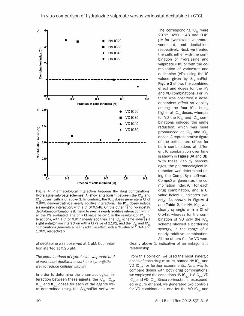
Figure 4. Pharmacological interaction between the drug combinations. Hydralazine-valproate schemes (A) show antagonism between the IC20 and IC30 doses, with a CI above 3. In contrast, the IC40 doses generate a CI of 0.999, demonstrating a nearly additive interaction. The IC50 doses induce a synergistic interaction, with a CI 0f 0.548. On the other hand, vorinostat-decitabinecombinations (B) tend to exert a nearly additive interaction within all the ICs evaluated. The only CI value below 1 is the resulting of IC20 in-teractions, with a CI of 0.957 (nearly additive). The IC30 scheme induces a slight antagonism interaction with a CI value of 1.192, and the IC40 and IC50 combinations generate a nearly additive effect with a CI value of 1.074 and 1.069, respectively.
The corresponding IC50 were 29.95, 450, 1.48 and 0.49 µM for hydralazine, valproate, vorinostat, and decitabine, respectively. Next, we treated the cells either with the com-bination of hydralazine and valproate (HV) or with the co- mbination of vorinostat and decitabine (VD), using the IC values given by SigmaPlot. Figure 2 shows the combined effect and doses for the HV and VD combinations. For HV there was observed a dose-dependent effect on viability among the four ICs, being higher at IC50 doses, whereas for VD the IC20 and IC30 com- binations induced the same reduction, which was more pronounced at IC40 and IC50 doses. A representative figure of the cell culture effect for both combinations at differ-ent IC combination over time is shown in Figure 3A and 3B. With these viability percent-ages, the pharmacological in- teraction was determined us- ing the CompuSyn software. CompuSyn generates the co- mbination index (CI) for each drug combination, and a CI value below 1 indicates syn-ergy. As shown in Figure 4 and Table 2, for HV, IC50 was clearly synergic with a CI of 0.548, whereas for the com- bination of VD only the IC20 scheme showed a borderline synergy, in the range of a nearly additive combination. All the others CIs for VD were
In vitro comparison of hydralazine valproate versus vorinostat decitabine in CTCL
11 Am J Blood Res 2018;8(2):5-16
the other for the VD IC50 schemes. Therefore, we used the same volume of ethanol required for vorinostat at IC20 dose for its control, while employing the same volume of ethanol requir- ed for vorinostat at IC50 dose for its control. Regarding HV combinations, because both hy- dralazine and valproate are resuspended in distilled water, we used only one control with the same volume of water as the employed in the HV IC50 dose for comparison.
Hydralazine-valproate and vorinostat-decitabine combinations significantly promote cell death as demonstrated by flow cytometry
To further investigate the effect of these treat-ments in cell cycle, flow cytometry assays with propidium iodide were performed with IC20 and IC50 doses of the HV and VD schemes. For both combinations at IC20 of doses, there were no statistically significant differences in cell cycle distribution. However, when treated with IC50 doses, both couple of drugs significantly incre- ased the percentage of cell death, in a very similar manner (Figure 5).
The hydralazine-valproate IC50 drug scheme promotes a maintained reduction in the clono-genic capacity of treated cells over time
To determine the cellular effects of these inter-actions upon the clonogenic capacity of treat-
ed cells, clonogenic assays with cellular viabili-ty evaluation were performed for both combina-tions with the IC20 and IC50 doses. As shown in Figure 6, a small effect on cellular viability was observed for the IC20 doses for HV, but it was clearly more marked and statistically signifi-cant when IC50 doses were employed. On the other hand, no viability reduction was obser- ved with either IC20 or IC50 doses for VD.
Healthy PMNCs are resistant to both hydrala-zine-valproate and vorinostat-decitabine com-binations
Finally, to determine which of these combina-tions would affect more to normal cells, freshly isolated peripheral blood mononuclear cells (PBMNCs) were treated with both HV and VD combinations with either IC20 or IC50 doses, as shown in Figure 7. No differences in cellular viability were seen with any of the drug sche- mes at IC20 concentrations, as compared with the respective controls. However, there was observed a higher decrease in cellular viability with both treatments at IC50 doses, being slight-ly more marked with the combination of VD, although none of them demonstrated a statisti-cally significant difference.
Discussion
In this in vitro study in which the antitumor effects of different epigenetic agents were evaluated in the Hut78 CTCL cancer cell line, the results show that each of the DNMTi and HDACi exerts growth inhibition, mostly by induc-ing apoptosis as shown in the cell cycle distri-bution. However, in the combination of HV the interaction is more synergic and also it inhibits the clonogenic capacity of cells over time. Additionally, the HV combination seems to af- fect in a minor degree the viability of periphe- ral blood mononuclear cells.
The therapy of CTCL is challenging since even with the use of HDACi as single agents the re- sponse rates are below 40%. Beyond the clini-cal study with hydralazine and valproate in CTCL [13], there are yet no clinical head-to-he- ad comparisons of different HDACi, nor clinical studies of any HDACi combined with any DN- MTi for CTCL. However, preclinical studies in a model of CTCL demonstrate that the HDACi romidepsin and the DNMTi azacitidine are syn-ergic in their epigenetic modulatory effects and apoptosis [12]. Likewise, but in a model of dif-
Table 2. Combinatory index (CI) values per drug combinationDrug combination fa CI
HV IC20 0.146746 3.408HV IC30 0.241617 3.329HV IC40 0.531805 0.999HV IC50 0.726923 0.548VD IC20 0.507458 0.957VD IC30 0.525263 1.192VD IC40 0.671353 1.074VD IC50 0.76435 1.069
Drug interactions were evaluated with the Cal-cusyn software by comparing the individual drug and the drug combinations effects on cellular viability. CI values >1 are indicative of antago-nistic relationships, while <1 are synergistic. CI values close to 1 are summative. HV: hydralazine + valproate; VD: vorinostat + decitabine; fa: frac-tion of affected cells, or percentage of reduction in cellular viability as compared with controls; CI: combinatory index.
In vitro comparison of hydralazine valproate versus vorinostat decitabine in CTCL
12 Am J Blood Res 2018;8(2):5-16
fuse large B-cell lymphoma (DLBCL), the combi-nation of panobinostat with decitabine also results in synergic growth inhibition and apop-tosis [10]. The results here reported on the higher synergy demonstrated by the pharmaco-logical interaction with hydralazine and valpro-ate support the results of a recently reported
On the other hand, in estrogen receptor (ER) negative breast cancer cells, decitabine and trichostatin A increase up to 300-400 fold the expression of the ER gene [16]. In ovarian can-cer, decitabine and vorinostat show G2/M ar- rest and apoptosis in cancer cell lines, while in xenografts they induce the expression of
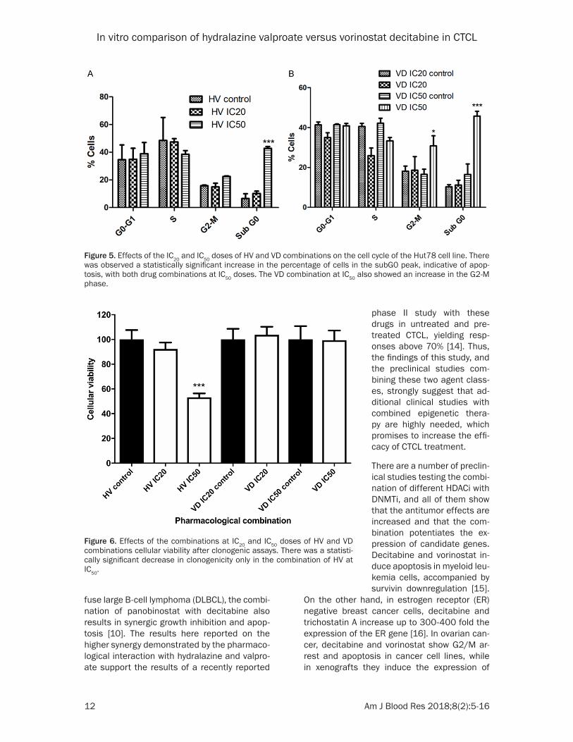
Figure 5. Effects of the IC20 and IC50 doses of HV and VD combinations on the cell cycle of the Hut78 cell line. There was observed a statistically significant increase in the percentage of cells in the subG0 peak, indicative of apop-tosis, with both drug combinations at IC50 doses. The VD combination at IC50 also showed an increase in the G2-M phase.
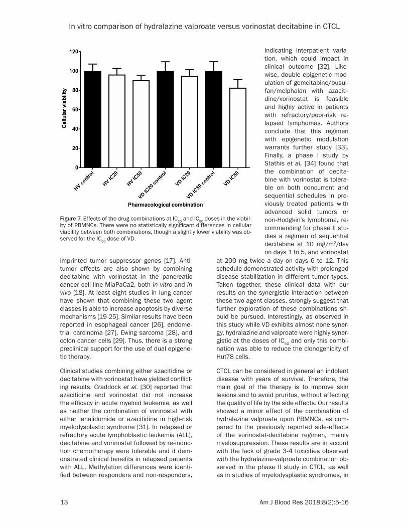
Figure 6. Effects of the combinations at IC20 and IC50 doses of HV and VD combinations cellular viability after clonogenic assays. There was a statisti-cally significant decrease in clonogenicity only in the combination of HV at IC50.
phase II study with these drugs in untreated and pre-treated CTCL, yielding resp- onses above 70% [14]. Thus, the findings of this study, and the preclinical studies com-bining these two agent class-es, strongly suggest that ad- ditional clinical studies with combined epigenetic thera- py are highly needed, which promises to increase the effi-cacy of CTCL treatment.
There are a number of preclin-ical studies testing the combi-nation of different HDACi with DNMTi, and all of them show that the antitumor effects are increased and that the com- bination potentiates the ex- pression of candidate genes. Decitabine and vorinostat in- duce apoptosis in myeloid leu-kemia cells, accompanied by survivin downregulation [15].
In vitro comparison of hydralazine valproate versus vorinostat decitabine in CTCL
13 Am J Blood Res 2018;8(2):5-16
imprinted tumor suppressor genes [17]. Anti- tumor effects are also shown by combining decitabine with vorinostat in the pancreatic cancer cell line MiaPaCa2, both in vitro and in vivo [18]. At least eight studies in lung cancer have shown that combining these two agent classes is able to increase apoptosis by diverse mechanisms [19-25]. Similar results have been reported in esophageal cancer [26], endome-trial carcinoma [27], Ewing sarcoma [28], and colon cancer cells [29]. Thus, there is a strong preclinical support for the use of dual epigene-tic therapy.
Clinical studies combining either azacitidine or decitabine with vorinostat have yielded conflict-ing results. Craddock et al. [30] reported that azacitidine and vorinostat did not increase the efficacy in acute myeloid leukemia, as well as neither the combination of vorinostat with either lenalidomide or azacitidine in high-risk myelodysplastic syndrome [31]. In relapsed or refractory acute lymphoblastic leukemia (ALL), decitabine and vorinostat followed by re-induc-tion chemotherapy were tolerable and it dem-onstrated clinical benefits in relapsed patients with ALL. Methylation differences were identi-fied between responders and non-responders,
at 200 mg twice a day on days 6 to 12. This schedule demonstrated activity with prolonged disease stabilization in different tumor types. Taken together, these clinical data with our results on the synergistic interaction between these two agent classes, strongly suggest that further exploration of these combinations sh- ould be pursued. Interestingly, as observed in this study while VD exhibits almost none syner-gy, hydralazine and valproate were highly syner-gistic at the doses of IC50 and only this combi-nation was able to reduce the clonogenicity of Hut78 cells.
CTCL can be considered in general an indolent disease with years of survival. Therefore, the main goal of the therapy is to improve skin lesions and to avoid pruritus, without affecting the quality of life by the side effects. Our results showed a minor effect of the combination of hydralazine valproate upon PBMNCs, as com- pared to the previously reported side-effects of the vorinostat-decitabine regimen, mainly myelosuppression. These results are in accord with the lack of grade 3-4 toxicities observed with the hydralazine-valproate combination ob- served in the phase II study in CTCL, as well as in studies of myelodysplastic syndromes, in
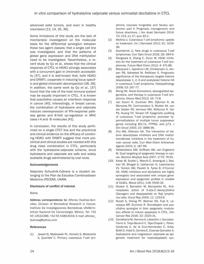
Figure 7. Effects of the drug combinations at IC20 and IC50 doses in the viabil-ity of PBMNCs. There were no statistically significant differences in cellular viability between both combinations, though a slightly lower viability was ob-served for the IC50 dose of VD.
indicating interpatient varia-tion, which could impact in clinical outcome [32]. Like- wise, double epigenetic mod-ulation of gemcitabine/busul-fan/melphalan with azaciti-dine/vorinostat is feasible and highly active in patients with refractory/poor-risk re- lapsed lymphomas. Authors conclude that this regimen with epigenetic modulation warrants further study [33]. Finally, a phase I study by Stathis et al. [34] found that the combination of decita- bine with vorinostat is tolera-ble on both concurrent and sequential schedules in pre- viously treated patients with advanced solid tumors or non-Hodgkin’s lymphoma, re- commending for phase II stu- dies a regimen of sequential decitabine at 10 mg/m2/day on days 1 to 5, and vorinostat
In vitro comparison of hydralazine valproate versus vorinostat decitabine in CTCL
14 Am J Blood Res 2018;8(2):5-16
advanced solid tumors, and even in healthy volunteers [13, 14, 35, 36].
Some limitations of this study are the lack of mechanistic investigation on the molecular basis for the differential synergism between these two agent classes; that a single cell line was investigated, and that the patterns of global gene expression and DNA methylation need to be investigated. Nevertheless, a re- cent study by Qu et al., shows that the clinical response of CTCL to HDACi strongly associates with a concurrent gain in chromatin accessibil-ity [37], and it is well-known that, both HDACi and DNMTi, cooperate in inducing locus specif-ic and global chromatin decompaction [38, 39]. In addition, the same work by Qu et al., [37] found that the role of the host immune system may be equally important in CTCL. It is known that azacitidine causes an interferon response in cancer [40]. Interestingly, in breast cancer, the combination of hydralazine and valproate induces overexpression of IFN-response path-way genes and 8-fold up-regulation in MHC class I-A and -B molecules [41].
In conclusion, the results of this study perfo- rmed on a single CTCT line and the preclinical and clinical evidence on the efficacy of combin-ing HDACi with DNMTi suggest that more pre-clinical and clinical studies are needed with this drug class combination in CTCL, particularly with the hydralazine-valproate scheme, since hydralazine and valproate are safe and widely available drugs administered by oral route.
Acknowledgements
Alejandro Schcolnik-Cabrera is a student be- longing to the Plan de Estudios Combinadosen Medicina (PECEM), UNAM.
Disclosure of conflict of interest
None.
Address correspondence to: Alfonso Dueñas-Gon- zález, Division of Biomedical Research in Cancer, Instituto De Investigaciones Biomédicas UNAM/In- stituto Nacional De Cancerología, México. Tel: +52 55 13532386; +52 55 54853034; E-mail: [email protected]
References
[1] Jawed SI, Myskowski PL, Horwitz S, Moskowitz A, Querfeld C. Primary cutaneous T-cell lym-
phoma (mycosis fungoides and Sezary syn-drome): part II. Prognosis, management, and future directions. J Am Acad Dermatol 2014; 70: 223, e1-17; quiz 40-2.
[2] Wollina U. Cutaneous T cell lymphoma: update on treatment. Int J Dermatol 2012; 51: 1019-36.
[3] Scarisbrick JJ. New drugs in cutaneous T-cell lymphomas. Curr Opin Oncol 2016; 28: 384-9.
[4] Rangwala S, Zhang C, Duvic M. HDAC inhibi-tors for the treatment of cutaneous T-cell lym-phomas. Future Med Chem 2012; 4: 471-86.
[5] Marquard L, Gjerdrum LM, Christensen IJ, Jen-sen PB, Sehested M, Ralfkiaer E. Prognostic significance of the therapeutic targets histone deacetylase 1, 2, 6 and acetylated histone H4 in cutaneous T-cell lymphoma. Histopathology 2008; 53: 267-77.
[6] Wong HK. Novel biomarkers, dysregulated epi-genetics, and therapy in cutaneous T-cell lym-phoma. Discov Med 2013; 16: 71-8.
[7] van Doorn R, Zoutman WH, Dijkman R, de Menezes RX, Commandeur S, Mulder AA, van der Velden PA, Vermeer MH, Willemze R, Yan PS, Huang TH, Tensen CP. Epigenetic profiling of cutaneous T-cell lymphoma: promoter hy- permethylation of multiple tumor suppressor genes including BCL7a, PTPRG, and p73. J Clin Oncol 2005; 23: 3886-96.
[8] Zhu WG, Otterson GA. The interaction of his-tone deacetylase inhibitors and DNA methyl-transferase inhibitors in the treatment of hu-man cancer cells. Curr Med Chem Anticancer Agents 2003; 3: 187-99.
[9] Hellebrekers DM, Griffioen AW, van Engeland M. Dual targeting of epigenetic therapy in can-cer. Biochim Biophys Acta 2007; 1775: 76-91.
[10] Kalac M, Scotto L, Marchi E, Amengual J, Ses-han VE, Bhagat G, Ulahannan N, Leshchenko VV, Temkin AM, Parekh S, Tycko B, O’Connor OA. HDAC inhibitors and decitabine are highly synergistic and associated with unique gene-expression and epigenetic profiles in models of DLBCL. Blood 2011; 118: 5506-16.
[11] Shaker S, Bernstein M, Momparler RL. Anti-neoplastic action of 5-aza-2’-deoxycytidine (Dacogen) and depsipeptide on Raji lympho- ma cells. Oncol Rep 2004; 11: 1253-6.
[12] Rozati S, Cheng PF, Widmer DS, Fujii K, Le- vesque MP, Dummer R. Romidepsin and aza- citidine synergize in their epigenetic modula-tory effects to induce apoptosis in CTCL. Clin Cancer Res 2016; 22: 2020-31.
[13] Candelaria M, Herrera A, Labardini J, Gonzalez-Fierro A, Trejo-Becerril C, Taja-Chayeb L, Perez-Cardenas E, de la Cruz-Hernandez E, Arias-Bofill D, Vidal S, Cervera E, Duenas-Gonzalez A. Hydralazine and magnesium valproate as epi-genetic treatment for myelodysplastic syn-
In vitro comparison of hydralazine valproate versus vorinostat decitabine in CTCL
15 Am J Blood Res 2018;8(2):5-16
drome. Preliminary results of a phase-II trial. Ann Hematol 2011; 90: 379-87.
[14] Espinoza-Zamora JR, Labardini-Mendez J, So-sa-Espinoza A, Lopez-Gonzalez C, Vieyra-Gar-cia M, Candelaria M, Lozano-Zavaleta V, Tole-dano-Cuevas DV, Zapata-Canto N, Cervera E, Duenas-Gonzalez A. Efficacy of hydralazine and valproate in cutaneous T-cell lymphoma, a phase II study. Expert Opin Investig Drugs 2017; 26: 481-7.
[15] Brodska B, Otevrelova P, Holoubek A. Decita- bine and SAHA-induced apoptosis is accompa-nied by survivin downregulation and potentiat-ed by ATRA in p53-deficient cells. Oxid Med Cell Longev 2014; 2014: 165303.
[16] Yang X, Phillips DL, Ferguson AT, Nelson WG, Herman JG, Davidson NE. Synergistic activa-tion of functional estrogen receptor (ER)-alpha by DNA methyltransferase and histone deacet-ylase inhibition in human ER-alpha-negative breast cancer cells. Cancer Res 2001; 61: 7025-9.
[17] Chen MY, Liao WS, Lu Z, Bornmann WG, Hen-nessey V, Washington MN, Rosner GL, Yu Y, Ahmed AA, Bast RC Jr. Decitabine and suber-oylanilide hydroxamic acid (SAHA) inhibit gr- owth of ovarian cancer cell lines and xeno-grafts while inducing expression of imprinted tumor suppressor genes, apoptosis, G2/M ar-rest, and autophagy. Cancer 2011; 117: 4424-38.
[18] Susanto JM, Colvin EK, Pinese M, Chang DK, Pajic M, Mawson A, Caldon CE, Musgrove EA, Henshall SM, Sutherland RL, Biankin AV, Scar-lett CJ. The epigenetic agents suberoylanilide hydroxamic acid and 5AZA2’ deoxycytidine de-crease cell proliferation, induce cell death and delay the growth of MiaPaCa2 pancreatic can-cer cells in vivo. Int J Oncol 2015; 46: 2223-30.
[19] Zhu WG, Lakshmanan RR, Beal MD, Otterson GA. DNA methyltransferase inhibition enhanc-es apoptosis induced by histone deacetylase inhibitors. Cancer Res 2001; 61: 1327-33.
[20] Luszczek W, Cheriyath V, Mekhail TM, Borden EC. Combinations of DNA methyltransferase and histone deacetylase inhibitors induce DNA damage in small cell lung cancer cells: correla-tion of resistance with IFN-stimulated gene ex-pression. Mol Cancer Ther 2010; 9: 2309-21.
[21] Chen M, Voeller D, Marquez VE, Kaye FJ, Steeg PS, Giaccone G, Zajac-Kaye M. Enhanced gr- owth inhibition by combined DNA methylation/HDAC inhibitors in lung tumor cells with si-lenced CDKN2A. Int J Oncol 2010; 37: 963-71.
[22] Chai G, Li L, Zhou W, Wu L, Zhao Y, Wang D, Lu S, Yu Y, Wang H, McNutt MA, Hu YG, Chen Y, Yang Y, Wu X, Otterson GA, Zhu WG.HDAC in-hibitors act with 5-aza-2’-deoxycytidine to in-
hibit cell proliferation by suppressing removal of incorporated abases in lung cancer cells. PLoS One 2008; 3: e2445.
[23] Boivin AJ, Momparler LF, Hurtubise A, Mompar-ler RL. Antineoplastic action of 5-aza-2’-deoxy-cytidine and phenylbutyrate on human lung carcinoma cells. Anticancer Drugs 2002; 13: 869-74.
[24] Kaminskyy VO, Surova OV, Vaculova A, Zhivoto-vsky B. Combined inhibition of DNA methyl-transferase and histone deacetylase restores caspase-8 expression and sensitizes SCLC cells to TRAIL. Carcinogenesis 2011; 32: 1450-8.
[25] Shieh JM, Tang YA, Hu FH, Huang WJ, Wang YJ, Jen J, Liao SY, Lu YH, Yeh YL, Wang TW, Lin P, Wang YC. A histone deacetylase inhibitor en-hances expression of genes inhibiting Wnt pathway and augments activity of DNA de-methylation reagent against nonsmall-cell lung cancer. Int J Cancer 2017; 140: 2375-86.
[26] Ahrens TD, Timme S, Hoeppner J, Ostendorp J, Hembach S, Follo M, Hopt UT, Werner M, Busch H, Boerries M, Lassmann S. Selective inhibi-tion of esophageal cancer cells by combination of HDAC inhibitors and azacytidine. Epigenetics 2015; 10: 431-45.
[27] Xu S, Ren J, Chen HB, Wang Y, Liu Q, Zhang R, Jiang SW, Li J. Cytostatic and apoptotic effects of DNMT and HDAC inhibitors in endometrial cancer cells. Curr Pharm Des 2014; 20: 1881-7.
[28] Hurtubise A, Bernstein ML, Momparler RL. Pre-clinical evaluation of the antineoplastic action of 5-aza-2’-deoxycytidine and different histone deacetylase inhibitors on human Ewing’s sar-coma cells. Cancer Cell Int 2008; 8: 16.
[29] Yang D, Torres CM, Bardhan K, Zimmerman M, McGaha TL, Liu K. Decitabine and vorinostat cooperate to sensitize colon carcinoma cells to Fas ligand-induced apoptosis in vitro and tu-mor suppression in vivo. J Immunol 2012; 188: 4441-9.
[30] Craddock CF, Houlton AE, Quek LS, Ferguson P, Gbandi E, Roberts C, Metzner M, Garcia-Martin N, Kennedy A, Hamblin A, Raghavan M, Nagra S, Dudley L, Wheatley K, McMullin MF, Pillai SP, Kelly RJ, Siddique S, Dennis M, Cavenagh JD, Vyas P. Outcome of azacitidine therapy in acute myeloid leukemia is not improved by concurrent vorinostat therapy but is predict- ed by a diagnostic molecular signature. Clin Cancer Res 2017; 23: 6430-40.
[31] Sekeres MA, Othus M, List AF, Odenike O, Stone RM, Gore SD, Litzow MR, Buckstein R, Fang M, Roulston D, Bloomfield CD, Moseley A, Nazha A, Zhang Y, Velasco MR, Gaur R, Atallah E, Attar EC, Cook EK, Cull AH, Rauh MJ, Appel-baum FR, Erba HP. Randomized phase II study
In vitro comparison of hydralazine valproate versus vorinostat decitabine in CTCL
16 Am J Blood Res 2018;8(2):5-16
of azacitidine alone or in combination with le-nalidomide or with vorinostat in higher-risk my-elodysplastic syndromes and chronic myelo-monocytic leukemia: north american inter- group study swog S1117. J Clin Oncol 2017; 35: 2745-53.
[32] Burke MJ, Lamba JK, Pounds S, Cao X, Ghodke-Puranik Y, Lindgren BR, Weigel BJ, Verneris MR, Miller JS. A therapeutic trial of decitabine and vorinostat in combination with chemother-apy for relapsed/refractory acute lymphoblas-tic leukemia. Am J Hematol 2014; 89: 889-95.
[33] Nieto Y, Valdez BC, Thall PF, Jones RB, Wei W, Myers A, Hosing C, Ahmed S, Popat U, Shpall EJ, Qazilbash M, Gulbis A, Anderlini P, Shah N, Bashir Q, Alousi A, Oki Y, Fanale M, Dabaja B, Pinnix C, Champlin R, Andersson BS. Double epigenetic modulation of high-dose chemo-therapy with azacitidine and vorinostat for pa-tients with refractory or poor-risk relapsed lym-phoma. Cancer 2016; 122: 2680-8.
[34] Stathis A, Hotte SJ, Chen EX, Hirte HW, Oza AM, Moretto P, Webster S, Laughlin A, Stayner LA, McGill S, Wang L, Zhang WJ, Espinoza-Delgado I, Holleran JL, Egorin MJ, Siu LL. Phase I study of decitabine in combination with vorinostat in patients with advanced solid tumors and non-Hodgkin’s lymphomas. Clin Cancer Res 2011; 17: 1582-90.
[35] Bauman J, Shaheen M, Verschraegen CF, Be-linsky SA, Houman Fekrazad M, Lee FC, Rabi-nowitz I, Ravindranathan M, Jones DV Jr. A phase i protocol of hydralazine and valproic acid in advanced, previously treated solid can-cers. Transl Oncol 2014; [Epub ahead of print].
[36] Garces-Eisele SJ, Cedillo-Carvallo B, Reyes-Nunez V, Estrada-Marin L, Vazquez-Perez R, Juarez-Calderon M, Guzman-Garcia MO, Due-nas-Gonzalez A, Ruiz-Arguelles A. Genetic se-lection of volunteers and concomitant dose adjustment leads to comparable hydralazine/valproate exposure. J Clin Pharm Ther 2014; 39: 368-75.
[37] Qu K, Zaba LC, Satpathy AT, Giresi PG, Li R, Jin Y, Armstrong R, Jin C, Schmitt N, Rahbar Z, Ueno H, Greenleaf WJ, Kim YH, Chang HY. Chromatin accessibility landscape of cutane-ous T cell lymphoma and dynamic response to hdac inhibitors. Cancer Cell 2017; 32: 27-41, e4.
[38] Li J, Hao D, Wang L, Wang H, Wang Y, Zhao Z, Li P, Deng C, Di LJ. Epigenetic targeting drugs po-tentiate chemotherapeutic effects in solid tu-mor therapy. Sci Rep 2017; 7: 4035.
[39] Ghoshal K, Datta J, Majumder S, Bai S, Dong X, Parthun M, Jacob ST. Inhibitors of histone de- acetylase and DNA methyltransferase syner-gistically activate the methylated metallothio-nein I promoter by activating the transcription factor MTF-1 and forming an open chromatin structure. Mol Cell Biol 2002; 22: 8302-19.
[40] Chiappinelli KB, Strissel PL, Desrichard A, Li H, Henke C, Akman B, Hein A, Rote NS, Cope LM, Snyder A, Makarov V, Budhu S, Slamon DJ, Wolchok JD, Pardoll DM, Beckmann MW, Zahn-ow CA, Merghoub T, Chan TA, Baylin SB, Strick R. Inhibiting DNA methylation causes an inter-feron response in cancer via dsRNA including endogenous retroviruses. Cell 2015; 162: 974-86.
[41] Arce C, Perez-Plasencia C, Gonzalez-Fierro A, de la Cruz-Hernandez E, Revilla-Vazquez A, Chavez-Blanco A, Trejo-Becerril C, Perez-Carde-nas E, Taja-Chayeb L, Bargallo E, Villarreal P, Ramirez T, Vela T, Candelaria M, Camargo MF, Robles E, Duenas-Gonzalez A. A proof-of-princi-ple study of epigenetic therapy added to neo-adjuvant doxorubicin cyclophosphamide for locally advanced breast cancer. PLoS One 2006; 1: e98.
Recommended