Int J Clin Exp Med 2016;9(7):14283-14293www.ijcem.com /ISSN:1940-5901/IJCEM0025001
Original ArticleComparison of pyrosequencing and sanger sequencing for HBV DNA genotyping and resistance mutations
Yongqing Tong1,2*, Bei Liu3*, Hui Liu4*, Hongyun Zheng2, Jian Gu1, Hang Liu2, Yali Ding5, Chunhua Song5, Yan Li1,2
Departments of 1Clinical Laboratory, 2Clinical Molecular Diagnostic Center, Renmin Hospital of Wuhan University, 99 Ziyang Road of Wuchang District, Wuhan 430060, China; 3Department of Pathology Affiliated Tianyou Hospital of Wuhan University of Science and Technology, Wuhan 430064, China; 4Eastern Hepatobiliary Surgery Hospital, Second Military Medical University, Shanghai 200438, China; 5Pennsylvania State University College of Medicine and Hershey Medical Center, Penn State Hershey Children’s Hospital, PO Box 850, 500 University Drive, Hershey, Pennsylvania 17033, U.S.A. *Equal contributors.
Received January 27, 2016; Accepted June 8, 2016; Epub July 15, 2016; Published July 30, 2016
Abstract: Drug resistance gene mutations in Hepatitis B virus (HBV) are the main reason for failure of currently used therapeutic nucleoside analogues. Two methods-Pyrosequencing and Sanger sequencing, are most commonly used for HBV genotyping and identification of its mutations, but their advantages of the two methods are undefined. Herein, the two methods were used to identify the HBV genotypes and drug-resistance mutations in the sera speci-men of 138 HBV patients treated with nucleoside analogues. It had no significant difference in the detective rate of HBV genotypes B or C between the two methods, but the Pyrosequencing had an error rate of 7.25% for HBV geno-typing but Sanger sequencing showed no mistakes. Sanger sequencing also had a lower failure rate and a signifi-cantly higher detection rate for the common drug-resistance mutations of HBV compared with the Pyrosequencing, and it could detect unknown new mutations in clinical samples. We also found that the Sanger sequencing had significant higher detection rate for single and multiple drug resistance mutations than the Pyrosequencing. In summary, the results indicated that the Sanger sequencing is a more reliable method with a lower failure rate and a higher detection rate for drug-resistance mutations in HBV patients’ samples, particularly in that with long-term anti-virus treatment.
Keywords: HBV, genotype, resistance, pyrosequencing, sanger sequencing
Introduction
It is estimated that more than 350 million peo-ple are chronically infected with Hepatitis B virus (HBV), three-quarters of whom reside in the Asia Pacific region, particularly in China [1]. HBV infected patients are at risk for the devel-opment of cirrhosis, hepatic decompensation, and hepatocellular carcinoma (HCC). Every year there are approximately 600,000HBY related deaths [2]. HBV infection is an important global public issue, and remains a disease of signifi-cant global health burden.
Antiviral therapy is the efficient way to prevent bad clinical outcomes of HBV infection. Currently, there are two types of anti-HBV drugs: interferon-alpha (IFN-α) and nucleoside ana-logs. IFN-α is highly efficient in preventing repli-
cation of the virus by stimulating leukocytes to secrete antiviral proteins, but has numerous side effects and the administration by injection is inconvenient. Nucleoside analogs include Lamivudine (LAM) [3-6], Telbivudine (LdT) [7], Entecavir (ETV) [8], Adefovir (ADV) [9, 10], Tenofovir (TDF) [9] and Emtricitabine (FTC) [11]. They are orally administered effective anti-HBV agents. They combine with HBV polymerase to prevent the replication of HBV DNA. These nucleoside analogs are well tolerated and easy to consume, and has reduced liver toxicity with fewer side effect profile. However, the big limita-tion is emerging drug resistance due to HBV DNA genetic mutations.
Hepatitis B virus (HBV) is a double-stranded DNA molecule with approximately 3200 base pairs (bp), consisting of S, C, P, X4 open reading
Comparison two methods for HBV typing and resistance
14284 Int J Clin Exp Med 2016;9(7):14283-14293
frames [12]. HBV lacks RNA polymerase and reverse transcriptase correction function, and is a highly variable virus. One or more nucleo-tides may incur reverse transcriptase mutation (s) during its replication [13]. The rate of nucleo-tide substitution per site is estimated to be 1.4 × 10⁵-3.2 × 10⁵ per year [14, 15]. This results in the emergence of HBV genotypes and subgeno-types. It has identified at least eight different genotypes (A-H) that differ in more than 8% of the genome [16]. It is reported that HBV geno-types affect clinical outcomes in chronic HBV patients [17]. Therefore, a reliable and applica-ble gold standard method for HBV genotyping is very important for HBV clinical application.
Moreover, HBV mutation can occur in any geno-types and also any one area of its four open reading frame, such as pre-c region, c gene pro-moter mutation and HBV polymerase gene region [18, 19]. The mutations of HBV can natu-rally occur in a chronic persistent infection, but also in the immune pressure, even during in the anti-viral drugs. Nucleoside analogues (NAs), such as Lamivudine, Adefovir, Entecavir, Tenofovir and Famciclovir, have been widely used in patients with chronic hepatitis B (CHB) as antiviral therapy, to inhibit HBV reverse tran-scriptase activity and prevent replication of viral nucleic acid. But long-term use can cause HBV P gene mutations which led to drug resis-tance [20, 21]. Therefore identifying the HBV genotypes and mutations could aid HBV diag-nosis and direct clinical treatment.
To date, there are over ten codons associated with primary antiviral drug resistance in CHB [22-24], which map to five of the functional domains of polymerase (Pol) gene: A-domain at codons rtL80, rtV84, and rtS85A; B-domain at codons rtI169, rtV173, rtL180, rtA181, and rtT184; C-domain at codons rtS202, rtM204 and rtV/L/M207; D-domain at codon rtV214, rtQ215S and rtN236; and E-domain at codon rtM250. It is reported that different types of anti-HBV nucleoside analogues induce muta-tions on the specific codons. Lamivudine-resistant amino acids have been described at positions rtA181T and rtM204V/I/S [3, 25], Adefovir at positions rtV84M, rtS85A, rtL80V/I, rtA181V/T, rtV214A, rtQ215S and rtN236T [26-28], Entecavir at positions I169T, V173L, rtS202I, rtL180M, rtT184S, and rtM204V/I [29-31], Tenofovir at positions rtL180M,
rtA181IV, rtA194T, rtM204V, rtV214A and rtQ215S [32-34], Famciclovir at positions rtG173L, rtL180M, rtV/L/M207I [35, 36]. Therefore identifying the HBV mutation in patients’ sample could help understand drug resistance of the patients and direct clinical therapy.
Many methods have been used to detect HBV drug resistance and genotype in recent years, such as direct sequencing, gene ChIP, real-time PCR et al [37-39]. Recently even it is reported to use the HiSeq sequencing to identify the genotyping and mutations of HBV [40]. Comparison of these methods, HiSeq sequenc-ing could obtain thousands of replicates for every nucleotide and help to detect very low fre-quency HBV mutation variants, but this method has drawbacks of high cost, complex for data analysis and difficulty for data explanation. The most common method is direct PCR sequenc-ing. This method can provide nucleotide sequences to identify the virus genotype and variants intuitively. Both Sanger and Pyro- sequencing are direct PCR sequencing meth-ods, which has high-throughput, simple opera-tion and high detection sensitivity [41]. However, there are a lot of test fails using pyro-sequencing to detect HBV mutations. Here, we examined the HBV genotypes and drug-resis-tance mutations in 138 HBV patients with Sanger and pyrophosphate direct PCR sequenc-ing, and compared the advantage of the two methods, and found that Sanger sequencing is more reliable for HBV genotyping, has high sen-sitivity and low failure rate for identify the drug-resistance mutations in clinical samples.
Materials and methods
Ethics statement
This study was approved by the Institutional Review Board of Renmin Hospital, Wuhan University School of Medicine. A written informed consent was obtained from each par-ticipant in accordance with the Ethics Committee of the Renmin Hospital of the Wuhan University.
Patients and plasma preparations
A total of 138 samples each 3-5 ml in volume (anticoagulated with EDTA) was obtained from the peripheral blood. All individuals had quanti-
Comparison two methods for HBV typing and resistance
14285 Int J Clin Exp Med 2016;9(7):14283-14293
tative real-time PCR (qPCR) confirmed HBV infection at the Department of Infectious Diseases, Renmin Hospital of Wuhan University, and were treated with nucleoside analogs(NAs), such as Lamivudine, Adefovir, Entecavir, Tenofovir or/and Famciclovirfor over one year duration. The HBV DNA titers of all patients were ≥ 103 IU/ml in the peripheral blood. The patients ages ranged from 22-67 years (medi-an, 42.9 years), and 43 patients had a history of interferon therapy of more than 6 months. All samples were centrifuged for 5 min at 3,000 g, and the supernatants were collected and stored at -70°C.
DNA extraction and PCR
DNA was extracted from each plasma sample with the UltraSens Virus Kit (QIAamp, German), according to the recommendations of the man-ufacturer. Briefly, 0.8 ml Buffer AC and 5.6 μl carrier RNA solution was pipetted on top of 1 ml plasma. After mixing and incubating at room temperature for 10 min, it was centrifuged and the supernatant was discarded. Then 300 μl Buffer AR and 20 μl proteinase K was added and vortexed. Afterwards, 300 μl Buffer AB was added and mixed thoroughly by vortexing and the sample was transferred to a QIAamp spin column. The column was centrifuged and the flow through was discarded, the silica pellet was washed with 500 μl Buffer AW1 and 500 μl Buffer AW2. The nucleic acids were eluted in 30 μl Buffer AVE and stored at -70°C. The PCR was designed to amplify the DNA fragment of the full-length sequences of RT (aa 1-344), wherein the primer sequence of the upstream primer was 5’-CCAGAGTGAGGGGCCTATATT-3’ (F1), and the downstream primer sequence was 5’-GCGAGCAAAACAAGCTGCTA-3’ (R1), the amplification length was 1270 bp. The PCR reaction was done as follows: 94°C (3 min); 94°C (30 sec), 56°C (50 sec), 72°C (120 sec)- for 35 cycles; then 72°C for 10 min. The PCR products were electrophoresed on a 1.2% agarose gel for gel purification and stored at -70°C.
Pyrosequencing
The second PCR was performed with the PyroMark PCR Master Mix kit (Qiagen, German) using the purified PCR products from above, fol-lowing the instructions of the manufacturer. For the second PCR, the primer sequence of the
upstream primer was 5’-TATTCCCATCCCATC- RTCYTG-3’ (F2) and the downstream primer sequence was 5’-GCATATAAAGGCATCARRG- CA-3’ (R2). The primer sequences used for pyrosequencing common NAs resistant mu- tant HBV detection, wherein the primer sequence is: sequencing primer 1, 5’-CRTC- TTGGGCTTTMGS-3’ (for detecting rtI169, rtV173); sequencing primer 2, 5’-AGTGGGCC- TCAGYCCGTTTC-3’ (for detecting rtL180, rtA181, rtT184); sequencing primer 3, 5’-CAT- TTGTTCAGTGGTTCGYMG-3’ (for detecting rtA- 194, rtS202, rtM204); sequencing primer 4, 5’-TACCAATTTTCTKTTRTC-3’ (for detecting rtN236, rtN250). The PCR primer F2 was biotinylated to allow immobilization of the PCR product on streptavidin-coated beads and preparation of single-stranded DNA for pyrose-quencing. After PCR, sample preparation was done with PyroMark PCR Kit (Qiagen, German) according to the instructions of the manufac-turer. When finished the optimal sample prepa-ration using the PyroMark Q24 kit (Qiagen, German) to pyrosequencing analyze. The result-ing complete sequences were analyzed for the HBV DNA mutation.
Sanger sequencing
The purified amplification products (PCR with F1 and R1) were sequenced with an ABI PRISM Big Dye 3.1 terminator cycle sequencing kit (ABI, USA). The sequencing primer of the upstream primer was same as F1, downstream of the primer sequence was same as R1.The sequencing reaction mixture contained 2 μl of Terminator Ready Reaction Mix, 6 μl of 2.5 Sequencing Buffer, 3 μl of template, 8 μl of deionized water, and 1 μl of either of the two PCR primers, primers F1 and R1. The cycle sequencing profile was 25 cycles of 96°C for 10 s, 50°C for 5 s, and 60°C for 4 min, followed by incubation at 4°C. The sequencing frag-ments were purified with 70% ethanol, 95% ethanol, and 3 M sodium acetate. Sequencing was performed on an ABI Prism 3130 Genetic Analyzer with ABI Prism 3130 Collection and Sequencing Analysis software. The sequences generated by the forward and reverse sequenc-ing primers were assembled and analyzed with the software program Sequencher 5.2.3 (Gene Codes Corporation, USA). The resulting com-plete sequences were translated into amino acid sequences to analyze the HBV DNA mutation.
Comparison two methods for HBV typing and resistance
14286 Int J Clin Exp Med 2016;9(7):14283-14293
HBV genotype and RT region mutation analy-ses
The genotypes and RT region mutation were analyzed using the HBV sequences available in the NCBI database (http://www.ncbi.nlm.nih.gov/projects/genotyping), which contains 23 HBV DNA reference sequence for 8 HBV sub-types, including Subtype A (Accession No. X02763, X51970, AF090842), Subtype B (Accession No. D00329, AF100309, AB0- 33554), Subtype C (Accession No. X04615, M12906, AB014381), Subtype D (Accession No. X65259, M32138, X85254), Subtype E (Accession No. X75657, AB032431), Subtype F (Accession No. X69798, AB036910, AF- 223965), Subtype G (Accession No. AF160501, AB064310, AF405706), and Subtype H (Accession No. AY090454, AY090457, AY- 090460).
Statistical analysis
Statistical analysis was performed with the IBM SPSS 20 (SPSS Inc., Chicago, USA). Distributions of continuous variables were analyzed by the Kruskal-Wallis test. For qualitative parameters,
the difference for two groups was analyzed using a χ2 test. A two-tailed P-value of less than 0.05 was considered to indicate statistical significance.
Results
Sanger sequencing is better for HBV genotyp-ing than pyrosequencing
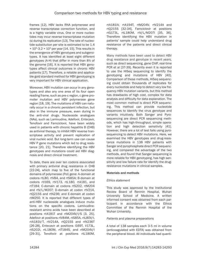
The pyrosequencing method only read about 60 bp short DNA, and we used multiple primers to sequence the HBV RT regions, and then used the resulted multiple short HBV DNA sequenc-es in RT regions to do HBV genotyping by align-ing with the 23 HBV DNA reference sequences in database (http://www.ncbi.nlm.nih.gov/proj-ects/genotyping). With this method, we found that HBV genotype B and C were detected in 12.32% (17 cases) and 87.68% (121 cases), respectively in the 138 samples (Figure 1A).
Sanger sequencing method could read approxi-mately 1100 bp long DNA sequences for the sequence primer, with which the DNA sequence for the full-length of HBV RT region could be obtained. Therefore the full length DNA
Figure 1. Results of HBV genotyping. A. The statistical data for HBV genotypes identified by Pyrosequencing and Sanger sequencing methods; B. HBV genotype B identified by alignment of Sanger DNA sequences of the patients’ samples with that of HBV database; C. HBV genotype C identified by alignment of Sanger DNA sequences of the pa-tients’ samples with that of HBV database; D. HBV genotype D identified by alignment of Sanger DNA sequences of the patients’ samples with that of HBV database; E. HBV genotype B/C hybrid type identified by alignment of Sanger DNA sequences of the patients’ samples with that of HBV database.
Comparison two methods for HBV typing and resistance
14287 Int J Clin Exp Med 2016;9(7):14283-14293
sequence of the RT region was aligned with the reference HBV nucleic acid database for geno-typing (Figure 1B-E). With this method, we found HBV genotype B was detected 10.87% (15/138) in the cohort, and also 86.23% (119/138) C, 0.72% (1/138) D, 2.17% (3/138) B/C hybrid were detected. Statistical analysis showed that there was no difference in the detection rate of HBV genotypes B or C between the Sanger and pyrosequencing methods.
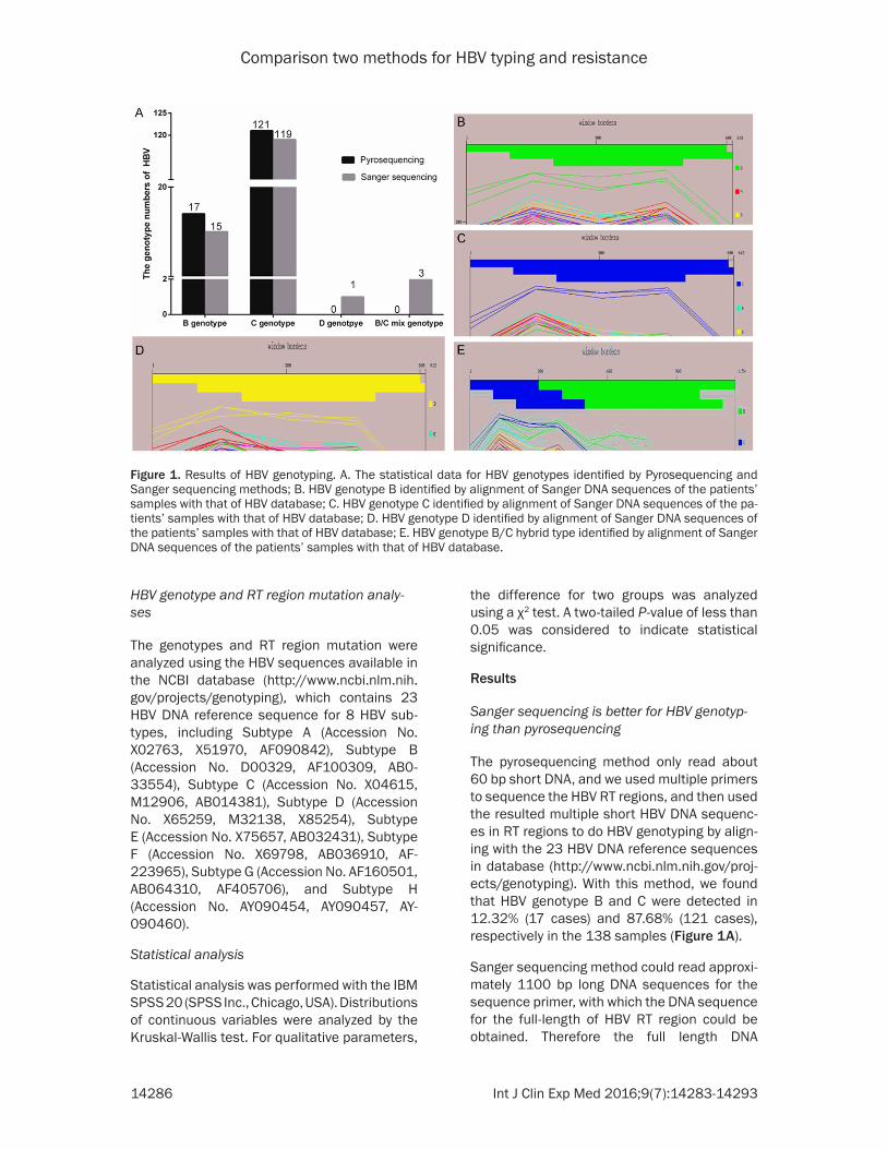
Moreover, with pyrosequencing, 10 samples were shown to be wrongly genotyped, the error rate was 7.25% (10/138), in which the 3 patients with genotype B was classified as C, and 3 with genotype C classified as B, 2 with genotype B/C as B, 1 with genotype B/C as C and 1 with genotype B as D (Table 1). However, no mistakes for genotyping were found with Sanger sequencing. Furthermore, Sanger sequencing could also read the full length of the HBV S region. By aligning the database with full-length of S region, we could obtain more accurate reference sequences (Table 1), which benefit the analysis of drug-resistance muta-tions in the HBV samples.
Pyrosequencing has higher failure rate for de-tection of drug-resistance mutations
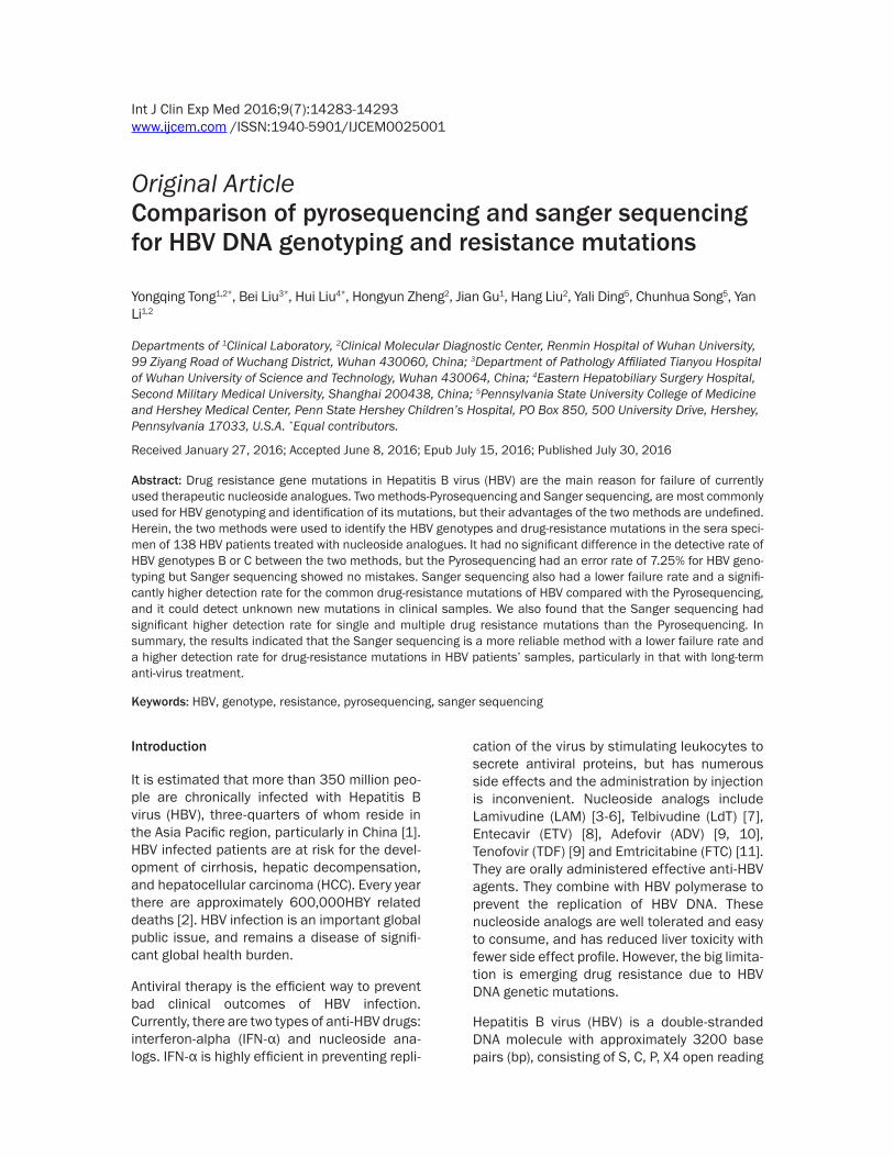
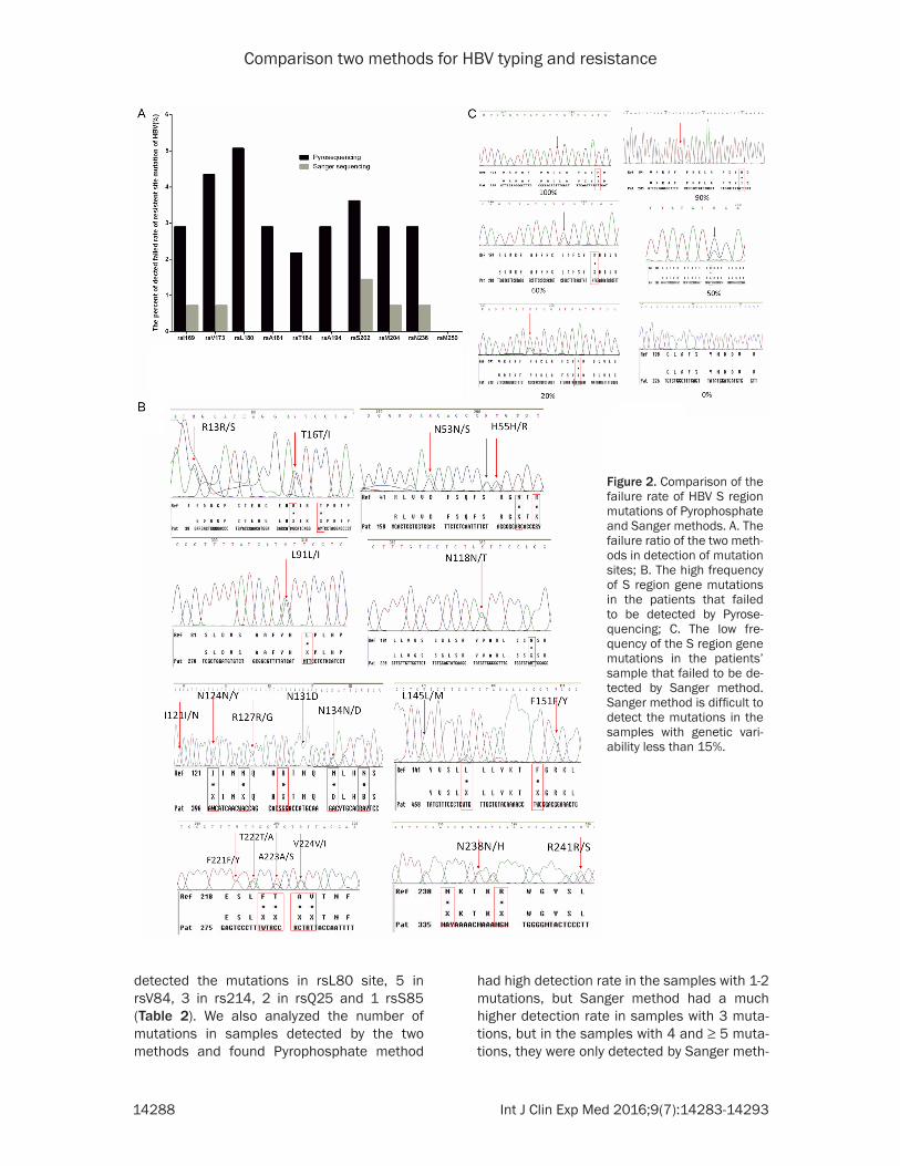
Both Pyrophosphate and Sanger methods can fail to detect the drug resistant mutation in HBV patients’ samples. We compared the fail-ure rate of the methods at 10 common muta-tion sites: rtI169, rtV173, rtL180, rtA181, rtT184, rtA194, rtS202, rtM204, rtN236 and rtM250. The failure rate for pyrophosphate
method was 2.90%, 4.35%, 5.07%, 2.90%, 2.17%, 2.90%, 3.62%, 2.90%, 2.90% and 0%, respectively; for Sanger method, they were 0.72%, 0.72%, 0, 0, 0, 0, 1.45%, 0.72%, 0.72% and 0%, respectively (Figure 2A). The failure rate for pyrophosphate method was sig-nificantly higher than that of Sanger method except for the rsM250 site where the failure rate was 0% for both methods.
Moreover, with Sanger sequencing method, many mutations un-related to nucleoside analog drug resistance, were detected in samples where the pyrophosphate method could not detect any mutations (Figure 2B). Moreover,
Table 1. The different results of the genotype of HBV detected by pyrosequencing and by Sanger sequencing
SamplePyrosequencing Sanger sequencing
Error rateGenotype Accession
No. Genotype Accession No.
4 C --- B AF100309 7.25%28 B --- B/C AB01438130 B --- B/C X0461549 B --- D X6525967 B --- C AB01438182 C --- B AF100309101 C --- B/C AF100309105 B --- C AB014381117 B --- C X04615122 C --- B AF100309
we found many samples where the drug-resis-tance mutations were not detected by pyro-phosphate method were from the patients who had received interferon treatment for more than 6 months. We also found that a few sam-ples were detected with low frequency of drug-resistance mutations using the pyrophosphate method but were negative with Sanger method (Figure 2C).
Sanger sequencing has higher rate for detec-tion of HBV drug-resistance mutations in pa-tients’ samples
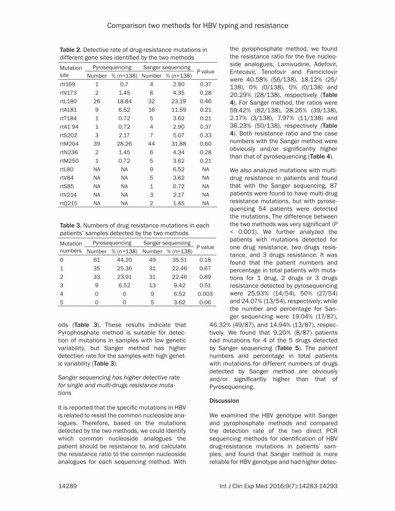
With pyrosequencing, the detection rate for the 10 common drug-resistance mutations of HBV was obviously lower than by Sanger sequenc-ing. The detection rate for the mutations : rtI169, rtV173, rtL180, rtA181, rtT184, rtA194, rtS202, rtM204, rtN236 and rtM250, with pyrophosphate method was 0.72%, 1.45%, 18.84%, 6.52%, 0.72%, 0.72%, 2.17%, 28.26%, 1.45% and 0.72%, respectively which was obvi-ously lower than by the Sanger sequencing: 2.90%, 4.35%, 23.19%, 11.59%, 3.62%, 2.90%, 5.07%, 31.88%, 4.34% and 3.62%, respectively (Table 2). Moreover, with Sanger sequencing, mutations in other drug-resistance sites such as rtL80, rtV84, rtS85, rtV214 and rtQ215 were also detected, and the frequency of the mutations was 9, 5, 1, 3 and 2, respec-tively, in the 138 patients’ samples (Table 2). These five mutation sites are usually not ana-lyzed by the Pyrophosphate method (Table 2). But Sanger sequencing covered full-length of RT region that includes those 5 sites. In our data we found that in up to 9 samples we
Comparison two methods for HBV typing and resistance
14288 Int J Clin Exp Med 2016;9(7):14283-14293
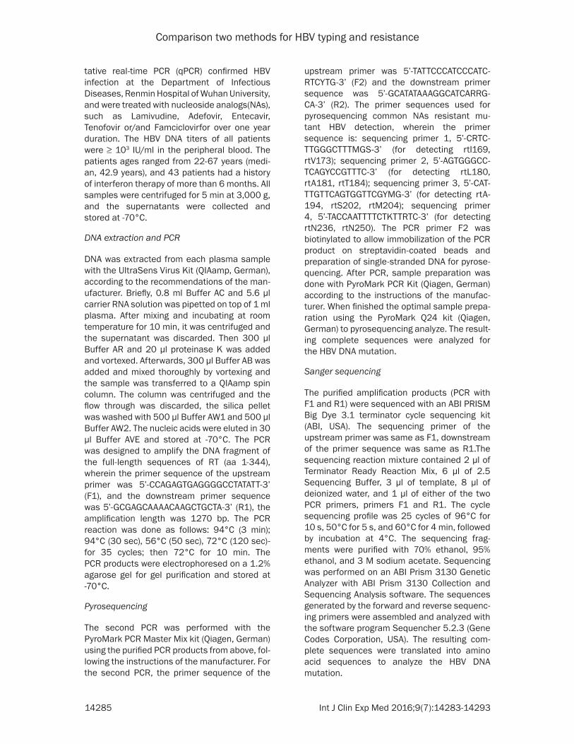
Figure 2. Comparison of the failure rate of HBV S region mutations of Pyrophosphate and Sanger methods. A. The failure ratio of the two meth-ods in detection of mutation sites; B. The high frequency of S region gene mutations in the patients that failed to be detected by Pyrose-quencing; C. The low fre-quency of the S region gene mutations in the patients’ sample that failed to be de-tected by Sanger method. Sanger method is difficult to detect the mutations in the samples with genetic vari-ability less than 15%.
detected the mutations in rsL80 site, 5 in rsV84, 3 in rs214, 2 in rsQ25 and 1 rsS85 (Table 2). We also analyzed the number of mutations in samples detected by the two methods and found Pyrophosphate method
had high detection rate in the samples with 1-2 mutations, but Sanger method had a much higher detection rate in samples with 3 muta-tions, but in the samples with 4 and ≥ 5 muta-tions, they were only detected by Sanger meth-
Comparison two methods for HBV typing and resistance
14289 Int J Clin Exp Med 2016;9(7):14283-14293
ods (Table 3). These results indicate that Pyrophosphate method is suitable for detec-tion of mutations in samples with low genetic variability, but Sanger method has higher detection rate for the samples with high genet-ic variability (Table 3).
Sanger sequencing has higher detective rate for single and multi-drugs resistance muta-tions
It is reported that the specific mutations in HBV is related to resist the common nucleoside ana-logues. Therefore, based on the mutations detected by the two methods, we could identify which common nucleoside analogues the patient should be resistance to, and calculate the resistance ratio to the common nucleoside analogues for each sequencing method. With
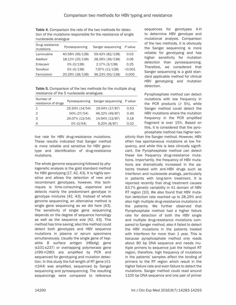
the pyrophosphate method, we found the resistance ratio for the five nucleo-side analogues, Lamivudine, Adefovir, Entecavir, Tenofovir and Famciclovir were 40.58% (56/138), 18.12% (25/ 138), 0% (0/138), 0% (0/138) and 20.29% (28/138), respectively (Table 4). For Sanger method, the ratios were 59.42% (82/138), 28.26% (39/138), 2.17% (3/138), 7.97% (11/138) and 36.23% (50/138), respectively (Table 4). Both resistance ratio and the case numbers with the Sanger method were obviously and/or significantly higher than that of pyrosequencing (Table 4).
We also analyzed mutations with multi-drug resistance in patients and found that with the Sanger sequencing, 87 patients were found to have multi-drug resistance mutations, but with pyrose-quencing 54 patients were detected the mutations. The difference between the two methods was very significant (P < 0.001). We further analyzed the patients with mutations detected for one drug resistance, two drugs resis-tance, and 3 drugs resistance. It was found that the patient numbers and percentage in total patients with muta-tions for 1 drug, 2 drugs or 3 drugs resistance detected by pyrosequencing were 25.93% (14/54), 50% (27/54) and 24.07% (13/54), respectively; while the number and percentage for San- ger sequencing were 19.04% (17/87),
Table 2. Detective rate of drug-resistance mutations in different gene sites identified by the two methodsMutation site
Pyrosequencing Sanger sequencingP value
Number % (n=138) Number % (n=138)rtI169 1 0.7 4 2.90 0.37rtV173 2 1.45 6 4.35 0.28rtL180 26 18.84 32 23.19 0.46rtA181 9 6.52 16 11.59 0.21rtT184 1 0.72 5 3.62 0.21rtA1 94 1 0.72 4 2.90 0.37rtS202 3 2.17 7 5.07 0.33rtM204 39 28.26 44 31.88 0.60rtN236 2 1.45 6 4.34 0.28rtM250 1 0.72 5 3.62 0.21rtL80 NA NA 9 6.52 NArtV84 NA NA 5 3.62 NArtS85 NA NA 1 0.72 NArtV214 NA NA 3 2.17 NArtQ215 NA NA 2 1.45 NA
Table 3. Numbers of drug resistance mutations in each patients’ samples detected by the two methodsMutation numbers
Pyrosequencing Sanger sequencingP value
Number % (n=138) Number % (n=138)0 61 44.20 49 35.51 0.181 35 25.36 31 22.46 0.672 33 23.91 31 22.46 0.893 9 6.52 13 9.42 0.514 0 0 9 6.52 0.0035 0 0 5 3.62 0.06
46.32% (49/87), and 14.94% (13/87), respec-tively. We found that 9.20% (8/87) patients had mutations for 4 of the 5 drugs detected by Sanger sequencing (Table 5). The patient numbers and percentage in total patients with mutations for different numbers of drugs detected by Sanger method are obviously and/or significantly higher than that of Pyrosequencing.
Discussion
We examined the HBV genotype with Sanger and pyrophosphate methods and compared the detection rate of the two direct PCR sequencing methods for identification of HBV drug-resistance mutations in patients’ sam-ples, and found that Sanger method is more reliable for HBV genotype and had higher detec-
Comparison two methods for HBV typing and resistance
14290 Int J Clin Exp Med 2016;9(7):14283-14293
tive rate for HBV drug-resistance mutations. These results indicated that Sanger method is more reliable and sensitive for HBV geno- type and identification of drug-resistance mutations.
The whole genome sequencing followed by phy-logenetic analysis is the gold standard method for HBV genotyping [17, 42, 43]. It is highly sen-sitive and allows the detection of new and recombinant genotypes, however, this tech-niques is time-consuming, expensive and detects mainly the predominant genotype in genotype mixtures [42, 43]. Instead of whole genome sequencing, an alternative method is single gene sequencing as we did here [43]. The sensitivity of single gene sequencing depends on the degree of sequence homology as well as the sequence size [42, 43]. This method has time saving; also this method could detect both genotypes and HBV sequence mutations in plasma or serum specimens simultaneously. Usually the single gene of hep-atitis B surface antigen (HBsAg) gene (s101-s237) or overlapping polymerase gene (rt99-rt280) are amplified by PCR and sequenced for genotyping and mutation detec-tion. In this study the full-length of RT gene (rt1-rt344) was amplified, sequenced by Sanger sequencing and pyrosequencing. The resulting sequencings were compared to reference
sequences for genotypes A-H to determine HBV genotype and mutational analysis. Comparison of the two methods, it is obviously the Sanger sequencing is more reliable for genotyping and has higher sensitivity for mutation detection than pyrosequencing. Therefore, we considered that Sanger sequencing is a gold stan-dard applicable method for clinical HBV genotyping and mutation detection.
Pyrophosphate method can detect mutations with low frequency in the PCR products (≥ 5%), while Sanger method could detect the HBV mutations where the mutation frequency in the PCR amplified fragment is over 15%. Based on this, it is considered that the pyro-phosphate method has higher sen-
Table 4. Comparison the rate of the two methods for detec-tion of the mutations responsible for the resistance of single nucleoside analogueDrug resistance mutations Pyrosequencing Sanger sequencing P value
Lamivudine 40.58% (56/138) 59.42% (82/138) 0.03Adefovir 18.12% (25/138) 28.26% (39/138) 0.06Entecavir 0% (0/138) 2.17% (3/138) 0.25Tenofovir 0% (0/138) 7.97% (11/138) <0.001Famciclovir 20.29% (28/138) 36.23% (50/138) 0.005
Table 5. Comparison of the two methods for the multiple drug resistance of the 5 nucleoside analoguesNumber of resistance of drugs Pyrosequencing Sanger sequencing P value
1 25.93% (14/54) 19.04% (17/87) 0.532 50% (27/54) 46.32% (49/87) 0.493 24.07% (13/54) 14.94% (13/87) 0.184 0% (0/54) 9.20% (8/87) 0.02
sitivity than the Sanger method. However, HBV often has spontaneous mutations at low fre-quency, and while this is less clinically signifi-cant, the Pyrophosphate method can detect these low frequency drug-resistance muta-tions. Importantly, the frequency of HBV muta-tions are dramatically increased in the pa- tients treated with anti-HBV drugs such as interferon and nucleoside analogs, particularly in patients with long-term treatment. It is reported recently that drug treatment causes 63.7% genetic variability in A1 domain of HBV RT region [10]. We also found that HBV muta-tion detection rate reached up to 28.29% and also high multiple drug-resistance mutations in the patients. We further observed that Pyrophosphate method had a higher failure rate for detection of both the HBV single and multiple drug-resistance mutations com-pared to Sanger method, also it failed to detect the HBV mutations in the patients treated with Interferon for more than 1 year. This is because pyrophosphate method only reads about 80 bp DNA sequence and needs mu- ltiple primers to sequence just the hotspot RT region, therefore, high frequency of mutations in the patients’ samples affect the binding of primers to the RT region which result in the higher failure rate and even failure to detect the mutations. Sanger method could read around 1100 bp DNA sequence and one pair of primer
Comparison two methods for HBV typing and resistance
14291 Int J Clin Exp Med 2016;9(7):14283-14293
could amplify and sequence full-length of RT region, the high frequency of the mutations barely affects the binding of the primers. Also, Sanger method could detect the mutations in the sites of rsL80, rsV84, rs214, rsQ25 and rsS85, which are not analyzed with Pyro- phosphate sequencing method. Therefore, Sanger method is also a more reliable and applicable method for drug resistance muta-tion, particularly for the patients with long-term anti-virus treatment with Interferon and/or and nucleoside analogs
In summary, we examined the HBV genotypes and drug-resistance mutations in 138 HBV patients with Sanger and Pyrophosphate direct PCR sequencing. We found that that Pyro- phosphate method is suitable for detecting mutations in HBV hotspot sites with low genetic variability, but Sanger method has higher detec-tion rate and lower failure rate for drug-induced high frequency of HBV mutations in HBV patients’ samples.
Acknowledgements
This work supported by the Hubei Provin- cial Natural Science Foundation of China (2010CDB06903), National Natural Science Foundation of China (81000771), National Key Basic Research Program of China-973 Program (2012CB526706) and the National Natural Science Foundation of China (81271694).
Disclosure of conflict of interest
None.
Address correspondence to: Dr. Yan Li, Department of Clinical Laboratory, Renmin Hospital of Wuhan University, 99 Ziyang Road of Wuchang District, Wuhan 430060, China. Tel: 86-27-88041911-88258; Fax: 86-27-88071553; E-mail: [email protected]; Dr. Chunhua Song, Pennsylvania State University College of Medicine and Hershey Medical Center, Penn State Hershey Children’s Hospital, PO Box 850, 500 University Drive, Hershey, PA 17033, U.S.A. Tel: 717-531-1841; Fax: 717-531-4789; E-mail: [email protected]
References
[1] Liaw YF and Chu CM. Hepatitis B virus infec-tion. Lancet 2009; 373: 582-592.
[2] Lavanchy D. Hepatitis B virus epidemiology, disease burden, treatment, and current and
emerging prevention and control measures. J Viral Hepat 2004; 11: 97-107.
[3] Allen MI, Deslauriers M, Andrews CW, Tipples GA, Walters KA, Tyrrell DL, Brown N and Condreay LD. Identification and characteriza-tion of mutations in hepatitis B virus resistant to lamivudine. Lamivudine Clinical Investigation Group. Hepatology 1998; 27: 1670-1677.
[4] Yeh CT, Chien RN, Chu CM and Liaw YF. Clearance of the original hepatitis B virus YMDD-motif mutants with emergence of dis-tinct lamivudine-resistant mutants during pro-longed lamivudine therapy. Hepatology 2000; 31: 1318-1326.
[5] Bozdayi AM, Uzunalimoglu O, Turkyilmaz AR, Aslan N, Sezgin O, Sahin T, Bozdayi G, Cinar K, Pai SB, Pai R, Bozkaya H, Karayalcin S, Yurdaydin C and Schinazi RF. YSDD: a novel mutation in HBV DNA polymerase confers clini-cal resistance to lamivudine. J Viral Hepat 2003; 10: 256-265.
[6] Lau GK and Leung N. Forty-eight weeks treat-ment with clevudine 30 mg qd versus lamivu-dine 100 mg qd for chronic hepatitis B infec-tion: a double-blind randomized study. Korean J Hepatol 2010; 16: 315-320.
[7] Locarnini S. Molecular virology and the devel-opment of resistant mutants: implications for therapy. Semin Liver Dis 2005; 25 Suppl 1: 9-19.
[8] Locarnini S. Primary resistance, multidrug re-sistance, and cross-resistance pathways in HBV as a consequence of treatment failure. Hepatol Int 2008; 2: 147-151.
[9] Lacombe K, Ollivet A, Gozlan J, Durantel S, Tran N, Girard PM and Zoulim F. A novel hepa-titis B virus mutation with resistance to adefo-vir but not to tenofovir in an HIV-hepatitis B vi-rus-co-infected patient. AIDS 2006; 20: 2229-2231.
[10] Gomes-Gouvea MS, Ferreira AC, Teixeira R, Andrade JR, Ferreira AS, Barros LM, Rezende RE, Nastri AC, Leite AG, Piccoli LZ, Galvan J, Conde SR, Soares MC, Kliemann DA, Bertolini DA, Kunyoshi AS, Lyra AC, Oikawa MK, de Araujo LV, Carrilho FJ, Mendes-Correa MC and Pinho JR. HBV carrying drug-resistance muta-tions in chronically infected treatment-naive patients. Antivir Ther 2015; 20: 387-395.
[11] Gish RG, Trinh H, Leung N, Chan FK, Fried MW, Wright TL, Wang C, Anderson J, Mondou E, Snow A, Sorbel J, Rousseau F and Corey L. Safety and antiviral activity of emtricitabine (FTC) for the treatment of chronic hepatitis B infection: a two-year study. J Hepatol 2005; 43: 60-66.
[12] Delius H, Gough NM, Cameron CH and Murray K. Structure of the hepatitis B virus genome. J Virol 1983; 47: 337-343.
Comparison two methods for HBV typing and resistance
14292 Int J Clin Exp Med 2016;9(7):14283-14293
[13] Wang JC, Nickens DG, Lentz TB, Loeb DD and Zlotnick A. Encapsidated hepatitis B virus re-verse transcriptase is poised on an ordered RNA lattice. Proc Natl Acad Sci U S A 2014; 111: 11329-11334.
[14] Alam MM, Zaidi SZ, Shaukat S, Sharif S, Angez M, Naeem A, Saleha S, Butt JA and Malik SA. Common genotypes of Hepatitis B virus preva-lent in injecting drug abusers (addicts) of North West Frontier Province of Pakistan. Virol J 2007; 4: 63.
[15] Orito E, Mizokami M, Ina Y, Moriyama EN, Kameshima N, Yamamoto M and Gojobori T. Host-independent evolution and a genetic classification of the hepadnavirus family based on nucleotide sequences. Proc Natl Acad Sci U S A 1989; 86: 7059-7062.
[16] Zekri AR, Hafez MM, Mohamed NI, Hassan ZK, El-Sayed MH, Khaled MM and Mansour T. Hepatitis B virus (HBV) genotypes in Egyptian pediatric cancer patients with acute and chronic active HBV infection. Virol J 2007; 4: 74.
[17] Guirgis BS, Abbas RO and Azzazy HM. Hepatitis B virus genotyping: current methods and clini-cal implications. Int J Infect Dis 2010; 14: e941-953.
[18] Preikschat P, Gunther S, Reinhold S, Will H, Budde K, Neumayer HH, Kruger DH and Meisel H. Complex HBV populations with mutations in core promoter, C gene, and pre-S region are as-sociated with development of cirrhosis in long-term renal transplant recipients. Hepatology 2002; 35: 466-477.
[19] Ouneissa R, Bahri O, Alaya-Bouafif NB, Chouaieb S, Ben Yahia A, Sadraoui A, Hammami W, Filali N, Azzouz MM, Mami NB and Triki H. Frequency and clinical significance of core promoter and precore region mutations in Tunisian patients infected chronically with hepatitis B. J Med Virol 2012; 84: 1719-1726.
[20] Papatheodoridis GV, Dimou E and Papadimi- tropoulos V. Nucleoside analogues for chronic hepatitis B: antiviral efficacy and viral resis-tance. Am J Gastroenterol 2002; 97: 1618-1628.
[21] Ayres A, Bartholomeusz A, Lau G, Lam KC, Lee JY and Locarnini S. Lamivudine and Famciclovir resistant hepatitis B virus associated with fatal hepatic failure. J Clin Virol 2003; 27: 111-116.
[22] Locarnini S. Molecular virology of hepatitis B virus. Semin Liver Dis 2004; 24 Suppl 1: 3-10.
[23] Devi U and Locarnini S. Hepatitis B antivirals and resistance. Curr Opin Virol 2013; 3: 495-500.
[24] Bartholomeusz A, Tehan BG and Chalmers DK. Comparisons of the HBV and HIV polymerase, and antiviral resistance mutations. Antivir Ther 2004; 9: 149-160.
[25] Horiike N, Duong TN, Michitaka K, Joko K, Hiasa Y, Konishi I, Yano M and Onji M. Characteristics of lamivudine-resistant hepati-tis B virus (HBV) strains with and without breakthrough hepatitis in patients with chronic hepatitis B evaluated by serial HBV full-ge-nome sequences. J Med Virol 2007; 79: 911-918.
[26] Liu Y, Li X, Xin S, Xu Z, Chen R, Yang J, Liu L, Wong VW, Yang D, Chan HL and Xu D. The rtA181S mutation of hepatitis B virus primarily confers resistance to adefovir dipivoxil. J Viral Hepat 2015; 22:328-34.
[27] Wu ZP, Hang T, Gao YT, Li Y, Liu T, Jing L, Liu L and Du Z. Resistance mutation patterns of hepatitis B virus in patients with suboptimal response to adefovir dipivoxil therapy after la-mivudine resistance. Zhonghua Gan Zang Bing Za Zhi 2010; 18: 498-501.
[28] Santantonio T, Fasano M, Durantel S, Barraud L, Heichen M, Guastadisegni A, Pastore G and Zoulim F. Adefovir dipivoxil resistance patterns in patients with lamivudine-resistant chronic hepatitis B. Antivir Ther 2009; 14: 557-565.
[29] Guo XF, Zhang CX, Liu Y, Wu F and Luo X. Drug-resistant genes at hepatitis B virus polymerase region during entecavir treatment. Acta Academiae Medicinae Sinicae 2013; 35: 444-446.
[30] Tenney DJ, Levine SM, Rose RE, Walsh AW, Weinheimer SP, Discotto L, Plym M, Pokorno- wski K, Yu CF, Angus P, Ayres A, Bartholomeusz A, Sievert W, Thompson G, Warner N, Locarnini S and Colonno RJ. Clinical emergence of ente-cavir-resistant hepatitis B virus requires addi-tional substitutions in virus already resistant to Lamivudine. Antimicrob Agents Chemother 2004; 48: 3498-3507.
[31] Nagasaki F, Niitsuma H, Ueno Y, Inoue J, Kogure T, Fukushima K and Shimosegawa T. The high incidence of the emergence of ente-cavir-resistant mutants among patients infect-ed with lamivudine-resistant hepatitis B virus. Tohoku J Exp Med 2007; 213: 181-186.
[32] Ahn SH, Park YK, Park ES, Kim JH, Kim DH, Lim KH, Jang MS, Choe WH, Ko SY, Sung IK, Kwon SY and Kim KH. The impact of the hepatitis B virus polymerase rtA181T mutation on replica-tion and drug resistance is potentially affected by overlapping changes in surface gene. J Virol 2014; 88: 6805-6818.
[33] Pastor R, Habersetzer F, Fafi-Kremer S, Doffoel M, Baumert TF, Gut JP, Stoll-Keller F and Schvoerer E. Hepatitis B virus mutations po-tentially conferring adefovir/tenofovir resis-tance in treatment-naive patients. World J Gastroenterol 2009; 15: 753-755.
[34] Liu Y, Wang CM, Cheng J, Liang ZL, Zhong YW, Ren XQ, Xu ZH, Zoulim F and Xu DP. Hepatitis B
Comparison two methods for HBV typing and resistance
14293 Int J Clin Exp Med 2016;9(7):14283-14293
virus in tenofovir-naive Chinese patients with chronic hepatitis B contains no mutation of rtA194T conferring a reduced tenofovir sus-ceptibility. Chin Med J (Engl) 2009; 122: 1585-1586.
[35] Seigneres B, Pichoud C, Ahmed SS, Hantz O, Trepo C and Zoulim F. Evolution of hepatitis B virus polymerase gene sequence during famci-clovir therapy for chronic hepatitis B. J Infect Dis 2000; 181: 1221-1233.
[36] Locarnini SA. Hepatitis B virus surface antigen and polymerase gene variants: potential viro-logical and clinical significance. Hepatology 1998; 27: 294-297.
[37] Lindstrom A, Odeberg J and Albert J. Pyrosequencing for detection of lamivudine-resistant hepatitis B virus. J Clin Microbiol 2004; 42: 4788-4795.
[38] Yang ZJ, Tu MZ, Liu J, Wang XL and Jin HZ. Comparison of amplicon-sequencing, pyrose-quencing and real-time PCR for detection of YMDD mutants in patients with chronic hepati-tis B. World J Gastroenterol 2006; 12: 7192-7196.
[39] Mello FC, Lago BV, Lewis-Ximenez LL, Fernandes CA and Gomes SA. Detection of mixed populations of wild-type and YMDD hep-atitis B variants by pyrosequencing in acutely and chronically infected patients. BMC Microbiol 2012; 12: 96.
[40] Han Y, Zhang Y, Mei Y, Wang Y, Liu T, Guan Y, Tan D, Liang Y, Yang L and Yi X. Analysis of hepatitis B virus genotyping and drug resis-tance gene mutations based on massively par-allel sequencing. J Virol Methods 2013; 193: 341-347.
[41] Fan J, Zhang Y, Xiong H, Wang Y and Guo X. Nucleotide analogue-resistant mutations in hepatitis B viral genomes found in hepatitis B patients. J Gen Virol 2015; 96: 663-670.
[42] Valsamakis A. Molecular testing in the diagno-sis and management of chronic hepatitis B. Clin Microbiol Rev 2007; 20: 426-439.
[43] Norder H, Courouce AM, Coursaget P, Echevarria JM, Lee SD, Mushahwar IK, Robertson BH, Locarnini S and Magnius LO. Genetic diversity of hepatitis B virus strains de-rived worldwide: genotypes, subgenotypes, and HBsAg subtypes. Intervirology 2004; 47: 289-309.
Recommended