Abstract. Osteochondroma is the most common benign bonetumor and usually occurs in the metaphyseal region of thelong bones. This tumor takes the form of a cartilage-cappedbony outgrowth on the surface of the bone. The vast majority(85% ) of osteochondromas present as solitary, nonhereditarylesions. Approximately 15% of osteochondromas occur asmultiple lesions in the context of hereditary multipleosteochondromas (HMOs), a disorder that is inherited in anautosomal dominant manner. Most lesions appear in childrenand adolescents as painless, slow-growing masses. However,depending on the location of the osteochondroma, significantsymptoms may occur as a result of complications such asfracture, bony deformity, mechanical joint problems andvascular or neurologic compromise. Malignant transformationof osteochondromas can occur later in adulthood but rarelymetastasize. The treatment of choice for osteochondroma issurgical unless the skeleton is still immature. Pathogeneticanalysis showed that HMOs are caused by mutations in eitherof two genes: exostosis (multiple)-1 (EXT1), which is locatedon chromosome 8q24.11–q24.13 or exostosis (multiple)-2(EXT2), which is located on chromosome 11p11–12. Recently,biallelic inactivation of the EXT1 locus was described innonhereditary osteochondromas. The EXT1 and EXT2 proteinsfunction in the biosynthesis of heparin sulfate proteoglycans(HSPGs) which are multifunctional proteins involved inseveral growth signaling pathways in the normal epiphysealgrowth plate. Reduced EXT1 or EXT2 expression inosteochondromas is associated with disordered cellular
distribution of HSPGs, resulting in defective endochondralossification which is likely to be involved in the formation ofosteochondromas. Here the clinical, radiological, pathologicaland pathogenetic features and the treatment modalities ofosteochondroma are reviewed.
Osteochondroma is the most common benign bone tumor (1-5). According to the WHO classification, this lesion is definedas a cartilage-capped bony projection arising on the externalsurface of bone containing a marrow cavity that is continuouswith that of the underlying bone (3, 4). Osteochondromaoccurs in 3% of the general population and it accounts formore than 30% of all benign bone tumors and 10-15% of allbone tumors (1-26). The vast majority of these tumors presentas solitary, nonhereditary lesions. Approximately 15% ofosteochondromas occur in the context of hereditary multipleosteochondromas (HMOs), a disorder that is inherited in anautosomal dominant manner. Solitary osteochondromas havea tendency to appear at metaphyses of the long bones,especially the femur, humerus, tibia, spine and hip, althoughevery part of the skeleton can be affected (1-5).Osteochondroma is usually symptomless and is found
incidentally (1-5, 8-15). Malignant transformation of asolitary osteochondroma may occur in 1-2% of patients,while for osteochondromas in the setting of HMO syndromethe occurrence is between 1% and 25% (5-7). The diagnosisof an osteochondroma requires radiological depiction and, insome cases, particularly if there is a suspicion of malignancy,histological examination is also needed (26-61). Thetreatment of choice for osteochondroma is surgical unless theskeleton is still immature; for a symptomatic solitary lesion,a partial excision is suggested (1, 5, 6, 9, 18).A large number of studies using cell biology, molecular
biology and immunohistochemical methods analyzed themechanisms involved in the pathogenesis of osteochondroma(62-114). It has been shown that HMO are caused by mutations
633
Correspondence to: Panagiotis Kitsoulis, MD, 21 October 28 St,45332, Ioannina, Greece. Tel: +30 2651079354, e-mail: [email protected] / [email protected]
Key Words: Osteochondroma, HMO, exostosis, chondrosarcoma,imaging, review.
in vivo 22: 633-646 (2008)
Review
Osteochondromas: Review of the Clinical,Radiological and Pathological Features
PANAGIOTIS KITSOULIS1, VASSILIKI GALANI1, KALLIOPI STEFANAKI2, GEORGIOS PARASKEVAS3,GEORGIOS KARATZIAS1, NIKI JOHN AGNANTIS4 and MARIA BAI4
1Department of Anatomy, Histology and Embryology, and4Department of Pathology Medical School, University of Ioannina;2Department of Pathology, Agia Sophia Children’s Hospital, Athens;
3Department of Anatomy, Medical School, Aristotle University of Thessaloniki, Greece
0258-851X/2008 $2.00+.40
in either of two genes: exostosis (multiple)-1 (EXT1), which islocated on chromosome 8q24.11–q24.13 or exostosis(multiple)-2 (EXT2), which is located on chromosome11p11–12 (75-81). Recently, biallelic inactivation of the EXT1locus was described in nonhereditary osteochondromas (104).The epidimiological, clinical, radiological, histological
and pathogenetic features and the treatment modalities ofosteochondroma are reviewed here.
Epidemiology
Osteochondromas are usually found in adolescents or children,rarely in infants or newborns (8). There is no predilection formales or females as far as solitary osteochondromas areconcerned. HMO syndrome affects males more often thanfemales (9) and is usually found in Caucasians rather than inother races, affecting 0.9-2 individuals per 100,000 ofpopulation. About 65% of patients have family members withautosomal dominant transmission of HMO genes (10-12). TheHMO syndrome comes to clinical attention during the firstdecade of life in more than 80% of patients (13, 14). Solitaryosteochondromas show a predilection for the metaphyses ofthe long tubular bones, especially the femur (30% ), humerus(26% ) and tibia (43% ). Lesions are rare in the carpal andtarsal bones, patella, sternum, skull and spine (15).
Clinical Features
Osteochondroma is usually symptomless and, therefore, theonly clinical symptom is a painless slow-growing mass onthe involved bone (16). However, significant symptoms mayoccur as a result of complications such as fracture, bonydeformity or mechanical joint problems. An osteochondromacan occur near a nerve or blood vessel, the commonest beingthe popliteal nerve and artery. The affected limb can exhibitnumbness, weakness, loss of pulse or changes in colour (17).Although rare, periodic changes in blood flow can alsooccur. Vascular compression, arterial thrombosis, aneurysm,pseudoaneurysm formation and venous thrombosis arecommon complications and lead to claudication, pain, acuteischemia, and signs of phlebitis, while nerve compressionoccurs in about 20% of patients (18, 19). The tumor can befound under a tendon, resulting in pain during relevantmovement and thus causing restriction of joint motion. Painis also present as a result of bursal inflammation or swelling,or even due to a fracture of the basis of the tumor’s stalk (4).Generally, the normal function and movement can be limitedand asymmetry may be also noted in a slowly and inwardlygrowing osteochondroma. If there is a tumor at the spinalcolumn, there may be kyphosis, or spondylolisthesis if it isclose to the intervertebral space (20). The clinical signs ofmalignant transformation are pain, swelling and anenlargement of the mass.
The hereditary multiple exostosis (HME) syndromeusually presents during the first decade of life or even innewborns. The manifestations include limb undergrowth withnormal height, ankle valgus, genu valgum and anomalies ofthe radio and ulnar deviation. Patients may present withmetacarpal, metatarsal and phalangeal shortening,anisomelia, coxa valga, scoliosis and asymmetry of thepectoral and pelvic girdles. Subluxation of the talus or thehip are common symptoms. Tibiofibular synostosis can alsotake place. Spinal compression syndrome may also be seen(21). Lesions that arise in the head and neck may beassosiated with facial asymmetry and dysfunction of themasseters (22, 23). An inwardly growing osteochondromacan cause injuries of the viscera such as hemothorax,obstruction of the intenstine or the urinary tract anddysphagia (24, 25).
Radiological Features
Apart from a detailed history and a careful physicalexamination, the diagnosis of an osteochondroma alsorequires radiological imaging.
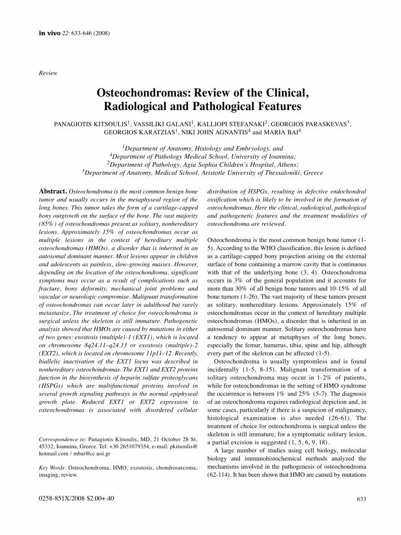
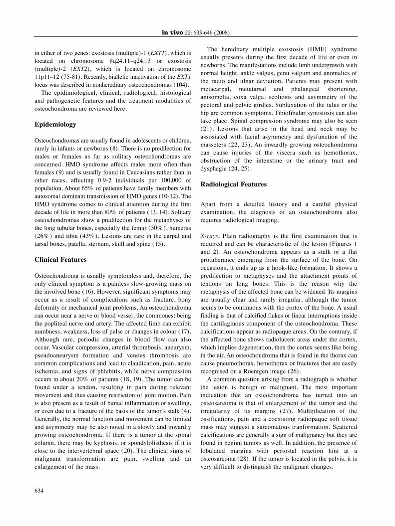
X-rays. Plain radiography is the first examination that isrequired and can be characteristic of the lesion (Figures 1and 2). An osteochondroma appears as a stalk or a flatprotuberance emerging from the surface of the bone. Onoccasions, it ends up as a hook-like formation. It shows apredilection to metaphyses and the attachment points oftendons on long bones. This is the reason why themetaphysis of the affected bone can be widened. Its marginsare usually clear and rarely irregular, although the tumorseems to be continuous with the cortex of the bone. A usualfinding is that of calcified flakes or linear interruptions insidethe cartilaginous component of the osteochondroma. Thesecalcifications appear as radiopaque areas. On the contrary, ifthe affected bone shows radiolucent areas under the cortex,which implies degeneration, then the cortex seems like beingin the air. An osteochondroma that is found in the thorax cancause pneumothorax, hemothorax or fractures that are easilyrecognised on a Roentgen image (26).A common question arising from a radiograph is whether
the lesion is benign or malignant. The most importantindication that an osteochondroma has turned into anosteosarcoma is that of enlargement of the tumor and theirregularity of its margins (27). Multiplication of theossifications, pain and a coexisting radiopaque soft tissuemass may suggest a sarcomatous tranformation. Scatteredcalcifications are generally a sign of malignancy but they arefound in benign tumors as well. In addition, the presence oflobulated margins with periostal reaction hint at aosteosarcoma (28). If the tumor is located in the pelvis, it isvery difficult to distinguish the malignant changes.
in vivo 22: 633-646 (2008)
634
The HME syndrome can present as bilateral lesions thatwiden a metaphysis. Tarsal and carpal bones are oftenaffected and are shown with a mass emerging from theirepiphysis. Joint disturbance and growth anomalies are easilyrecognized in plain radiography.
Computed tomography. Computed tomography is a veryaccurate method for depicting osteochondromas of thespinal column, shoulder and pelvis. In particular, ifcompressive myelopathy has taken place, CT myelographyis the examination of choice. CT can depict the bony lesionin detail, as well as showing the presence of calcifications.Its ability in distinguishing an osteochondroma from anosteosarcoma has been a matter of debate (29). Thecriterion that is used is the thickness of the cartilaginouscap of the tumor, given that an osteosarcoma has a thickerone. CT is currently thought to be unreliable on this
subject, as underestimation of the thickness is usual (30).The disadvantage of CT is that it cannot estimate themetabolic activity, a serious indication of malignancy ofany tumor.
Ultrasound. Ultrasound is the examination of choice wherethere is suspicion of aneurysms or pseudoaneurysms andarterial or venous thrombosis. It is an accurate method forexamining the cartilaginous cap of the osteochondroma as anhypoechoic area above the cortex of the relevant bone (27,31). It is also the only way to pinpoint a bursitis. However,ultrasound cannot depict the cap if there is an inwarddevelopment of the tumor.
Nuclear medicine. Scintigraphic methods are being used inorder to examine the metabolic activity of the tumor. A poormetabolic activity is only present in benign lesions. Thallium201 is used to detect a malignant transformation of HME. Itis important to know that it is still impossible to distinguishmalignant ossifications, hyperemia and an osteoblasticreaction in chondrosarcoma via scintigraphy (32).
Kitsoulis et al: Osteochondroma-Clinical, Radiological and Pathological Features (Review)
635
Figure 1. X-ray: A typical lesion involving the right femur. Note theprotuberance on the external surface of the femoral bone. Linearcalcifications inside the tumor lesion are also obvious.
Figure 2. X-ray: Anteroposterior radiograph of a tibial osteochondroma.Note the protuberance on the external surface of the tibial bone.
Angiography. Angiography is often used for vascular lesionscaused by an osteochondroma (33, 34). Aneurysms,thrombosis and occlusions are not rare. These lesions arecaused by the ossified cartilaginous cap. Neovascularity,which characterizes malignant lesions, can also be detectedin a malignant transformation using angiography.
Magnetic resonance imaging. MRI is the most preciseimaging method for symptomatic cases of bone masses as itcan depict the exact morphology of a tumor, arterial andvenous compromise and nervous lesions. Additionally, MRIcan demonstrate a probable recurrence if a malignant tumoris diagnosed. In order to get full depiction of the tumor,MRI is performed in coronal, sagittal, paracoronal andparasagittal planes.MRI is first of all used in order to verify the continuity of
the palpable mass with the cortex of the affected bone andto differentiate an osteochondroma from other surface bonelesions (35). The cartilaginous cap, because it is rich inwater, presents a high signal on T2-weighted MRI and a lowone on T1-weighted. It is usual to detect above it a lowsignal zone of the perichondrium. T2-weighted MRI ispreferable because it provides better differentiation of signalintensities.A short time inversion recovery (STIR) depiction can
reveal accompanying edema in chondrosarcomas (36). Ifmusculoskeletal complications occur, a T1-weighted seriesis recommended. If a high signal is obtained, then there ismuscular damage because T1 relaxation time is shorter. If aT2-weighted series shows a high signal, then there iscertainly edema around the lesion.MRI can also depict vascular complications caused by the
tumor (37). For example, a pseudoaneurysm will present asa nonhomogenous formation and a thrombosis as an onion-shaped formation inside the lumen of a vessel. If denervationof a muscle takes place, an MRI shows a high signalintensity, as fatty tissue will have taken the place of themuscle cells. In addition, a differentiation in the signaling ofa nerve may suggest its supression or damage (38-41). AnMRI image can easily demonstrate lesions of the spinalcolumn or the cranium, something that is not possible forother methods. A bursitis is the only case where an MRI cangive a false-positive indication (42-45).Distinguishing a malignant from a benign lesion is a
challenge for MRI. MRI can nevertheless be used toaccurately diagnose even a low-grade osteosarcoma (46, 47).Again, the thickness of the cartilaginous cap is the basiccriterion. In this respect, Woertler et al. (48) suggested thatcartilage cap thickness exceeding 2 cm in adults and 3 cm inchildren should raise the suspicion of malignanttransformation. A chondrosarcoma is also characterized bylow T1 signal after intravenous contrast infusion, somethingthat is rarely recorded in a benign cartilaginous tumor.
Nowadays, using gadolinium it is also possible to undertakea dynamic examination of neovascularization, which is muchmore preferable in differentiating an osteosarcoma from anosteochondroma (49-51). All the aforementioned reasonsjustify the view that MRI is the gold standard technique fordetecting a malignant transformation (48-52).
Pathology
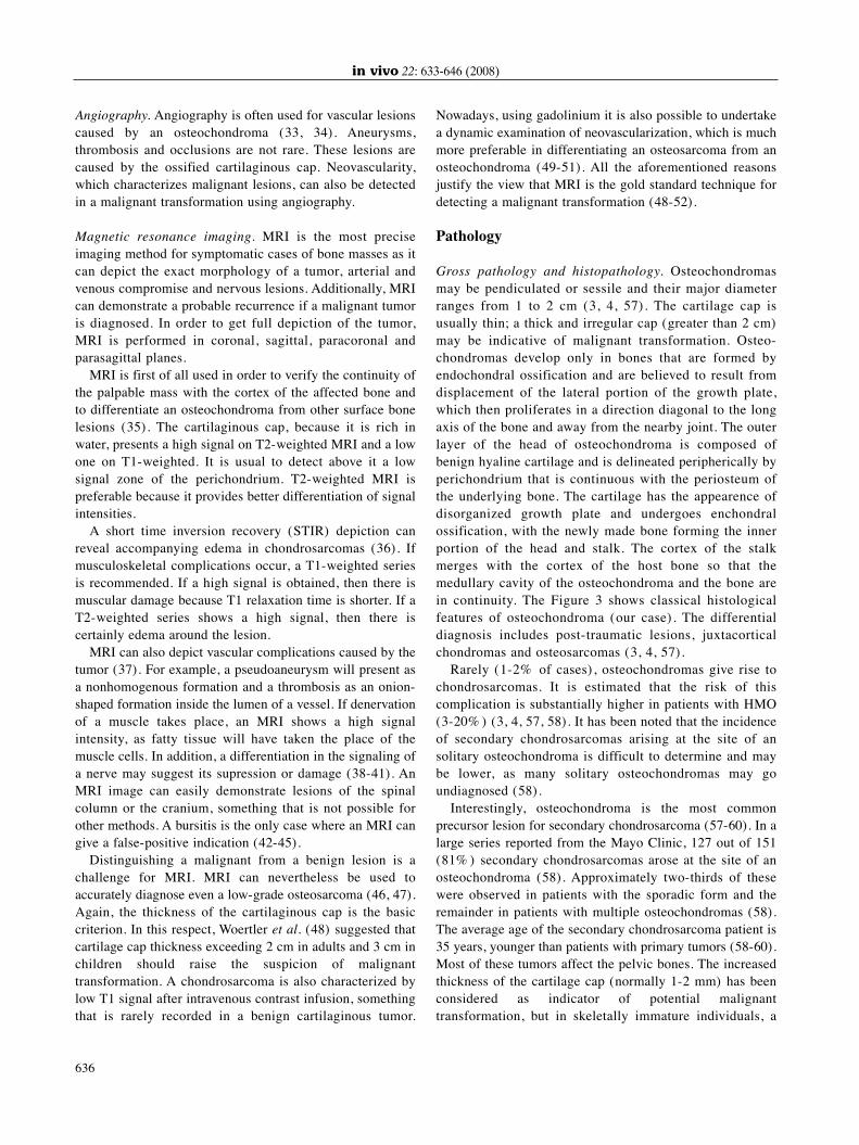
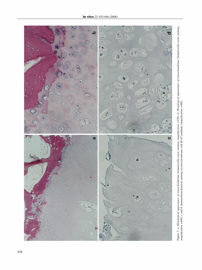
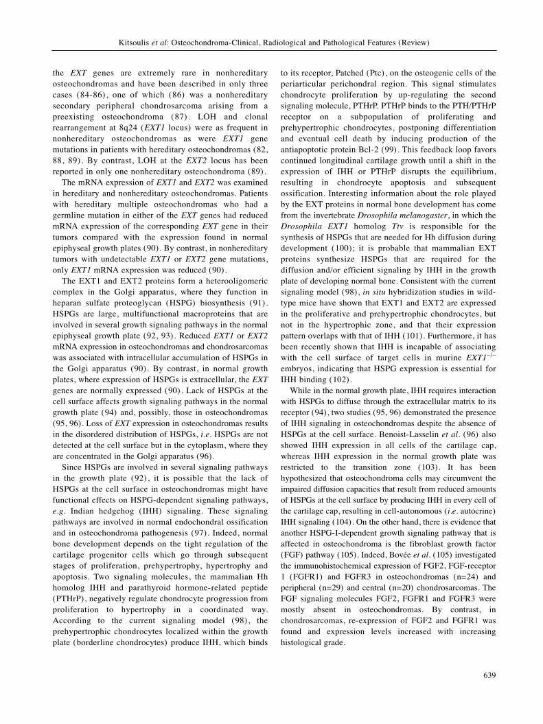
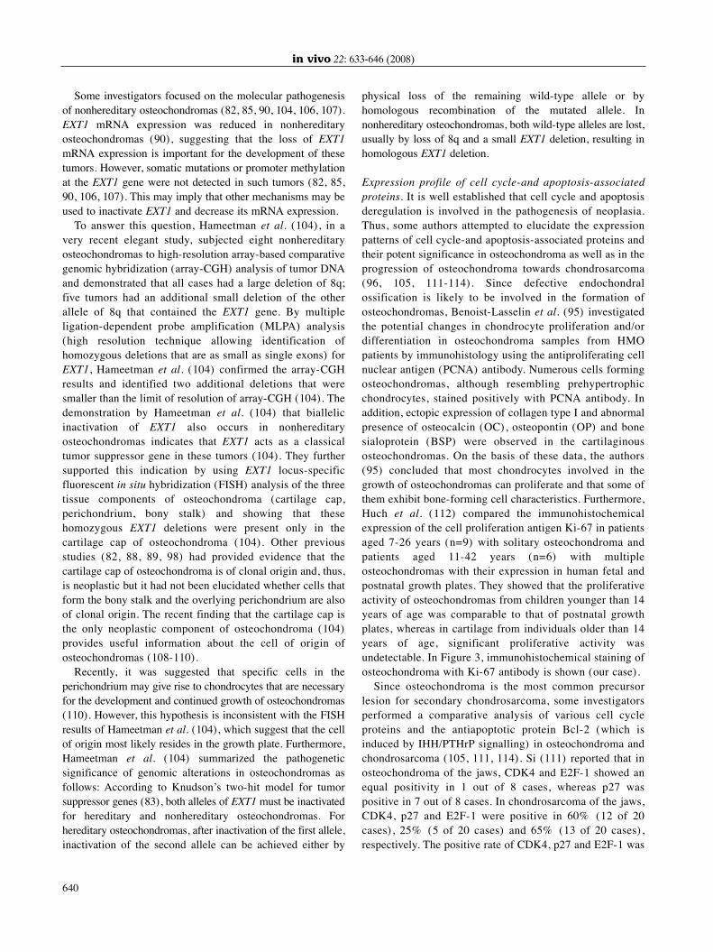
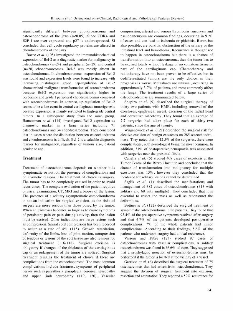
Gross pathology and histopathology. Osteochondromasmay be pendiculated or sessile and their major diameterranges from 1 to 2 cm (3, 4, 57). The cartilage cap isusually thin; a thick and irregular cap (greater than 2 cm)may be indicative of malignant transformation. Osteo-chondromas develop only in bones that are formed byendochondral ossification and are believed to result fromdisplacement of the lateral portion of the growth plate,which then proliferates in a direction diagonal to the longaxis of the bone and away from the nearby joint. The outerlayer of the head of osteochondroma is composed ofbenign hyaline cartilage and is delineated peripherically byperichondrium that is continuous with the periosteum ofthe underlying bone. The cartilage has the appearence ofdisorganized growth plate and undergoes enchondralossification, with the newly made bone forming the innerportion of the head and stalk. The cortex of the stalkmerges with the cortex of the host bone so that themedullary cavity of the osteochondroma and the bone arein continuity. The Figure 3 shows classical histologicalfeatures of osteochondroma (our case). The differentialdiagnosis includes post-traumatic lesions, juxtacorticalchondromas and osteosarcomas (3, 4, 57).Rarely (1-2% of cases), osteochondromas give rise to
chondrosarcomas. It is estimated that the risk of thiscomplication is substantially higher in patients with HMO(3-20% ) (3, 4, 57, 58). It has been noted that the incidenceof secondary chondrosarcomas arising at the site of ansolitary osteochondroma is difficult to determine and maybe lower, as many solitary osteochondromas may goundiagnosed (58).Interestingly, osteochondroma is the most common
precursor lesion for secondary chondrosarcoma (57-60). In alarge series reported from the Mayo Clinic, 127 out of 151(81% ) secondary chondrosarcomas arose at the site of anosteochondroma (58). Approximately two-thirds of thesewere observed in patients with the sporadic form and theremainder in patients with multiple osteochondromas (58).The average age of the secondary chondrosarcoma patient is35 years, younger than patients with primary tumors (58-60).Most of these tumors affect the pelvic bones. The increasedthickness of the cartilage cap (normally 1-2 mm) has beenconsidered as indicator of potential malignanttransformation, but in skeletally immature individuals, a
in vivo 22: 633-646 (2008)
636

cartilage cap of up to 2 cm might be identified. In addition toa thick cartilage cap, other findings that should raisesuspicion of malignant transformation are recent growth ofan exostosis in an adult proximal skeletal location, irregularmineralization, the presence of soft tissue bands, a grosslyirregular surface, cystic changes, loss of architecture ofcartilage, myxoid changes, necrosis, increased cellularity,mitotic activity and atypia of chondrocytes (3, 58). Mostsecondary chondrosarcomas present histological features oflow-grade malignancy (59). These tumors generally carry agood prognosis and surgical treatment without adjuvantchemotherapy or irradiation is the treatment of choice (59).Secondary osteosarcomas arising within an osteochondromaare extremely rare (61).
Expression profile of chondrocytic differentiation-associatedproteins. There is growing evidence that, besidesconventional histological criteria, analysis of theextracellular matrix gene expression pattern, in particularsubtyping of collagen gene expression profiles usingimmunohistochemical and in situ hybridization methods, ishelpful for the definition and identification of differentphenotypes of normal and pathological chondrocytic cells(63-74). On that basis, it was suggested that the expressionprofiles of the collagen types might play an important rolein classification and diagnosis of chondrogenic neoplasiasof the skeleton (63).Accumulating data suggest that the cellular phenotypes
of normal chondrocyte differentiation so far decribedduring fetal chondroneogenesis and in fetal growth platecartilage (chondroprogenitor cells, mature chondrocytes,hypertrophic chondrocytes) display different expressionprofiles of the collagen types (63, 65, 66). Indeed, normalchondroprogenitor cells are characterized by the expressionof their specific gene product, the alternative splice variantof collagen COL2 and COL2A (65, 66). Normal maturechondrocytes express the typical cartilage collagen typesCOL2B, COL9, and COL11 as well as aggrecan and linkprotein (63). However, the expression of these collagentypes is not specific for cartilage since they are alsoobserved in a few other tissues such as the vitreous body(63). Hypertrophic chondrocytes are characterized by theexpression of their unique gene product COL10 (67).Normally, terminally differentiated hypertrophicchondrocytes become to a large extent apoptotic beforethey get replaced by ingrowing bone forming cells whichreplace the pre-existing cartilaginous matrix by a bonematrix (63). Interestingly, terminally differentiatedhypertrophic chondrocytes that survive and undergoposthypertrophic differentiation to osteoblast-like cells(which start to express COL1) have so far only beenidentified in the chick (68, 69). “Dedifferentiated”chondrocytes, a phenotype so far only identified in vitro,
express COL1 and COL3, but not the cartilage-typicalcollagen types (COL2, COL9, COL11) nor aggrecanproteoglycan (63, 70).The characteristic feature of osteochondromas, enchon-
dromas and conventional chondrosarcomas is the presenceof neoplastic cells displaying a chondrocytic cell shape andthe gene expression profile of mature fetal chondrocytes;these neoplastic cells are responsible for the formation ofthe characteristic hyaline cartilage-like extracellular tumormatrix (71-74). Neoplastic chondrocytes in vivo exhibit thefull differentiation expression profile of their physiologicalcounterparts. In chondrogenic neoplasias, besides thephenotype of mature chondrocytes, hypertrophic celldifferentiation with the expression of COL10 is observed(71, 73). The expression of COL1 without COL3expression in differentiated neoplastic chondrocytesrepresents experimental evidence of the potential ofmammalian chondrocytes to undergo posthypertrophicdifferentiation to osteoblast-like cells in vivo andimplicates the deposition of bone matrix componentswithin pre-existing cartilaginous tumor matrix (71).Whereas enchondromas and conventional chondrosarcomasexhibit a random cellular differentiation pattern,osteochondromas are characterized by a highly structuredtissue organization. In osteochondromas, mesenchymal celllayers of fibrous appearance overlay cartilaginous tissue,with chondrocytic cells expressing COL2 and the aggrecanproteoglycan; in the deep zone, chondrocytic cells becomehypertrophic, express COL10 and endochondralossification is observed (73, 74).
Molecular pathology and pathogenesis. The neoplasticnature of osteochondroma has only recently come to lightwith the discovery of the loss of heterozygosity (LOH) atthe EXT1 locus as well as other clonal abnormalities in asubset of these tumors (75-79). Multiple osteochondromasis caused by mutations in either of two genes: exostosis(multiple)-1 EXT1, Online Mendelian Inheritance in Man(OMIM) No. 133700, which is located on chromosome8q24.11–q24.13, or exostosis (multiple)-2 (EXT2; OMIMNo. 133701), which is located on chromosome 11p11–12(75-77). Both genes are ubiquitously expressed (75-77).Most of the germline mutations that have been identifiedin EXT1 and EXT2 result in premature truncation of theEXT proteins and loss of protein function (78). Mosthereditary osteochondromas demonstrate heterozygousmutations in EXT1 or EXT2 (79-81). Some hereditaryosteochondromas, in addition to carrying an EXT1mutation, exhibit loss of the remaining wild-type allele ofEXT1 (82); this is consistent with Knudson’s two-hitmodel of tumorigenesis (83) and indicates that EXT1 actsas a classical tumor suppressor gene in multipleosteochondromas. On the other hand, somatic mutations in
Kitsoulis et al: Osteochondroma-Clinical, Radiological and Pathological Features (Review)
637
in vivo 22: 633-646 (2008)
638
Figure3.a:Histologicalappearanceofosteochondroma
(hematoxylin-eosinstaining,magnification×250);b:Histologicalappearanceofosteochondroma
(hematoxylin-eosinstaining,
magnification×
400);candd:Imm
unohistochemicalstainingofosteochondroma
withKi-67antibody(magnification×
400).
the EXT genes are extremely rare in nonhereditaryosteochondromas and have been described in only threecases (84-86), one of which (86) was a nonhereditarysecondary peripheral chondrosarcoma arising from apreexisting osteochondroma (87). LOH and clonalrearrangement at 8q24 (EXT1 locus) were as frequent innonhereditary osteochondromas as were EXT1 genemutations in patients with hereditary osteochondromas (82,88, 89). By contrast, LOH at the EXT2 locus has beenreported in only one nonhereditary osteochondroma (89).The mRNA expression of EXT1 and EXT2 was examined
in hereditary and nonhereditary osteochondromas. Patientswith hereditary multiple osteochondromas who had agermline mutation in either of the EXT genes had reducedmRNA expression of the corresponding EXT gene in theirtumors compared with the expression found in normalepiphyseal growth plates (90). By contrast, in nonhereditarytumors with undetectable EXT1 or EXT2 gene mutations,only EXT1 mRNA expression was reduced (90).The EXT1 and EXT2 proteins form a heterooligomeric
complex in the Golgi apparatus, where they function inheparan sulfate proteoglycan (HSPG) biosynthesis (91).HSPGs are large, multifunctional macroproteins that areinvolved in several growth signaling pathways in the normalepiphyseal growth plate (92, 93). Reduced EXT1 or EXT2mRNA expression in osteochondromas and chondrosarcomaswas associated with intracellular accumulation of HSPGs inthe Golgi apparatus (90). By contrast, in normal growthplates, where expression of HSPGs is extracellular, the EXTgenes are normally expressed (90). Lack of HSPGs at thecell surface affects growth signaling pathways in the normalgrowth plate (94) and, possibly, those in osteochondromas(95, 96). Loss of EXT expression in osteochondromas resultsin the disordered distribution of HSPGs, i.e. HSPGs are notdetected at the cell surface but in the cytoplasm, where theyare concentrated in the Golgi apparatus (96).Since HSPGs are involved in several signaling pathways
in the growth plate (92), it is possible that the lack ofHSPGs at the cell surface in osteochondromas might havefunctional effects on HSPG-dependent signaling pathways,e.g. Indian hedgehog (IHH) signaling. These signalingpathways are involved in normal endochondral ossificationand in osteochondroma pathogenesis (97). Indeed, normalbone development depends on the tight regulation of thecartilage progenitor cells which go through subsequentstages of proliferation, prehypertrophy, hypertrophy andapoptosis. Two signaling molecules, the mammalian Hhhomolog IHH and parathyroid hormone-related peptide(PTHrP), negatively regulate chondrocyte progression fromproliferation to hypertrophy in a coordinated way.According to the current signaling model (98), theprehypertrophic chondrocytes localized within the growthplate (borderline chondrocytes) produce IHH, which binds
to its receptor, Patched (Ptc), on the osteogenic cells of theperiarticular perichondral region. This signal stimulateschondrocyte proliferation by up-regulating the secondsignaling molecule, PTHrP. PTHrP binds to the PTH/PTHrPreceptor on a subpopulation of proliferating andprehypertrophic chondrocytes, postponing differentiationand eventual cell death by inducing production of theantiapoptotic protein Bcl-2 (99). This feedback loop favorscontinued longitudinal cartilage growth until a shift in theexpression of IHH or PTHrP disrupts the equilibrium,resulting in chondrocyte apoptosis and subsequentossification. Interesting information about the role playedby the EXT proteins in normal bone development has comefrom the invertebrate Drosophila melanogaster, in which theDrosophila EXT1 homolog Ttv is responsible for thesynthesis of HSPGs that are needed for Hh diffusion duringdevelopment (100); it is probable that mammalian EXTproteins synthesize HSPGs that are required for thediffusion and/or efficient signaling by IHH in the growthplate of developing normal bone. Consistent with the currentsignaling model (98), in situ hybridization studies in wild-type mice have shown that EXT1 and EXT2 are expressedin the proliferative and prehypertrophic chondrocytes, butnot in the hypertrophic zone, and that their expressionpattern overlaps with that of IHH (101). Furthermore, it hasbeen recently shown that IHH is incapable of associatingwith the cell surface of target cells in murine EXT1–/–embryos, indicating that HSPG expression is essential forIHH binding (102).While in the normal growth plate, IHH requires interaction
with HSPGs to diffuse through the extracellular matrix to itsreceptor (94), two studies (95, 96) demonstrated the presenceof IHH signaling in osteochondromas despite the absence ofHSPGs at the cell surface. Benoist-Lasselin et al. (96) alsoshowed IHH expression in all cells of the cartilage cap,whereas IHH expression in the normal growth plate wasrestricted to the transition zone (103). It has beenhypothesized that osteochondroma cells may circumvent theimpaired diffusion capacities that result from reduced amountsof HSPGs at the cell surface by producing IHH in every cell ofthe cartilage cap, resulting in cell-autonomous (i.e. autocrine)IHH signaling (104). On the other hand, there is evidence thatanother HSPG-I-dependent growth signaling pathway that isaffected in osteochondroma is the fibroblast growth factor(FGF) pathway (105). Indeed, Bovée et al. (105) investigatedthe immunohistochemical expression of FGF2, FGF-receptor1 (FGFR1) and FGFR3 in osteochondromas (n=24) andperipheral (n=29) and central (n=20) chondrosarcomas. TheFGF signaling molecules FGF2, FGFR1 and FGFR3 weremostly absent in osteochondromas. By contrast, inchondrosarcomas, re-expression of FGF2 and FGFR1 wasfound and expression levels increased with increasinghistological grade.
Kitsoulis et al: Osteochondroma-Clinical, Radiological and Pathological Features (Review)
639
Some investigators focused on the molecular pathogenesisof nonhereditary osteochondromas (82, 85, 90, 104, 106, 107).EXT1 mRNA expression was reduced in nonhereditaryosteochondromas (90), suggesting that the loss of EXT1mRNA expression is important for the development of thesetumors. However, somatic mutations or promoter methylationat the EXT1 gene were not detected in such tumors (82, 85,90, 106, 107). This may imply that other mechanisms may beused to inactivate EXT1 and decrease its mRNA expression.To answer this question, Hameetman et al. (104), in a
very recent elegant study, subjected eight nonhereditaryosteochondromas to high-resolution array-based comparativegenomic hybridization (array-CGH) analysis of tumor DNAand demonstrated that all cases had a large deletion of 8q;five tumors had an additional small deletion of the otherallele of 8q that contained the EXT1 gene. By multipleligation-dependent probe amplification (MLPA) analysis(high resolution technique allowing identification ofhomozygous deletions that are as small as single exons) forEXT1, Hameetman et al. (104) confirmed the array-CGHresults and identified two additional deletions that weresmaller than the limit of resolution of array-CGH (104). Thedemonstration by Hameetman et al. (104) that biallelicinactivation of EXT1 also occurs in nonhereditaryosteochondromas indicates that EXT1 acts as a classicaltumor suppressor gene in these tumors (104). They furthersupported this indication by using EXT1 locus-specificfluorescent in situ hybridization (FISH) analysis of the threetissue components of osteochondroma (cartilage cap,perichondrium, bony stalk) and showing that thesehomozygous EXT1 deletions were present only in thecartilage cap of osteochondroma (104). Other previousstudies (82, 88, 89, 98) had provided evidence that thecartilage cap of osteochondroma is of clonal origin and, thus,is neoplastic but it had not been elucidated whether cells thatform the bony stalk and the overlying perichondrium are alsoof clonal origin. The recent finding that the cartilage cap isthe only neoplastic component of osteochondroma (104)provides useful information about the cell of origin ofosteochondromas (108-110).Recently, it was suggested that specific cells in the
perichondrium may give rise to chondrocytes that are necessaryfor the development and continued growth of osteochondromas(110). However, this hypothesis is inconsistent with the FISHresults of Hameetman et al. (104), which suggest that the cellof origin most likely resides in the growth plate. Furthermore,Hameetman et al. (104) summarized the pathogeneticsignificance of genomic alterations in osteochondromas asfollows: According to Knudson’s two-hit model for tumorsuppressor genes (83), both alleles of EXT1 must be inactivatedfor hereditary and nonhereditary osteochondromas. Forhereditary osteochondromas, after inactivation of the first allele,inactivation of the second allele can be achieved either by
physical loss of the remaining wild-type allele or byhomologous recombination of the mutated allele. Innonhereditary osteochondromas, both wild-type alleles are lost,usually by loss of 8q and a small EXT1 deletion, resulting inhomologous EXT1 deletion.
Expression profile of cell cycle-and apoptosis-associatedproteins. It is well established that cell cycle and apoptosisderegulation is involved in the pathogenesis of neoplasia.Thus, some authors attempted to elucidate the expressionpatterns of cell cycle-and apoptosis-associated proteins andtheir potent significance in osteochondroma as well as in theprogression of osteochondroma towards chondrosarcoma(96, 105, 111-114). Since defective endochondralossification is likely to be involved in the formation ofosteochondromas, Benoist-Lasselin et al. (95) investigatedthe potential changes in chondrocyte proliferation and/ordifferentiation in osteochondroma samples from HMOpatients by immunohistology using the antiproliferating cellnuclear antigen (PCNA) antibody. Numerous cells formingosteochondromas, although resembling prehypertrophicchondrocytes, stained positively with PCNA antibody. Inaddition, ectopic expression of collagen type I and abnormalpresence of osteocalcin (OC), osteopontin (OP) and bonesialoprotein (BSP) were observed in the cartilaginousosteochondromas. On the basis of these data, the authors(95) concluded that most chondrocytes involved in thegrowth of osteochondromas can proliferate and that some ofthem exhibit bone-forming cell characteristics. Furthermore,Huch et al. (112) compared the immunohistochemicalexpression of the cell proliferation antigen Ki-67 in patientsaged 7-26 years (n=9) with solitary osteochondroma andpatients aged 11-42 years (n=6) with multipleosteochondromas with their expression in human fetal andpostnatal growth plates. They showed that the proliferativeactivity of osteochondromas from children younger than 14years of age was comparable to that of postnatal growthplates, whereas in cartilage from individuals older than 14years of age, significant proliferative activity wasundetectable. In Figure 3, immunohistochemical staining ofosteochondroma with Ki-67 antibody is shown (our case).Since osteochondroma is the most common precursor
lesion for secondary chondrosarcoma, some investigatorsperformed a comparative analysis of various cell cycleproteins and the antiapoptotic protein Bcl-2 (which isinduced by IHH/PTHrP signalling) in osteochondroma andchondrosarcoma (105, 111, 114). Si (111) reported that inosteochondroma of the jaws, CDK4 and E2F-1 showed anequal positivity in 1 out of 8 cases, whereas p27 waspositive in 7 out of 8 cases. In chondrosarcoma of the jaws,CDK4, p27 and E2F-1 were positive in 60% (12 of 20cases), 25% (5 of 20 cases) and 65% (13 of 20 cases),respectively. The positive rate of CDK4, p27 and E2F-1 was
in vivo 22: 633-646 (2008)
640
significantly different between chondrosarcoma andosteochondroma of the jaws (p<0.05). Since CDK4 andE2F-1 are over expressed and p27 is underexpressed, Siconcluded that cell cycle regulatory proteins are altered inchondrosarcoma of the jaws.Bovee et al. (105) investigated the immunohistochemical
expression of Bcl-2 as a diagnostic marker for malignancy inosteochondromas (n=24) and peripheral (n=29) and central(n=20) chondrosarcomas. Bcl-2 was mostly absent inosteochondromas. In chondrosarcomas, expression of Bcl-2was found and expression levels were found to increase withincreasing histological grade. Up-regulation of Bcl-2characterized malignant transformation of osteochondromabecause Bcl-2 expression was significantly higher inborderline and grade I peripheral chondrosarcomas comparedwith osteochondromas. In contrast, up-regulation of Bcl-2seems to be a late event in central cartilaginous tumorigenesisbecause expression is mainly restricted to high-grade centraltumors. In a subsequent study from the same group,Hameetman et al. (114) investigated Bcl-2 expression asdiagnostic marker in a large series including 71osteochondromas and 34 chondrosarcomas. They concludedthat in cases where the distinction between osteochondromaand chondrosarcoma is difficult, Bcl-2 is a valuable diagnosticmarker for malignancy, regardless of tumour size, patientgender or age.
Treatment
Treatment of osteochondroma depends on whether it issymptomatic or not, on the presence of complications andon cosmetic reasons. The treatment of choice is surgery.The tumor has to be completely excised in order to avoidrecurrences. The complete evaluation of the patient requiresphysical examination, CT, MRI and a biopsy of the lesion.The presence of a solitary asymptomatic osteochondromais not an indication for surgical excision, as the risks ofsurgery are more serious than those posed by the tumor.When an exostosis becomes so large as to cause symptomsof persistent pain or pain during activity, then the lesionmust be excised. Other indications are nerve lesions suchas compression. Spinal cord compression has been recordedto occur at a rate of 4% (115). Growth retardation,deformity of the limbs, loss of joint motion, compressionof tendons or lesions of the soft tissue are also reasons forsurgical treatment (116-118). Surgical excision isobligatory if changes of the thickness of the cartilaginouscap or an enlargement of the tumor are noticed. Surgicaltreatment remains the treatment of choice if there arecomplications from the osteochondroma. The most commoncomplications include fractures, symptoms of peripheralnerves such as paresthesia, paraplegia, peroneal neuropathyand upper limb neuropathy (119, 120). Vascular
compression, arterial and venous thrombosis, aneurysm andpseudoaneurysm are common findings, occurring in 91%of cases and can lead to ischemia or phlebitis. Rarer, butalso possible, are bursitis, obstruction of the urinary or theintestinal tract and hemothorax. Recurrence is thought notto happen in osteochondroma but there is a chance oftransformation into an osteosarcoma, thus the tumor has tobe excised totally without leakage of myxomatous tissue orpart of the cartilaginous cap. Chemotherapy andradiotherapy have not been proven to be effective, but indedifferentiated tumors are the only choice as theirprognosis is worse. Metastases are unusual, occurring inapproximately 3-7% of patients, and most commonly affectthe lungs. The treatment results of a large series ofosteochondromas are summarized below.Shapiro et al. (9) described the surgical therapy of
thirty-two patients with HME, including removal of theexostoses, epiphyseal arrest, excision of the radial headand corrective osteotomy. They found that an average of2.7 surgeries had taken place for each of thirty-twopatients, since the age of twenty.Wirganowicz et al. (121) described the surgical risk for
elective excision of benign exostoses on 285 osteochondro-masa. They noted that in 12.5% of the surgeries, there werecomplications, with neurological being the most common. Inaddition, 33% of postoperative neurapraxia was associatedwith surgeries near the proximal fibula.Canella et al. (5) studied 408 cases of exostosis at the
Tumor Centre of the Rizzoli Institute and concluded that thechance of transformation into malignancy for multipleexostoses was 13% , however they concluded that theincidence for solitary lesions cannot be determined.Saglik et al. (1) described the manifestations and
management of 382 cases of osteochondromas (313 withsolitary and 69 with multiple). They concluded that it isessential to resect the mass as well as reconstruct thedeformities.Bottner et al. (122) described the surgical treatment of
symptomatic osteochondroma in 86 patients. They found that93.4% of the pre-operative symptoms resolved after surgeryand that 4.7% of the patients developed postoperativecomplications; 7% of the whole patients had minorcomplications. According to their findings, 5.8% of thepatients who undertook surgery had a local recurrence.Vasseur and Fabre (123) studied 97 cases of
osteochondromas with vascular complications. A solitaryosteochondroma was found in 66.6% of them. They suggestedthat a prophylactic resection of osteochondromas must beperformed if the tumor is located at the vicinity of a vessel.Garrison et al. (6) described the surgical treatment of 75
osteosarcomas that had arisen from osteochondromas. Theysuggest the division of surgical treatment into excision,resection and amputation. They reported a 52% recurrence for
Kitsoulis et al: Osteochondroma-Clinical, Radiological and Pathological Features (Review)
641
the entire group, of whom 70% had multiple osteocartilaginousexostoses and 31% solitary osteochondromas. They alsoreported that the recurrence rate was 78% for excisional and15% for resectional therapy and that benign osteochondromashave a recurrence rate of 2% .Pierz et al. (124) studied 43 patients with HME, twenty
of whom had a family history of HME. Twenty-seven out oftheir 43 patients required between one to five surgeries tocontrol their lesions.Shin et al. (125) evaluated the outcomes of the Sauve-
Kapandji technique for HMO in 29 patients. They concludedthat in young patients, a simple excision of the tumor mayimprove the range of movement of the forearm but it cannotcontrol the progress of the disease.Most authors conclude that the treatment of choice for
osteochondroma is surgical (1, 5, 6, 9, 115-125). The tumorshould be excised when symptoms or complications havepresented. A prophylactic resection is suggested only if thelesion lies next to a vessel. An osteochondroma must becompletely excised, without leakage of myxomatous tissueor part of the cartilaginous cap, especially when asarcomatous change is suspected. In addition to resection,reconstructive techniques have to be undertaken.Chemotherapy and radiotherapy are suggested indedifferentiated tumors.
Prognostic factors. Excision of osteochondroma is usuallycurative (3). Recurrence may be seen when the removal isincomplete. Multiple recurrences or recurrence in a well-excised lesion should raise the suspicion of malignancy.
Conclusion
Osteochondroma is the most common benign bone tumorand the most common precursor for secondarychondrosarcoma. Eighty-five percent of osteochondromaspresent as solitary lesions and 15% as multiple due toHMO syndrome. The HMO syndrome is caused bymutations of the EXT1 and EXT2 genes. The tumor usuallypresents at the external surface of long tubular bones andis rarely symptomatic. Several complications can occurincluding nerve suppression, aneurysms, thrombosis, bonedeformities and fractures. The diagnosis is done usingX-rays, CT, MRI, ultrasound, angiography, scintigraphyand histological examination. The tumor appears as aprotuberance on the bone surface while calcified flakes canbe present as well. The change in metabolic activity of thetumor, its enlargement, the irregularity of its margins and acap thickness more than 2 cm in adults and 3 cm inchildren are signs of malignant transformation. The alreadymentioned complications, esthetic reasons and the suspicionof malignant transformation (1-2% ) are indications forsurgical treatment. A complete surgical resection of the
tumor is the treatment of choice. No myxomatous tissue norpieces of the cartilaginous cap should be left. Severalreconstructive surgeries might then be needed, while in5.8% of the cases a recurrence can occur. The Sauve-Kapandji technique is preferable for HMO and aprophylactic resection should be undertaken only if thetumor exists in the proximity of vessels. Chemotherapy andradiotherapy are used only in dedifferentiated tumors.
References1 Saglik Y, Altay M, Unai VS, Basari K and Yildiz Y:
Manifestations and management of osteochondromas: aretrospective analysis of 382 patients. Acta Orthop Belg 72:748-755, 2006.
2 Porter DE and Simpson AHRW: The neoplastic pathogenesis ofsolitary and multiple osteochondromas. J Pathol 188: 119-125,1999.
3 Khurana J, Abdul-Karim F and Bovée JVMG:Osteochondroma. In: World Health Organization Classificationof Tumours. Pathology and Genetics of Tumours of Soft Tissueand Bone. Fletcher CDM, Unni KK and Mertens F (eds.).Lyon, France: IARC pp. 234-236, 2002.
4 Bovée JVMG and Hogendoorn PCW: Multiple Osteochondromas.In: World Health Organization Classification of Tumours.Pathology and Genetics of Tumours of Soft Tissue and Bone.Fletcher CDM, Unni KK and Mertens F (eds.). Lyon, France,IARC pp. 360-362, 2002.
5 Canella P, Gardin F and Borriani S: Exostosis: development,evolution and relationship to malignant degeneration. Ital JOrthop Traumatol 7: 293-298, 1981.
6 Garrison RS, Unni KK, McLeod RA, Pritchard DJ and DahlinDC: Chondrosarcoma arising in osteochondroma. Cancer 49:1890-1897, 1982.
7 Peterson HA: Multiple hereditary osteochondromata. ClinOrthop 239: 222-230, 1989.
8 Seibert JJ, Rossi NP and McCarthy EF: A primary rib tumor ina newborn. J Pediatr Surg 11: 1031-1032, 1976.
9 Sharipo F, Simon S and Glimcber MJ: Hereditary multipleexostoses. Anthropometric, roentgenographic, and clinicalaspects. J Bone Joint Surg 61: 815-824, 1979.
10 Volpi N, Dotti MT, Giannini F, Cappelh B, Terrosi Vagnoli P andFederico A: Familial multiple exostoses syndrome: a phacomatosisof bone tissue. Acta Neurol Scand 8: 516-527, 1986.
11 Wood VE, Sauser D and Mudge D: The treatment of hereditarymultiple exostosis of the upper extremity. J Hand Surg 10: 505-513, 1985.
12 Morton KS: On the question of recurrence of osteochondroma.J Bone Joint Surg 46: 723-725, 1964.
13 Hennekam RCM: Hereditary multiple exostoses. J Med Genet28: 262-266, 1991.
14 Nawata K, Teshima R, Minamizaki T and Yamamoto K: Kneedeformities in multiple hereditary exostoses: A longitudinalradiographic study. Clin Orthop 313: 194-199, 1995.
15 Solomon L: Hereditary multiple exostosis. J. Bone Joint Surg45: 292-304, 1963.
16 Chrisman OD and Goldenberg RR: Untreated solitaryosteochondroma. Report of two cases. J Bone Joint Surg Am50: 508-512, 1968.
in vivo 22: 633-646 (2008)
642
17 Van Oost J, Feyen J and Opheide J: Compartment syndromeassociated with an osteocartilaginous exostosis. Acta OrthopBelg 62: 233-235, 1996.
18 Cardelia JM, Dormans JP, Drummond DS, Davidson RS,Duhaime C and Sutton L: Proximal fibular osteochondromawith associated peroneal nerve palsy: a review of six cases. JPediatr Orthop 15: 574-577, 1995.
19 Vallance R, Hamblen DL and Kelly IG: Vascular complicationsof osteochondroma. Clin Radiol 36: 639-642, 1985.
20 Quirini GE, Meyer JR, Herman M and Russell EJ:Osteochondroma of the thoracic spine: an unusual cause ofspinal cord compression. Am J Neuradiol 17: 961-964, 1996.
21 Pandya NK, Auerbach JD, Baldwin K, Lackman RD and ChinKR: Spinal cord compression in a patient with multiple hereditaryexostoses caused by breast adenocarcinoma metastatic to osteo-chondromas of the spine: case report. Spine 31: 920-924, 2006.
22 Koga M, Toyofuku S, Nakamura Y, Yoshiura K and KusukawaJ: Osteochondroma in the mandibular condyle that caused facialasymmetry: a case report. Cranio 24: 67-70, 2006.
23 Koehl GL and Tilson HB: Osteochondromas associated withfacial asymmetry and masticatory dysfunction: report of twocases. J Oral Surg 35: 934-939, 1977.
24 Huang HR, Lin TY and Wong KS: Costal exostosis presentingwith hemothorax: report of one case. Eur J Pediatr 165: 342-343, 2006.
25 Castells L, Comas P, Gonzalez A, Vargas V, Guardia J and GifreL: Case report: haemothorax in hereditary multiple exostosis.Br J Radiol 66: 269-270, 1993.
26 Markove RC and Huvos AG: Cartilaginous tumors of the ribs.Cancer 27: 794-801, 1971.
27 Park YK, Yang MH, Ryu KN and Chung DW: Dedifferentiatedchondrosarcoma arising in an osteochondroma. Skeletal Radiol24: 617-619, 1995.
28 Murphey MD, Choi JJ and Kransdorf MJ: Imaging ofosteochondroma: variants and complications with radiologic-pathologic correlation. Radiographics 20: 1407-1434, 2000.
29 Kenney PJ, GlIula LA and Murphy WA: The use of computedtomography to distinguish osteochondroma and chondrosarcoma.Radiology 139: 129-137, 1981.
30 Hudson TM, Springfield DS, Spanier SS, Enneking WF andHamlin DJ: Benign exostoses and exostotic chondrosarcomas:evaluation of cartilage thickness by CT. Radiology 152: 595-599,1984.
31 Malighem J, VandeBerg B, Noel H and Maldague B: Benignosteochondromas and exostosis chondrosarcomas: evaluation ofcartilage cap thikness by ultrasound. Skeletal Radiol 21: 33-37,1992.
32 Hudson TM, Chew FS and Manaster BJ: Scintigraphy of benignexostoses and exostotic chondrosarcomas. Am J Roentgenol140: 581-586, 1983.
33 Matsushita M, Nishikimi N, Sakurai T and Nimura Y:Pseudoaneurysm of the popliteal artery caused by exostosis ofthe femur: case report and review of the literature. J Vasc Surg32: 201-204, 2000.
34 Shore RM, Poznanski AK, Anandappa EC and Dias LS: Arterialand venous compromise by an osteochondroma. Pediatr Radiol24: 39-40, 1994.
35 Mehta M, White LM, Knapp T, Kandel RA, Wunder JS andBell RS: MR imaging of symptomatic osteochondromas withpathological correlation. Skeletal Radiol 27: 427-436, 1998
36 Beltran J and Rosenberg ZS: Diagnosis of compressive andentrapment neuropathies of the upper extremity: value of MRimaging. Am J Roentgenol 163: 525-531, 1994.
37 Karasick D, Schweitzer ME and Eschelman DJ: Symptomaticosteochondromas imaging features. Am J Roentgenol 168:1507-1512, 1997.
38 Vinstein A and Franken JR: Hereditary multiple exostoses:Report of a case with spinal cord compression. Am JRoentgenol 112: 405-408, 1971.
39 Crim JR and Seeger LL: Diagnosis of low-grade chondrosarcoma:Devil’s advocate. Radiology 189: 503-504, 1993.
40 Geimaerdt MJ, Bloem JL, Eulderink F, Hogendoom PC andTaminiau AH: Cartilaginous tumors: correlation of gadolinium-enhanced MR imaging and histopathologic findings. Radiology186: 813-817, 1993.
41 De Beuckeleer LH L, De Schepper AMA and Ramon F:Magnetic resonance imaging of cartilaginous tumors: Is ituseful or necessary? Skeletal Radiol 25: 137-141, 1996.
42 Schofield TD, Pitcher JD and Youngberg R: Synovialchondromatosis simulating neoplastic degeneration of osteochon-droma: findings on MR and CT. Skeletal Radiol 23: 99-102, 1994
43 Van der Woude HJ, Verstraete KL, Hogendoorn PCW, TaminiauAHM, Hermans J and Bloem JL: Musculoskeletal tumors: Doesfast dynamic contrast-enhanced MR imaging contribute to thecharacterization? Radiology 208: 821-828, 1998.
44 Lee JK, Yao L and Wirth CR: MR imaging of solitaryosteochondromas: report of eight cases. Am J Roentgenol 149:557-560, 1987.
45 Geirnaerdt MJ, Bloem JL, Eulderink F, Hogendoorn PC andTaminiau AH: Cartilaginous tumors: correlation of gadolinium-enhanced MR imaging and histopathologic findings. Radiology186: 813-817, 1993.
46 Geirnaerdt MJ, Hogendoorn PC, Bloem JL, Taminiau AH andvan der Woude HJ: Cartilaginous tumors: fast contrast-enhancedMR imaging. Radiology 214: 539-546, 2000.
47 Castriota-Scanderbeg A, Bonetti MG, Cammisa M andDallapiccola B: Spontaneous regression of exostoses: two casereports. Pediatr Radiol 25: 544-548, 1995.
48 Woertler K, Lindner N, Gosheger G, Brinkschmidt C andHeindel W: Osteochondroma: MR imaging of tumor-relatedcomplications. Eur Radiol 10: 832-840, 2000.
49 Sato K, Kodera T, Kitai R and Kubota T: Osteochondroma ofthe skull base: MRI and histological correlation. Neuroradiology38: 41-43, 1996.
50 Cirak B, Karabulut N and Palaoglu S: Cervical osteochondromaas a cause of spinal cord compression in a patient with hereditarymultiple exostoses: Computed tomography and magneticresonance imaging findings. Australas Radiol Sep 46: 309-311,2002.
51 Golfieri R, Baddeley H and Pringle JS: Primary bone tumors.MR morphologic appearance correlated with pathologicexaminations. Acta Radiol 32: 290-298, 1991.
52 Uri DS, Dalinka MK and Kneeland JB: Muscle impingement:MR imaging of a painful complication of osteochondromas.Skeletal Radiol 25: 689-692, 1996.
53 Giudici MA, Moser RP and Kransdorf MJ: Cartilaginous bonetumors. Radiol Clin North Am 31: 237-259, 1993.
54 Griffiths HJ, Thompson RC, Galloway HR, Everson LI and SuhJS: Bursitis in association with solitary osteochondromaspresenting as mass lesions. Skeletal Radiol 20: 513-516, 1991.
Kitsoulis et al: Osteochondroma-Clinical, Radiological and Pathological Features (Review)
643
55 Wicklund LC, Pauli RM, Johnston D and Hecht JT: Naturalhistory study of hereditary multiple exostoses. Am J Med Genet55: 43-46, 1995.
56 Schmale GA, Conrad III EU and Raskind WH: The naturalhistory of hereditary multiple exostosis. J Bone Joint Surg Am76: 986-992, 1994.
57 Cotran RS, Kumar V and Collins T: Pathologic Basis ofDisease. Philadelphia, WB Saunders, pp. 1237-1238, 1999.
58 Horvai A and Unni KK: Premalignant conditions of bone. JOrthop Sci 11: 412-423, 2006.
59 Ahmed AR, Tan TS, Unni KK, Collins MS, Wenger DE, SimFH: Secondary chondrosarcoma: report of 107 patients. ClinOrthop 411: 193-206, 2003.
60 Unni KK: Cartilaginous lesions of bone. J Orthop Sci 6: 457-472, 2001.
61 Bovee JV, Sakkers RJ, Geirnaerdt MJ, Tamimiau AH andHogendoorn PC: Intermediate grade osteosarcoma andchondrosarcoma arising in an osteochondroma: a case report ofa patient with hereditary multiple exostoses. J Clin Pathol 55:226-229, 2002.
62 Bovee JV, Cleton-Jansen AM, Wuyts W, Caethoven G, TamimiauAH, Bakker E, Van Hul W. Cornehsse CJ and Hogendoorn PC:EXT-mutation analysis and loss of heterozygosiy in sporadic andhereditary osteochondromas and secondary chondrosarcomas.Am J Hum Genet 65: 689-698, 1999.
63 Aigner T: Towards a new understanding and classification ofchondrogenic neoplasias of the skeleton – biochemistry and cellbiology of chondrosarcoma and its variants. Virchows Arch 441:219-230, 2002.
64 Cancedda R, Descalzi-Cancedda F and Castagnola P:Chondrocyte differentiation. Int Rev Cytol 159: 265-358,1995.
65 Sandell LJ, Morris NP, Robbins JR and Goldring MB:Alternatively spliced type II procollagen mRNAs define distinctpopulations of cells during vertebral development: differentialexpression of the amino-propeptide. J Cell Biol 114: 1307-131,1991.
66 Vornehm SI, Dudhia J, Von der Mark K and Aigner T:Expression of collagen types IX and XI as well as other majorcartilage matrix components by human fetal chondrocytes invivo. Matrix Biol 15: 91-98, 1996.
67 Reichenberger E, Aigner T, von der Mark K, Stöss H and BertlingW: In situ hybridization studies on the expression of type Xcollagen in fetal human cartilage. Dev Biol 148: 562-572, 1991.
68 Cancedda FD, Gentili C, Manduca P and Cancedda R:Hypertrophic chondrocytes undergo further differentiation inculture. J Cell Biol 117: 427-435, 1992.
69 Roach HI, Erenpreisa J and Aigner T: Osteogenic differentiationof hypertrophic chondrocytes involves asymmetric cell divisionsand apoptosis. J Cell Biol 131: 483-494, 1995.
70 Benya PD, Padilla SR and Nimni ME: Independent regulationof collagen types by chondrocytes during the loss ofdifferentiated function in culture. Cell 15: 1313-1321, 1978.
71 Aigner T, Dertinger S, Vornehm SI, Dudhia J, von der Mark Kand Kirchner T: Phenotypic diversity of neoplastic chondrocytesand extracellular matrix gene expression in cartilaginousneoplasms. Am J Pathol 150: 2133-2141, 1997.
72 Aigner T, Dertinger S, Neureiter D and Kirchner T: De-differentiated chondrosarcoma is not a “de-differentiated”chondrosarcoma. Histopathology 33: 11-19, 1998.
73 Aigner T, Frischholz S, Dertinger S, Beier F, Girkontaité I andvon der Mark K: Type X collagen expression and hypertrophicdifferentiation in chondrogenic neoplasms. Histochem Cell Biol107: 435-440, 1997.
74 Aigner T, Dietz U, Stöß H and von der Mark K: Differentialexpression of collagen types I, II, III, and X in humanosteophytes. Lab Invest 73: 236-243, 1995.
75 Ahn J, Ludecke H-J, Lindow S, Horton WA, Lee B, WagnerMJ, Horsthemke B and Wells DE: Cloning of the putativetumour suppressor gene for hereditary multiple exostoses(EXT1). Nat Genet 11: 137-143, 1995.
76 Wuyts W, Van Hul W, Wauters J, Nemtsova M, Reyniers E andVan Hul E: Positional cloning of a gene involved in hereditarymultiple exostoses. Hum Mol Genet 5: 1547-1557, 1996.
77 Stickens D, Clines G, Burbee D, Ramos P, Thomas S, HogueD, Hecht JT, Lovett M and Evans GA: The EXT2 multipleexostoses gene defines a family of putative tumour suppressorgenes. Nat Genet 14: 25-32, 1996.
78 Zak BM, Crawford BE and Esko JD: Hereditary multipleexostoses and heparan sulfate polymerization. Biochim BiophysActa 1573: 346-355, 2002.
79 Hall CR, Cole WG, Haynes R and Hecht JT: Reevaluation of agenetic model for the development of exostosis in hereditarymultiple exostosis. Am J Med Genet 112: 1-5, 2002.
80 Bernard MA, Hogue DA, Cole WG, Sanford T, Snuggs MB,Montufar-Solis D, Duke PJ, Carson DD, Scott A, Van Winkle WBand Hecht JT: Cytoskeletal abnormalities in chondrocytes withEXT1 and EXT2 mutations. J Bone Miner Res 15: 442-450, 2000.
81 Legeai-Mallet L, Rossi A, Benoist-Lasselin C, Piazza R, MalletJF, Delezoide AL, Munnich A, Bonaventure J and ZylberbergL: EXT1 gene mutation induces chondrocyte cytoskeletalabnormalities and defective collagen expression in theexostoses. J Bone Miner Res 15: 1489-1500, 2000.
82 Bovée JVMG, Cleton-Jansen AM, Wuyts W, Caethoven G,Taminiau AHM and Bakker E: EXT-mutation analysis and loss ofheterozygosity in sporadic and hereditary osteochondromas andsecondary chondrosarcomas. Am J Hum Genet 65: 689-698, 1999.
83 Knudson AG Jr: Mutation and cancer: statistical study ofretinoblastoma. Proc Natl Acad Sci USA 68: 820-823, 1971.
84 Hecht JT, Hall CR, Snuggs M, Hayes E, Haynes R and ColeWG: Heparan sulfate abnormalities in exostosis growth plates.Bone 31: 199-204, 2002.
85 Bernard MA, Hall CE, Hogue DA, Cole WG, Scott A, SnuggsMB, Clines GA, Ludecke HJ, Lovett M, Van Winkle WB andHecht JT: Diminished levels of the putative tumor suppressorproteins EXT1 and EXT2 in exostosis chondrocytes. Cell MotilCytoskeleton 48: 149-162, 2001.
86 Hecht JT, Hogue D, WangY, Blanton SH, Wagner M, Strong LC,Raskind W, Hansen MF and Wells D: Hereditary multipleexostoses (EXT): mutational studies of familial EXT1 cases andEXT-associated malignancies. Am J Hum Genet 60: 80-86, 1997.
87 Bertoni F, Bacchini P, Hogendoorn PCW: Chondrosarcoma. In:World Health Organisation Classification of Tumours.Pathology and Genetics of Tumours of Soft Tissue and Bone.Fletcher CDM, Unni KK and Mertens F (eds.). Lyon, France,IARC pp. 247-51, 2002.
88 Mertens F, Rydholm A, Kreicbergs A, Willen H, Jonsson K,Heim S, Mitelman F and Mandahl N: Loss of chromosomeband 8q24 in sporadic osteocartilaginous exostoses. GenesChromosom Cancer 9: 8-12, 1994.
in vivo 22: 633-646 (2008)
644
89 Bridge JA, Nelson M, Orndal C, Bhatia P and Neff JR: Clonalkaryotypic abnormalities of the hereditary multiple exostoseschromosomal loci 8q24.1 (EXT1) and 11p11-12 (EXT2) inpatients with sporadic and hereditary osteochondromas. Cancer82: 1657-1663, 1998.
90 Hameetman L, David G, Yavas A, White SJ, Taminiau AHM,Cleton-Jansen AM, Hogendoorn PC and Bovee JV: DecreasedEXT expression and intracellular accumulation of HSPG inosteochondromas and peripheral chondrosarcomas. J Pathol211: 399-409, 2007.
91 Esko JD and Selleck SB: Order out of chaos: assembly ofligand-binding sites in heparan sulfate. Annu Rev Biochem 71:435-471, 2002.
92 Knudson CB and Knudson W: Cartilage proteoglycans. SeminCell Dev Biol 12: 69-78, 2001.
93 Selleck SB: Proteoglycans and pattern formation. Sugarbiochemistry meets developmental genetics. Trends Genet 16:206-212, 2000.
94 Koziel L, Kunath M, Kelly OG and Vortkamp A: Ext1-dependent heparan sulfate regulates the range of IHHsignaling during endochondral ossification. Dev Cell 6: 801-813, 2004.
95 Hameetman L, Rozeman LB, Lombaerts M, Oosting J,Taminiau AHM, Cleton-Jansen AM, Bovee JV and HogendoornPC: Peripheral chondrosarcoma progression is accompanied bydecreased Indian hedgehog (IHH) signalling. J Pathol 209: 501-511, 2006.
96 Benoist-Lasselin C, de Margerie E, Gibbs L, Cormier S, SilveC, Nicolas G, LeMerrer M, Mallet JF, Munnich A, BonaventureJ, Zylberberg L and Legeai-Mallet L: Defective chondrocyteproliferation and differentiation in osteochondromas of MHEpatients. Bone 39: 17-26, 2006.
97 Duncan G, McCormick C and Tufaro F: The link betweenheparan sulfate and hereditary bone disease: finding a functionfor the EXT family of putative tumor suppressor proteins. JClin Invest 108: 511-516, 2001.
98 Stickens D and Evans GA: A sugar fix for bone tumors? NatGenet 19: 110-111, 1998.
99 Horton WE, Feng L and Adams C: Chondrocyte apoptosis indevelopment, ageing and disease. Matrix Biol 17: 107-115,1998.
100 Bellaiche Y, The I and Perrimon N: Tout-velu is a Drosophilahomologue of the putative tumour suppressor Ext-1 and isneeded for Hh diffusion. Nature 394: 85-88, 1998.
101 Stickens D, Brown D and Evans GA: EXT genes aredifferentially expressed in bone and cartilage during mouseembryogenesis. Dev Dyn 218: 452-464, 2000.
102 Lin X, Wei G, Shi Z, Dryer L, Esko JD, Wells DE and MatzukMM: Disruption of gastrulation and heparan sulfatebiosynthesis in EXT1-deficient mice. Dev Biol 224: 299-311,2000.
103 Vortkamp A, Lee K, Lanske B, Segre GV, Kronenberg HM andTabin CJ: Regulation of rate of cartilage differentiation byIndian hedgehog and PTH-related protein. Science 273: 613-622, 1996.
104 Hameetman L, Szuhai K, Yavas A, Knijnenburg J, van Duin M,van Dekken H, Taminiau HM, Cleton-Jansen AM, BovéeJVMG and Hogendoorn PCW: The role of EXT1 innonhereditary osteochondroma: Identification of homozygousdeletions. J Natl Cancer Inst 99: 396-406, 2007.
105 Bovée JVMG, Van den Broek LJCM, Cleton-Jansen AM andHogendoorn PCW: Up-regulation of PTHrP and Bcl-2expression characterizes the progression of osteochondromatowards peripheral chondrosarcoma and is a late event in centralchondrosarcoma. Lab Invest 80: 1925-1933, 2000.
106 Ropero S, Setien F, Espada J, Fraga MF, Herranz M, Asp J,Benassi M S, Franchi A, Patiño A, Ward LS, Bovee J, CigudosaJC, Wim W and Esteller M: Epigenetic loss of the familialtumor-suppressor gene exostosin-1 (EXT1) disrupts heparansulfate synthesis in cancer cells. Hum Mol Genet 13: 2753-2765, 2004.
107 Tsuchiya T, Osanai T, Ogose A, Tamura G, Chano T, KanekoY, Ishikawa A, Orui H, Wada T, Ikeda T, Namba M, TakigawaM, Kawashima H, Hotta T, Tsuchiya A, Ogino T andMotoyama T: Methylation status of EXT1 and EXT2 promotersand two mutations of EXT2 in chondrosarcoma. Cancer GenetCytogenet 158: 148-155, 2005.
108 Porter DE and Simpson AHRW: The neoplastic pathogenesis ofsolitary and multiple osteochondromas. J Pathol 188: 119-125,1999.
109 Bovée JVMG and Hogendoorn PCW: Letter to the editor.Review article entitled "The neoplastic pathogenesis of solitaryand multiple osteochondromas". J Pathol 190: 516-517, 2000.
110 Hecht JT, Hayes E, Haynes R, Cole WG, Long RJ, Farach-Carson MC and Carson DD: Differentiation-induced loss ofheparan sulfate in human exostosis-derived chondrocytes.Differentiation 73: 212-221, 2005.
111 Si XH: Expression of cell cycle-associated proteins CDK4, p27and E2F-1 in chondrosarcoma of the jaws. Chin J Dent Res 3:40-43, 2000.
112 Huch K, Mordstein V, Stove J, Nerlich AG, Amholdt H, DellingG, Puhl W, Gunther KP and Brenner RE: Expression ofcollagen type I, II, X and Ki-67 in osteochondroma comparedto human growth plate cartilage. Eur J Histochem 46: 249-258,2002.
113 Chano T, Ishizawa M, Matsumoto K, Morimoto S, Hukuda Sand Okabe H: The identity of proliferating cells in bone tumorswith cartilaginous components: evaluation by double-immunohistochemical staining using proliferating cell nuclearantigen and S-100 protein. Eur J Histochem 39: 21-30, 1995.
114 Hameetman L, Kok P, Eilers PH, Cleton-Jansen AM,Hogendoorn PC and Bovee JV: The use of Bcl-2 and PTHLHimmunohistochemistry in the diagnosis of peripheral chondro-sarcoma in a clinicopathological setting. Virchows Arch 446:430-437, 2005.
115 Oztuk C, Tezer M and Hamzaoglu A: Solitary osteochondromaof the cervical spine causing spinal cord compression. ActaOrthop Belg 73: 133-136, 2007.
116 Crandal B, Field LL, Sparkes RS and Spence MA: Hereditarymultiple exostoses report of a familly. Clin Orthop 190: 217-219, 1984.
117 Madigan R, Worral T and McClain EJ: Cervical cord compressionin Hereditary Multiple Exostosis: Review of literature and reportof case. J Bone Joint Surg Am 56: 401-404, 1974.
118 Humbert ET, Mehlman C and Crawford AH: Two cases ofosteochondroma recurrence after surgical resection. Am JOrthop 30: 62-64, 2001.
119 Watson LW and Torch MA: Peroneal nerve palsy secondary tocompression from an osteochondroma. Orthopedics 16: 707-709, 1993.
Kitsoulis et al: Osteochondroma-Clinical, Radiological and Pathological Features (Review)
645

120 Smithuis T: Exostosis bursata: report of case. J Bone Joint Surg46: 544-545, 1964.
121 Wirganowicz P, Watts Z and Hugh G: Surgical risk for electiveexcision of benign exostoses (tumors). J Pediatr Orthop 17:455-459, 1997.
122 Bottner F, Rodl R, Kordish I, Winklemann W, Gosheger G andLindner N: Surgical treatment of symptomatic osteochondroma.A three-to eight-year follow- up study. J Bone Joint Surg Br85: 1161-1165, 2003.
123 Vasseur MA and Fabre O: Vascular complication ofosteochondromas. J Vasc Surg 31: 532-538, 2000.
124 Pierz K A, Stieber J, Kusumi K and Dormans JP: HereditaryMultiple Exostoses: One center experience and review ofetiology. Clin Orthop 401: 49-59, 2002.
125 Shin K, Jones NF and Lawrence JF: Treament of multiplehereditary osteochondromas of the forearm in children. J BoneJoint Surg 88: 255-260, 2006.
Received December 14, 2007Revised May 26, 2008Accepted June 3, 2008
in vivo 22: 633-646 (2008)
646
Recommended