Pathophysiology and prevention of photoaging
the role of melanin, reactive oxygen species
and infiltrating neutrophils
Feiko Rijken

ISBN: 978-94-6169-093-7
Printing and layout: Optima Grafische Communicatie, Rotterdam, The Netherlands
Pathophysiology and prevention of photoagingthe role of melanin, reactive oxygen species
and infiltrating neutrophils
Pathofysiologie en preventie van veroudering van de huid onder invloed van zonlicht
de rol van melanine, reactieve zuurstofdeeltjes en
infiltrerende neutrofiele granulocyten
(met een samenvatting in het Nederlands)
Proefschrift
ter verkrijging van de graad van doctor aan de Universiteit Utrecht
op gezag van de rector magnificus, prof.dr. G.J. van der Zwaan,
ingevolge het besluit van het college voor promoties in het openbaar te verdedigen
op dinsdag 5 juli 2011 des middags te 4.15 uur
door
Feiko Rijken
geboren op 5 september 1972 te distrikt Brokopondo, Suriname

Promotor Prof.dr. C.A.F.M. Bruijnzeel-Koomen
Co-promotor Dr. P.L.B. Bruijnzeel

Carpe diem

Contents
Chapter 1 General Introduction 9
Chapter 2 Responses of black and white skin to solar-simulating radiation: differences in DNA photodamage, infiltrating neutrophils, proteolytic enzymes induced, keratinocyte activation and IL-10 expression
41
Chapter 2b UV-protection by melanin and hyperkeratosis in vitiligo patients with skin phototype VI: 2 case reports
59
Chapter 3 Skin-infiltrating neutrophils following exposure to solar-simulating radiation could play an important role in photoaging of human skin
67
Chapter 4 Photoaging-associated matrix metalloproteinases: how relevant are they to the photoaging process?
83
Chapter 5 Solar elastosis results from elastic fiber degradation and can be induced by neutrophil elastase and reactive oxygen species: an immunohistochemical study of black and white skin
103
Chapter 6 Photoaged skin: the role of neutrophils, preventive measures, and potential pharmacological targets
125
Chapter 7 The protective effect of sunscreens, topical vitamin E 6% cream and betamethasone 0.1% cream on solar-simulating radiation-induced erythema and neutrophil influx
137
Chapter 8 Summary of Results and General Discussion 151
Nederlandse Samenvatting 171
Dankwoord 179
Bibliografie en Curriculum Vitae 187
Abbreviations 191


Chapter 1
General Introduction

11
General Introduction
I. Clinical and microscopic characteristics of photoaged skin
‘Photoaging’ or ‘extrinsic aging’ is the process by which sunlight or artificial ultraviolet radiation (UV) gradually induces clinical and histological changes in the skin. Photo-aging, in fact, superimposes intrinsic physiological skin-aging otherwise known as ‘chronoaging’.1 The clinical appearance of photoaged skin can vary considerably between individuals. Typical photoaged skin, however, is characterized by dryness, a rough tex-ture, irregular pigmentation, telangiectasia, yellowish color, plaque-like thickening, loss of skin tone, deep creases and fine wrinkles.2 One of the primary affected sites is the back of the neck which is easily and often unintentionally exposed to the sun.3
The skin changes seen in UV-protected chronoaged skin differ considerably from typical photoaged skin. UV-protected chronoaged skin is relatively smooth, pale and atrophic.1;4
Histological changes in the skin due to chronic UV exposure involve the epidermis and the dermis. The epidermis may be relatively normal or show alterations such as epi-dermal hyperplasia or atrophy, disappearance of dermal papillae, thickening of the base-ment membrane, focally increased numbers and irregular distribution of melanocytes and melanosomes, atypical keratinocytes, parakeratosis, and thickening of the stratum corneum. These epidermal changes are responsible for the irregular pigmentation and roughening of the skin surface.2 Ultimately UV-induced damage to the epidermal cells can lead to malignant transformation.5;6
The most conspicuous dermal histological defect, and the hallmark of photoaged skin, is an accumulation of elastotic material in the mid and upper dermis (so-called solar elastosis). By electron microscopy, fully developed photoaged skin shows alternating areas of fibrous, granular and homogenous elastotic material.3;7;8 The fibrous areas are believed to consist of increased numbers of thickened and tangled elastic fibers. The granular and homogenous areas are thought to be the result of fragmentation of these thickened and tangled fibers. Other observations in the mid and upper dermis include the presence of deformed collagen fibers, a decrease in the total amount of collagen, increased amounts of ground substance, and dilated blood vessels.1;4
In normal skin, elastic and collagen fibers form a complex network providing a biomechanical scaffold for cell attachment and anchorage of macromolecules. Elastic and collagen fibers thus provide tensile strength and resistance to the skin and help maintain its shape and form.9;10
Collagen molecules, the building blocks of collagen fibers, are synthesized by fibro-blasts. A collagen molecule comprises three polypeptide chains (α-chains) which form a unique triple helix structure.9;11 The formation of collagen fibrils and fibers is basically a self-assembly process which is mostly determined by the intrinsic properties of the individual collagen molecules.9 There are more than twenty genetically distinct collagen
Chapter 1
12
molecules (collagen types) in human tissues. Numerous collagen types can be found in the skin including types I, III, IV and VII.11;12 Collagen molecules constitute ±70% of dry weight of the skin.
Elastic fibers are also synthesized by fibroblasts. In the upper dermis the elastic fiber system is organized in a characteristic fashion: so-called elaunin fibers form bundles running parallel to the epidermis and bundles originating in the middle dermis running radially towards the epidermis. Terminal elastic fibers, known as oxytalan fibers, run perpendicular to the epidermis and end in the basement membrane zone.7;10 Elastic fibers consist of two major components: elastin and microfibrils.10;13;14 Elastin makes up 90% of a mature, normal elastic fiber.7 Microfibrils are biochemically made up of fibrillin-1, fibril-lin-2, fibrillin-3 and other microfibril-associated glycoproteins.13 During the synthesis of elastic fibers microfibrils are layed down as scaffold on which elastin is deposited.7;10;15 On electron-microscopic examination, the cross section of a young elastic fiber shows an oval or flattened, speckled structure. The electron-dense speckles are formed by concen-trations of microfibrils which are surrounded by amorphous, relatively electron-lucent elastin. The periphery of the elastic fiber is also rich in electron-dense microfibrils. As the elastic fiber matures, the electron-dense speckles can become obscured by further depo-sition of elastin.7;10 The ratio of elastin to microfibrils (‘level of elastinization’) decreases as elastic fibers approach the epidermis. Elaunin fibers are only partially ‘elastinized’ while oxytalan fibers consist purely of microfibrils.7
On electron-microscopic examination of photoaged skin the lower dermis shows relatively normal elastic fibers with a speckled amorphous core surrounded by electron-dense microfibrils. Depending on the degree of UV damage the following changes of elastic fibers can be observed in the mid and upper dermis: (1) enlarged electron-dense zones within the elastic fiber, (2) reduced peripheral electron-dense microfibrils, (3) loss of fibrous structure, (4) alternating areas with granular and amorphous elastotic material.7
Immunohistochemical light- and electron-microscopic studies of photoaged skin consistently show elastin positivity of the elastotic material in photoaged skin indicating that it originates from elastic fibers. However, there are contrasting reports on microfibril staining in photoaged skin. Although some studies have described fibrillin-1 protein staining, Suwabe et al.16 did not find significant fibrillin-1 staining in degenerated elastic fibers of severely photoaged facial skin, and Matsuta et al.3 could not detect anti-micro-fibril HB-8 (HB-8, anti-human thoracic duct lymphocyte monoclonal antibody which is cross reactive to elastic fiber micorfibrils) staining in photodamaged skin in the neck.
The origin of the elastotic material in photoaged skin has been (and still is) subject of much debate. Hypotheses include that the elastotic material could be (1) a degradation product of elastic fibers,17 (2) a degradation product of collagen fibers,18 (3) a degrada-tion product of both collagen and elastic fibers,19 and (4) an abnormal product of UV-stimulated fibroblasts.20 Braverman et al.7 found decreased amounts of collagen in areas of

13
General Introduction
elastosis, but found no morphological evidence that collagen degeneration participated in the production of the elastotic material. Based on the immunohistochemical staining pat-terns of multiple markers of the extracellular matrix (ECM), several groups concluded that the elastotic material in photoaged skin must mainly be derived from degenerated elastic fibers.3;16;17 Debelle and Tamburro14 stated that: ‘mature elastin is extremely stable, and its turnover is so slow it can be considered that elastin lasts for the entire lifespan of the host’.
In summary, photoaging involves many changes in the epidermis and the dermis. The ECM damage in the dermis, so-called solar elastosis, is the most conspicuous feature of photoaged skin.
Chapter 1
14
II. Acute and chronic UV-induced events and the pathophysiology of photoaging
Acute and chronic effects of UV
Most of the harmful effects of sunlight are attributed to the ultraviolet part of the solar spectrum (wavelengths 100-400 nm).21;22 Solar UV is classically divided into UVA (320-400 nm), UVB (280-320 nm) and UVC (100-280 nm). UVA is further subdivided into UVA1 (340-400 nm) and UVA2 (320-340 nm).23 The stratospheric ozone layer, formed 10-40 km above the earth’s surface, prevents all UVC and 70-90% of UVB from reaching the earth’s surface. Individual exposure to the remaining solar UV depends on geographi-cal location, altitude, time of the year, time of the day and cloud cover.24
UV can damage cellular and extracellular components in the skin.25;26 A single expo-sure to UV can lead to a clinical sunburn or ‘sunburn reaction’. This is characterized by the four cardinal signs of an inflamed tissue: rubor, tumor, calor and dolor. Histologically, an inflammatory infiltrate consisting of various cell types is observed.27 The presumable function of a sunburn reaction is to remove and repair UV-damaged tissue. Chronic exposure to UV can lead to the clinical and histological changes discussed above (i.e., photoaged skin) and (pre)malignant skin lesions.
Although the pathophysiology of photoaging has been studied extensively and attrac-tive hypothetical models have been proposed, the exact mechanisms (and their relative contribution) are not yet fully understood. Furthermore, the precise action spectrum for photoaging of human skin has not yet been determined. UVA could be more important than UVB.28 This is based on the following data: daily and year-round exposure to UVA is 10-100 times higher than exposure to UVB, UVA penetrates the skin more deeply,29 and UVA is a powerful inducer of reactive oxygen species (ROS) in the skin.30 On the other hand, UVB photons are 600-1000 times more energetic than UVA photons31 and, despite being strongly filtered by the epidermis, 10-15% of UVB still penetrates into the dermis.21 Furthermore, UVB is also capable of inducing oxidative damage in the skin.32 Finally, UVB is believed to be mainly responsible for the sunburn reaction, sun tanning and photocarcinogenesis.5;28;31
Many studies investigating the acute and chronic effects of UV have used artificial UVB and/or UVA sources to conduct their experiments. Moreover, many studies are limited to in vitro work or were performed using animals. These studies have provided valuable data, yet one cannot unreservedly extrapolate these experimental results to sunlight-induced acute and/or chronic changes in human skin. There is a growing trend towards using solar simulator UV-sources which emit both UVB and UVA and whose emission spectra approach that of sunlight.21 Using solar simulators and performing ex-periments in vivo with human subjects are probably superior tools to investigate sunlight-induced skin changes, and are most likely to generate results relevant to photoaging of
15
General Introduction
human skin. Therefore, next to natural sunlight, solar-simulating radiation (SSR) should be the radiation of choice when investigating photoaging in vivo.
UV-induced photoproducts, ROS, signaling pathways, and inflammatory response
The damaging effects of UV on the skin are both direct and indirect.25;26 UV directly induces DNA-photoproducts, transforms trans- to cis-urocanic acid, and is a powerful generator of ROS.21 These direct actions trigger a cascade of events which ultimately give rise to the acute and chronic signs (clinical and histological) of UV-damaged skin. The precise sequence of all the events that take place and their relative contribution to the photoaging process remains a topic of study.
The heterocyclic bases of DNA are major UV-absorbing chromophores in the skin.33;34 Absorption of UV-photons leads to the formation of DNA photoproducts. The main directly induced DNA photoproducts are cyclobutane pyrimidine dimers and 6-4 photoproducts.33;35 The latter, although quantitatively less important, are more mutagenic. Thymine dimers form the major portion UV-induced cyclobutane pyrimidine dimers.33 UV, particularly UVA, also induces DNA photoproducts through generation of ROS which react with nucleic acids.33 Keratinocytes and other affected cells in the skin are generally well-equipped to repair DNA-photoproducts. However, excessive and/or recur-rent DNA damage can overwhelm the repair-systems resulting in permanent mutations in coding or regulatory DNA sequences.5;6 These permanent mutations can eventually give rise to malignant tumors, particularly when they occur in tumor suppressor genes or oncogenes.5;6 In addition to gene mutations, through induction of proinflammatory and immunoregulatory cytokines and chemokines, DNA photoproducts appear to trigger other events surrounding UV-induced skin changes, including immune suppression.21
Trans-urocanic acid is another major UV-absorbing chromophore in the skin.36 It is formed by deamination of histidine and accumulates in the epidermis due to a lack of the catabolic enzyme urocanase. Urocanic acid is a normal constituent of all skin phototypes (SPT, see further). Trans-urocanic acid absorbs UV and is transformed to cis-urocanic acid. Cis-urocanic acid can induce mast-cell degranulation, inhibit respiratory burst activity of neutrophils, and is believed to play an important role in UV-induced immune suppression.21;36;37
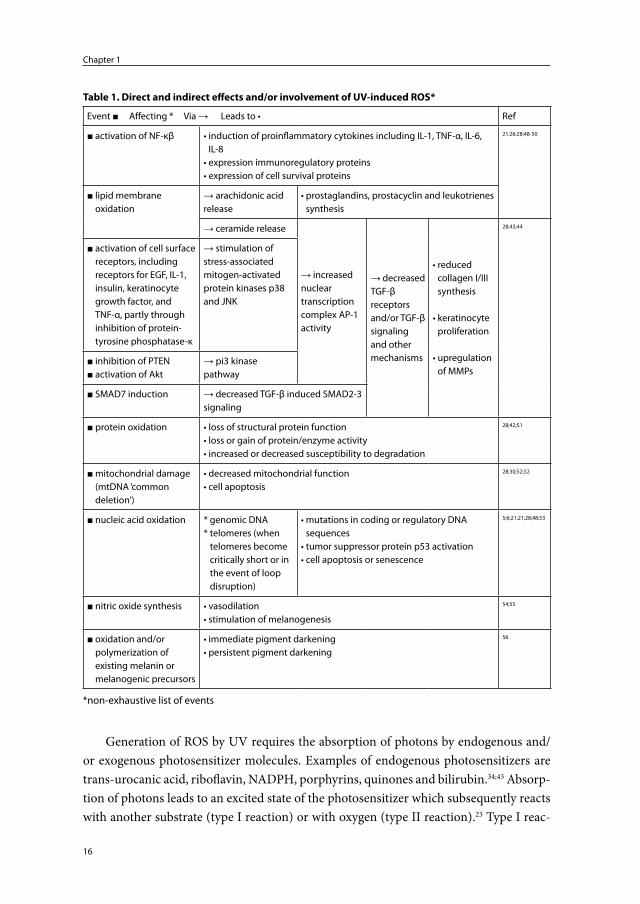
There is considerable evidence that ROS initiate many of the events following ir-radiation of the skin (summarized in Table 1). In vivo, under normal circumstances, low levels of ROS are generated continuously. These ROS are involved in signaling pathways, cell-activation, cell-proliferation and cell-differentiation.38-40 The body defends itself against excessive endogenous or exogenous oxidative stress with antioxidant enzymes and non-enzymic antioxidants.38;41 During UV exposure, similar to DNA damage and repair, this protective system can be overwhelmed resulting in direct and indirect damage to cellular and extracellular components, including the ECM proteins.42-45
Chapter 1
16
Generation of ROS by UV requires the absorption of photons by endogenous and/or exogenous photosensitizer molecules. Examples of endogenous photosensitizers are trans-urocanic acid, riboflavin, NADPH, porphyrins, quinones and bilirubin.34;43 Absorp-tion of photons leads to an excited state of the photosensitizer which subsequently reacts with another substrate (type I reaction) or with oxygen (type II reaction).23 Type I reac-
Table 1. Direct and indirect effects and/or involvement of UV-induced ROS*
Event ■ Affecting * Via → Leads to • Ref
■ activation of NF-κβ • induction of proinflammatory cytokines including IL-1, TNF-α, IL-6, IL-8
• expression immunoregulatory proteins• expression of cell survival proteins
21;26;28;48-50
■ lipid membrane oxidation
→ arachidonic acid release
• prostaglandins, prostacyclin and leukotrienes synthesis
→ ceramide release
→ increased nuclear transcription complex AP-1 activity
→ decreased TGF-β receptors and/or TGF-β signaling and other mechanisms
• reduced collagen I/III synthesis
• keratinocyte proliferation
• upregulation of MMPs
28;43;44
■ activation of cell surface receptors, including receptors for EGF, IL-1, insulin, keratinocyte growth factor, and TNF-α, partly through inhibition of protein-tyrosine phosphatase-κ
→ stimulation of stress-associated mitogen-activated protein kinases p38 and JNK
■ inhibition of PTEN■ activation of Akt
→ pi3 kinase pathway
■ SMAD7 induction → decreased TGF-β induced SMAD2-3 signaling
■ protein oxidation • loss of structural protein function• loss or gain of protein/enzyme activity• increased or decreased susceptibility to degradation
28;42;51
■ mitochondrial damage (mtDNA ‘common deletion’)
• decreased mitochondrial function• cell apoptosis
28;30;52;52
■ nucleic acid oxidation * genomic DNA* telomeres (when
telomeres become critically short or in the event of loop disruption)
• mutations in coding or regulatory DNA sequences
• tumor suppressor protein p53 activation• cell apoptosis or senescence
5;6;21;21;28;48;53
■ nitric oxide synthesis • vasodilation• stimulation of melanogenesis
54;55
■ oxidation and/or polymerization of existing melanin or melanogenic precursors
• immediate pigment darkening• persistent pigment darkening
56
*non-exhaustive list of events
17
General Introduction
tions produce radical ions, type II reactions produce ROS. Examples of ROS are singlet oxygen, superoxide anion, hydrogen peroxide and hydroxyl radical.46 The hydroxyl radi-cal, which is formed via superoxide anion and hydrogen peroxide, is highly active and has been detected in UV-exposed murine skin.47
Following UV exposure, one of the first observable events is a vasodilatory response resulting in an increased blood flow. Vasodilation and resultant erythema follow a biphasic pattern. Erythema is observed immediately following or during UV exposure and is seen again 1 hour after exposure, reaching a peak intensity after 24-48 hours.21;26 Although keratinocytes are primarily affected and stimulated by UV, all nucleated cells in the skin are capable of producing cytokines when stimulated or activated by UV. Key mediators of UV-induced erythema are thought to be prostaglandins (PGE2, PGF2α and other arachidonic acid metabolites) and nitric oxide (NO).21;48;54 Other inflammatory substances, however, probably also contribute to UV-induced erythema. In the initial phase of erythema development UV-stimulated release of vasoactive mediators by mast cells could play an important role. Resident mast cells can release proinflammatory and immunoregulatory cytokines (e.g., histamine, TNF-α) in response to UV exposure.57 The precise mechanisms behind UV-induced mast cell degranulation are unknown. Photosensitizers may be involved or, as Kulka et al.58 showed, particular neuropeptides (substance-P, VIP) may initiate mast cell degranulation and synthesis of chemokines.
NO, which is produced by keratinocytes following UV exposure and, as mentioned above, appears to play an important role in UV-induced erythema, may also be a key mediator in UV-induced melanogensis.55
As early as 30 minutes after UV exposure, depending on the dose and wavelengths of UV, dyskeratotic or apoptotic keratinocytes (sunburn cells) can be observed in the lower half of the epidermis. UV-induced apoptosis or ‘programmed cell death’ protects the body against UV-induced carcinogenesis.6 ‘Immediate apoptosis’, which is observed within 30-60 minutes after UV exposure, is caused by direct targeting of mitochondria by ROS. ‘Intermediate apoptosis’, starting within 4 hours of UV damage, involves receptor-triggered mechanisms (e.g., FasR). UV-induced genomic DNA damage is believed to be the trigger of ‘delayed apoptosis’ which commences later than 4 hours after UV exposure and entails new protein synthesis.30
Langerhans cells quickly migrate from the epidermis upon UV exposure. 72 hours after UV exposure (depending on the dose and wavelengths of UV) only 10% may remain in the epidermis.26
As mentioned earlier, the UV-induced inflammatory infiltrate contains different cell types. These cells, in order of entrance, are neutrophilic granulocytes, macrophages and T-lymphocytes.27;59-61 Leucocytes enter the skin a few hours after irradiation and the re-sponse generally resolves within 48-72 hours. Inflammatory cell-type, cell-numbers and the duration of inflammation are, however, dose-dependent and dependent on the UV
Chapter 1
18
source.62 Mobilization of inflammatory cells largely depends upon specific chemoattrac-tant stimuli (e.g., IL-8, LTB4, PAF, C5a) and appropriate adhesion molecule expression (e.g., E-selectin, P-selectin, ICAM-1, L-selectin, β2 integrins).63;64 UV-activated kerati-nocytes are capable of producing numerous cytokines including IL-1, TNF-α, IL-6, IL-8, and Gro-α. These cytokines, together with other soluble factors, are responsible for the recruitment of the inflammatory cells after UV irradiation.21;65;66 Dermal endothelial cells have been shown to express adhesion molecules after UV exposure.66;67
In summary, UV can: (1) damage cellular and extracellular components including nucleic acids, proteins, lipids and bilipid membranes, (2) activate signaling pathways that are related to proliferation, differentiation, senescence and tissue degradation, (3) cause depletion of cellular antioxidants and antioxidant enzymes, (4) activate the neuroen-docrine system and stimulate release of neuroendocrine mediators, (5) cause increased synthesis and release of proinflammatory mediators from a variety of skin cells, and (6) induce an inflammatory infiltrate. With respect to photoaging, UV-induced mediators may either directly damage the ECM or indirectly contribute to the skin changes ob-served: i.e., induced skin-infiltrating cells and stimulated resident cells may release their own mediators which can further damage the ECM.
Matrix metalloproteinases and photoaging
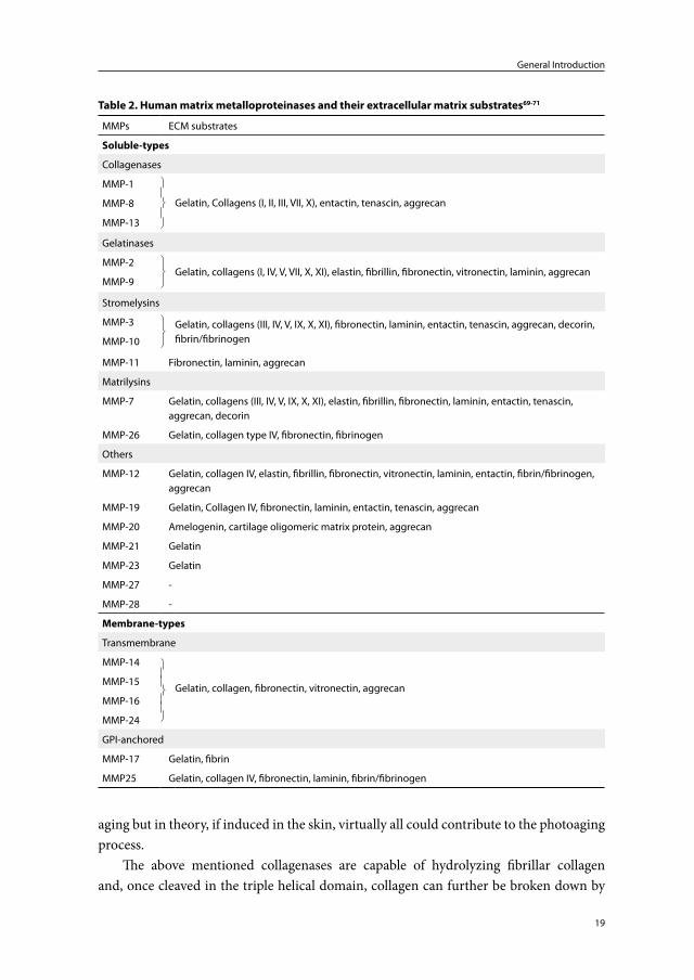
Matrix metalloproteinases (MMPs) are a group of structurally related zinc-dependent endopeptidases.68 Endopeptidases are enzymes that break peptide bonds within a protein molecule, in contrast to exopeptidases which cleave only the end parts of a polypeptide chain. The family of MMPs comprises over 20 members. MMPs are classified as col-lagenases, gelatinases, stromelysins, matrilysins, membrane-type MMPs and others69 (Table 2).
MMPs are multifunctional proteins. Collectively they can degrade essentially all extracellular matrix components including collagen fibers, elastic fibers, glycoproteins and proteoglycans.70;71 Furthermore, they are able to process many bioactive molecules: MMPs can cleave cell surface receptors, release apoptotic ligands, and activate or inacti-vate cytokines, chemokines and growth factors.71 MMPs are believed to play an important role in cell-proliferation, cell-differentiation, cell-migration, cell-adhesion, apoptosis and host defense.72 Keratinocytes, fibroblasts, endothelial cells, macrophages, mast cells, neutrophils and eosinophils are all capable of producing MMPs.73 MMPs can be induced in the skin by UVA, UVB, SSR, infrared radiation and natural sunlight.74-78
Each MMP has specific action loci whereby particular amino acid sequences are cleaved.70 However, the substrate specificity of MMPs can be broad and, consequently, cross reactivity between different MMPs is common (Table 2). The relative proteolytic capacity of different MMPs with respect to a particular substrate has not yet been fully elucidated. Not all the MMPs listed in Table 2 have been studied in the context of photo-
19
General Introduction
aging but in theory, if induced in the skin, virtually all could contribute to the photoaging process.
The above mentioned collagenases are capable of hydrolyzing fibrillar collagen and, once cleaved in the triple helical domain, collagen can further be broken down by
Table 2. Human matrix metalloproteinases and their extracellular matrix substrates69-71
MMPs ECM substrates
Soluble-types
Collagenases
MMP-1
Gelatin, Collagens (I, II, III, VII, X), entactin, tenascin, aggrecanMMP-8
MMP-13
Gelatinases
MMP-2
Gelatin, collagens (I, IV, V, VII, X, XI), elastin, fibrillin, fibronectin, vitronectin, laminin, aggrecanMMP-9
Stromelysins
MMP-3
Gelatin, collagens (III, IV, V, IX, X, XI), fibronectin, laminin, entactin, tenascin, aggrecan, decorin, fibrin/fibrinogenMMP-10
MMP-11 Fibronectin, laminin, aggrecan
Matrilysins
MMP-7 Gelatin, collagens (III, IV, V, IX, X, XI), elastin, fibrillin, fibronectin, laminin, entactin, tenascin, aggrecan, decorin
MMP-26 Gelatin, collagen type IV, fibronectin, fibrinogen
Others
MMP-12 Gelatin, collagen IV, elastin, fibrillin, fibronectin, vitronectin, laminin, entactin, fibrin/fibrinogen, aggrecan
MMP-19 Gelatin, Collagen IV, fibronectin, laminin, entactin, tenascin, aggrecan
MMP-20 Amelogenin, cartilage oligomeric matrix protein, aggrecan
MMP-21 Gelatin
MMP-23 Gelatin
MMP-27 -
MMP-28 -
Membrane-types
Transmembrane
MMP-14
Gelatin, collagen, fibronectin, vitronectin, aggrecanMMP-15
MMP-16
MMP-24
GPI-anchored
MMP-17 Gelatin, fibrin
MMP25 Gelatin, collagen IV, fibronectin, laminin, fibrin/fibrinogen
Chapter 1
20
gelatinases and stromelysins. The combined actions of interstitial collagenase (MMP-1), gelatinase A (MMP-2), gelatinase B (MMP-9) and stromelysin 1 (MMP-3) can fully degrade collagen type I (Dermal collagen consists of ±80% collagen type I and 10-20% collagen type III11) and components of the elastic network.70 These MMPs, together with neutrophil collagenase (MMP-8), macrophage elastase (MMP-12) and matrilysin (MMP-7), have particularly been studied with respect to the pathophysiology of photoaging. MMP-8, which is a major product of neutrophils, has similar substrates compared to MMP-1: these substrates include collagens type I, II, III, VII, X, gelatin, various glyco-proteins and proteoglycans.71 MMP-8, however, cleaves collagen type I faster than type III, while MMP-1 shows greater selectivity for collagen type III compared to type I.73 MMP-12 is mainly produced by macrophages.73;79 Its substrates includes fibronectin, fibrin/fibrinogen, laminin, and proteoglycans. Furthermore, MMP-12 together with MMP-2, MMP-7 and MMP-9, are capable of damaging components of the elastic-fiber network.69;80-82
The integrity of healthy human skin is largely dependent on a tightly regulated homeostasis of the collagen and elastic fiber networks.83 Under normal conditions, the activity of MMPs is controlled by transcriptional regulation, pro-enzyme activation, and inhibitors of MMPs. MMPs are inhibited by α2-macroglobulin and tissue inhibitors of metalloproteinases (TIMPs).69;70 The latter multifunctional proteins can inactivate spe-cific MMPs. An imbalance between MMP and TIMP synthesis, resulting in an excess of (activated) MMPs, can lead to extracellular matrix degradation. Investigators have shown that following UV exposure certain MMPs are upregulated and particular TIMPs are downregulated.75;84;85 Thus, the cells and mediators involved in UV-induced synthesis, activation and release of MMPs and TIMPs could play an important role in photoaging.
Neutrophils and photoaging
Neutrophils are the most abundant type of white blood cells and are a hallmark of tissue inflammation.86 Neutrophils react within an hour of tissue injury and rapidly migrate toward the site of tissue damage. Neutrophils are capable of releasing a wide variety of products including proteolytic enzymes, antimicrobial products, ROS, cytokines and chemokines. These products are stored in azurophilic-, specific-, tertiary-, and secretory vesicles, or are newly formed.87-89 Carl Nathan90 collected considerable scientific evidence to support the following views with respect to the functions of neutrophils: (1) neutro-phils often have an important role in launching immune responses, (2) neutrophils help to heal tissues as well as destroying them, (3) neutrophils give instructions with as much specificity as a lymphocyte or neuron, albeit with a specificity of a different kind, (4) neutrophils integrate information with a circuitry of awe-inspiring design, to tailor its responses to its spatial and temporal context, (5) neutrophils offer potential opportunities for selective pharmacological intervention, to both promote and restrain inflammation.
21
General Introduction
It has long been recognized that neutrophils can damage tissues.91;92 Furthermore, neutrophils and their products have been identified as a major cause of tissue destruction in a number of conditions, including acute lung injury and emphysema,93;94 rheumatoid arthritis,95 periodontitis,96;97 and wound infection98. Despite their potency, however, neutrophils have only been reported as likely important contributors to photoaging in a murine model.99
An important immunohistochemical marker and major product of neutrophils, is neutrophil elastase.100;101 Together with cathepsin G and proteinase-3, neutrophil elas-tase belongs to the group of serine proteinases produced by neutrophils.94 These serine proteinases are stored in azurophilic granules and are normally rapidly and irreversibly inhibited by powerful plasma antiproteinases (α1-antitrypsin, α2-macroglobulin and secretory leucoproteinase inhibitor) present in blood and the interstitium.92 Neutrophil elastase is a potent proteolytic enzyme capable of cleaving elastin, fibrillin, collagen (types I-IV), fibronectin, laminin, vitronectin and proteoglycans.94;102;103 The biological functions of neutrophil elastase include: (1) involvement in migration of neutrophils by means of focal proteolysis, (2) killing of microorganisms, and (3) degradation or activation of vari-ous proteins including ECM proteins, receptors, cytokines and chemokines.94;101;104
Neutrophils also express MMPs, namely; MMP-8 and MMP-9. MMP-8 and MMP-9 are stored in specific and tertiary vesicles in an inactive form.105 MMP-12 expression by neutrophils has also been reported.106
The probable mechanisms by which neutrophil-derived proteolytic enzymes are ac-tivated and prevented from being inactivated by antiproteinases, and cause ECM damage are reviewed by Weiss92 and Chua.104 It involves a complex interaction with nicotinamide adenine dinucleotide phosphate (NADPH) oxidase-derived oxygen metabolites and it occurs at or just outside the plasma membranes of neutrophils. The following section summarizes the events that take place: activated neutrophils generate ROS by means of the membrane-associated NADPH oxidase enzyme complex. Following a series of reac-tions catalyzed by superoxide dismutase and myeloperoxidase and involving superoxide anion and hydrogen peroxide, hypochlorous acid is formed. Hypochlorous acid is a highly active product and instantaneously reacts with amines (present in the immediate vicinity of the neutrophil or released by the cell itself) forming a group of oxidants known as chloramines. These chloramines are responsible for breaking down the antiproteinase shield surrounding tissue-infiltrating neutrophils (chloramines oxidize and inactivate plasma antiproteinases such as α1-antitrypsin) and are responsible for activating particu-lar proteolytic enzymes released from neutrophil secretory vesicles (chloramines oxidize and activate MMP-8 and MMP-9). In addition, MMP-8 and MMP-9 can themselves hydrolyse and inactivate α1-antitrypsin and neutrophil elastase can inactivate TIMPs. MMP-8, MMP-9 and neutrophil elastase released from neutrophils are thus able to damage the ECM. In conditions where there is a deficiency of antiproteinases (e.g., α1-
Chapter 1
22
antitrypsin deficiency) or in case of massive neutrophil infiltration (e.g., infected wounds with abscess formation) proteolytic enzymes are active beyond the immediate vicinity of the neutrophil and cause accelerated and/or severe tissue destruction.
In summary, neutrophils are potent cells capable of generating and releasing multiple mediators which can directly or indirectly damage the ECM. Since neutrophils infiltrate the skin following UV exposure they should be studied more closely with respect to the pathophysiology of photoaging.
Current hypothetical model of photoaging
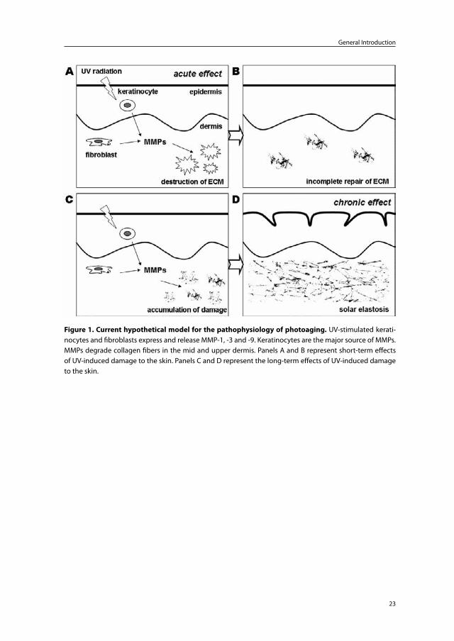
A hypothetical model for the pathophysiology of photoaging has been presented by Fisher et al.76;85 They suggested that skin damage after a single exposure to UV is only partially repaired and that microscopic damage accumulates after each exposure, eventu-ally leading to the clinical and histopathological signs of photoaged skin. Fisher et al.76;85 postulated that this microscopic damage is caused by specific MMPs. They showed that MMP-1, MMP-3 and MMP-9 are induced following exposure of human skin to suber-ythemogenic doses of UVB. As mentioned above, these MMPs are capable of breaking down skin collagens. On the basis of (1) the localization of immunohistochemically stained MMPs, (2) in situ hybridisation of mRNA, (3) in situ zymography, and (4) lit-erature data, they concluded that the MMPs were fibroblast and keratinocyte derived. The expression of MMP protein by fibroblasts following UV exposure was based on the morphology of MMP-positive cells in in vivo experiments and on in vitro data. In their in vivo studies, MMP mRNA was mostly located in the epidermis and MMP protein was also mainly induced in this compartment.85 They concluded that keratinocytes are the major source of MMPs. Because the ECM damage observed in photoaged skin is located mostly in the mid and upper dermis, they proposed that keratinocyte-derived MMPs diffuse from the epidermis into the dermis (where most of the MMP enzyme activity had been detected). In a separate study, Fisher et al.77 studied neutrophil-derived MMP-8. MMP-8 was induced in skin exposed to 2 MED of UVB and 2 MED of SSR. On the basis of the finding that trans-retinoic acid failed to inhibit MMP-8 expression following UV exposure, but did inhibit degradation of collagen type I (indirect detection method), they concluded that MMP-8 was not enzymatically active and therefore did not contribute to photoaging.
Thus, the current hypothetical model proclaims that fibroblast- and particularly keratinocyte-derived MMPs are key mediators in the pathophysiology of photoaging (Figure 1).
23
General Introduction
Figure 1. Current hypothetical model for the pathophysiology of photoaging. UV-stimulated kerati-nocytes and fibroblasts express and release MMP-1, -3 and -9. Keratinocytes are the major source of MMPs. MMPs degrade collagen fibers in the mid and upper dermis. Panels A and B represent short-term effects of UV-induced damage to the skin. Panels C and D represent the long-term effects of UV-induced damage to the skin.
Chapter 1
24
III. Skin phototypes (SPT) and natural defenses against sunlight/UV
Natural defenses of human skin against sunlight/UV107
The basic mechanisms by which the skin protects itself against the damaging effect of sunlight/UV are summarized below:(1) The stratum corneum absorbs, reflects and scatters UV. UV-absorbing products in
the stratum corneum include urocanic acid and particular amino acids of keratins such as histidine, tyrosine and tryptophan.
(2) Genetically determined constitutive and facultative (see further) skin pigmentation. Melanin absorbs and scatters UV, and is a free radical/ROS scavenger.
(3) Naturally occurring antioxidants such as lipophilic carotenoids, alpha-tocopherol (vitamin E), glutathione and ascorbic acid (vitamin C) react with and neutralize a variety of free radicals/ROS.
(4) Antioxidant enzymes such as catalase, superoxide dismutase and gluthatione per-oxidase/reductase deactivate ROS.
(5) DNA repair mechanisms repair UV-induced DNA damage.(6) Epidermal hyperplasia and hyperkeratosis increase the physical barrier to UV.
Skin color and SPT, sunburn reaction and photoaging
Skin color is the result of a blend of colors due to various chromophores present in the skin: melanin (brown), oxyhaemoglobin (red), deoxygenated haemoglobin (blue) and carotene (yellow-orange). Melanin, however, is the most important contributor to skin color.108
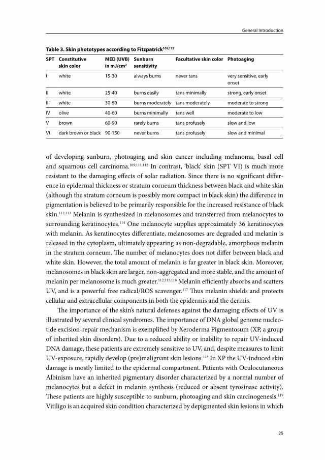
SPT is a concept that was introduced in 1975 to establish one’s sensitivity to UV.107;109 It was used to determine a safe starting dose for UV therapy. SPT is based on the his-tory of reactions of a person’s skin to sun exposure. To determine the SPT the following questions need to be answered:109 (1) How does the skin respond to 45-60 minutes of sun exposure at noon, in the northern latitudes (20º to 45º), in the early summer (+/- 90-120 mJ/cm² of UVB)? (2) How intense is the sunburn reaction (pain, erythema, oedema)? (3) How much tan has developed after 7 days? Based on this history and on the constitutive skin color, people are categorized into six phototypes (originally 4 later 6 skin phototypes - Table 3). Constitutive skin color is the degree of pigmentation of non-stimulated, sun-protected skin. It is genetically determined and reflects the inher-ent content and distribution of melanin in the epidermis.107;108 Tan or facultative skin color is the inducible skin pigmentation. The degree of pigmentation raised above the constitutional level varies according to genetically determined tanning ability and his-tory of UV exposure.107;108;110 Whereas hair and eye color are unreliable guides to sunburn sensitivity, SPT is a good predictor of a person’s susceptibility to the acute and chronic damaging effects of UV. The lower SPTs (SPT I-III, ‘white’ skin) are at far greater risk
25
General Introduction
of developing sunburn, photoaging and skin cancer including melanoma, basal cell and squamous cell carcinoma.109;111;112 In contrast, ‘black’ skin (SPT VI) is much more resistant to the damaging effects of solar radiation. Since there is no significant differ-ence in epidermal thickness or stratum corneum thickness between black and white skin (although the stratum corneum is possibly more compact in black skin) the difference in pigmentation is believed to be primarily responsible for the increased resistance of black skin.112;113 Melanin is synthesized in melanosomes and transferred from melanocytes to surrounding keratinocytes.114 One melanocyte supplies approximately 36 keratinocytes with melanin. As keratinocytes differentiate, melanosomes are degraded and melanin is released in the cytoplasm, ultimately appearing as non-degradable, amorphous melanin in the stratum corneum. The number of melanocytes does not differ between black and white skin. However, the total amount of melanin is far greater in black skin. Moreover, melanosomes in black skin are larger, non-aggregated and more stable, and the amount of melanin per melanosome is much greater.112;115;116 Melanin efficiently absorbs and scatters UV, and is a powerful free radical/ROS scavenger.117 Thus melanin shields and protects cellular and extracellular components in both the epidermis and the dermis.
The importance of the skin’s natural defenses against the damaging effects of UV is illustrated by several clinical syndromes. The importance of DNA global genome nucleo-tide excision-repair mechanism is exemplified by Xeroderma Pigmentosum (XP, a group of inherited skin disorders). Due to a reduced ability or inability to repair UV-induced DNA damage, these patients are extremely sensitive to UV, and, despite measures to limit UV-exposure, rapidly develop (pre)malignant skin lesions.118 In XP the UV-induced skin damage is mostly limited to the epidermal compartment. Patients with Oculocutaneous Albinism have an inherited pigmentary disorder characterized by a normal number of melanocytes but a defect in melanin synthesis (reduced or absent tyrosinase activity). These patients are highly susceptible to sunburn, photoaging and skin carcinogenesis.119 Vitiligo is an acquired skin condition characterized by depigmented skin lesions in which
Table 3. Skin phototypes according to Fitzpatrick109;112
SPT Constitutiveskin color
MED (UVB)in mJ/cm2
Sunburnsensitivity
Facultative skin color Photoaging
I white 15-30 always burns never tans very sensitive, early onset
II white 25-40 burns easily tans minimally strong, early onset
III white 30-50 burns moderately tans moderately moderate to strong
IV olive 40-60 burns minimally tans well moderate to low
V brown 60-90 rarely burns tans profusely slow and low
VI dark brown or black 90-150 never burns tans profusely slow and minimal

Chapter 1
26
melanocytes are lost from the epidermis.120 Persons with SPT VI and vitiligo offer a natural disease model to further substantiate the photoprotective properties of melanin.
In summary, different SPTs show a different susceptibility to UV-induced skin changes including photoaging. Melanin directly absorbs UV photons and scavenges UV-induced ROS, and appears to play a major role in the skin’s defense against UV-induced skin changes.
27
General Introduction
IV. Prevention and treatment of sunburn reaction and photoaging
Prevention and treatment methods for sunburned skin and/or photoaged skin described in literature include the following measures, products and interventions: behavioral changes, clothing and hats, sunscreens, DNA-damage repair enzymes, antioxidants, dietary lipids, osmolytes, hydroxy acids, fluorouracil, imiquimod, retinoids, botulinum toxin, chemical peels, dermabrasion, photodynamic therapy, laser therapy, injectables, and surgical procedures.28;121;122
A Cochrane review123 entitled ‘interventions for photodamaged skin’ assessed the ef-fects of topically applied treatments, systemic drug treatments, laser therapy and surgical procedures on photodamaged skin. The selection criteria for this systemic review were: (1) randomized controlled trials which compared drug or surgical interventions with no treatment or with placebo or with another drug, and (2) studies must include adults with mild, moderate or severe photodamaged facial or forearm skin. Thirty studies of variable quality were included. Based on these thirty studies the author’s conclusions were: (1) Topical tretinoin cream (a retinoid) improves skin changes (fine and coarse wrinkles, roughness, freckles and pigmentation) associated with prolonged sun exposure. The greatest improvements occurred with higher concentrations of tretinoin, but at the expense of greater local irritation. (2) More evidence is needed before any recommenda-tions can be made on oral or topical polysaccharide- or hydroxy- acids. (3) It is unclear how useful surgical, laser or chemical peels are in the absence of suitable control groups. Both techniques lead to pain and redness after the procedure. The authors did not find any randomized controlled trials on the effectiveness of application of sunscreens or wearing of protective clothing in preventing photoaging of the skin.
Retinoids, corticosteroids, vitamin E and sunscreens
Because retinoids, corticosteroids, vitamin E and sunscreens are often mentioned and/or have been studied extensively with respect to prevention and management of sunburn reactions and/or photoaging they are discussed in more detail.
Retinoids
There are a significant number of studies investigating the effects of retinoids on pho-toaging. The improvement of human photoaged skin following treatment with topical retinoids, although modest, has been shown to be highly statistically significant.28;124-126 The described (long term) effects of topical retinoids on the skin are: (1) reduction and redistribution of epidermal melanin, (2) improved ultrastructural characteristics of the epidermis, (3) increased anchoring fibrils, (4) increased deposition of papillary dermal collagen, and (5) increased vascularity in the papillary dermis. Kossard et al.127investigated the effect of daily application of 0.05% tretinoin for 6 months on photoaged skin. They
Chapter 1
28
found that tretinoin treatment particularly affected the epidermis and had no effect on the degree of solar elastosis.
The effects of retinoids are probably mediated through binding to nuclear retinoic acid receptors and subsequent binding of the ligand/receptor complex to specific sites on particular genes. Several effects are proposed to result from an inhibition of the induction of activator protein-1 (AP-1). Inhibition of AP-1 activity and reduced TGF-β signaling in keratinocytes and fibroblasts leads to a reduced expression of MMPs and increased ex-pression of collagen types I and III.128 This mechanism (i.e., inhibition of AP-1 activity by retinoids), together with the hypothesis that keratinocyte- and fibroblast-derived MMPs are responsible for ECM damage leading to solar elastosis, are often quoted as the basis of the effectiveness of retinoids in the treatment of photoaging. Application of retinoids does not inhibit infiltration of neutrophils following skin exposure to erythemogenic doses of UVB and SSR.77
Corticosteroids
(Gluco)corticosteroids are cholesterol derivatives which are normally synthesized in the adrenal cortex. Corticosteroids are involved in a wide range of physiologic systems including carbohydrate metabolism, protein catabolism, lipid catabolism, electrolyte and water homeostatis, stress response, immune response and regulation of inflam-mation.129;130 Corticosteroids are fat soluble and easily diffuse across lipid membranes into the cytoplasmic compartment of cells where they bind to receptor proteins. The corticosteroid-binding receptor is activated when two hormone molecules are bound. Subsequently, the hormone-receptor complex is transferred to the nucleus where it exerts its functions by influencing transcription.129;130
Chronic use of corticosteroids induces atrophy of the skin.129;131 Considering this catabolic effect, it is logical that topical or systemic steroids are not used to treat photo-aged skin. Corticosteroids have, however, been used to treat sunburned skin and several studies have shown that UV-induced erythema can be suppressed by topical corticoste-roids.132;133 Other authors failed to show an effect of topical and systemic corticosteroids on UV-induced erythema.134;135 The consensus of opinion with respect to the management of an acute sunburn is not to administer corticosteroids.136 Topical corticosteroids are widely used for different inflammatory skin conditions, including those where infiltra-tion of neutrophils is involved.137 Fisher et al.77 investigated the effect of a powerful topical steroid on UV-induced MMP upregulation and on infiltration of neutrophils and found that both were inhibited. The effect of (topical) steroids on UV-induced ROS has, to our knowledge, not been studied.
29
General Introduction
Vitamin E
As mentioned earlier, the skin contains both antioxidant enzymes and non-enzymatic antioxidants. Vitamin E or tocopherol is an example of the latter. It is an essential nutrient and a major lipophilic antioxidant in plasma, membranes and tissues.138;139 There are eight naturally occurring tocopherols of which α-tocopherol is the most important, both in quantity and activity.139 α-Tocopherol is easily oxidized and thus inactivated. The acetate derivative of α-tocopherol is, however, more stable. Relative vitamin E activity of specific tocopherols depends on biochemical reactivity, resorption from the gut, cellular uptake and metabolic turnover. Vitamin E scavenges free radicals/ROS and plays a particularly important role in protecting polyunsaturated fatty acids against oxidation.138;139 Poly-unsaturated fatty acids are major constituents of cytoplasmic- and mitochondrial-lipid membranes. In the presence of iron or toxins, ROS can be generated from polyunsatu-rated fatty acids and initiate a chain reaction. Vitamin E can neutralize ROS and the chain reaction may thus be prevented.138;139
The effect of topical and systemic vitamin E on sunburn reaction and photoaging has been studied quite extensively in both animals and humans.140 Numerous studies have demonstrated that topical and/or systemic vitamin E administration prior to UV exposure is capable of reducing acute skin responses such as erythema and oedema (particularly when combined with vitamin C).141-144 Conversely, other studies have shown little or no effect of vitamin E on UV-induced erythema.140;145 Several murine studies have shown a protective effect against UV-induced wrinkle formation.140 In general, the acetate derivative of α-tocopherol, although more stable, is less protective.140
Both topical and systemic vitamin E products are being marketed as anti-aging products. Although there are in vitro and in vivo, animal and human data supporting a positive effect, randomized controlled trials are lacking.
Sunscreens
Sunscreens prevent UV from penetrating the skin. Sunscreens contain UV filters that can be grouped into two broad categories: organic filters (or ‘chemical’ filters) which absorb UV photons and inorganic filters (or ‘physical’ filters) which reflect UV photons.24;28 Organic filters are usually ‘invisible’ and thus cosmetically appealing. Absorption of UV-photons by organic filters releases heat and may lead to the formation of active products capable of interacting with cutaneous molecules. Although first generation organic filters tended to be unstable, current filters are photostable. There is a long list of safe organic filters presently available, which absorb UV along a wide range of wavelengths such that a combination of filters can achieve adequate UVA and UVB protection.24;28 Zinc oxide and titanium dioxide are examples of inorganic filters. In addition to reflecting UV, these compounds also reflect visible light, making them cosmetically less attractive. Their ad-vantage is, however, that they are chemically inert and protect against UVA, UVB and vis-
Chapter 1
30
ible light.24;28 Modern sunscreens contain both organic and inorganic filters. Nanoparticle technology has facilitated the production of cosmetically more acceptable sunscreens that also contain inorganic filters. Skin protection factor (SPF) is a widely used concept that refers to a sunscreen’s protection against UV-induced (mostly UVB) erythema.
Sunscreens have been shown to effectively prevent or reduce UV-induced erythe-ma.146 Also, sunscreens can prevent UV-induced neutrophil infiltration. Murine studies have demonstrated that sunscreens can prevent solar elastosis.147 As mentioned earlier there are few if any randomized controlled trials investigating the efficacy of sunscreens in preventing or slowing down photoaging of human skin.
31
General Introduction
V. Outline of this thesis
The primary objectives of this thesis are to determine the role of neutrophils in the patho-physiology of photoaging, and to critically examine the current hypothetical model for the pathophysiology of photoaging.Secondary objectives are:(i) To determine the expression, activity and origin of photoaging-associated proteo-
lytic enzymes in ‘young’ and ‘elderly’ skin, different skin phototypes (‘black’ and ‘white’), sun-exposed and sun-protected skin, and SSR-exposed skin.
(ii) To show that in chronically sun-exposed skin elastic fiber breakdown is more im-portant than collagen breakdown.
(iii) To provide further evidence for the role of ROS in the pathophysiology of photoag-ing.
(iv) To determine the protective effect of skin pigmentation, sunscreens, topical vitamin E, and betamethasone against SSR-induced erythema and neutrophil influx.
The current hypothetical model for the pathophysiology of photoaging focuses on colla-gen degradation and states that keratinocyte- and fibroblast-derived MMPs play a crucial role in the dermal ECM damage seen in photoaged skin. However, solar elastosis, the histopathologic hallmark of photoaged skin, is characterized by deposition of elastotic material in the mid and upper dermis. This elastotic material is most likely derived from degraded elastic fibers. Neutrophils infiltrate the skin following UV exposure but have not received much attention with respect to the pathophysiology of photoaging. Neutrophils, however, are capable of generating and releasing proteolytic enzymes which can dam-age both collagen and elastic fibers. Skin pigmentation, melanin, appears to be a major protective factor against the acute and chronic damaging effects of UV on the skin. The mechanisms by which skin pigmentation protects against photoaging have not been fully elucidated. A possible mechanism could be protection against UV-induced neutrophil influx. Multiple commercially available products claim that they are effective in slowing down or even reversing the photoaging process. These products include sunscreens and ROS scavengers such as topical vitamin E.
In chapters 2 and 4 we compare black and white skin with respect to the expres-sion of photoaging-associated proteolytic enzymes and other SSR-induced responses. In chapters 2, 3 and 4 data is shown supporting an important role for neutrophils in the pathophysiology of photoaging. The same data poses questions regarding the current hy-pothetical model. In chapter 5 we analyze collagen types and elastic fiber components in different skin phototypes. In addition, we investigate the effect of neutrophils, neutrophil elastase, ROS, and direct UV exposure on elastic fiber staining. In chapter 6 we review the probable role of neutrophils in photoaging, and we discuss preventive measures and

Chapter 1
32
potential pharmacological targets in this respect. In chapter 7 we investigate the effect of sunscreens, topical betamethasone and topical vitamin E on SSR-induced erythema and neutrophil influx.
33
General Introduction
References
1. Yaar M. Clinical and histological features of intrinsic versus extrinsic skin aging. In: Skin aging (Gilchrest BA, Krutmann J, eds), 1st edn. Springer, 2006: 9-21.
2. Gilchrest BA. Skin aging and photoaging: an overview. J.Am.Acad.Dermatol. 1989; 21: 610-3.
3. Matsuta M, Izaki S, Ide C et al. Light and electron microscopic immunohistochemistry of solar elastosis: a study with cutis rhomboidalis nuchae. J.Dermatol. 1987; 14: 364-74.
4. Oikarinen A. The aging of skin: chronoaging versus photoaging. Photodermatol.Photoim-munol.Photomed. 1990; 7: 3-4.
5. Black HS, deGruijl FR, Forbes PD et al. Photocarcinogenesis: an overview. J.Photochem.Photobiol.B 1997; 40: 29-47.
6. Hoeijmakers JH. DNA damage, aging, and cancer. N.Engl.J.Med. 2009; 361: 1475-85.7. Braverman IM, Fonferko E. Studies in cutaneous aging: I. The elastic fiber network. J.Invest
Dermatol. 1982; 78: 434-43.8. Tsuji T. The surface structural alterations of elastic fibers and elastotic material in solar
elastosis: a scanning electron microscopic study. J.Cutan.Pathol. 1984; 11: 300-8.9. Kadler KE, Holmes DF, Trotter JA et al. Collagen fibril formation. Biochem.J. 1996; 316 ( Pt
1): 1-11.10. Kielty CM, Sherratt MJ, Shuttleworth CA. Elastic fibres. J.Cell Sci. 2002; 115: 2817-28.11. Nimni ME. Collagen: structure, function, and metabolism in normal and fibrotic tissues.
Semin.Arthritis Rheum. 1983; 13: 1-86.12. Garrone R, Lethias C, Le GD. Distribution of minor collagens during skin development.
Microsc.Res.Tech. 1997; 38: 407-12.13. Kielty CM, Wess TJ, Haston L et al. Fibrillin-rich microfibrils: elastic biopolymers of the
extracellular matrix. J.Muscle Res.Cell Motil. 2002; 23: 581-96.14. Debelle L, Tamburro AM. Elastin: molecular description and function. Int.J.Biochem.Cell
Biol. 1999; 31: 261-72.15. Ledvina M, Velebny V. Biochemistry of elastic fibers in normal and pathological tissues. Sb
Ved.Pr Lek.Fak.Karlovy Univerzity Hradci Kralove 1982; 25: 75-101.16. Suwabe H, Serizawa A, Kajiwara H et al. Degenerative processes of elastic fibers in sun-
protected and sun-exposed skin: immunoelectron microscopic observation of elastin, fibrillin-1, amyloid P component, lysozyme and alpha1-antitrypsin. Pathol.Int. 1999; 49: 391-402.
17. Chen VL, Fleischmajer R, Schwartz E et al. Immunochemistry of elastotic material in sun-damaged skin. J.Invest Dermatol. 1986; 87: 334-7.
18. Mitchell RE. Chronic solar dermatosis: a light and electron microscopic study of the der-mis. J.Invest Dermatol. 1967; 48: 203-20.
19. Niebauer G, Stockinger L. Senile elastosis: histochemical and electron microscopic studies. Arch.Klin.Exp.Dermatol. 1965; 221: 122-43.
20. Nurnberger F, Schober E, Marsch WC et al. Actinic elastosis in black skin. A light- and electronmicroscopic study. Arch.Dermatol.Res. 1978; 262: 7-14.
21. Clydesdale GJ, Dandie GW, Muller HK. Ultraviolet light induced injury: immunological and inflammatory effects. Immunol.Cell Biol. 2001; 79: 547-68.
22. Wharton JR, Cockerell CJ. The sun: a friend and enemy. Clin.Dermatol. 1998; 16: 415-9.
Chapter 1
34
23. Moseley H. Elementary Photobiology and Photophysics. In: Photodermatology (Ferguson J, Dover JS, eds), 1st edn. Mmanson, 2006: 9-13.
24. Moseley H. Photoprotection. In: Photodermatology (Ferguson J, Dover JS, eds), 1st edn. Manson, 2006: 21-8.
25. Trautinger F. Mechanisms of photodamage of the skin and its functional consequences for skin ageing. Clin.Exp.Dermatol. 2001; 26: 573-7.
26. Hruza LL, Pentland AP. Mechanisms of UV-induced inflammation. J.Invest Dermatol. 1993; 100: 35S-41S.
27. Soter NA. Acute effects of ultraviolet radiation on the skin. Semin.Dermatol. 1990; 9: 11-5.28. Yaar M, Gilchrest BA. Photoageing: mechanism, prevention and therapy. Br.J.Dermatol.
2007; 157: 874-87.29. Krutmann J, Gilchrest BA. Photoaging of skin. In: Skin aging (Gilchrest BA, Krutmann J,
eds), 1st edn. Springer, 2006: 33-43.30. Klotz LO, Holbrook NJ, Sies H. UVA and singlet oxygen as inducers of cutaneous signaling
events. Curr.Probl.Dermatol. 2001; 29: 95-113.31. Parrish JA, Jaenicke KF, Anderson RR. Erythema and melanogenesis action spectra of
normal human skin. Photochem.Photobiol. 1982; 36: 187-91.32. Masini V, Noel-Hudson MS, Wepierre J. Free-radical damage by UV or hypoxanthine-
xanthine oxidase in cultured human skin fibroblasts: Protective effects of two human plasma fractions. Toxicol.In Vitro 1994; 8: 235-42.
33. Ravanat JL, Douki T, Cadet J. Direct and indirect effects of UV radiation on DNA and its components. J.Photochem.Photobiol.B 2001; 63: 88-102.
34. Young AR. Chromophores in human skin. Phys.Med.Biol. 1997; 42: 789-802.35. Balajee AS, May A, Bohr VA. DNA repair of pyrimidine dimers and 6-4 photoproducts in
the ribosomal DNA. Nucleic Acids Res. 1999; 27: 2511-20.36. Gibbs NK, Tye J, Norval M. Recent advances in urocanic acid photochemistry, photobiol-
ogy and photoimmunology. Photochem.Photobiol.Sci. 2008; 7: 655-67.37. Noonan FP, De Fabo EC. Immunosuppression by ultraviolet B radiation: initiation by
urocanic acid. Immunol.Today 1992; 13: 250-4.38. Sauer H, Wartenberg M, Hescheler J. Reactive oxygen species as intracellular messengers
during cell growth and differentiation. Cell Physiol Biochem. 2001; 11: 173-86.39. Nathan C. Specificity of a third kind: reactive oxygen and nitrogen intermediates in cell
signaling. J.Clin.Invest 2003; 111: 769-78.40. Bergendi L, Benes L, Durackova Z et al. Chemistry, physiology and pathology of free
radicals. Life Sci. 1999; 65: 1865-74.41. Pinnell SR. Cutaneous photodamage, oxidative stress, and topical antioxidant protection.
J.Am.Acad.Dermatol. 2003; 48: 1-19.42. Sander CS, Chang H, Salzmann S et al. Photoaging is associated with protein oxidation in
human skin in vivo. J.Invest Dermatol. 2002; 118: 618-25.43. Scharffetter-Kochanek K, Wlaschek M, Brenneisen P et al. UV-induced reactive oxygen
species in photocarcinogenesis and photoaging. Biol.Chem. 1997; 378: 1247-57.44. Rittie L, Fisher GJ. UV-light-induced signal cascades and skin aging. Ageing Res.Rev. 2002;
705-20.45. Witt EH, Motchnik P, Packer L. Evidence for UV light as an oxidative stressor in skin. In:
Oxidative stress in dermatology (Fuchs J, Packer L, eds). New York: Marcel Dekker, Inc, 1993: 29-44.
35
General Introduction
46. Darr D, Fridovich I. Free radicals in cutaneous biology. J.Invest Dermatol. 1994; 102: 671-5.47. Masaki H, Atsumi T, Sakurai H. Detection of hydrogen peroxide and hydroxyl radicals in
murine skin fibroblasts under UVB irradiation. Biochem.Biophys.Res.Commun. 1995; 206: 474-9.
48. Terui T, Okuyama R, Tagami H. Molecular events occurring behind ultraviolet-induced skin inflammation. Curr.Opin.Allergy Clin.Immunol. 2001; 1: 461-7.
49. Legrand-Poels S, Schoonbroodt S, Matroule JY et al. Nf-kappa B: an important transcrip-tion factor in photobiology. J.Photochem.Photobiol.B 1998; 45: 1-8.
50. Sarkar D, Fisher PB. Molecular mechanisms of aging-associated inflammation. Cancer Lett. 2006; 236: 13-23.
51. Shacter E. Protein oxidative damage. Methods Enzymol. 2000; 319: 428-36.52. Berneburg M, Plettenberg H, Krutmann J. Photoaging of human skin. Photodermatol.
Photoimmunol.Photomed. 2000; 16: 239-44.53. Cerutti PA. Oxy-radicals and cancer. Lancet 1994; 344: 862-3.54. Deliconstantinos G, Villiotou V, Stravrides JC. Release by ultraviolet B (u.v.B) radiation of
nitric oxide (NO) from human keratinocytes: a potential role for nitric oxide in erythema production. Br.J.Pharmacol. 1995; 114: 1257-65.
55. Weller R. Nitric oxide: a key mediator in cutaneous physiology. Clin.Exp.Dermatol. 2003; 28: 511-4.
56. Miyamura Y, Coelho SG, Wolber R et al. Regulation of human skin pigmentation and responses to ultraviolet radiation. Pigment Cell Res. 2007; 20: 2-13.
57. Walsh LJ. Ultraviolet B irradiation of skin induces mast cell degranulation and release of tumour necrosis factor-alpha. Immunol.Cell Biol. 1995; 73: 226-33.
58. Kulka M, Sheen CH, Tancowny BP et al. Neuropeptides activate human mast cell degranu-lation and chemokine production. Immunology 2008; 123: 398-410.
59. Di Nuzzo S, de Rie MA, van der Loos CM et al. Solar-simulated ultraviolet irradiation induces selective influx of CD4+ T lymphocytes in normal human skin. Photochem.Photo-biol. 1996; 64: 988-93.
60. Gilchrest BA, Soter NA, Hawk JL et al. Histologic changes associated with ultraviolet A--induced erythema in normal human skin. J.Am.Acad.Dermatol. 1983; 9: 213-9.
61. Hawk JL, Murphy GM, Holden CA. The presence of neutrophils in human cutaneous ultraviolet-B inflammation. Br.J.Dermatol. 1988; 118: 27-30.
62. Lee PL, van Weelden H, Bruijnzeel PL. Neutrophil infiltration in normal human skin after exposure to different ultraviolet radiation sources. Photochem.Photobiol. 2008; 84: 1528-34.
63. Middleton J, Patterson AM, Gardner L et al. Leukocyte extravasation: chemokine transport and presentation by the endothelium. Blood 2002; 100: 3853-60.
64. Witko-Sarsat V, Rieu P, scamps-Latscha B et al. Neutrophils: molecules, functions and pathophysiological aspects. Lab Invest 2000; 80: 617-53.
65. Strickland I, Rhodes LE, Flanagan BF et al. TNF-alpha and IL-8 are upregulated in the epidermis of normal human skin after UVB exposure: correlation with neutrophil accumu-lation and E-selectin expression. J.Invest Dermatol. 1997; 108: 763-8.
66. Kondo S. The roles of cytokines in photoaging. J.Dermatol.Sci. 2000; 23 Suppl 1: S30-S36.67. Norris P, Poston RN, Thomas DS et al. The expression of endothelial leukocyte adhesion
molecule-1 (ELAM-1), intercellular adhesion molecule-1 (ICAM-1), and vascular cell adhesion molecule-1 (VCAM-1) in experimental cutaneous inflammation: a comparison of ultraviolet B erythema and delayed hypersensitivity. J.Invest Dermatol. 1991; 96: 763-70.
Chapter 1
36
68. Nagase H, Woessner JF, Jr. Matrix metalloproteinases. J.Biol.Chem. 1999; 274: 21491-4.69. Nagase H, Murphy G. Encyclopedia of Biological Chemistry, 1st edn., Vol. 2. 2004: 657-64.70. Birkedal-Hansen H, Moore WG, Bodden MK et al. Matrix metalloproteinases: a review.
Crit Rev.Oral Biol.Med. 1993; 4: 197-250.71. McCawley LJ, Matrisian LM. Matrix metalloproteinases: they’re not just for matrix any-
more!. Curr.Opin.Cell Biol. 2001; 13: 534-40.72. Parks WC, Wilson CL, Lopez-Boado YS. Matrix metalloproteinases as modulators of
inflammation and innate immunity. Nat.Rev.Immunol. 2004; 4: 617-29.73. Kahari VM, Saarialho-Kere U. Matrix metalloproteinases in skin. Exp.Dermatol. 1997; 6:
199-213.74. Schroeder P, Lademann J, Darvin ME et al. Infrared Radiation-Induced Matrix Metallopro-
teinase in Human Skin: Implications for Protection. J.Invest Dermatol. 2008.75. Scharffetter K, Wlaschek M, Hogg A et al. UVA irradiation induces collagenase in human
dermal fibroblasts in vitro and in vivo. Arch.Dermatol.Res. 1991; 283: 506-11.76. Fisher GJ, Datta SC, Talwar HS et al. Molecular basis of sun-induced premature skin ageing
and retinoid antagonism. Nature 1996; 379: 335-9.77. Fisher GJ, Choi HC, Bata-Csorgo Z et al. Ultraviolet irradiation increases matrix metal-
loproteinase-8 protein in human skin in vivo. J.Invest Dermatol. 2001; 117: 219-26.78. Cho S, Lee MJ, Kim MS et al. Infrared plus visible light and heat from natural sunlight
participate in the expression of MMPs and type I procollagen as well as infiltration of inflammatory cell in human skin in vivo. J.Dermatol.Sci. 2008; 50: 123-33.
79. Shapiro SD, Kobayashi DK, Ley TJ. Cloning and characterization of a unique elasto-lytic metalloproteinase produced by human alveolar macrophages. J.Biol.Chem. 1993; 268: 23824-9.
80. Chung JH, Seo JY, Lee MK et al. Ultraviolet modulation of human macrophage metal-loelastase in human skin in vivo. J.Invest Dermatol. 2002; 119: 507-12.
81. Saarialho-Kere U, Kerkela E, Jeskanen L et al. Accumulation of matrilysin (MMP-7) and macrophage metalloelastase (MMP-12) in actinic damage. J.Invest Dermatol. 1999; 113: 664-72.
82. Shapiro SD. Elastolytic metalloproteinases produced by human mononuclear phagocytes. Potential roles in destructive lung disease. Am.J.Respir.Crit Care Med. 1994; 150: S160-S164.
83. Pearce RH, Grimmer BJ. Age and the chemical constitution of normal human dermis. J.Invest Dermatol. 1972; 58: 347-61.
84. Quan T, Qin Z, Xia W et al. Matrix-degrading metalloproteinases in photoaging. J.Investig.Dermatol.Symp.Proc. 2009; 14: 20-4.
85. Fisher GJ, Wang ZQ, Datta SC et al. Pathophysiology of premature skin aging induced by ultraviolet light. N.Engl.J.Med. 1997; 337: 1419-28.
86. Witko-Sarsat V, Rieu P, scamps-Latscha B et al. Neutrophils: molecules, functions and pathophysiological aspects. Lab Invest 2000; 80: 617-53.
87. Faurschou M, Borregaard N. Neutrophil granules and secretory vesicles in inflammation. Microbes.Infect. 2003; 5: 1317-27.
88. Borregaard N, Cowland JB. Granules of the human neutrophilic polymorphonuclear leukocyte. Blood 1997; 89: 3503-21.
89. Witko-Sarsat V, Rieu P, scamps-Latscha B et al. Neutrophils: molecules, functions and pathophysiological aspects. Lab Invest 2000; 80: 617-53.
37
General Introduction
90. Nathan C. Neutrophils and immunity: challenges and opportunities. Nat.Rev.Immunol. 2006; 6: 173-82.
91. Janoff A. Neutrophil proteases in inflammation. Annu.Rev.Med. 1972; 23: 177-90.92. Weiss SJ. Tissue destruction by neutrophils. N.Engl.J.Med. 1989; 320: 365-76.93. Gadek JE. Adverse effects of neutrophils on the lung. Am.J.Med. 1992; 92: 27S-31S.94. Kawabata K, Hagio T, Matsuoka S. The role of neutrophil elastase in acute lung injury.
Eur.J.Pharmacol. 2002; 451: 1-10.95. Edwards SW, Hallett MB. Seeing the wood for the trees: the forgotten role of neutrophils in
rheumatoid arthritis. Immunol.Today 1997; 18: 320-4.96. Ozcaka O, Bicakci N, Pussinen P et al. Smoking and matrix metalloproteinases, neutrophil
elastase and myeloperoxidase in chronic periodontitis. Oral Dis. 2011; 17: 68-76.97. Golub LM, Sorsa T, Lee HM et al. Doxycycline inhibits neutrophil (PMN)-type matrix me-
talloproteinases in human adult periodontitis gingiva. J.Clin.Periodontol. 1995; 22: 100-9.98. Singer AJ, McClain SA. Persistent wound infection delays epidermal maturation and
increases scarring in thermal burns. Wound.Repair Regen. 2002; 10: 372-7.99. Starcher B, Conrad M. A role for neutrophil elastase in solar elastosis. Ciba Found.Symp.
1995; 192: 338-46.100. Lammers AM, van de Kerkhof PC, Schalwijk J et al. Elastase, a marker for neutrophils in
skin infiltrates. Br.J.Dermatol. 1986; 115: 181-6.101. Shapiro SD. Neutrophil elastase: path clearer, pathogen killer, or just pathologic? Am.J.Respir.
Cell Mol.Biol. 2002; 26: 266-8.102. Travis J. Structure, function, and control of neutrophil proteinases. Am.J.Med. 1988; 84:
37-42.103. Kielty CM, Woolley DE, Whittaker SP et al. Catabolism of intact fibrillin microfibrils by
neutrophil elastase, chymotrypsin and trypsin. FEBS Lett. 1994; 351: 85-9.104. Chua F, Laurent GJ. Neutrophil elastase: mediator of extracellular matrix destruction and
accumulation. Proc.Am.Thorac.Soc. 2006; 3: 424-7.105. Murphy G, Bretz U, Baggiolini M et al. The latent collagenase and gelatinase of human
polymorphonuclear neutrophil leucocytes. Biochem.J. 1980; 192: 517-25.106. Ilumets H, Rytila P, Demedts I et al. Matrix metalloproteinases -8, -9 and -12 in smokers
and patients with stage 0 COPD. Int.J.Chron.Obstruct.Pulmon.Dis. 2007; 2: 369-79.107. Pathak MA, Fitzpatrick TB. Preventive treatment of sunburn, dermatoheliosis, and skin
cancer with sun-protective agents. In: Fitzpatrick’s dermatology in general medicine (Fitz-patrick TB, Eisen AZ, Wolff K et al, eds), 4th edn. McGraw Hill, 1993: 1689-717.
108. Fitzpatrick TB, Ortonne JP. Normal skin color and general considerations of pigmentary disorders. In: Fitzpatrick’s dermatology in general medicine (Freedberg IM, Eisen AZ, Wolff K et al, eds), 6th edn. McGraw-Hill, 2007: 819-26.
109. Fitzpatrick TB. The validity and practicality of sun-reactive skin types I through VI. Arch.Dermatol. 1988; 124: 869-71.
110. Lorincz AL. Physiological and pathological changes in skin from sunburn and suntan. JAMA 1960; 173: 1227-31.
111. Godar DE. UV doses worldwide. Photochem.Photobiol. 2005; 81: 736-49.112. Taylor SC. Skin of color: biology, structure, function, and implications for dermatologic
disease. J.Am.Acad.Dermatol. 2002; 46: S41-S62.113. Kaidbey KH, Agin PP, Sayre RM et al. Photoprotection by melanin--a comparison of black
and Caucasian skin. J.Am.Acad.Dermatol. 1979; 1: 249-60.
Chapter 1
38
114. Vancoillie G, Lambert J, Nayaert JM. Melanocyte biology and its implications for the clini-cian. Eur.J.Dermatol. 1999; 9: 241-51.
115. Olson RL, Gaylor J, Everett MA. Skin color, melanin, and erythema. Arch.Dermatol. 1973; 108: 541-4.
116. McDonald CJ. Structure and function of the skin. Are there differences between black and white skin? Dermatol.Clin. 1988; 6: 343-7.
117. Meredith P, Sarna T. The physical and chemical properties of eumelanin. Pigment Cell Res. 2006; 19: 572-94.
118. Lichon V, Khachemoune A. Xeroderma pigmentosum: beyond skin cancer. J.Drugs Derma-tol. 2007; 6: 281-8.
119. Okulicz JF, Shah RS, Schwartz RA et al. Oculocutaneous albinism. J.Eur.Acad.Dermatol.Venereol. 2003; 17: 251-6.
120. Le PC, Boissy RE. Vitiligo. Semin.Cutan.Med.Surg. 1997; 16: 3-14.121. Sachs DL, Voorhees JJ. Age-reversing drugs and devices in dermatology. Clin.Pharmacol.
Ther. 2011; 89: 34-43.122. Stern RS. Clinical practice. Treatment of photoaging. N.Engl.J.Med. 2004; 350: 1526-34.123. Samuel M., Brooke RC, and Griffiths CE. Interventions for photodamaged skin. The Co-
chrane Library, Wiley, 2007; 4: 1-101.124. Weiss JS, Ellis CN, Headington JT et al. Topical tretinoin improves photoaged skin. A
double-blind vehicle-controlled study. JAMA 1988; 259: 527-32.125. Griffiths CE, Russman AN, Majmudar G et al. Restoration of collagen formation in photo-
damaged human skin by tretinoin (retinoic acid). N.Engl.J.Med. 1993; 329: 530-5.126. Gilchrest BA. A review of skin ageing and its medical therapy. Br.J.Dermatol. 1996; 135:
867-75.127. Kossard S, Anderson P, Davies A et al. Histological evaluation of the effect of 0.05%
tretinoin in the treatment of photo damaged skin. Geographic differences in elastosis in baseline biopsies. Australas.J.Dermatol. 1993; 34: 89-95.
128. Fisher GJ, Kang S, Varani J et al. Mechanisms of photoaging and chronological skin aging. Arch.Dermatol. 2002; 138: 1462-70.
129. Lester RS. Corticosteroids. Clin.Dermatol. 1989; 7: 80-97.130. Jackson S, Gilchrist H, Nesbitt LT, Jr. Update on the dermatologic use of systemic glucocor-
ticosteroids. Dermatol.Ther. 2007; 20: 187-205.131. Fisher DA. Adverse effects of topical corticosteroid use. West J.Med. 1995; 162: 123-6.132. Duteil L, Queille-Roussel C, Lorenz B et al. A randomized, controlled study of the safety
and efficacy of topical corticosteroid treatments of sunburn in healthy volunteers. Clin.Exp.Dermatol. 2002; 27: 314-8.
133. Ljunggren B, Moller H. Influence of corticosteroids on ultraviolet light erythema and pigmentation in man. Arch.Dermatol.Forsch. 1973; 248: 1-12.
134. Greenwald JS, Parrish JA, Jaenicke KF et al. Failure of systemically administered corticoste-roids to suppress UVB-induced delayed erythema. J.Am.Acad.Dermatol. 1981; 5: 197-202.
135. Russo PM, Schneiderman LJ. Effect of topical corticosteroids on symptoms of clinical sunburn. J.Fam.Pract. 1978; 7: 1129-32.
136. Han A, Maibach HI. Management of acute sunburn. Am.J.Clin.Dermatol. 2004; 5: 39-47.137. Callen JP. Neutrophilic dermatoses. Dermatol.Clin. 2002; 20: 409-19.138. Nachbar F, Korting HC. The role of vitamin E in normal and damaged skin. J.Mol.Med.
1995; 73: 7-17.
39
General Introduction
139. Brigelius-Flohe R, Traber MG. Vitamin E: function and metabolism. FASEB J. 1999; 13: 1145-55.
140. Thiele JJ, Hsieh SN, Ekanayake-Mudiyanselage S. Vitamin E: critical review of its current use in cosmetic and clinical dermatology. Dermatol.Surg. 2005; 31: 805-13.
141. Dreher F, Maibach H. Protective effects of topical antioxidants in humans. Curr.Probl.Dermatol. 2001; 29: 157-64.
142. Thiele JJ, Hsieh SN, Ekanayake-Mudiyanselage S. Vitamin E: critical review of its current use in cosmetic and clinical dermatology. Dermatol.Surg. 2005; 31: 805-13.
143. Fuchs J, Kern H. Modulation of UV-light-induced skin inflammation by D-alpha-tocoph-erol and L-ascorbic acid: a clinical study using solar simulated radiation. Free Radic.Biol.Med. 1998; 25: 1006-12.
144. Stahl W, Mukhtar H, Afaq F et al. Vitamins and polyphenols in systmeic photoprotection. In: Skin aging (Gilchrest,BA, Yaar,M, eds), 1st edn. 2006: 113-21.
145. Werninghaus K, Meydani M, Bhawan J et al. Evaluation of the photoprotective effect of oral vitamin E supplementation. Arch.Dermatol. 1994; 130: 1257-61.
146. Edwards EK, Jr., Edwards EK, Sr. Sunburn and sunscreens: an update and review. Mil.Med. 1990; 155: 381-3.
147. Boyd AS, Naylor M, Cameron GS et al. The effects of chronic sunscreen use on the histo-logic changes of dermatoheliosis. J.Am.Acad.Dermatol. 1995; 33: 941-6.

Chapter 2
Responses of black and white skin to solar-simulating
radiation: differences in DNA photodamage, infiltrating
neutrophils, proteolytic enzymes induced, keratinocyte
activation and IL-10 expression
Feiko Rijken, Piet L.B. Bruijnzeel, Huib van Weelden and Rebecca C. M. Kiekens
Department of Dermatology, University Medical Center Utrecht, Utrecht, the Netherlands
Journal of Investigative Dermatology 2004 Jun; 122(6): 1448-55
Chapter 2
42
Abstract
Black skin is more resistant to the deleterious effects of ultraviolet radiation than white skin. A higher melanin content and a different melanosomal dispersion pattern in the epidermis are thought to be responsible for this. Our purpose was to compare skin re-sponses in black and white skin following exposure to solar-simulating radiation (SSR) to further investigate the photoprotective properties of black skin. Six volunteers of skin phototype I-III (white) were exposed to (doses measured directly with a Waldmann UV detector device) 12 000-18 000 mJ per cm2 (2 MED) of SSR and compared with six vol-unteers of skin phototype VI (black) exposed to 18 000 mJ per cm2 (<1 MED) of SSR. The presence and distribution of skin pigment, DNA photodamage, infiltrating neutrophils, photoaging associated proteolytic enzymes, keratinocyte activation, and the source of interleukin 10 (IL-10) in skin biopsies taken before and after exposure were studied. In all white skinned subjects, 12 000-18 000 mJ per cm2 of SSR induced DNA damage in epidermal and dermal cells, an influx of neutrophils, active proteolytic enzymes, and dif-fuse keratinocyte activation. Additionally, in three of the white skinned volunteers IL-10 positive neutrophils were found to infiltrate the epidermis. Except for DNA damage in the suprabasal epidermis, none of these changes was found in black skinned subjects. Increased skin pigmentation appears to be primarily responsible for the observed differ-ences in skin responses. Our data could provide an explanation as to why black skin is less susceptible to sunburn, photoaging, and skin carcinogenesis.
43
Responses of black and white skin to SSR
Introduction
Black skin is better protected against the damaging effects of ultraviolet radiation (UV) than white skin. These effects include sunburn (acute effect), photoaging, and skin carci-nogenesis (chronic effects).1 The mechanisms underlying the photoprotective properties of black skin are still poorly understood. A higher melanin content, however, and a dif-ferent melanosomal dispersion pattern in the epidermis ensuring a greater barrier to UV, are thought to be responsible.1-3
Documented effects of UV on the skin, which have, or may be compared between persons of different skin color are as follows: DNA photodamage, infiltration of inflam-matory cells, induction of photoaging-associated proteolytic enzymes, keratinocyte proliferation, and immune suppression.
A previous investigation has studied the protective effect of skin pigment with re-spect to UV-induced DNA damage.4 Persons with skin phototypes (SPT) IV and II were exposed to repeated suberythemogenic doses of solar-simulating radiation (SSR) and the outcome was compared. It appeared that the levels of DNA damage induced were depen-dent on the physical (in Joules per cm2 rather than the erythema effective dose (in MED or minimal erythemal dose) of SSR. Although the two groups differed in terms of the mean minimal erythemal dose (IV>II), the amount of epidermal DNA damage induced was similar in both groups after exposure to equal physical doses of SSR. A more recent study has described a difference in the distribution of UV-induced DNA damage between subjects with SPT I/II and SPT V/VI.5 In the former, DNA photoproducts were found in both the epidermis and the upper dermis. In the latter, however, DNA photoproducts were restricted to post-mitotic cells in the upper epidermis.
Inflammatory cells infiltrate the skin following UV exposure, and neutrophilic granulocytes are known to be present in sunburned skin.6 Neutrophils are potent pro-ducers of a wide array of proteolytic enzymes, including neutrophil elastase and matrix metalloproteinase 9 (MMP-9).7 These products are potentially capable of inflicting seri-ous extracellular matrix damage.8-10 MMP-1, produced by keratinocytes and fibroblasts, is the matrix metalloproteinase most often associated with extracellular matrix damage in the pathogenesis of photoaging of the skin.11;12
UV radiation can activate keratinocytes and induce (hyper) proliferation in vivo as demonstrated by increased epidermal thickness following UV exposure.13 This may be regarded as an adaptive process since a thicker epidermis serves as a more effective physical barrier. Activated, hyperproliferative keratinocytes express a different set of cytokeratins (K6, K16, and K17) as compared with basal (K5, K14) and differentiating keratinocytes (K1, K2, and K10).14 Immunohistochemical staining of these proteins thus enables the possibility of distinguishing between different activation/proliferation states of keratinocytes.
Chapter 2
44
Selgrade et al.15 have previously investigated UV-induced immunosuppression in subjects with different SPT. They demonstrated that the degree of immunosuppression induced with a solar simulator was dependent on the erythemal reactivity. Subjects with a weak or no erythemal response (mainly SPT IV-VI) showed a flat dose-response curve, i.e., no erythema and no immunosuppression.
The cytokine associated with UV-induced immunosuppression is interleukin 10 (IL-10).16 IL-10-producing inflammatory cells infiltrate the epidermis following exposure to erythemogenic doses of UV.17 IL-10-producing cells may be involved in skin carcino-genesis16;18;19 and thus play a role in the difference found between black and white skinned persons in (UV associated) skin cancer incidence.
The purpose of this study was to further compare skin responses in black and white skin after UV exposure. A solar simulator was used as a UV source because it best ap-proaches the emission spectrum of natural sunlight. The following parameters were investigated: SSR-induced DNA photodamage, infiltrating neutrophils, the presence and enzymatic activity of (selected) proteolytic enzymes, keratinocyte activation markers, and the presence and source of IL-10.
Materials and Methods
Volunteers, irradiation procedures, and biopsy processing
The skin characteristics of six healthy white- (SPT I-III: mean age 24.5 y, SEM 1.06) and six healthy black-(SPT VI: mean age 24.3 y, SEM 0.99) skinned volunteers are summarized in (Table 1). Six other white skinned volunteers (SPT I-III: mean age 25.3, SEM 3.21) were additionally recruited to study skin responses after exposure to different erythema effective doses of SSR (Table I, volunteers 13-18). All white subjects were of Dutch origin. Black subjects were of West-African and Afro-(South) American descent. The medical ethical committee of the University Medical Center Utrecht approved the study. Informed con-sents were obtained from all volunteers prior to commencing the experimental procedures.
For the irradiation protocols, CLEO Natural lamps (Philips, Eindhoven, the Nether-lands) were used. This solar simulator emits predominantly UVA and 3% UVB. The ratio of UVB/UVA is comparable to mid-day sunlight during summer at 52°N, Amsterdam. The spectral energy distribution shows a continuum ranging from 290 to 420 nm, with a maximum around 355 nm. Relative spectral distribution measurements were performed with a calibrated standard UV-visible spectrometer (model 742, Optronic Laboratories, Orlando, FL, USA).
Throughout the experiments irradiation of the skin was monitored with a UV detec-tor device (Waldmann, Schwenningen, Germany). The output at the surface of the skin was kept at 6.0 mW per cm2 by adjusting the distance of the source to the exposed skin
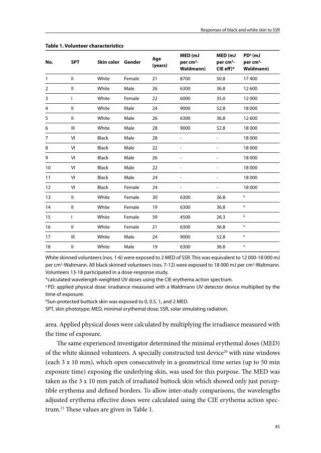
45
Responses of black and white skin to SSR
area. Applied physical doses were calculated by multiplying the irradiance measured with the time of exposure.
The same experienced investigator determined the minimal erythemal doses (MED) of the white skinned volunteers. A specially constructed test device20 with nine windows (each 3 x 10 mm), which open consecutively in a geometrical time series (up to 50 min exposure time) exposing the underlying skin, was used for this purpose. The MED was taken as the 3 x 10 mm patch of irradiated buttock skin which showed only just percep-tible erythema and defined borders. To allow inter-study comparisons, the wavelengths adjusted erythema effective doses were calculated using the CIE erythema action spec-trum.21 These values are given in Table 1.
Table 1. Volunteer characteristics
No. SPT Skin color GenderAge (years)
MED (mJ per cm2-Waldmann)
MED (mJ per cm2-CIE eff)*
PDa (mJ per cm2- Waldmann)
1 II White Female 21 8700 50.8 17 400
2 II White Male 26 6300 36.8 12 600
3 I White Female 22 6000 35.0 12 000
4 II White Male 24 9000 52.8 18 000
5 II White Male 26 6300 36.8 12 600
6 III White Male 28 9000 52.8 18 000
7 VI Black Male 28 - - 18 000
8 VI Black Male 22 - - 18 000
9 VI Black Male 26 - - 18 000
10 VI Black Male 22 - - 18 000
11 VI Black Male 24 - - 18 000
12 VI Black Female 24 - - 18 000
13 II White Female 30 6300 36.8 b
14 II White Female 19 6300 36.8 b
15 I White Female 39 4500 26.3 b
16 II White Female 21 6300 36.8 b
17 III White Male 24 9000 52.8 b
18 II White Male 19 6300 36.8 b
White skinned volunteers (nos. 1-6) were exposed to 2 MED of SSR. This was equivalent to 12 000-18 000 mJ per cm2-Waltmann. All black skinned volunteers (nos. 7-12) were exposed to 18 000 mJ per cm2-Waltmann. Volunteers 13-18 participated in a dose-response study.*calculated wavelength weighted UV doses using the CIE erythema action spectrum.a PD: applied physical dose: irradiance measured with a Waldmann UV detector device multiplied by the time of exposure.bSun-protected buttock skin was exposed to 0, 0.5, 1, and 2 MED.SPT, skin phototype; MED, minimal erythemal dose; SSR, solar simulating radiation.
Chapter 2
46
Following MED determination, the first group of white skinned volunteers (1-6, Table 1) was irradiated with a dose of 2 MED. Three areas of approximately 2 x 2 cm on the buttock were exposed and 4 mm punch biopsies were taken from the irradiated sites after 6, 24, and 48 h. A control biopsy was taken from a patch of non-irradiated buttock skin. The six other white skinned volunteers were irradiated with increasing doses of SSR (0, 0.5, 1, and 2 MED) and biopsies were taken after 24 h.
Black skinned volunteers were irradiated with a fixed physical dose of SSR (18 000 mJ per cm2-Walmann) corresponding to 2-3 MED of the white skinned volunteers. As with the first group of white skinned volunteers, three 2 x 2 cm areas on the buttock were exposed, and biopsies were taken after 6, 24, and 48 h including a control biopsy.
The biopsies were snap-frozen in liquid nitrogen, embedded in Tissuetek (Sakura, Torrance, CA, USA), and stored at -80°C. In order to stain the skin immunohisto-chemically and evaluate enzymatic activity in situ, biopsies were cut into 6 μm thick skin sections with a freezing microtome and mounted on 3-aminopropyl tri-ethoxy silane (A3648, Sigma, St Louis, Missouri)-coated slides. Subsequently, the slides were left to dry and either put on Silicagel (MERCK, Darmstadt, Germany) to be used the following day or temporarily stored at -80°C.
Immunohistochemistry
To investigate UV-induced DNA photodamage thymine dimers were analyzed. The following steps (1-8) summarize the immunohistochemical staining procedure: skin sections were fixed, respectively, in (1) acetone containing 0.06% hydrogen peroxide, (2) 25% acetic acid-PBS solution, and (3) 0.07 M sodium hydroxide-70% ethanol-phosphate-buffered saline (PBS) solution. Next, the sections were consecutively incubated with (4) 10% normal rabbit serum (NRS), (5) monoclonal mouse anti-thymine dimers (Kamiya Biomedical Company, Seattle, Washington; diluted 1:2000 in 1% NRS), (6) biotinylated rabbit anti-mouse IgG1 (ZYMED, San Francisco, California; diluted 1:200 in 1% NRS), and (7) alkaline phosphatase (AP)-conjugated avidin-biotin complex (DAKO A/S, Glos-trup, Denmark; diluted 1:50 in PBS). (8) AP activity was demonstrated using naphthol ASBI (Sigma) as a substrate and new fuchsine (Sigma) 10 mg per 100 ml as chromogen dissolved in 0.1 M Tris-HCL + 0.05 M MgCl2 (pH 8.5) resulting in a pink staining. En-dogenous AP activity was inhibited by adding levamisole (35 mg per 100 mL) to the above reaction mixture.
The presence of neutrophils in skin sections was demonstrated by staining cells using monoclonal mouse anti-human neutrophil elastase (NP57, DAKO A/S; diluted 1:50 in 1% normal human serum (NhuS)/1% normal horse serum (NhoS)). Sections were fixed in dry acetone and pre-incubated with 10% NhuS/10% NhoS. Subsequently, the sections were incubated with mouse anti-human neutrophil elastase (primary antibody). This was followed by incubation with biotinylated horse anti-mouse immunoglobulin (secondary
47
Responses of black and white skin to SSR
antibody obtained from Vector, Burlingame, California; diluted 1:800) and AP-labeled streptavidine (Boehringer Mannheim GmbH, Mannheim, Germany; diluted 1:300). Both primary and secondary antibodies were diluted with 1% NhuS/1% NhoS. AP activity was detected as described above. Sections were counterstained with Mayer’s hematoxylin.
MMP-9 and -1, cytokeratins 6, 16, and 17 were detected by performing practically identical staining procedures as described above for neutrophil elastase, differing only in the primary antibodies used. These were (all monoclonal antibodies): mouse anti-human MMP-9 (Neomarkers, Fremont, California; diluted 1:50), mouse anti-human MMP-1 (Oncogene, Boston, Massachusetts; diluted 1:50) and mouse anti-human cytokeratin 6, 16, and 17 (all three purchased from Novocastra, Newcastle, UK; diluted 1:50).
IL-10 expression was analyzed using monoclonal mouse anti-human IL-10 (In-struchemie, Delfzijl, the Netherlands; diluted 1:300). Frozen skin sections were dried in air and fixed in 100 mL dry acetone containing 0.06% hydrogen peroxide. After pre-incubating with 0.1% bovine serum albumin (BSA)-PBS, the sections were incubated overnight at 4°C with the IL-10 primary antibodies (diluted 1:300 in PBS-0.1% BSA). The following day, the sections were incubated with (step 1) biotinylated horse anti-mouse immunoglobulin (diluted 1:800 in 1% BSA-PBS) and (step 2) horseradish peroxidase-conjugated avidin-biotin complex (DAKO A/S; diluted 1:50 in 1% BSA-PBS). Staining was executed with 3-amino-ethyl-carbazole (AEC) purchased from Sigma (20 mg AEC in 5 ml DMF added to 95 mL of acetate buffer and 100 μL hydrogen peroxide). Finally, counterstaining was performed with Mayer's hematoxylin.
To stain both IL-10 and neutrophil elastase on the same slide, skin sections were first incubated with anti IL-10 antibodies as described above. Following incubation with biotinylated horse anti-mouse immunoglobulin (see above: step 1 of the second day in the IL-10 single staining protocol), sections were incubated with alkaline phosphatase-conjugated avidin-biotin complex (diluted 1:50 in 1% BSA–PBS). Next, the skin sections were incubated with 10% normal mouse serum (NMS) followed by incubation with horseradish peroxidase-conjugated neutrophil elastase NP57 (DAKO A/S; diluted 1:4 in 1% NMS) and ultimately staining was performed with Fast Bleu BB salt (Sigma) and AEC. Fast Bleu BB salt staining solution was prepared by adding 35 mg of levamisol, 25 mg of fast blue BB Salt, 12,5 mg of naphthol-ASMX (Sigma) dissolved in 1 mL of DMF to 100 mL of 0,05 M magnesiumchloride Tris-HCl.
(Note: The times of incubation and washing steps with PBS-Tween (0.05%) have been omitted in the descriptions for the purpose of clarity. New fuchsine, AEC, and fast bleuBB salt enzymatic activity was halted with distilled water.)
Microscopic evaluation
Immunohistochemically stained skin sections were examined by light microscopy at x200 and x400 magnification. For quantification of cell numbers, stained cells in an area
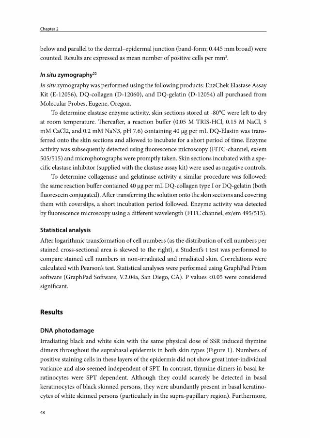
Chapter 2
48
below and parallel to the dermal–epidermal junction (band-form; 0.445 mm broad) were counted. Results are expressed as mean number of positive cells per mm2.
In situ zymography22
In situ zymography was performed using the following products: EnzChek Elastase Assay Kit (E-12056), DQ-collagen (D-12060), and DQ-gelatin (D-12054) all purchased from Molecular Probes, Eugene, Oregon.
To determine elastase enzyme activity, skin sections stored at -80°C were left to dry at room temperature. Thereafter, a reaction buffer (0.05 M TRIS-HCl, 0.15 M NaCl, 5 mM CaCl2, and 0.2 mM NaN3, pH 7.6) containing 40 μg per mL DQ-Elastin was trans-ferred onto the skin sections and allowed to incubate for a short period of time. Enzyme activity was subsequently detected using fluorescence microscopy (FITC-channel, ex/em 505/515) and microphotographs were promptly taken. Skin sections incubated with a spe-cific elastase inhibitor (supplied with the elastase assay kit) were used as negative controls.
To determine collagenase and gelatinase activity a similar procedure was followed: the same reaction buffer contained 40 μg per mL DQ-collagen type I or DQ-gelatin (both fluorescein conjugated). After transferring the solution onto the skin sections and covering them with coverslips, a short incubation period followed. Enzyme activity was detected by fluorescence microscopy using a different wavelength (FITC channel, ex/em 495/515).
Statistical analysis
After logarithmic transformation of cell numbers (as the distribution of cell numbers per stained cross-sectional area is skewed to the right), a Student’s t test was performed to compare stained cell numbers in non-irradiated and irradiated skin. Correlations were calculated with Pearson’s test. Statistical analyses were performed using GraphPad Prism software (GraphPad Software, V.2.04a, San Diego, CA). P values <0.05 were considered significant.
Results
DNA photodamage
Irradiating black and white skin with the same physical dose of SSR induced thymine dimers throughout the suprabasal epidermis in both skin types (Figure 1). Numbers of positive staining cells in these layers of the epidermis did not show great inter-individual variance and also seemed independent of SPT. In contrast, thymine dimers in basal ke-ratinocytes were SPT dependent. Although they could scarcely be detected in basal keratinocytes of black skinned persons, they were abundantly present in basal keratino-cytes of white skinned persons (particularly in the supra-papillary region). Furthermore,
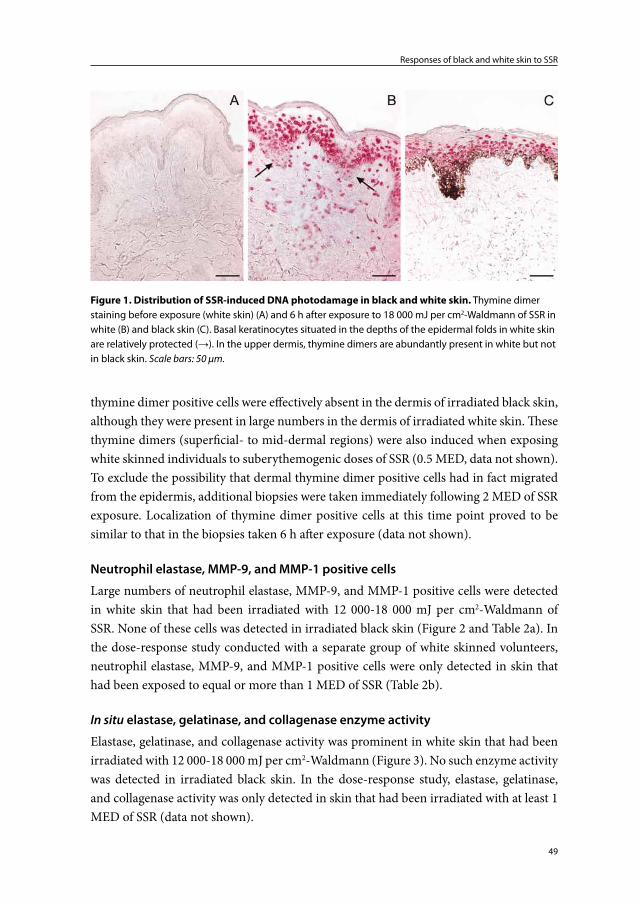
49
Responses of black and white skin to SSR
thymine dimer positive cells were effectively absent in the dermis of irradiated black skin, although they were present in large numbers in the dermis of irradiated white skin. These thymine dimers (superficial- to mid-dermal regions) were also induced when exposing white skinned individuals to suberythemogenic doses of SSR (0.5 MED, data not shown). To exclude the possibility that dermal thymine dimer positive cells had in fact migrated from the epidermis, additional biopsies were taken immediately following 2 MED of SSR exposure. Localization of thymine dimer positive cells at this time point proved to be similar to that in the biopsies taken 6 h after exposure (data not shown).
Neutrophil elastase, MMP-9, and MMP-1 positive cells
Large numbers of neutrophil elastase, MMP-9, and MMP-1 positive cells were detected in white skin that had been irradiated with 12 000-18 000 mJ per cm2-Waldmann of SSR. None of these cells was detected in irradiated black skin (Figure 2 and Table 2a). In the dose-response study conducted with a separate group of white skinned volunteers, neutrophil elastase, MMP-9, and MMP-1 positive cells were only detected in skin that had been exposed to equal or more than 1 MED of SSR (Table 2b).
In situ elastase, gelatinase, and collagenase enzyme activity
Elastase, gelatinase, and collagenase activity was prominent in white skin that had been irradiated with 12 000-18 000 mJ per cm2-Waldmann (Figure 3). No such enzyme activity was detected in irradiated black skin. In the dose-response study, elastase, gelatinase, and collagenase activity was only detected in skin that had been irradiated with at least 1 MED of SSR (data not shown).
Figure 1. Distribution of SSR-induced DNA photodamage in black and white skin. Thymine dimer staining before exposure (white skin) (A) and 6 h after exposure to 18 000 mJ per cm2-Waldmann of SSR in white (B) and black skin (C). Basal keratinocytes situated in the depths of the epidermal folds in white skin are relatively protected (→). In the upper dermis, thymine dimers are abundantly present in white but not in black skin. Scale bars: 50 μm.
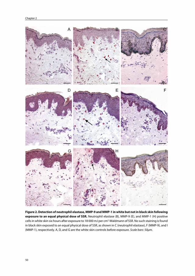
Chapter 2
50
Figure 2. Detection of neutrophil elastase, MMP-9 and MMP-1 in white but not in black skin following exposure to an equal physical dose of SSR. Neutrophil elastase (B), MMP-9 (E), and MMP-1 (H) positive cells in white skin six hours after exposure to 18 000 mJ per cm2-Waldmann of SSR. No such staining is found in black skin exposed to an equal physical dose of SSR, as shown in C (neutrophil elastase), F (MMP-9), and I (MMP-1), respectively. A, D, and G are the white skin controls before exposure. Scale bars: 50μm.
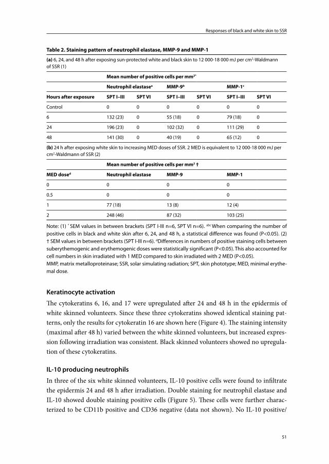
51
Responses of black and white skin to SSR
Keratinocyte activation
The cytokeratins 6, 16, and 17 were upregulated after 24 and 48 h in the epidermis of white skinned volunteers. Since these three cytokeratins showed identical staining pat-terns, only the results for cytokeratin 16 are shown here (Figure 4). The staining intensity (maximal after 48 h) varied between the white skinned volunteers, but increased expres-sion following irradiation was consistent. Black skinned volunteers showed no upregula-tion of these cytokeratins.
IL-10 producing neutrophils
In three of the six white skinned volunteers, IL-10 positive cells were found to infiltrate the epidermis 24 and 48 h after irradiation. Double staining for neutrophil elastase and IL-10 showed double staining positive cells (Figure 5). These cells were further charac-terized to be CD11b positive and CD36 negative (data not shown). No IL-10 positive/
Table 2. Staining pattern of neutrophil elastase, MMP-9 and MMP-1
(a) 6, 24, and 48 h after exposing sun-protected white and black skin to 12 000-18 000 mJ per cm2-Waldmann of SSR (1)
Mean number of positive cells per mm2*
Neutrophil elastasea MMP-9b MMP-1c
Hours after exposure SPT I–III SPT VI SPT I–III SPT VI SPT I–III SPT VI
Control 0 0 0 0 0 0
6 132 (23) 0 55 (18) 0 79 (18) 0
24 196 (23) 0 102 (32) 0 111 (29) 0
48 141 (30) 0 40 (19) 0 65 (12) 0
(b) 24 h after exposing white skin to increasing MED doses of SSR. 2 MED is equivalent to 12 000-18 000 mJ per cm2-Waldmann of SSR (2)
Mean number of positive cells per mm2 †
MED dosed Neutrophil elastase MMP-9 MMP-1
0 0 0 0
0.5 0 0 0
1 77 (18) 13 (8) 12 (4)
2 248 (46) 87 (32) 103 (25)
Note: (1) * SEM values in between brackets (SPT I-III n=6, SPT VI n=6). abc When comparing the number of positive cells in black and white skin after 6, 24, and 48 h, a statistical difference was found (P<0.05). (2) † SEM values in between brackets (SPT I-III n=6). dDifferences in numbers of positive staining cells between suberythemogenic and erythemogenic doses were statistically significant (P<0.05). This also accounted for cell numbers in skin irradiated with 1 MED compared to skin irradiated with 2 MED (P<0.05).MMP, matrix metalloproteinase; SSR, solar simulating radiation; SPT, skin phototype; MED, minimal erythe-mal dose.
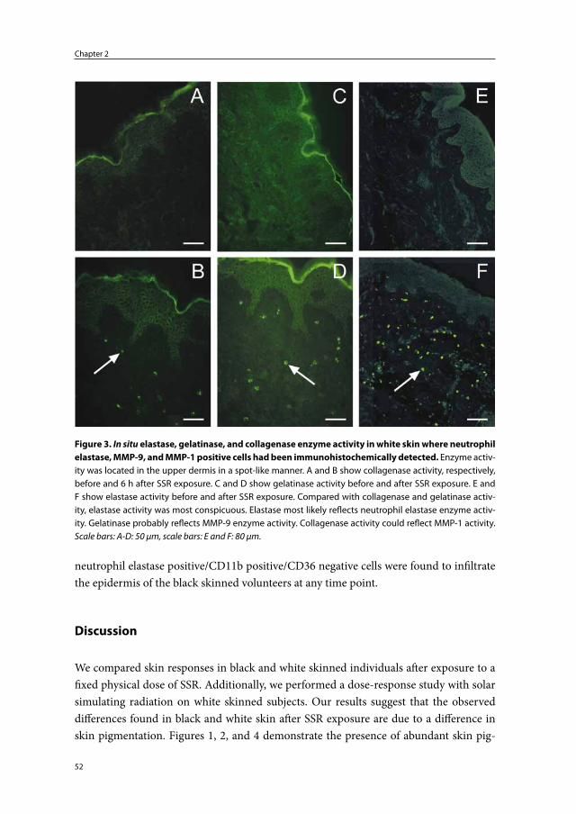
Chapter 2
52
neutrophil elastase positive/CD11b positive/CD36 negative cells were found to infiltrate the epidermis of the black skinned volunteers at any time point.
Discussion
We compared skin responses in black and white skinned individuals after exposure to a fixed physical dose of SSR. Additionally, we performed a dose-response study with solar simulating radiation on white skinned subjects. Our results suggest that the observed differences found in black and white skin after SSR exposure are due to a difference in skin pigmentation. Figures 1, 2, and 4 demonstrate the presence of abundant skin pig-
Figure 3. In situ elastase, gelatinase, and collagenase enzyme activity in white skin where neutrophil elastase, MMP-9, and MMP-1 positive cells had been immunohistochemically detected. Enzyme activ-ity was located in the upper dermis in a spot-like manner. A and B show collagenase activity, respectively, before and 6 h after SSR exposure. C and D show gelatinase activity before and after SSR exposure. E and F show elastase activity before and after SSR exposure. Compared with collagenase and gelatinase activ-ity, elastase activity was most conspicuous. Elastase most likely reflects neutrophil elastase enzyme activ-ity. Gelatinase probably reflects MMP-9 enzyme activity. Collagenase activity could reflect MMP-1 activity. Scale bars: A-D: 50 μm, scale bars: E and F: 80 μm.
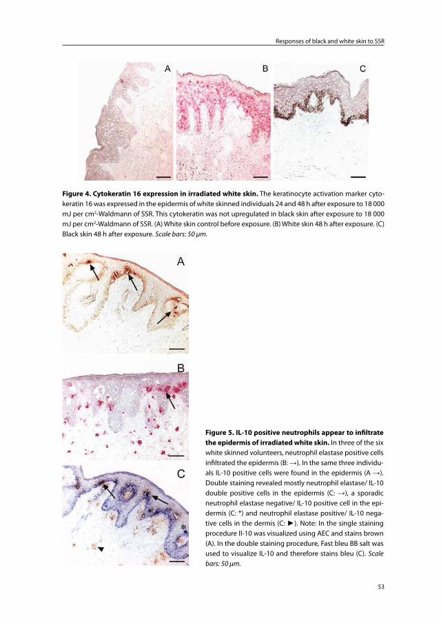
53
Responses of black and white skin to SSR
Figure 5. IL-10 positive neutrophils appear to infiltrate the epidermis of irradiated white skin. In three of the six white skinned volunteers, neutrophil elastase positive cells infiltrated the epidermis (B: →). In the same three individu-als IL-10 positive cells were found in the epidermis (A →). Double staining revealed mostly neutrophil elastase/ IL-10 double positive cells in the epidermis (C: →), a sporadic neutrophil elastase negative/ IL-10 positive cell in the epi-dermis (C: *) and neutrophil elastase positive/ IL-10 nega-tive cells in the dermis (C: ►). Note: In the single staining procedure Il-10 was visualized using AEC and stains brown (A). In the double staining procedure, Fast bleu BB salt was used to visualize IL-10 and therefore stains bleu (C). Scale bars: 50 µm.
Figure 4. Cytokeratin 16 expression in irradiated white skin. The keratinocyte activation marker cyto-keratin 16 was expressed in the epidermis of white skinned individuals 24 and 48 h after exposure to 18 000 mJ per cm2-Waldmann of SSR. This cytokeratin was not upregulated in black skin after exposure to 18 000 mJ per cm2-Waldmann of SSR. (A) White skin control before exposure. (B) White skin 48 h after exposure. (C) Black skin 48 h after exposure. Scale bars: 50 μm.
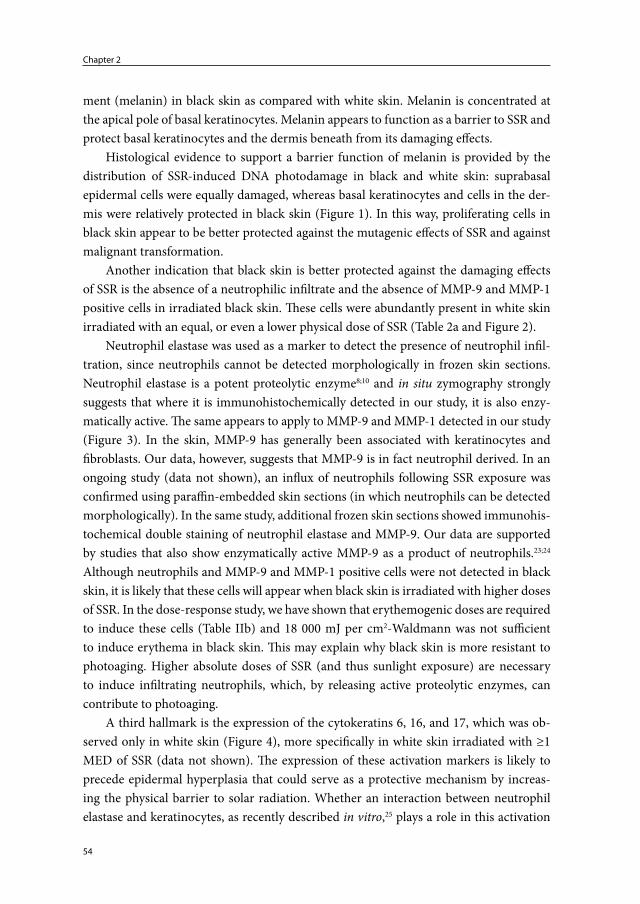
Chapter 2
54
ment (melanin) in black skin as compared with white skin. Melanin is concentrated at the apical pole of basal keratinocytes. Melanin appears to function as a barrier to SSR and protect basal keratinocytes and the dermis beneath from its damaging effects.
Histological evidence to support a barrier function of melanin is provided by the distribution of SSR-induced DNA photodamage in black and white skin: suprabasal epidermal cells were equally damaged, whereas basal keratinocytes and cells in the der-mis were relatively protected in black skin (Figure 1). In this way, proliferating cells in black skin appear to be better protected against the mutagenic effects of SSR and against malignant transformation.
Another indication that black skin is better protected against the damaging effects of SSR is the absence of a neutrophilic infiltrate and the absence of MMP-9 and MMP-1 positive cells in irradiated black skin. These cells were abundantly present in white skin irradiated with an equal, or even a lower physical dose of SSR (Table 2a and Figure 2).
Neutrophil elastase was used as a marker to detect the presence of neutrophil infil-tration, since neutrophils cannot be detected morphologically in frozen skin sections. Neutrophil elastase is a potent proteolytic enzyme8;10 and in situ zymography strongly suggests that where it is immunohistochemically detected in our study, it is also enzy-matically active. The same appears to apply to MMP-9 and MMP-1 detected in our study (Figure 3). In the skin, MMP-9 has generally been associated with keratinocytes and fibroblasts. Our data, however, suggests that MMP-9 is in fact neutrophil derived. In an ongoing study (data not shown), an influx of neutrophils following SSR exposure was confirmed using paraffin-embedded skin sections (in which neutrophils can be detected morphologically). In the same study, additional frozen skin sections showed immunohis-tochemical double staining of neutrophil elastase and MMP-9. Our data are supported by studies that also show enzymatically active MMP-9 as a product of neutrophils.23;24 Although neutrophils and MMP-9 and MMP-1 positive cells were not detected in black skin, it is likely that these cells will appear when black skin is irradiated with higher doses of SSR. In the dose-response study, we have shown that erythemogenic doses are required to induce these cells (Table IIb) and 18 000 mJ per cm2-Waldmann was not sufficient to induce erythema in black skin. This may explain why black skin is more resistant to photoaging. Higher absolute doses of SSR (and thus sunlight exposure) are necessary to induce infiltrating neutrophils, which, by releasing active proteolytic enzymes, can contribute to photoaging.
A third hallmark is the expression of the cytokeratins 6, 16, and 17, which was ob-served only in white skin (Figure 4), more specifically in white skin irradiated with ≥1 MED of SSR (data not shown). The expression of these activation markers is likely to precede epidermal hyperplasia that could serve as a protective mechanism by increas-ing the physical barrier to solar radiation. Whether an interaction between neutrophil elastase and keratinocytes, as recently described in vitro,25 plays a role in this activation
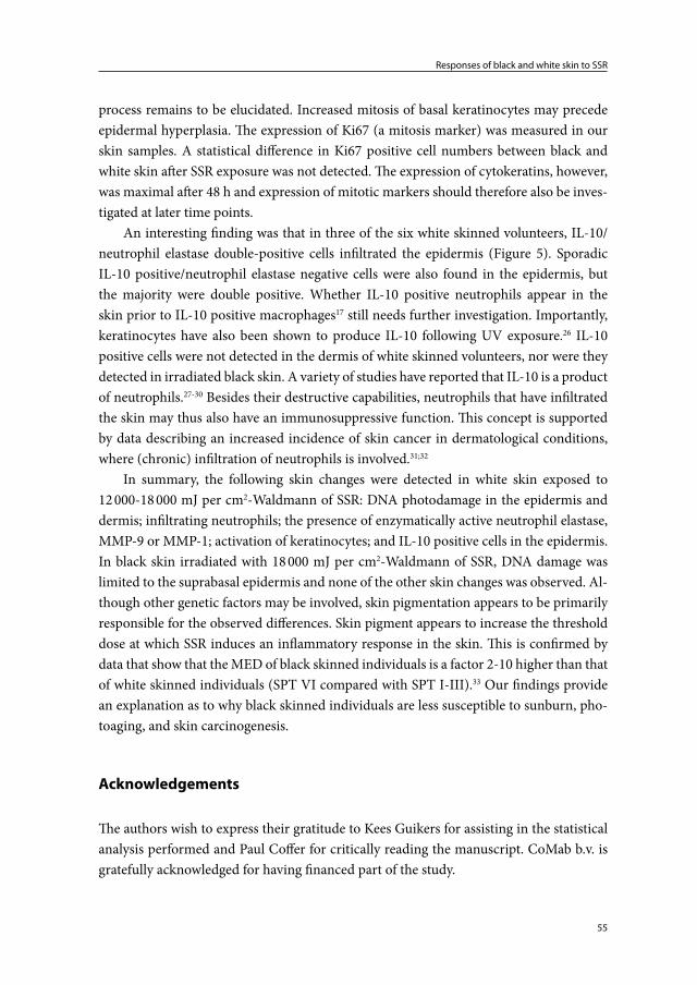
55
Responses of black and white skin to SSR
process remains to be elucidated. Increased mitosis of basal keratinocytes may precede epidermal hyperplasia. The expression of Ki67 (a mitosis marker) was measured in our skin samples. A statistical difference in Ki67 positive cell numbers between black and white skin after SSR exposure was not detected. The expression of cytokeratins, however, was maximal after 48 h and expression of mitotic markers should therefore also be inves-tigated at later time points.
An interesting finding was that in three of the six white skinned volunteers, IL-10/neutrophil elastase double-positive cells infiltrated the epidermis (Figure 5). Sporadic IL-10 positive/neutrophil elastase negative cells were also found in the epidermis, but the majority were double positive. Whether IL-10 positive neutrophils appear in the skin prior to IL-10 positive macrophages17 still needs further investigation. Importantly, keratinocytes have also been shown to produce IL-10 following UV exposure.26 IL-10 positive cells were not detected in the dermis of white skinned volunteers, nor were they detected in irradiated black skin. A variety of studies have reported that IL-10 is a product of neutrophils.27-30 Besides their destructive capabilities, neutrophils that have infiltrated the skin may thus also have an immunosuppressive function. This concept is supported by data describing an increased incidence of skin cancer in dermatological conditions, where (chronic) infiltration of neutrophils is involved.31;32
In summary, the following skin changes were detected in white skin exposed to 12 000-18 000 mJ per cm2-Waldmann of SSR: DNA photodamage in the epidermis and dermis; infiltrating neutrophils; the presence of enzymatically active neutrophil elastase, MMP-9 or MMP-1; activation of keratinocytes; and IL-10 positive cells in the epidermis. In black skin irradiated with 18 000 mJ per cm2-Waldmann of SSR, DNA damage was limited to the suprabasal epidermis and none of the other skin changes was observed. Al-though other genetic factors may be involved, skin pigmentation appears to be primarily responsible for the observed differences. Skin pigment appears to increase the threshold dose at which SSR induces an inflammatory response in the skin. This is confirmed by data that show that the MED of black skinned individuals is a factor 2-10 higher than that of white skinned individuals (SPT VI compared with SPT I-III).33 Our findings provide an explanation as to why black skinned individuals are less susceptible to sunburn, pho-toaging, and skin carcinogenesis.
Acknowledgements
The authors wish to express their gratitude to Kees Guikers for assisting in the statistical analysis performed and Paul Coffer for critically reading the manuscript. CoMab b.v. is gratefully acknowledged for having financed part of the study.
Chapter 2
56
References
1. Taylor SC. Skin of color: biology, structure, function, and implications for dermatologic disease. J.Am.Acad.Dermatol. 2002; 46: S41-S62.
2. Ito S, Wakamatsu K. Quantitative analysis of eumelanin and pheomelanin in humans, mice, and other animals: a comparative review. Pigment Cell Res. 2003; 16: 523-31.
3. Kaidbey KH, Agin PP, Sayre RM et al. Photoprotection by melanin--a comparison of black and Caucasian skin. J.Am.Acad.Dermatol. 1979; 1: 249-60.
4. Sheehan JM, Cragg N, Chadwick CA et al. Repeated ultraviolet exposure affords the same protection against DNA photodamage and erythema in human skin types II and IV but is associated with faster DNA repair in skin type IV. J.Invest Dermatol. 2002; 118: 825-9.
5. Fisher GJ, Kang S, Varani J et al. Mechanisms of photoaging and chronological skin aging. Arch.Dermatol. 2002; 138: 1462-70.
6. McGregor JM, Hawk JLM. Acute effects of ultraviolet radiation on the skin. In: Fitzpatrick’s dermatology in general medicine (Freedberg IM, Eisen AZ, Wolff K et al, eds), 5th edn. New York: McGraw-Hill, 1999: 1555-61.
7. Ford Bainton D. Developmental Biology of Neutrophils and Eosinophils. In: Inflammation: Basic Principles and Clinical Correlates (Gallin JI, Snyderman R, eds). Philadelphia: Lip-pincott Williams & Wilkins, 1999: 13-33.
8. Doring G. The role of neutrophil elastase in chronic inflammation. Am.J.Respir.Crit Care Med. 1994; 150: S114-S117.
9. Opdenakker G, Van den Steen PE, Dubois B et al. Gelatinase B functions as regulator and effector in leukocyte biology. J Leukoc.Biol. 2001; 69: 851-9.
10. Shapiro SD. Neutrophil elastase: path clearer, pathogen killer, or just pathologic?. Am.J.Respir.Cell Mol.Biol. 2002; 26: 266-8.
11. Fisher GJ, Datta SC, Talwar HS et al. Molecular basis of sun-induced premature skin ageing and retinoid antagonism. Nature 1996; 379: 335-9.
12. Fisher GJ, Wang ZQ, Datta SC et al. Pathophysiology of premature skin aging induced by ultraviolet light. N.Engl.J.Med. 1997; 337: 1419-28.
13. de Winter S, Vink AA, Roza L et al. Solar-simulated skin adaptation and its effect on subse-quent UV-induced epidermal DNA damage. J.Invest Dermatol. 2001; 117: 678-82.
14. Freedberg IM, Tomic-Canic M, Komine M et al. Keratins and the keratinocyte activation cycle. J.Invest Dermatol. 2001; 116: 633-40.
15. Selgrade MK, Smith MV, Oberhelman-Bragg LJ et al. Dose response for UV-induced im-mune suppression in people of color: differences based on erythemal reactivity rather than skin pigmentation. Photochem.Photobiol. 2001; 74: 88-95.
16. Asadullah K, Sabat R, Wiese A et al. Interleukin-10 in cutaneous disorders: implications for its pathophysiological importance and therapeutic use. Arch.Dermatol.Res. 1999; 291: 628-36.
17. Kang K, Hammerberg C, Meunier L et al. CD11b+ macrophages that infiltrate human epidermis after in vivo ultraviolet exposure potently produce IL-10 and represent the major secretory source of epidermal IL-10 protein. J.Immunol. 1994; 153: 5256-64.
18. Alamartine E, Berthoux P, Mariat C et al. Interleukin-10 promoter polymorphisms and susceptibility to skin squamous cell carcinoma after renal transplantation. J.Invest Derma-tol. 2003; 120: 99-103.
57
Responses of black and white skin to SSR
19. Kim J, Modlin RL, Moy RL et al. IL-10 production in cutaneous basal and squamous cell carcinomas. A mechanism for evading the local T cell immune response. J.Immunol. 1995; 155: 2240-7.
20. Boonstra HE, van Weelden H, Toonstra J et al. Polymorphous light eruption: A clinical, photobiologic, and follow-up study of 110 patients. J.Am.Acad.Dermatol. 2000; 42: 199-207.
21. McKinlay AT, Diffey BL. Reference spectrum for ultraviolet induced erythema in huma skin. CIE-J Res Note 1987; 17-22.
22. Oh LY, Larsen PH, Krekoski CA et al. Matrix metalloproteinase-9/gelatinase B is required for process outgrowth by oligodendrocytes. J.Neurosci. 1999; 19: 8464-75.
23. Choi KH, Lee HB, Jeong MY et al. The role of matrix metalloproteinase-9 and tissue inhibi-tor of metalloproteinase-1 in cryptogenic organizing pneumonia. Chest 2002; 121: 1478-85.
24. Golub LM, Sorsa T, Lee HM et al. Doxycycline inhibits neutrophil (PMN)-type matrix me-talloproteinases in human adult periodontitis gingiva. J.Clin.Periodontol. 1995; 22: 100-9.
25. Rogalski C, Meyer-Hoffert U, Proksch E et al. Human leukocyte elastase induces keratino-cyte proliferation in vitro and in vivo. J.Invest Dermatol. 2002; 118: 49-54.
26. Curiel-Lewandrowski C, Venna SS, Eller MS et al. Inhibition of the elicitation phase of contact hypersensitivity by thymidine dinucleotides is in part mediated by increased ex-pression of interleukin-10 in human keratinocytes. Exp.Dermatol. 2003; 12: 145-52.
27. Glowacka E, Banasik M, Lewkowicz P et al. The effect of LPS on neutrophils from patients with high risk of type 1 diabetes mellitus in relation to IL-8, IL-10 and IL-12 production and apoptosis in vitro. Scand.J.Immunol. 2002; 55: 210-7.
28. Koller M, Clasbrummel B, Kollig E et al. Major injury induces increased production of interleukin-10 in human granulocyte fractions. Langenbecks Arch.Surg. 1998; 383: 460-5.
29. Shimonkevitz R, Bar-Or D, Harris L et al. Granulocytes, including neutrophils, synthesize IL-10 after traumatic pancreatitis: case report. J Trauma 2000; 48: 165-8.
30. Spuck S, Schaaf B, Wiedorn KH et al. G-CSF application in patients with severe bacterial pneumonia increases IL-10 expression in neutrophils. Respir.Med. 2003; 97: 51-8.
31. Mackie RM. Epidermal skin tumours. In: Rook/Wilkinson/Ebling: Textbook of Dermatology (Champion RH, Burton JL, Burns T et al, eds), 5th edn. Oxford: Blackwell Science Ltd, 1998: 1651-93.
32. Wallach D. Neutrophilic dermatoses: an overview. Clin.Dermatol. 2000; 18: 229-31.33. Fitzpatrick TB. The validity and practicality of sun-reactive skin types I through VI. Arch.
Dermatol. 1988; 124: 869-71.

Chapter 2b
UV-protection by melanin and hyperkeratosis in vitiligo
patients with skin phototype VI: 2 case reports
Feiko Rijken1,2, Ilse C. Bihari1, Huib van Weelden1 and Piet L.B. Bruijnzeel1,3
Department of Dermatology, University Medical Center Utrecht, Utrecht, the Netherlands1
Department of Dermatology, Diakonessenhuis, Utrecht, the Netherlands2
AstraZeneca, Loughborough (Charnwood), United Kingdom3
Study to be completed
Chapter 2b
60
Abstract
Black skin [skin phototype (SPT) VI] is more resistant to photoaging than white skin (SPT I-III). Furthermore, it has been shown that black skin is more resistant to UV-induced neutrophil influx than white skin. Neutrophils are potent cells capable of generating and releasing proteolytic enzymes which can damage the extracellular matrix. Thereby, neutrophils could contribute to photoaging.Vitiligo is an acquired skin disease characterized by depigmentation of the skin caused by loss of melanocytes from the epidermis. Melanocytes synthesize melanin which is trans-ferred to surrounding keratinocytes and protects the skin against the damaging effects of UV. To show that melanin does indeed have a photoprotective effect and can prevent UV-induced neutrophil influx, vitiligo in patients with SPT VI could be a helpful natural disease model. Here we present two case reports that indicate that heavy melanization of the epidermis but also strong hyperkeratosis can prevent UV-induced neutrophil influx.
61
UV protection by melanin and hyperkeratosis
Introduction
Vitiligo is an acquired skin disease characterized by depigmented (white) macular lesions in which melanocytes are lost from the epidermis. The disease is classified according to the extent of skin involvement and the distribution of depigmentation. Several hypotheses with respect to the pathophysiology of vitiligo have been proposed:1-4 (1) the autoimmune hypothesis, whereby cytotoxic T-cells or antibody-mediated cytotoxicity cause destruc-tion of melanocytes; (2) the autocytotoxicity hypothesis, in which cytotoxic precursors or metabolites of melanin induce melanocytic cell death; (3) the neural hypothesis, wherein neural activity plays a role in the destruction of melanocytes; (4) the genetic hypothesis, in which melanocytes have a genetic defect that is triggered at some point and impedes their growth and differentiation. Considering the different physiologic and pathologic findings in vitiligo, multiple mechanisms may be involved (the heterogenous hypothesis).
Light microscopic examination of ‘active’ vitiligo shows absence of/or degenerative melanocytes, and may show focal vacuolar degeneration and some perivascular inflam-matory changes in the dermis. Longstanding vitiligo, however, only shows an absence of melanocytes and is often only recognized by a pathologist when specific immunohisto-chemical stains (to demonstrate the absence of melanocytes) are performed.1;5
There is conflicting data with respect to vitiligo and sunburn sensitivity. A number of studies have shown that the minimal erythema dose (MED) of vitiligenous skin is not significantly lower than the MED of normal skin. One study showed that this was due to relative hyperkeratosis in lesional skin as compared to adjacent normal skin.6 Stud-ies investigating the MED of vitiligenous skin, however, have mainly included patients with fair skin (SPT I-IV) in whom the difference in pigmentation between lesional and normal skin is often quite subtle, particularly when the areas of skin investigated are sun-protected (i.e., not tanned). Certainly patients with black skin (SPT VI) and vitiligo claim that their vitiligenous skin ‘burns’ more easily than their normally pigmented skin. Thus, vitiligenous skin could be more susceptible to a sunburn reaction.
Literature is also not entirely clear on whether vitiligenous skin is more susceptible to photocarcinogenesis and photoaging.7-9 Generally, data suggests no increased risk of skin cancer and photoaging. This is rather surprising considering the fact that one of the most important barriers against UV (i.e., melanin) is lost. There are a number of possible explanations as to why signs of chronic UV damage may not be(come) obvious in viti-ligenous skin: (1) Vitiligo can be a temporary condition, hence there is not enough time to reveal a difference in susceptibility to photocarcinogenesis and photoaging between lesional and normal skin. (2) As touched on above, differences in skin pigmentation between lesional and normal skin can be subtle, therefore increased sensitivity to UV is marginal. (3) Compensatory protective mechanisms, such as hyperkeratosis, may oc-cur. (4) Persons with vitiligo avoid sun exposure to reduce increased contrast between
Chapter 2b
62
lesional and normal skin. (5) Social stigma encourages patients to cover or camouflage the affected areas, thus unwittingly protecting vitiligenous skin against UV. On the other hand, different modalities of UV-therapy are used to treat vitiligo, thus increasing UV exposure.1 The duration of UV-treatment is often extended due to the therapy-resistant and chronic character of the disease. However, normally therapy will be seized following a cumulative treatment of ±2 years and thus the total UV dose is still relatively low.
It has been shown that black skin (SPT VI) is more resistant to UV-induced neutro-phil influx than white skin (SPT I-III).10 Neutrophils are capable of generating powerful proteolytic enzymes which can damage the extracellular matrix11 and thereby could contribute to photoaging of the skin. To show that melanin does indeed have a photopro-tective effect and can prevent UV-induced neutrophil influx, vitiligo in patients with SPT VI could be a helpful natural disease model.
Vitiligo and photoaging: 2 case reports
To further investigate the photoprotective properties of melanin and the role of skin-infiltrating neutrophils in photoaging, a study involving vitiligo patients was set up. Recruitment of two groups of vitiligo patients was foreseen.Group 1: inclusion of elderly patients with SPT VI and stable, longstanding (>10 years) vitiligo on sun-exposed skin to study the occurrence of solar elastosis, the hallmark of photoaged skin. During a single visit punch biopsies would be taken from sun-exposed lesional and adjacent normal skin.Group 2: inclusion of vitiligo patients with SPT VI to study solar-simulating radiation (SSR)-induced neutrophil influx and DNA damage. Lesional and adjacent normal skin would be exposed to 18 000 mJ/cm2 of SSR (equivalent to 2-3 MED for persons with SPT I-III). 24 hours after SSR exposure punch biopsies would be taken from unexposed lesional and adjacent normal skin (controls) and from SSR-exposed lesional and adjacent normal skin.
After the study was approved by the medical ethical committee of the University Medical Center Utrecht patient recruitment was started.
Mainly due to a lack of patients with vitiligo and SPT VI in the region, we finally recruited only 2 patients. Both patients were included in group 2. Despite the fact that the study has not yet been completed, the preliminary data are worth reporting here.
The materials and methods with respect to staining of neutrophil elastase (as a marker of neutrophils) and staining of thymine dimers (as a marker for UV-induced DNA damage) are described in chapter 2.
63
UV protection by melanin and hyperkeratosis
Case reports
Patient 1 (female, age 34 yrs) had stable vitiligo for 20 years and was tested on the abdo-men. Exposure of depigmented skin and adjacent normal black skin to 18 000 mJ/cm2 of SSR induced erythema and neutrophil influx in depigmented skin only (Figure 1a). SSR-induced thymine dimers were distributed throughout the epidermis and upper dermis in depigmented skin, but were limited to suprabasal keratinocytes in adjacent normal black skin (Figure 1b).
Patient 2 (male, age 32 yrs) had vitiligo for 8 years on the dorsum of his hands and fingers. The inclusion of this patient was during a hot period in the summer (July 2008) and a few months earlier he had received UVB therapy. Exposure to 18 000 mJ/cm2 of SSR was not sufficient to induce erythema or neutrophil influx in either depigmented or adjacent nor-mal black skin. Interestingly, the control biopsy of his depigmented skin already showed thymine dimers which were particularly prominent in the upper dermis (Figure 2). Furthermore, his depigmented skin showed an extremely thickened stratum corneum. In accordance with the skin localization (i.e., dorsal hand) the stratum corneum of adjacent normal black skin was also thickened but not as much as his depigmented skin. Exposure to 18 000 mJ/cm2 of SSR increased the intensity of thymine dimers in depigmented skin and induced thymine dimers in suprabasal keratinocytes of adjacent normal black skin. However, compared to patient 1 (Figure 1b) staining of thymine dimers in the epidermis of both lesional and normal skin was much less intense (Figure 2).
Discussion
In chapter 2 we compared black and white skin and showed that strong pigmentation of the epidermis protects basal keratinocytes and the dermis beneath against directly induced DNA damage, and increases the damage threshold at which SSR induces neu-trophil influx. The results from patient 1 are in line with and further substantiate the results in chapter 2. The results from patient 2 are more surprising. It appeared that due to thickening of the stratum corneum (most likely caused by UVB therapy and/or the fact that is was mid-summer) the threshold for SSR-induced erythema and neutrophil influx was increased. The reactive thickening of the stratum corneum, however, did not fully protect against thymine dimer induction by SSR. Interestingly, thymine dimer induction in depigmented skin was particularly prominent in the dermal compartment. Thymine dimer induction in the epidermis of patient 2 in both depigmented and normal skin was increased following SSR exposure, but staining of thymine dimers in this compartment was not very intense. This could be explained by a preferential absorption of shorter UV wavelengths (UVB) by keratin,12 whereby the epidermal cells are relatively spared of
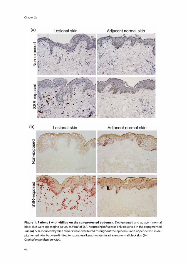
Chapter 2b
64
Figure 1. Patient 1 with vitiligo on the sun-protected abdomen. Depigmented and adjacent normal black skin were exposed to 18 000 mJ/cm2 of SSR. Neutrophil influx was only observed in the depigmented skin (a). SSR-induced thymine dimers were distributed throughout the epidermis and upper dermis in de-pigmented skin, but were limited to suprabasal keratinocytes in adjacent normal black skin (b).Original magnification: x200.
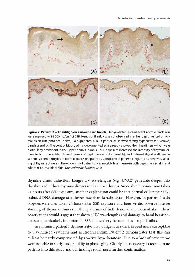
65
UV protection by melanin and hyperkeratosis
thymine dimer induction. Longer UV wavelengths (e.g., UVA2) penetrate deeper into the skin and induce thymine dimers in the upper dermis. Since skin biopsies were taken 24 hours after SSR exposure, another explanation could be that dermal cells repair UV-induced DNA damage at a slower rate than keratinocytes. However, in patient 1 skin biopsies were also taken 24 hours after SSR exposure and here we did observe intense staining of thymine dimers in the epidermis of both lesional and normal skin. These observations would suggest that shorter UV wavelengths and damage to basal keratino-cytes, are particularly important in SSR-induced erythema and neutrophil influx.
In summary, patient 1 demonstrates that vitiligenous skin is indeed more susceptible to UV-induced erythema and neutrophil influx. Patient 2 demonstrates that this can at least be partly compensated by reactive hyperkeratosis. Due to a lack of patients we were not able to study susceptibility to photoaging. Clearly it is necessary to recruit more patients into this study and our findings so far need further confirmation.
Figure 2. Patient 2 with vitiligo on sun-exposed hands. Depigmented and adjacent normal black skin were exposed to 18 000 mJ/cm2 of SSR. Neutrophil influx was not observed in either depigmented or nor-mal black skin (data not shown). Depigmented skin, in particular, showed strong hyperkeratosis (arrows, panels a and b). The control biopsy of his depigmented skin already showed thymine dimers which were particularly prominent in the upper dermis (panel a). SSR exposure increased the intensity of thymine di-mers in both the epidermis and dermis of depigmented skin (panel b), and induced thymine dimers in suprabasal keratinocytes of normal black skin (panel d). Compared to patient 1 (Figure 1b), however, stain-ing of thymine dimers in the epidermis of patient 2 was notably less intense in both depigmented skin and adjacent normal black skin. Original magnification: x200.
Chapter 2b
66
References
1. Kovacs SO. Vitiligo. J.Am.Acad.Dermatol. 1998; 38: 647-66.2. Le Poole C, Boissy RE. Vitiligo. Semin.Cutan.Med.Surg. 1997; 16: 3-14.3. Le Poole I, Wankowicz-Kalinska A, van den Wijngaard RM et al. Autoimmune aspects of
depigmentation in vitiligo. J.Investig.Dermatol.Symp.Proc. 2004; 9: 68-72.4. van den Boorn JG, Konijnenberg D, Dellemijn TA et al. Autoimmune destruction of skin
melanocytes by perilesional T cells from vitiligo patients. J Invest Dermatol. 2009; 129: 2220-32.
5. Nordlund JJ, Lerner AB. Vitiligo. Arch.Dermatol. 1982; 118: 5-8.6. Gniadecka M, Wulf HC, Mortensen NN et al. Photoprotection in vitiligo and normal skin.
A quantitative assessment of the role of stratum corneum, viable epidermis and pigmenta-tion. Acta Derm.Venereol. 1996; 76: 429-32.
7. Saarinen K, Lestringant GG, Masouye I et al. Actinic damage and squamous cell carcinoma in sun-exposed skin affected by vitiligo. Br.J.Dermatol. 2000; 143: 219-21.
8. Schallreuter KU, Tobin DJ, Panske A. Decreased photodamage and low incidence of non-melanoma skin cancer in 136 sun-exposed caucasian patients with vitiligo. Dermatology 2002; 204: 194-201.
9. Seo SL, Kim IH. Squamous cell carcinoma in a patient with generalized vitiligo. J.Am.Acad.Dermatol. 2001; 45: S227-S229.
10. Rijken F, Bruijnzeel PL, van Weelden H et al. Responses of black and white skin to solar-simulating radiation: differences in DNA photodamage, infiltrating neutrophils, proteolytic enzymes induced, keratinocyte activation, and IL-10 expression. J.Invest Dermatol. 2004; 122: 1448-55.
11. Weiss SJ. Tissue destruction by neutrophils. N.Engl.J.Med. 1989; 320: 365-76.12. Nielsen, K. P., Zhao, L., Stamnes, J. J., Stamnes, K., and Moan, J. The optics of human skin:
aspects important for human health. The Norwegian academy of science and letters: Solar radiation and human health. 2008; 35-46. 2008.
Chapter 3
Skin-infiltrating neutrophils following exposure to solar-
simulating radiation could play an important role in
photoaging of human skin
Feiko Rijken, Rebecca C.M. Kiekens and Piet L.B. Bruijnzeel
Department of Dermatology, University Medical Center Utrecht, Utrecht, the Netherlands
British Journal of Dermatology 2005 Feb; 152(2): 321-8
Chapter 3
68
Abstract
Background The pathophysiology of photoaging of the skin has been studied extensively. Matrix metalloproteinases (MMPs) originating from keratinocytes and fibroblasts are thought to play a primary role in this process. Although neutrophils are potent producers of a wide array of proteolytic substances and are present in sunburned skin, their contri-bution to the pathophysiology of photoaging has been described only in murine studies.Objectives To determine the role of neutrophils in photoaging of human skin.Methods Healthy white-skinned volunteers were recruited and their sun-protected buttock skin was exposed to solar-simulating radiation (SSR) in dose-response and time-course studies. Punch biopsies were taken and the influx of neutrophils and the expression of neutrophil elastase and MMPs was studied using immunohistochemical techniques and in situ zymography.Results Neutrophil elastase and MMPs were detected only in skin irradiated with ery-themogenic doses (≥ 1 minimal erythema doses) of SSR. Immunohistochemical double staining demonstrated neutrophils to be the major source of MMP-1, MMP-8 and MMP-9. In situ zymography showed elastase, collagenase and gelatinase enzyme activity in those cells.Conclusions Our study suggests that neutrophils participate in the process of photoag-ing of human skin as they infiltrate the skin and release enzymatically active elastase (neutrophil elastase), collagenase (MMP-1) and gelatinase (MMP-9).
69
Skin-infiltrating neutrophils could play an important role in photoaging
Introduction
Solar radiation can damage human skin. Acute overexposure to solar radiation causes a clinical sunburn.1 Chronic exposure can lead to skin changes such as plaque-like thick-ening, loss of skin tone, deep furrowing and fine wrinkle formation. Collectively, these changes may be termed ‘photoaged skin’ or a ‘solar scar’.2
The pathophysiology of photoaging has been investigated extensively. A hypothetical model was proposed by Fisher et al.3;4 They postulated that imperfect repair following skin matrix damage after exposure to sunlight accumulates and ultimately leads to the formation of a solar scar. It was demonstrated that suberythemal exposure(s) to ultra-violet (UV) B radiation induces three matrix metalloproteinases (MMPs) in human skin in vivo: collagenase (MMP-1), gelatinase B (MMP-9) and stromelysin-1 (MMP-3). The combined actions of these enzymes can fully degrade skin collagen. Based on the localization of in situ hybridized MMP mRNA, immunohistochemical staining of MMP protein and in situ determination of enzyme activity, it was concluded that the MMPs mainly originated from keratinocytes and fibroblasts.
Another cell that may contribute to sun-induced skin damage is the neutrophilic granulocyte. Neutrophils are present in sunburned skin.1 Moreover, they are potent producers of a wide array of proteolytic substances,5 including the pluripotent neutro-phil elastase,6;7 MMP-8 and MMP-9. Nevertheless, their contribution to photoaging in humans has been somewhat neglected. The role of neutrophil elastase was investigated in two murine studies8;9 and neutrophil elastase was recently studied as a product of fibro-blasts.10 In humans, only MMP-8 has been studied as a product of neutrophils.11 It was found that following a single exposure to UVB, skin-infiltrating neutrophils contained enzymatically inactive MMP-8 protein. MMP-8 mRNA could not be detected and it was concluded that MMP-8 does not contribute significantly to UV-induced skin collagen degradation. Similar to MMP-8, neutrophils isolated from human donor blood contained MMP-9 in an inactive latent form.12 However, the presence and enzymatic activity of MMP-9 within skin-infiltrating neutrophils following exposure to solar radiation or solar-simulating radiation (SSR) has not yet been studied. MMP-1 has, to our knowledge, not been convincingly demonstrated as a product of neutrophils.
To investigate the role of neutrophils in the pathophysiology of photoaging further, healthy volunteers were exposed to SSR in a dose-response and a time-course study. SSR was chosen because it approaches the emission spectrum of natural sunlight in the UV range. To study the presence and enzymatic activity of neutrophil elastase and MMPs, biopsies were taken, processed and subjected to immunohistochemical staining and in situ zymography.
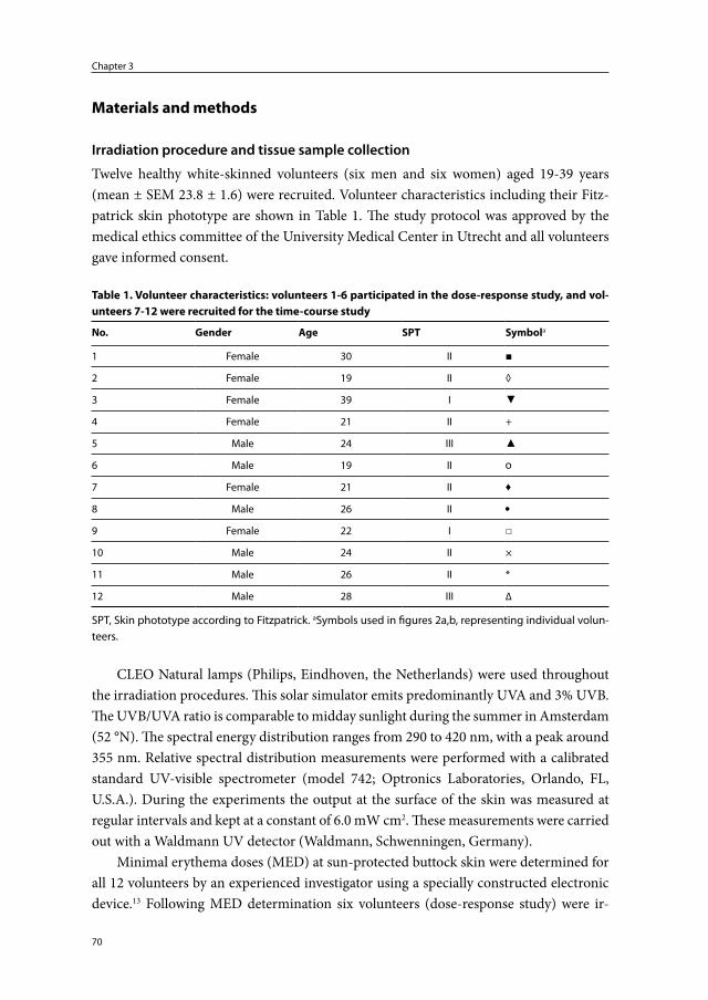
Chapter 3
70
Materials and methods
Irradiation procedure and tissue sample collection
Twelve healthy white-skinned volunteers (six men and six women) aged 19-39 years (mean ± SEM 23.8 ± 1.6) were recruited. Volunteer characteristics including their Fitz-patrick skin phototype are shown in Table 1. The study protocol was approved by the medical ethics committee of the University Medical Center in Utrecht and all volunteers gave informed consent.
CLEO Natural lamps (Philips, Eindhoven, the Netherlands) were used throughout the irradiation procedures. This solar simulator emits predominantly UVA and 3% UVB. The UVB/UVA ratio is comparable to midday sunlight during the summer in Amsterdam (52 °N). The spectral energy distribution ranges from 290 to 420 nm, with a peak around 355 nm. Relative spectral distribution measurements were performed with a calibrated standard UV-visible spectrometer (model 742; Optronics Laboratories, Orlando, FL, U.S.A.). During the experiments the output at the surface of the skin was measured at regular intervals and kept at a constant of 6.0 mW cm2. These measurements were carried out with a Waldmann UV detector (Waldmann, Schwenningen, Germany).
Minimal erythema doses (MED) at sun-protected buttock skin were determined for all 12 volunteers by an experienced investigator using a specially constructed electronic device.13 Following MED determination six volunteers (dose-response study) were ir-
Table 1. Volunteer characteristics: volunteers 1-6 participated in the dose-response study, and vol-unteers 7-12 were recruited for the time-course study
No. Gender Age SPT Symbola
1 Female 30 II ■
2 Female 19 II ◊
3 Female 39 I ▼
4 Female 21 II +
5 Male 24 III ▲
6 Male 19 II o
7 Female 21 II ♦
8 Male 26 II •
9 Female 22 I □
10 Male 24 II ×
11 Male 26 II *
12 Male 28 III ∆
SPT, Skin phototype according to Fitzpatrick. aSymbols used in figures 2a,b, representing individual volun-teers.
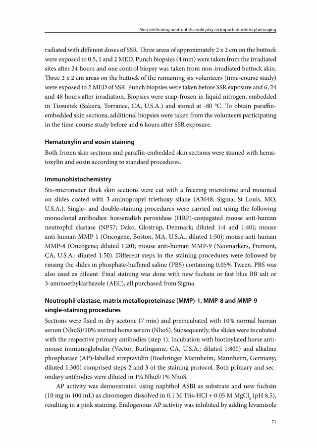
71
Skin-infiltrating neutrophils could play an important role in photoaging
radiated with different doses of SSR. Three areas of approximately 2 x 2 cm on the buttock were exposed to 0.5, 1 and 2 MED. Punch biopsies (4 mm) were taken from the irradiated sites after 24 hours and one control biopsy was taken from non-irradiated buttock skin. Three 2 x 2 cm areas on the buttock of the remaining six volunteers (time-course study) were exposed to 2 MED of SSR. Punch biopsies were taken before SSR exposure and 6, 24 and 48 hours after irradiation. Biopsies were snap-frozen in liquid nitrogen, embedded in Tissuetek (Sakura, Torrance, CA, U.S.A.) and stored at -80 °C. To obtain paraffin-embedded skin sections, additional biopsies were taken from the volunteers participating in the time-course study before and 6 hours after SSR exposure.
Hematoxylin and eosin staining
Both frozen skin sections and paraffin-embedded skin sections were stained with hema-toxylin and eosin according to standard procedures.
Immunohistochemistry
Six-micrometer thick skin sections were cut with a freezing microtome and mounted on slides coated with 3-aminopropyl triethoxy silane (A3648; Sigma, St Louis, MO, U.S.A.). Single- and double-staining procedures were carried out using the following monoclonal antibodies: horseradish peroxidase (HRP)-conjugated mouse anti-human neutrophil elastase (NP57; Dako, Glostrup, Denmark; diluted 1:4 and 1:40); mouse anti-human MMP-1 (Oncogene, Boston, MA, U.S.A.; diluted 1:50); mouse anti-human MMP-8 (Oncogene; diluted 1:20); mouse anti-human MMP-9 (Neomarkers, Fremont, CA, U.S.A.; diluted 1:50). Different steps in the staining procedures were followed by rinsing the slides in phosphate-buffered saline (PBS) containing 0.05% Tween. PBS was also used as diluent. Final staining was done with new fuchsin or fast blue BB salt or 3-aminoethylcarbazole (AEC), all purchased from Sigma.
Neutrophil elastase, matrix metalloproteinase (MMP)-1, MMP-8 and MMP-9 single-staining procedures
Sections were fixed in dry acetone (7 min) and preincubated with 10% normal human serum (NhuS)/10% normal horse serum (NhoS). Subsequently, the slides were incubated with the respective primary antibodies (step 1). Incubation with biotinylated horse anti-mouse immunoglobulin (Vector, Burlingame, CA, U.S.A.; diluted 1:800) and alkaline phosphatase (AP)-labelled streptavidin (Boehringer Mannheim, Mannheim, Germany; diluted 1:300) comprised steps 2 and 3 of the staining protocol. Both primary and sec-ondary antibodies were diluted in 1% NhuS/1% NhoS.
AP activity was demonstrated using naphthol ASBI as substrate and new fuchsin (10 mg in 100 mL) as chromogen dissolved in 0.1 M Tris-HCl + 0.05 M MgCl2 (pH 8.5), resulting in a pink staining. Endogenous AP activity was inhibited by adding levamisole
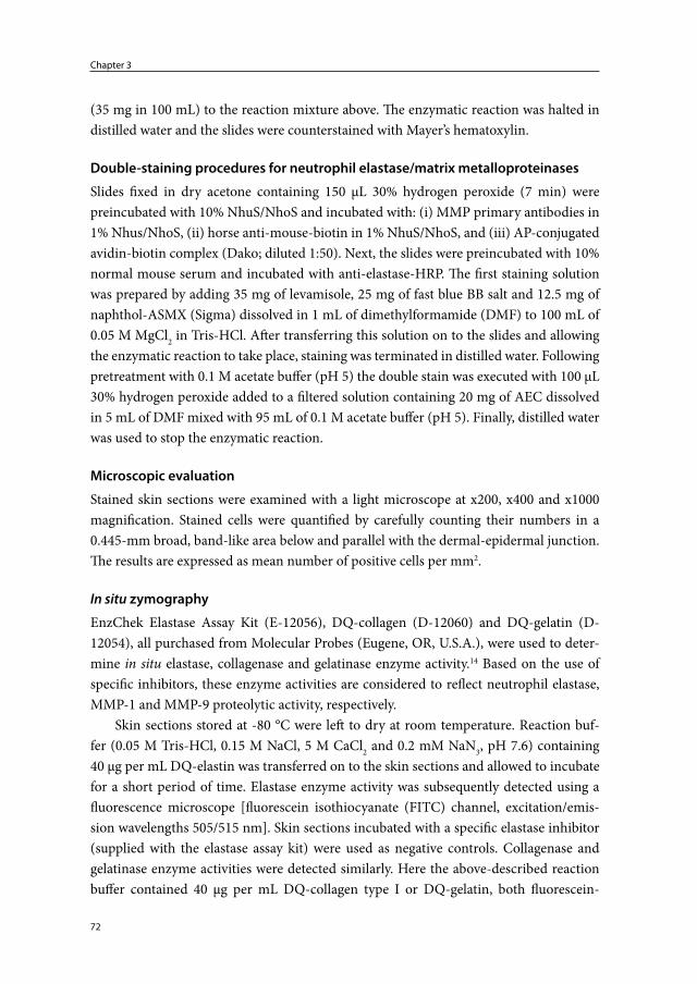
Chapter 3
72
(35 mg in 100 mL) to the reaction mixture above. The enzymatic reaction was halted in distilled water and the slides were counterstained with Mayer’s hematoxylin.
Double-staining procedures for neutrophil elastase/matrix metalloproteinases
Slides fixed in dry acetone containing 150 µL 30% hydrogen peroxide (7 min) were preincubated with 10% NhuS/NhoS and incubated with: (i) MMP primary antibodies in 1% Nhus/NhoS, (ii) horse anti-mouse-biotin in 1% NhuS/NhoS, and (iii) AP-conjugated avidin-biotin complex (Dako; diluted 1:50). Next, the slides were preincubated with 10% normal mouse serum and incubated with anti-elastase-HRP. The first staining solution was prepared by adding 35 mg of levamisole, 25 mg of fast blue BB salt and 12.5 mg of naphthol-ASMX (Sigma) dissolved in 1 mL of dimethylformamide (DMF) to 100 mL of 0.05 M MgCl2 in Tris-HCl. After transferring this solution on to the slides and allowing the enzymatic reaction to take place, staining was terminated in distilled water. Following pretreatment with 0.1 M acetate buffer (pH 5) the double stain was executed with 100 µL 30% hydrogen peroxide added to a filtered solution containing 20 mg of AEC dissolved in 5 mL of DMF mixed with 95 mL of 0.1 M acetate buffer (pH 5). Finally, distilled water was used to stop the enzymatic reaction.
Microscopic evaluation
Stained skin sections were examined with a light microscope at x200, x400 and x1000 magnification. Stained cells were quantified by carefully counting their numbers in a 0.445-mm broad, band-like area below and parallel with the dermal-epidermal junction. The results are expressed as mean number of positive cells per mm2.
In situ zymography
EnzChek Elastase Assay Kit (E-12056), DQ-collagen (D-12060) and DQ-gelatin (D-12054), all purchased from Molecular Probes (Eugene, OR, U.S.A.), were used to deter-mine in situ elastase, collagenase and gelatinase enzyme activity.14 Based on the use of specific inhibitors, these enzyme activities are considered to reflect neutrophil elastase, MMP-1 and MMP-9 proteolytic activity, respectively.
Skin sections stored at -80 °C were left to dry at room temperature. Reaction buf-fer (0.05 M Tris-HCl, 0.15 M NaCl, 5 M CaCl2 and 0.2 mM NaN3, pH 7.6) containing 40 µg per mL DQ-elastin was transferred on to the skin sections and allowed to incubate for a short period of time. Elastase enzyme activity was subsequently detected using a fluorescence microscope [fluorescein isothiocyanate (FITC) channel, excitation/emis-sion wavelengths 505/515 nm]. Skin sections incubated with a specific elastase inhibitor (supplied with the elastase assay kit) were used as negative controls. Collagenase and gelatinase enzyme activities were detected similarly. Here the above-described reaction buffer contained 40 µg per mL DQ-collagen type I or DQ-gelatin, both fluorescein-
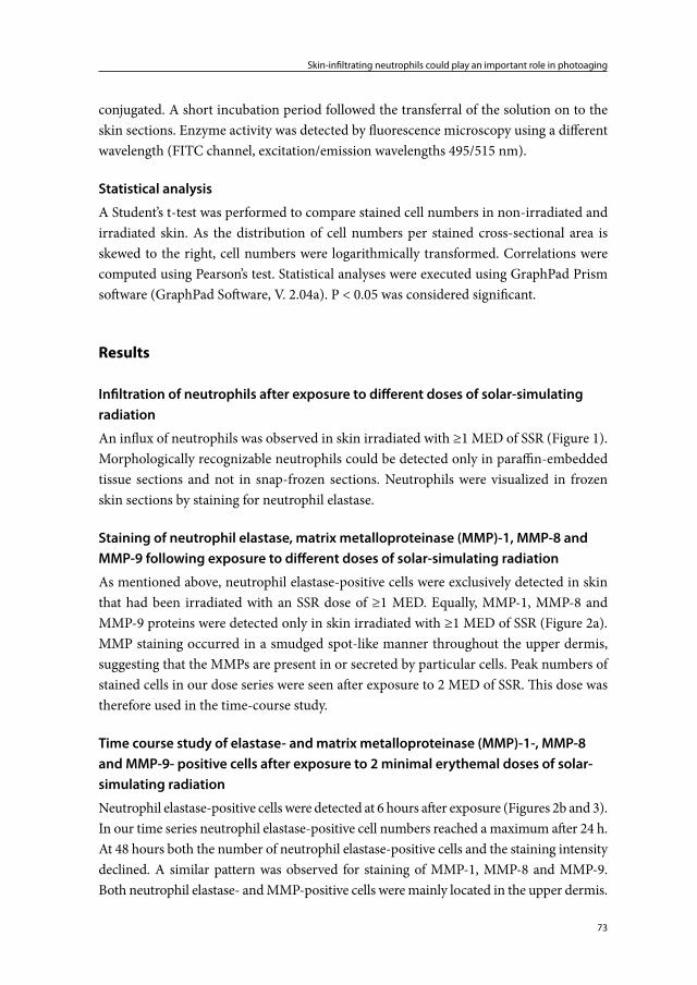
73
Skin-infiltrating neutrophils could play an important role in photoaging
conjugated. A short incubation period followed the transferral of the solution on to the skin sections. Enzyme activity was detected by fluorescence microscopy using a different wavelength (FITC channel, excitation/emission wavelengths 495/515 nm).
Statistical analysis
A Student’s t-test was performed to compare stained cell numbers in non-irradiated and irradiated skin. As the distribution of cell numbers per stained cross-sectional area is skewed to the right, cell numbers were logarithmically transformed. Correlations were computed using Pearson’s test. Statistical analyses were executed using GraphPad Prism software (GraphPad Software, V. 2.04a). P < 0.05 was considered significant.
Results
Infiltration of neutrophils after exposure to different doses of solar-simulating radiation
An influx of neutrophils was observed in skin irradiated with ≥1 MED of SSR (Figure 1). Morphologically recognizable neutrophils could be detected only in paraffin-embedded tissue sections and not in snap-frozen sections. Neutrophils were visualized in frozen skin sections by staining for neutrophil elastase.
Staining of neutrophil elastase, matrix metalloproteinase (MMP)-1, MMP-8 and MMP-9 following exposure to different doses of solar-simulating radiation
As mentioned above, neutrophil elastase-positive cells were exclusively detected in skin that had been irradiated with an SSR dose of ≥1 MED. Equally, MMP-1, MMP-8 and MMP-9 proteins were detected only in skin irradiated with ≥1 MED of SSR (Figure 2a). MMP staining occurred in a smudged spot-like manner throughout the upper dermis, suggesting that the MMPs are present in or secreted by particular cells. Peak numbers of stained cells in our dose series were seen after exposure to 2 MED of SSR. This dose was therefore used in the time-course study.
Time course study of elastase- and matrix metalloproteinase (MMP)-1-, MMP-8 and MMP-9- positive cells after exposure to 2 minimal erythemal doses of solar-simulating radiation
Neutrophil elastase-positive cells were detected at 6 hours after exposure (Figures 2b and 3). In our time series neutrophil elastase-positive cell numbers reached a maximum after 24 h. At 48 hours both the number of neutrophil elastase-positive cells and the staining intensity declined. A similar pattern was observed for staining of MMP-1, MMP-8 and MMP-9. Both neutrophil elastase- and MMP-positive cells were mainly located in the upper dermis.
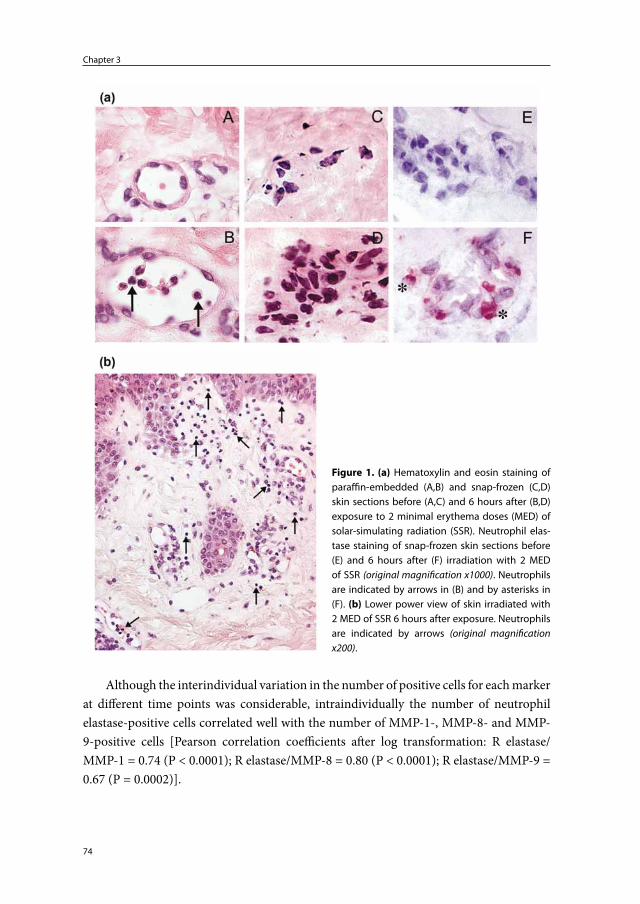
Chapter 3
74
Although the interindividual variation in the number of positive cells for each marker at different time points was considerable, intraindividually the number of neutrophil elastase-positive cells correlated well with the number of MMP-1-, MMP-8- and MMP-9-positive cells [Pearson correlation coefficients after log transformation: R elastase/MMP-1 = 0.74 (P < 0.0001); R elastase/MMP-8 = 0.80 (P < 0.0001); R elastase/MMP-9 = 0.67 (P = 0.0002)].
Figure 1. (a) Hematoxylin and eosin staining of paraffin-embedded (A,B) and snap-frozen (C,D) skin sections before (A,C) and 6 hours after (B,D) exposure to 2 minimal erythema doses (MED) of solar-simulating radiation (SSR). Neutrophil elas-tase staining of snap-frozen skin sections before (E) and 6 hours after (F) irradiation with 2 MED of SSR (original magnification x1000). Neutrophils are indicated by arrows in (B) and by asterisks in (F). (b) Lower power view of skin irradiated with 2 MED of SSR 6 hours after exposure. Neutrophils are indicated by arrows (original magnification x200).
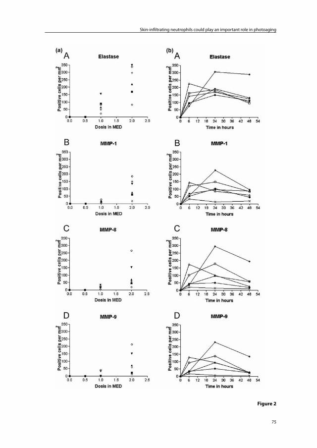
75
Skin-infiltrating neutrophils could play an important role in photoaging
Figure 2
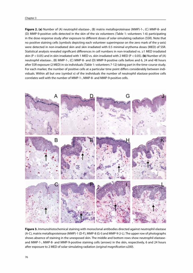
Chapter 3
76
Figure 3. Immunohistochemical staining with monoclonal antibodies directed against neutrophil elastase (A-C), matrix metalloproteinase (MMP)-1 (D-F), MMP-8 (G-I) and MMP-9 (J-L). The upper row of photographs shows absence of staining in the unexposed skin. The middle and bottom rows show neutrophil elastase- and MMP-1-, MMP-8- and MMP-9-positive staining cells (arrows) in the skin, respectively, 6 and 24 hours after exposure to 2 MED of solar-simulating radiation (original magnification x200).
Figure 2. (a) Number of (A) neutrophil elastase-, (B) matrix metalloproteinase (MMP)-1-, (C) MMP-8- and (D) MMP-9-positive cells detected in the skin of the six volunteers (Table 1: volunteers 1-6) participating in the dose-response study after exposure to different doses of solar-simulating radiation (SSR). Note that no positive staining cells (symbols depicting each volunteer superimpose on the zero mark of the y-axis) were detected in non-irradiated skin and skin irradiated with 0.5 minimal erythema doses (MED) of SSR. Statistical analysis revealed significant differences in cell numbers in non-irradiated vs. ≥1 MED irradiated skin (P < 0.05) and in skin irradiated with 1 MED vs. skin irradiated with 2 MED (P < 0.05). (b) Number of (A) neutrophil elastase-, (B) MMP-1-, (C) MMP-8- and (D) MMP-9-positive cells before and 6, 24 and 48 hours after SSR exposure (2 MED) in six individuals (Table 1: volunteers 7-12) taking part in the time-course study. For each marker, the number of positive cells at a particular time point differs considerably between indi-viduals. Within all but one (symbol x) of the individuals the number of neutrophil elastase-positive cells correlates well with the number of MMP-1-, MMP-8- and MMP-9-positive cells.
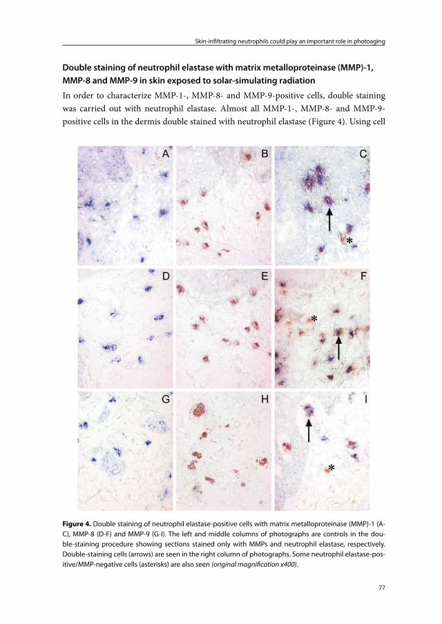
77
Skin-infiltrating neutrophils could play an important role in photoaging
Double staining of neutrophil elastase with matrix metalloproteinase (MMP)-1, MMP-8 and MMP-9 in skin exposed to solar-simulating radiation
In order to characterize MMP-1-, MMP-8- and MMP-9-positive cells, double staining was carried out with neutrophil elastase. Almost all MMP-1-, MMP-8- and MMP-9-positive cells in the dermis double stained with neutrophil elastase (Figure 4). Using cell
Figure 4. Double staining of neutrophil elastase-positive cells with matrix metalloproteinase (MMP)-1 (A-C), MMP-8 (D-F) and MMP-9 (G-I). The left and middle columns of photographs are controls in the dou-ble-staining procedure showing sections stained only with MMPs and neutrophil elastase, respectively. Double-staining cells (arrows) are seen in the right column of photographs. Some neutrophil elastase-pos-itive/MMP-negative cells (asterisks) are also seen (original magnification x400).
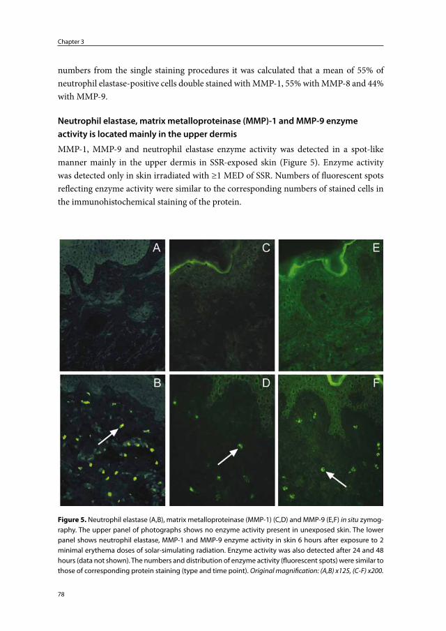
Chapter 3
78
numbers from the single staining procedures it was calculated that a mean of 55% of neutrophil elastase-positive cells double stained with MMP-1, 55% with MMP-8 and 44% with MMP-9.
Neutrophil elastase, matrix metalloproteinase (MMP)-1 and MMP-9 enzyme activity is located mainly in the upper dermis
MMP-1, MMP-9 and neutrophil elastase enzyme activity was detected in a spot-like manner mainly in the upper dermis in SSR-exposed skin (Figure 5). Enzyme activity was detected only in skin irradiated with ≥1 MED of SSR. Numbers of fluorescent spots reflecting enzyme activity were similar to the corresponding numbers of stained cells in the immunohistochemical staining of the protein.
Figure 5. Neutrophil elastase (A,B), matrix metalloproteinase (MMP-1) (C,D) and MMP-9 (E,F) in situ zymog-raphy. The upper panel of photographs shows no enzyme activity present in unexposed skin. The lower panel shows neutrophil elastase, MMP-1 and MMP-9 enzyme activity in skin 6 hours after exposure to 2 minimal erythema doses of solar-simulating radiation. Enzyme activity was also detected after 24 and 48 hours (data not shown). The numbers and distribution of enzyme activity (fluorescent spots) were similar to those of corresponding protein staining (type and time point). Original magnification: (A,B) x125, (C-F) x200.
79
Skin-infiltrating neutrophils could play an important role in photoaging
Discussion
When investigating the possible involvement of neutrophilic granulocytes in SSR-induced skin damage we made some unexpected observations. Although neutrophils are known to be present in sunburned skin, their morphology was completely absent in frozen skin sections. They could be recognized morphologically only in paraffin-embedded skin sec-tions. In frozen skin sections the presence of neutrophils is generally demonstrated by immunohistochemical staining of neutrophil elastase. Thus neutrophils were shown to be abundant in skin that had been exposed to a single dose of 2 MED of SSR (representing a radiation dose equivalent to sunburn) in both the paraffin-embedded skin sections and the frozen skin sections. The cells primarily infiltrated the dermis but also the epidermis. Neutrophils were not detected in skin irradiated with <1 MED of SSR. The fact that these cells lose their conspicuous morphology in frozen skin sections and the fact that they do not infiltrate suberythematous irradiated skin is probably the reason why they have received little attention with respect to the pathogenesis of photoaging of human skin.
Another surprising observation was that, contrary to the findings of Fisher et al.,3;4 we observed prominent MMP-1, MMP-8 and MMP-9 immunohistochemical staining only in skin that had been irradiated with ≥1 MED of SSR. Immunohistochemical double staining with neutrophil elastase suggested that these MMPs were neutrophil derived and not keratinocyte or fibroblast derived. Even though small amounts of neutrophil elastase may be present in macrophages and fibroblasts, together with the paraffin-embedded skin sections showing the presence of morphologically distinct neutrophils, our results strongly suggest that the MMPs are indeed neutrophil derived.
In all skin sections analyzed, the number of neutrophil elastase-positive cells ex-ceeded that of MMP-1-, MMP-8- and MMP-9-positive cells. One explanation is that not all neutrophils produce MMPs. Furthermore, we found that 12-20% of the neutrophil elastase-positive cells double stained with CD36, a marker for macrophages. CD36+ cells did not, however, double stain with the MMPs under study (data not shown).
Our in situ zymography results provide additional evidence that erythemogenic doses of SSR are required to induce MMPs in vivo. Elastase, collagenase and gelatinase activity was detected only in skin that had been irradiated with ≥1 MED. This finding bypasses the necessity to demonstrate mRNA for these enzymes, as the detection of mRNA in itself is no proof of the presence of enzymatically active protein.
MMP-9 as an extracellular matrix-damaging agent has previously been connected to infiltrating neutrophils in cryptogenic organizing pneumonia15 and adult periodontitis.16 In adult periodontitis investigators proclaimed MMP-9 to originate primarily from infiltrating neutrophils rather than resident fibroblasts, epithelial cells or macrophages. Cryptogenic organizing pneumonia is an interstitial lung disease characterized by the production, deposition and proteolysis of extracellular matrix components.
Chapter 3
80
MMP-1 as a product of neutrophils has, to our knowledge, not been shown convinc-ingly in previous studies. In an ongoing study we also detected MMP-1 protein in freshly isolated neutrophils. Many neutrophil products are stored in preformed granules5 and are probably not synthesized de novo as these cells infiltrate their target organ. Nielsen et al.17 investigated MMP-9 expression in human breast cancer and found that infiltrating neutrophils were an important source of MMP-9 protein. However, MMP-9 mRNA could not be detected in these cells. Similarly, studies at mRNA level may not show MMP-1 mRNA in neutrophils.
In contrast to MMP-8 and MMP-9, staining for MMP-1 showed subtle staining of the epidermis. This was particularly apparent in the double-stained skin sections where no counterstaining was performed. MMP-1 epidermal staining was present in all skin sec-tions, including the controls (taken from skin not exposed to UV radiation), and ended abruptly at the epidermal-dermal junction. MMP-1 is most likely to be constitutionally expressed in keratinocytes of the epidermis as in situ zymography showed a similar faint fluorescent pattern in the epidermis. Again, prominent MMP-1 enzyme activity and MMP-1 protein staining were detected only in skin exposed to ≥1 MED of SSR, and were particularly located in the dermis.
MMP-8 as a product of neutrophils was studied by Fisher et al.11 As mentioned in the introduction, it was not considered an important contributor to the photoaging process.
In another ongoing study we irradiated healthy freshly obtained breast reduction skin (similarly treated and incubated as human skin explants used in the investigations conducted by Boisnic et al.18) with equal and higher physical doses of SSR (doses applied in the in vivo experiments). Irradiated and incubated ex vivo skin was processed and skin sections were stained for the presence of MMP-1, MMP-8, MMP-9 and neutrophil elastase. None of these enzymes could be detected. As neutrophils are not present in breast reduction skin samples, these findings support our in vivo findings and support a possible role of infiltrating neutrophils in photoaging of the skin.
Fisher et al.3;4 proposed that elevated levels of keratinocyte- and fibroblast-derived MMPs following brief exposure to the sun every other day is the mechanism that leads to persistent breakdown of skin connective tissue and accelerated skin ageing. Yet, severe wrinkle formation and plaque-like thickening of the skin is particularly seen in outdoor workers and sun-worshippers: persons whose skin has frequently been burned (i.e., exposed to erythemogenic doses of SSR). Our results suggest a different mechanism in which exposure to erythemogenic doses of sunlight and possibly infiltrating neutrophils play an important role. However, further investigations with repeated exposures to ery-themogenic doses of SSR are required to confirm this hypothesis.

81
Skin-infiltrating neutrophils could play an important role in photoaging
Acknowledgements
The authors wish to express their gratitude to Huib van Weelden for helping to select and obtain the CLEO Natural lamps, Kees Guikers for assisting in the statistical analysis performed, and Edward Knol for critically reading the manuscript. CoMab b.v. is grate-fully acknowledged for having financed part of the study.
Chapter 3
82
References
1. McGregor JM, Hawk JLM. Acute effects of ultraviolet radiation on the skin. In: Fitzpatrick’s dermatology in general medicine (Freedberg IM, Eisen AZ, Wolff K et al, eds), 5th edn. New York: McGraw-Hill, 1999: 1555-61.
2. Yaar M, Gilchrest BA. Aging of the skin. In: Fitzpatrick’s dermatology in general medicine (Freedberg IM, Eisen AZ, Wolff K et al, eds), 5th edn. New York: McGraw-Hill, 1999: 1697-706.
3. Fisher GJ, Datta SC, Talwar HS et al. Molecular basis of sun-induced premature skin ageing and retinoid antagonism. Nature 1996; 379: 335-9.
4. Fisher GJ, Wang ZQ, Datta SC et al. Pathophysiology of premature skin aging induced by ultraviolet light. N.Engl.J.Med. 1997; 337: 1419-28.
5. Ford Bainton D. Developmental Biology of Neutrophils and Eosinophils. In: Inflammation: Basic Principles and Clinical Correlates (Gallin JI, Snyderman R, eds). Philadelphia: Lip-pincott Williams & Wilkins, 1999: 13-33.
6. Doring G. The role of neutrophil elastase in chronic inflammation. Am.J.Respir.Crit Care Med. 1994; 150: S114-S117.
7. Shapiro SD. Neutrophil elastase: path clearer, pathogen killer, or just pathologic? Am.J.Respir.Cell Mol.Biol. 2002; 26: 266-8.
8. Labat-Robert J, Fourtanier A, Boyer-Lafargue B et al. Age dependent increase of elastase type protease activity in mouse skin. Effect of UV-irradiation. J.Photochem.Photobiol.B 2000; 57: 113-8.
9. Starcher B, Conrad M. A role for neutrophil elastase in solar elastosis. Ciba Found.Symp. 1995; 192: 338-46.
10. Cavarra E, Fimiani M, Lungarella G et al. UVA light stimulates the production of cathepsin G and elastase-like enzymes by dermal fibroblasts: a possible contribution to the remodel-ing of elastotic areas in sun-damaged skin. Biol.Chem. 2002; 383: 199-206.
11. Fisher GJ, Choi HC, Bata-Csorgo Z et al. Ultraviolet irradiation increases matrix metal-loproteinase-8 protein in human skin in vivo. J.Invest Dermatol. 2001; 117: 219-26.
12. Murphy G, Bretz U, Baggiolini M et al. The latent collagenase and gelatinase of human polymorphonuclear neutrophil leucocytes. Biochem.J. 1980; 192: 517-25.
13. Boonstra HE, van Weelden H, Toonstra J et al. Polymorphous light eruption: A clinical, photobiologic, and follow-up study of 110 patients. J.Am.Acad.Dermatol. 2000; 42: 199-207.
14. Oh LY, Larsen PH, Krekoski CA et al. Matrix metalloproteinase-9/gelatinase B is required for process outgrowth by oligodendrocytes. J.Neurosci. 1999; 19: 8464-75.
15. Choi KH, Lee HB, Jeong MY et al. The role of matrix metalloproteinase-9 and tissue inhibi-tor of metalloproteinase-1 in cryptogenic organizing pneumonia. Chest 2002; 121: 1478-85.
16. Golub LM, Sorsa T, Lee HM et al. Doxycycline inhibits neutrophil (PMN)-type matrix me-talloproteinases in human adult periodontitis gingiva. J.Clin.Periodontol. 1995; 22: 100-9.
17. Nielsen BS, Sehested M, Kjeldsen L et al. Expression of matrix metalloprotease-9 in vascu-lar pericytes in human breast cancer. Lab Invest 1997; 77: 345-55.
18. Boisnic S, Branchet-Gumila MC, Le CY et al. Repair of UVA-induced elastic fiber and col-lagen damage by 0.05% retinaldehyde cream in an ex vivo human skin model. Dermatology 1999; 199 Suppl 1: 43-8.
Chapter 4
Photoaging-associated matrix metalloproteinases: how
relevant are they to the photoaging process?
Feiko Rijken1,2, Ilse C. Bihari1, Huib van Weelden1 and Piet L.B. Bruijnzeel1,3
Department of Dermatology, University Medical Center Utrecht, Utrecht, the Netherlands1
Department of Dermatology, Diakonessenhuis, Utrecht, the Netherlands2
AstraZeneca, Loughborough (Charnwood), United Kingdom3
Manuscript to be submitted
Chapter 4
84
Abstract
Background Sunlight-induced matrix metalloproteinases (MMPs) are believed to play an important role in the pathophysiology of photoaging. Keratinocyte- and fibroblast-derived MMP-1 has particularly been studied in this context. Erythemogenic doses of solar-simulating radiation (SSR) induce skin-infiltrating neutrophils which express MMPs, neutrophil elastase and other proteinases capable of contributing to photoaging.Objectives To reevaluate the role of MMP-1,-2,-3,-8,-9 and -12 and neutrophil elastase in photoaging.Methods Different skin samples were investigated: young and elderly, black and white, sun-protected and sun-exposed, and SSR-exposed. Peripheral blood neutrophilic cells were isolated from healthy donor blood. Immunohistochemistry and immunofluores-cence were used to demonstrate MMPs and neutrophil elastase expression. MMP-specific monoclonal antibodies were tested for specificity prior to use. To show functional activity of MMPs and neutrophil elastase in situ zymography was performed.Results Testing the specificity of the MMP monoclonal antibodies revealed monospeci-ficity for all monoclonal antibody batches except for one anti-MMP-1 batch. This batch appeared to be contaminated with anti-MMP-8 and was not used in further experiments. This finding implicates rectification of previously reported MMP-1 expression by infiltrat-ing neutrophils (Rijken et al, Br.J.Dermatol 2005 Feb; 152(2): 321-8). MMP-1 and MMP-3 staining and collagenase activity were mostly detected in the epidermis and showed a sharp demarcation between the epidermis and the dermis. Furthermore, MMP-1 and MMP-3 staining and collagenase activity appeared to be consistent in all skin samples and not increased in SSR-exposed skin. MMP-2 and MMP-12 could not be detected in any of the skin samples. Epidermal MMP-9 staining and gelatinase activity appeared consistent in all skin samples. However, prominent spot-like MMP-9 staining and related gelatinase activity were only detected in the dermis of white skin exposed to erythemogenic doses of SSR. These skin samples also showed coinciding spot-like MMP-8 and neutrophil elastase staining and elastase activity. Although neutrophil smears showed cytoplasmic collagenase activity, SSR-induced neutrophil-derived MMP-8 (in vivo detected) appeared enzymatically inactive.Conclusions Our study suggests that most MMPs, including MMP-1, are less relevant to the photoaging process than previously thought. Only neutrophil-derived MMP-9, together with neutrophil elastase, are likely important contributors to the dermal damage observed in photoaged skin.
85
How relevant are MMPs to the photoaging process?
Introduction
Sunlight-induced matrix metalloproteinases (MMPs) are believed to play an important role in the pathophysiology of photoaging.1;2 Interstitial collagenase, MMP-1, has been most studied in this context. Photoaged skin is characterized by damaged elastic and collagen fiber networks3;4 and MMPs are capable of degrading elastic and collagen fi-bers.5;6 MMPs have been shown to be induced in the skin by different electromagnetic wavelength spectra: natural sunlight, solar-simulating radiation (SSR), UVB, UVA, and infrared radiation (IR).
Fisher et al.,2;7 whose studies primarily focus on collagen degradation, showed that MMP-1, MMP-3 and MMP-9 can be induced in human skin following exposure to suberythemogenic doses of UVB. Induction of MMP-1, -3 and -9 by UVA and SSR has also been shown.1;8-10 Collectively MMP-1, -3 and -9 are capable of breaking down skin collagens and thereby could play a role in the pathophysiology of photoaging. Based on the localization of immunohistochemically stained MMPs, in situ hybridisation of mRNA, laser capture microdissecation coupled with real-time reverse transcriptase PCR, in situ zymography and literature data, Fisher et al.1;2 concluded that the UV-induced MMPs were fibroblast- and, predominantly, keratinocyte-derived. In their in vivo studies, MMP mRNA was mostly located in the epidermis and MMP protein was also mainly induced in this compartment2. The expression of MMP protein by fibroblasts was based on the morphology of MMP positive cells in the dermis and on in vitro data. Fisher et al.11 further showed that MMP-8 is induced following exposure of human skin to erythe-mogenic doses of UV. MMP-8 co-localized with UVB- and SSR-induced skin-infiltrating neutrophils. They suggested that MMP-8 does not contribute to photoaging since in their studies UV-induced MMP-8 appeared to be enzymatically inactive.
Based on our experimental findings we have previously suggested that SSR-induced, neutrophil-derived proteolytic enzymes could be important contributors to the photoag-ing process.12 Neutrophil elastase, in particular, may contribute to photoaging of the skin: neutrophil elastase is an important immunohistochemical marker of neutrophils13 and is a potent proteolytic enzyme capable of damaging the extracellular matrix.14 With respect to MMPs, our results differed from the results of Fisher et al. in that suberythemogenic doses of UV did not induce significant amounts of MMPs. Neutrophils infiltrate the skin following exposure to erythemogenic doses of natural sunlight, UVA, UVB and SSR.15-17 Neutrophils have been shown to express MMP-8, MMP-9, MMP-1218;19 and in our studies MMP-1 also co-localized with skin-infiltrating neutrophils.12 Although these proteolytic enzymes are normally stored in vesicles in an inactive form, our in situ zymogaphy studies suggested that neutrophil-derived MMPs are enzymatically active in the SSR-exposed skin.12;17;20
MMP-2 mRNA has been shown to be induced in the skin by PUVA, UVB and SSR.9;21 MMP-2 induction by SSR has also been shown by immunohistchemistry.22 MMP-12 induc-
Chapter 4
86
tion following exposure to UVB/UVA2 has been reported.23 MMP-2 and MMP-12 should be taken into account with respect to photoaging since they have elastolytic properties.6
Cho et al.15 showed that significant amounts of MMP-1 and MMP-9 mRNA and pro-tein are induced in sun-exposed, cloth-covered skin. Since clothes block the penetration of UV but do not block the penetration of IR, this indicates that IR induces MMP-1 and MMP-9. Based on the hypothesis of Fisher et al. concerning the pathophysiology of pho-toaging where MMP-1 and -9 play an important role, the authors concluded that IR must contribute to the photoaging process. In their experiments, exposure of cloth-covered skin to natural sunlight did not induce any skin-erythema and consequently neutrophils were not detected.
Taken together, different wavelength-spectra appear to be capable of inducing differ-ent MMPs in both resident and skin-infiltrating cells. SSR best approaches the UV spec-trum of natural sunlight. Therefore, next to natural sunlight which has the disadvantage that the dosimetry is difficult to control, SSR appears to be the radiation of choice when studying photoaging in vivo. Basal gene expression and induction by SSR of MMP-1, -2, -3, -8, -9, -12 mRNA has been studied.1 This particular study, however, did not show data on MMP protein expression and induction. In the present study we examined the expres-sion of the aforementioned MMPs and neutrophil elastase by immunohistochemistry in young and elderly skin, black and white skin, sun-protected (buttock) and sun-exposed (forearm) skin, and SSR-exposed skin. Black skin photoages at a much slower rate than white skin.24 Thus, a comparison of black and white skin is extremely useful in studying SSR-induced skin changes as differences observed herein may also be relevant to photo-aging. For the detection of MMP-1 we used monoclonal anti-MMP-1 antibodies from different companies and of different IgG isotypes. In addition, purified MMP proteins were used as controls for testing of staining specificity. Gelatinase, collagenase and elas-tase activity was studied by in situ zymography in both the collected skin samples and in neutrophils isolated from peripheral blood.
Materials and Methods
Volunteer inclusion, irradiation procedures, and skin sample collection and processing
Skin samples from 40 healthy volunteers were used in the experimental procedures. Volunteers from different age-groups (18 - 28yrs and 56 - 63yrs) and different skin photo-types ( white skin: SPT I, II, III and black skin: SPT VI) were included in the study. 4mm skin biopsies were taken from sun-protected buttock skin, sun-exposed forearm skin, and SSR-exposed buttock skin. Additionally, mamma reduction skin was obtained from the department of Plastic Surgery and healthy donor blood was obtained from the donor

87
How relevant are MMPs to the photoaging process?
service of the University Medical Center Utrecht. The study was approved by the medical ethical committee of the University Medical Center Utrecht and informed consent was obtained from all volunteers.
Sun-protected buttock skin of 18/40 volunteers was exposed to SSR. 12/18 (6 black- and 6 white-skinned subjects) of these volunteers participated in a time-course study (biopsies taken before, 6, 24 and 48 hours after irradiation of the skin with 2 MED of SSR). 6/18 volunteers participated in a dose-response study (biopsies taken 24 hours after irradiation with 0.5, 1 and 2 MED of SSR). CLEO Natural lamps (Philips, Eindhoven, the Netherlands) were used as a source of SSR. CLEO Natural lamps emit mostly UV-A and 3% UV-B. The spectral energy distribution of CLEO Natural lamps ranges from 290 nm to 420 nm, with a maximum around 355 nm. The MED of white-skinned (SPT I-III) subjects was determined using a specifically designed test device with nine 3 by 10mm windows that open consecutively exposing the underlying skin. Black-skinned subjects were ex-posed to 18 000 mJ/cm2 which is equivalent to 2-3 MEDs of white-skinned subjects.
Skin biopsies collected were snap frozen in liquid nitrogen and embedded in Tis-suetek (Sakura,Torrance, CA,USA). Biopsies were cut into 6μm thick skin sections with a freezing microtome, mounted on Superfrost® Plus coated slides (Thermo scientific, Waltham, MA, USA), and left to dry on Silicagel (MERCK, Darmstadt, Germany).
Immunohistochemical and immunofluorescence analyses
The antibodies used to detect different MMPs and neutrophil elastase by immunohis-tomchemistry are listed in Table 1a. Isotype (IgG1, IgG2a, IgG2b) control experiments were performed (obtained from ‘the binding site group ltd’, Birmingham, UK). Most of the immunohistochemically stained skin sections were not counterstained with hematoxylin because hematoxylin can obscure the stained MMPs.
To test for any cross-reactivity of the MMP-targeting-antibodies, the following control experiments using purified MMPs (listed in Table 1b) were performed (Figure 1a): (i) two, 8mm diameter, sun-protected, mamma-reduction skin sections were placed on a glass slide; (ii) undiluted, purified MMPs were applied onto one of the skin sec-tions and directly onto the glass slide, and left to dry in air; (iii) immunohistochemical staining procedures (described below) with all the MMP-targeting-antibodies used were performed to test whether these antibodies react specifically with their respective MMP proteins coated onto the glass slides.
Immunohistochemical staining procedures were as follows: (i) fixation of skin sec-tions in dry acetone containing 200μl hydrogen peroxide; (ii) pre-incubation with 10% normal human serum (NhuS)/normal horse serum (NhoS)/bovine serum albumin (BSA); (iii) incubation with mouse anti-human MMP (and anti-human neutrophil elastase)-specific immunoglobulin (diluted in 1% NhuS/BSA, see Table 1a for specific di-lutions); (iv) washing of skin sections in phosphate-buffered saline (PBS)/Tween 0.05%;
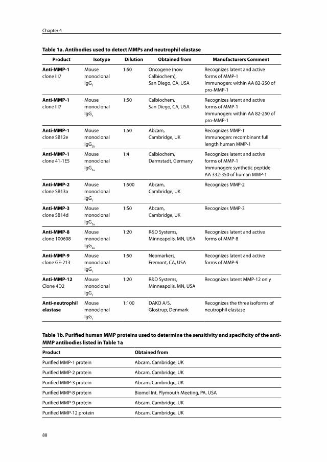
Chapter 4
88
Table 1a. Antibodies used to detect MMPs and neutrophil elastase
Product Isotype Dilution Obtained from Manufacturers Comment
Anti-MMP-1clone III7
Mouse monoclonal IgG1
1:50 Oncogene (now Calbiochem),San Diego, CA, USA
Recognizes latent and active forms of MMP-1Immunogen: within AA 82-250 of pro-MMP-1
Anti-MMP-1clone III7
Mouse monoclonal IgG1
1:50 Calbiochem,San Diego, CA, USA
Recognizes latent and active forms of MMP-1Immunogen: within AA 82-250 of pro-MMP-1
Anti-MMP-1clone SB12e
Mouse monoclonal IgG2b
1:50 Abcam,Cambridge, UK
Recognizes MMP-1Immunogen: recombinant full length human MMP-1
Anti-MMP-1clone 41-1E5
Mouse monoclonal IgG2a
1:4 Calbiochem,Darmstadt, Germany
Recognizes latent and active forms of MMP-1Immunogen: synthetic peptide AA 332-350 of human MMP-1
Anti-MMP-2clone SB13a
Mouse monoclonal IgG1
1:500 Abcam,Cambridge, UK
Recognizes MMP-2
Anti-MMP-3clone SB14d
Mouse monoclonal IgG2a
1:50 Abcam,Cambridge, UK
Recognizes MMP-3
Anti-MMP-8clone 100608
Mouse monoclonal IgG2a
1:20 R&D Systems,Minneapolis, MN, USA
Recognizes latent and active forms of MMP-8
Anti-MMP-9clone GE-213
Mouse monoclonal IgG1
1:50 Neomarkers,Fremont, CA, USA
Recognizes latent and active forms of MMP-9
Anti-MMP-12Clone 4D2
Mouse monoclonal IgG1
1:20 R&D Systems,Minneapolis, MN, USA
Recognizes latent MMP-12 only
Anti-neutrophil elastase
Mouse monoclonalIgG1
1:100 DAKO A/S, Glostrup, Denmark
Recognizes the three isoforms of neutrophil elastase
Table 1b. Purified human MMP proteins used to determine the sensitivity and specificity of the anti-MMP antibodies listed in Table 1a
Product Obtained from
Purified MMP-1 protein Abcam, Cambridge, UK
Purified MMP-2 protein Abcam, Cambridge, UK
Purified MMP-3 protein Abcam, Cambridge, UK
Purified MMP-8 protein Biomol Int, Plymouth Meeting, PA, USA
Purified MMP-9 protein Abcam, Cambridge, UK
Purified MMP-12 protein Abcam, Cambridge, UK
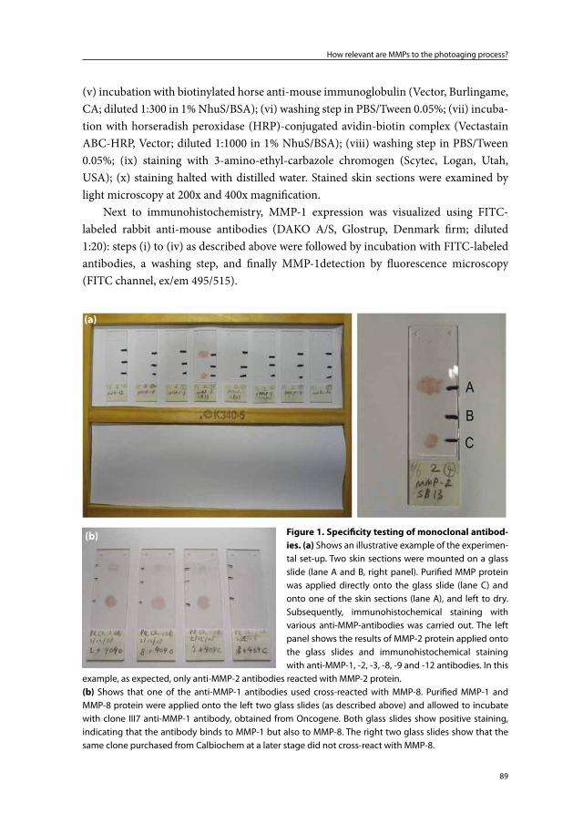
89
How relevant are MMPs to the photoaging process?
(v) incubation with biotinylated horse anti-mouse immunoglobulin (Vector, Burlingame, CA; diluted 1:300 in 1% NhuS/BSA); (vi) washing step in PBS/Tween 0.05%; (vii) incuba-tion with horseradish peroxidase (HRP)-conjugated avidin-biotin complex (Vectastain ABC-HRP, Vector; diluted 1:1000 in 1% NhuS/BSA); (viii) washing step in PBS/Tween 0.05%; (ix) staining with 3-amino-ethyl-carbazole chromogen (Scytec, Logan, Utah, USA); (x) staining halted with distilled water. Stained skin sections were examined by light microscopy at 200x and 400x magnification.
Next to immunohistochemistry, MMP-1 expression was visualized using FITC-labeled rabbit anti-mouse antibodies (DAKO A/S, Glostrup, Denmark firm; diluted 1:20): steps (i) to (iv) as described above were followed by incubation with FITC-labeled antibodies, a washing step, and finally MMP-1detection by fluorescence microscopy (FITC channel, ex/em 495/515).
Figure 1. Specificity testing of monoclonal antibod-ies. (a) Shows an illustrative example of the experimen-tal set-up. Two skin sections were mounted on a glass slide (lane A and B, right panel). Purified MMP protein was applied directly onto the glass slide (lane C) and onto one of the skin sections (lane A), and left to dry. Subsequently, immunohistochemical staining with various anti-MMP-antibodies was carried out. The left panel shows the results of MMP-2 protein applied onto the glass slides and immunohistochemical staining with anti-MMP-1, -2, -3, -8, -9 and -12 antibodies. In this
example, as expected, only anti-MMP-2 antibodies reacted with MMP-2 protein.(b) Shows that one of the anti-MMP-1 antibodies used cross-reacted with MMP-8. Purified MMP-1 and MMP-8 protein were applied onto the left two glass slides (as described above) and allowed to incubate with clone III7 anti-MMP-1 antibody, obtained from Oncogene. Both glass slides show positive staining, indicating that the antibody binds to MMP-1 but also to MMP-8. The right two glass slides show that the same clone purchased from Calbiochem at a later stage did not cross-react with MMP-8.
(b)
(a)
Chapter 4
90
Neutrophil isolation and in situ zymography
The following procedure was carried out to isolate neutrophils from healthy donor blood: (i) addition of 0.4% (w/v) trisodium citrate (pH 7.4) to anticoagulate the blood; (ii) cen-trifugation over isotonic Ficoll (Pharmacia, Uppsala, Sweden) to remove mononuclear cells; (iii) lysis of erythrocytes in an isotonic NH4Cl solution; (iv) washing and resuspen-sion of neutrophils in incubation buffer (20 mM Hepes, 132 mM NaCl, 6 mM KCl, 1 mM MgSO4, 1.2 mM KH2PO4, 5 mM glucose, 1 mM CaCl2 and 0.5% human serum albumin); (v) spinning down of neutropils (1000 RPM for 5 minutes).
Neutrophils were smeared onto Superfrost® Plus coated slides and left to dry on Silicagel.
EnzChek Elastase Assay Kit (E-12056), DQ-collagen (D-12060) and DQ-gelatin (D-12054) all purchased from Molecular Probes (Eugene, OR, USA) were used to determine in situ elastase, collagenase and gelatinase enzyme activity. Slides with mounted skin sections or isolated neutrophil smears were used in these experiments. Reaction buffer (0.05 M TRIS-HCl, 0.15 M NaCl, 5 mM CaCl2, and 0.2 mM NaN3, pH 7.6) containing 40 μg/ml DQ-Elastin was transferred onto the slides and allowed to incubate for a short period of time. Elastase enzyme activity was detected using a fluorescence microscope (FITC-channel, ex/em 505/515). 0.01-1 mM N-methoxysuccinyl-Ala-Ala-Pro-Val-chloromethyl ketone supplied with the elastase assay kit was used as a specific neutrophil elastase inhibitor. Collagenase and gelatinase enzyme activity were detected in a similar fashion. Here the above described reaction buffer contained 40μg/ml DQ-collagen type I or DQ-gelatin, both fluorescein conjugated. The solutions were transferred onto the skin sections and, following a short incubation time, enzyme activity was detected by fluores-cence microscopy using a slightly different wavelength (FITC channel, ex/em 495/515). 0.1-10 mM of 1,10-phenanthroline was used as a general metalloproteinase inhibitor. To inhibit collagenase, gelatinase and elastase activity, skin sections were incubated with the MMP and/or neutrophil elastase inhibitors for one hour before applying DQ-collagen, DQ-gelatin and DQ-elastin.
Results
Specificity of MMP-targeting-antibodies, isotype controls and non-specific staining of sebaceous glands
Prior to immunohistochemical staining of MMPs, the monoclonal antibodies listed in Table 1a were tested for specificity. All monoclonals listed reacted with their respective purified MMP proteins. Purified MMP proteins (Table 1b) clearly adhered to the glass slides both when applied directly and when applied onto skin sections that were already mounted on the glass slides. Figure 1a shows an illustrative example of the experimen-
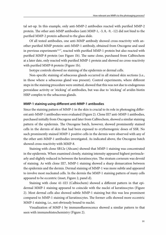
91
How relevant are MMPs to the photoaging process?
tal set-up. In this example, only anti-MMP-2 antibodies reacted with purified MMP-2 protein. The other anti-MMP antibodies (anti MMP-1, -3, 8, -9, -12) did not bind to the purified MMP-2 protein adhered to the glass slide.
Of all tested antibodies, one anti-MMP antibody showed cross-reactivity with an-other purified MMP protein: anti-MMP-1 antibody, obtained from Oncogene and used in previous experiments12;17, reacted with purified MMP-1 protein but also reacted with purified MMP-8 protein (see Figure 1b). The same clone, purchased from Calbiochem at a later date, only reacted with purified MMP-1 protein and showed no cross-reactivity with purified MMP-8 protein (Figure 1b).
Isotype controls showed no staining of the epidermis or dermal cells.Non-specific staining of sebaceous glands occurred in all stained skin sections (i.e.,
in those where a sebaceous gland was present). Control experiments, where different steps in the staining procedure were omitted, showed that this was not due to endogenous peroxidase activity or ‘sticking’ of antibodies, but was due to ‘sticking’ of avidin-biotin HRP complex to the sebaceous glands.
MMP-1 staining using different anti-MMP-1 antibodies
Since the staining pattern of MMP-1 in the skin is crucial to its role in photoaging differ-ent anti-MMP-1 antibodies were evaluated (Figure 2). Clone III7 anti-MMP-1 antibodies, purchased initially from Oncogene and later from Calbiochem, showed a similar staining pattern of the epidermis. The Oncogene batch, however, showed prominently stained cells in the dermis of skin that had been exposed to erythemogenic doses of SSR. No such prominently stained MMP-1 positive cells in the dermis were observed with any of the other anti-MMP-1 antibodies investigated. As indicated above, the Oncogene batch showed cross-reactivity with MMP-8.
Staining with clone SB12e (Abcam) showed that MMP-1 staining was concentrated in the epidermis. When examined closely, staining intensity appeared highest perinucle-arly and slightly reduced in between the keratinocytes. The stratum corneum was devoid of staining. As with clone III7, MMP-1 staining showed a sharp demarcation between the epidermis and the dermis. Dermal staining of MMP-1 was more subtle and appeared to involve most nucleated cells. In the dermis the MMP-1 staining pattern of many cells appeared to be eccentric (inset, Figure 2, panel d).
Staining with clone 41-1E5 (Calbiochem) showed a different pattern in that epi-dermal MMP-1 staining appeared to coincide with the nuclei of keratinocytes (Figure 2). Most dermal cells also showed subtle MMP-1 staining but this was less prominent compared to MMP-1 staining of keratinocytes. The former cells showed more eccentric MMP-1 staining, i.e., not obviously bound to nuclei.
Visualization of MMP-1 by immunofluorescence showed a similar pattern to that seen with immunohistochemistry (Figure 2).
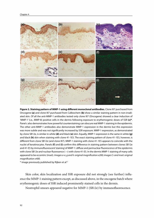
Chapter 4
92
Skin color, skin localization and SSR exposure did not strongly (see further) influ-ence the MMP-1 staining pattern except, as discussed above, in the oncogene batch where erythemogenic doses of SSR induced prominently stained cells in the dermis.
Neutrophil smears appeared negative for MMP-1 (SB12e) by immunofluorescence.
Figure 2. Staining pattern of MMP-1 using different monoclonal antibodies. Clone III7 purchased from Oncogene (a) and clone III7 purchased from Calbiochem (b) show a similar staining pattern in non-irradi-ated skin. Of all the anti-MMP-1 antibodies tested only clone III7 (Oncogene) showed a clear induction of ‘MMP-1’ (i.e., MMP-8) positive cells in the dermis following exposure to erythemogenic doses of SSR (c)*. Panel c also demonstrates how powerful counterstaining can obscure real MMP-1 staining in the epidermis. The other anti-MMP-1 antibodies also demonstrate MMP-1 expression in the dermis but this expression was more subtle and was not significantly increased by SSR exposure. MMP-1 expression, as demonstrated by clone SB12e, is similar in white (d) and black (e) skin. Equally, MMP-1 expression is the same in white (g) and black (h) skin when staining with clone 41-1E5. The exact staining pattern of clone 41-1E5, however, is different from clone SB12e (and clone III7). MMP-1 staining with clone 41-1E5 appears to coincide with the nuclei of keratinocytes. Panels (f) and (i) confirm this difference in staining pattern between clones SB12e and 41-E5 by immunofluorescent ‘staining’ of MMP-1: diffuse and perinuclear fluorescence of the epidermis with clone SB12e and nuclear fluoresence (→) with clone 41-E5. In the dermis MMP-1 staining of many cells appeared to be eccentric (inset). Images a-e, g and h: original magnification x200; images f, i and inset: original magnification x400.* image previously published by Rijken et al12
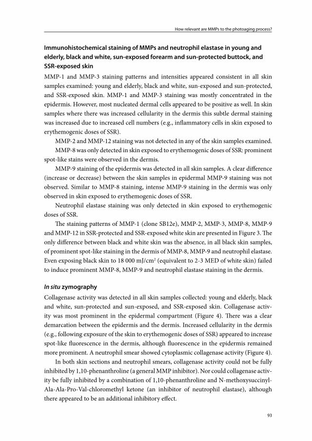
93
How relevant are MMPs to the photoaging process?
Immunohistochemical staining of MMPs and neutrophil elastase in young and elderly, black and white, sun-exposed forearm and sun-protected buttock, and SSR-exposed skin
MMP-1 and MMP-3 staining patterns and intensities appeared consistent in all skin samples examined: young and elderly, black and white, sun-exposed and sun-protected, and SSR-exposed skin. MMP-1 and MMP-3 staining was mostly concentrated in the epidermis. However, most nucleated dermal cells appeared to be positive as well. In skin samples where there was increased cellularity in the dermis this subtle dermal staining was increased due to increased cell numbers (e.g., inflammatory cells in skin exposed to erythemogenic doses of SSR).
MMP-2 and MMP-12 staining was not detected in any of the skin samples examined.MMP-8 was only detected in skin exposed to erythemogenic doses of SSR: prominent
spot-like stains were observed in the dermis.MMP-9 staining of the epidermis was detected in all skin samples. A clear difference
(increase or decrease) between the skin samples in epidermal MMP-9 staining was not observed. Similar to MMP-8 staining, intense MMP-9 staining in the dermis was only observed in skin exposed to erythemogenic doses of SSR.
Neutrophil elastase staining was only detected in skin exposed to erythemogenic doses of SSR.
The staining patterns of MMP-1 (clone SB12e), MMP-2, MMP-3, MMP-8, MMP-9 and MMP-12 in SSR-protected and SSR-exposed white skin are presented in Figure 3. The only difference between black and white skin was the absence, in all black skin samples, of prominent spot-like staining in the dermis of MMP-8, MMP-9 and neutrophil elastase. Even exposing black skin to 18 000 mJ/cm2 (equivalent to 2-3 MED of white skin) failed to induce prominent MMP-8, MMP-9 and neutrophil elastase staining in the dermis.
In situ zymography
Collagenase activity was detected in all skin samples collected: young and elderly, black and white, sun-protected and sun-exposed, and SSR-exposed skin. Collagenase activ-ity was most prominent in the epidermal compartment (Figure 4). There was a clear demarcation between the epidermis and the dermis. Increased cellularity in the dermis (e.g., following exposure of the skin to erythemogenic doses of SSR) appeared to increase spot-like fluorescence in the dermis, although fluorescence in the epidermis remained more prominent. A neutrophil smear showed cytoplasmic collagenase activity (Figure 4).
In both skin sections and neutrophil smears, collagenase activity could not be fully inhibited by 1,10-phenanthroline (a general MMP inhibitor). Nor could collagenase activ-ity be fully inhibited by a combination of 1,10-phenanthroline and N-methoxysuccinyl-Ala-Ala-Pro-Val-chloromethyl ketone (an inhibitor of neutrophil elastase), although there appeared to be an additional inhibitory effect.
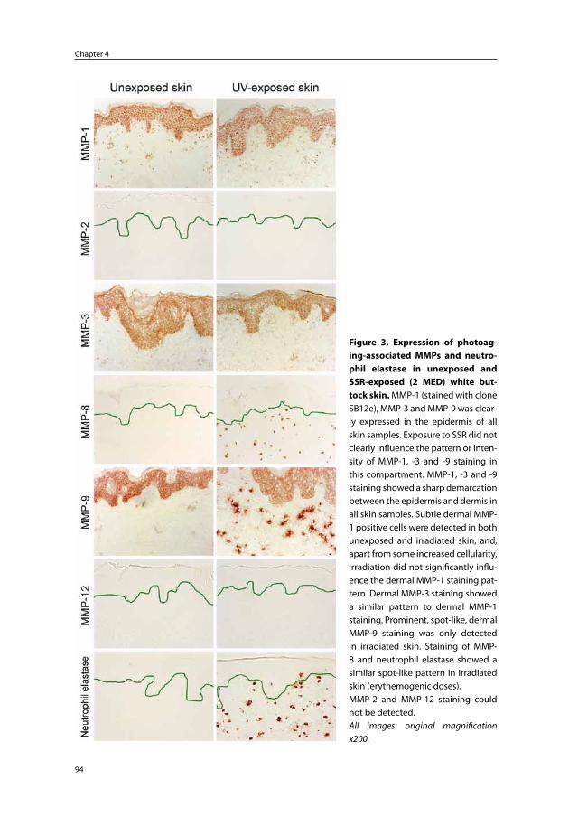
Chapter 4
94
Figure 3. Expression of photoag-ing-associated MMPs and neutro-phil elastase in unexposed and SSR-exposed (2 MED) white but-tock skin. MMP-1 (stained with clone SB12e), MMP-3 and MMP-9 was clear-ly expressed in the epidermis of all skin samples. Exposure to SSR did not clearly influence the pattern or inten-sity of MMP-1, -3 and -9 staining in this compartment. MMP-1, -3 and -9 staining showed a sharp demarcation between the epidermis and dermis in all skin samples. Subtle dermal MMP-1 positive cells were detected in both unexposed and irradiated skin, and, apart from some increased cellularity, irradiation did not significantly influ-ence the dermal MMP-1 staining pat-tern. Dermal MMP-3 staining showed a similar pattern to dermal MMP-1 staining. Prominent, spot-like, dermal MMP-9 staining was only detected in irradiated skin. Staining of MMP-8 and neutrophil elastase showed a similar spot-like pattern in irradiated skin (erythemogenic doses).MMP-2 and MMP-12 staining could not be detected.All images: original magnification x200.
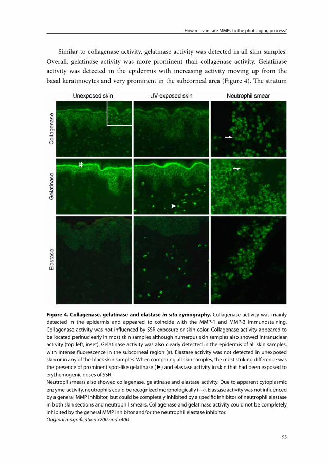
95
How relevant are MMPs to the photoaging process?
Similar to collagenase activity, gelatinase activity was detected in all skin samples. Overall, gelatinase activity was more prominent than collagenase activity. Gelatinase activity was detected in the epidermis with increasing activity moving up from the basal keratinocytes and very prominent in the subcorneal area (Figure 4). The stratum
Figure 4. Collagenase, gelatinase and elastase in situ zymography. Collagenase activity was mainly detected in the epidermis and appeared to coincide with the MMP-1 and MMP-3 immunostaining. Collagenase activity was not influenced by SSR-exposure or skin color. Collagenase activity appeared to be located perinuclearly in most skin samples although numerous skin samples also showed intranuclear activity (top left, inset). Gelatinase activity was also clearly detected in the epidermis of all skin samples, with intense fluorescence in the subcorneal region (#). Elastase activity was not detected in unexposed skin or in any of the black skin samples. When comparing all skin samples, the most striking difference was the presence of prominent spot-like gelatinase (►) and elastase activity in skin that had been exposed to erythemogenic doses of SSR.Neutropil smears also showed collagenase, gelatinase and elastase activity. Due to apparent cytoplasmic enzyme-activity, neutrophils could be recognized morphologically (→). Elastase activity was not influenced by a general MMP inhibitor, but could be completely inhibited by a specific inhibitor of neutrophil elastase in both skin sections and neutrophil smears. Collagenase and gelatinase activity could not be completely inhibited by the general MMP inhibitor and/or the neutrophil elastase inhibitor.Original magnification x200 and x400.
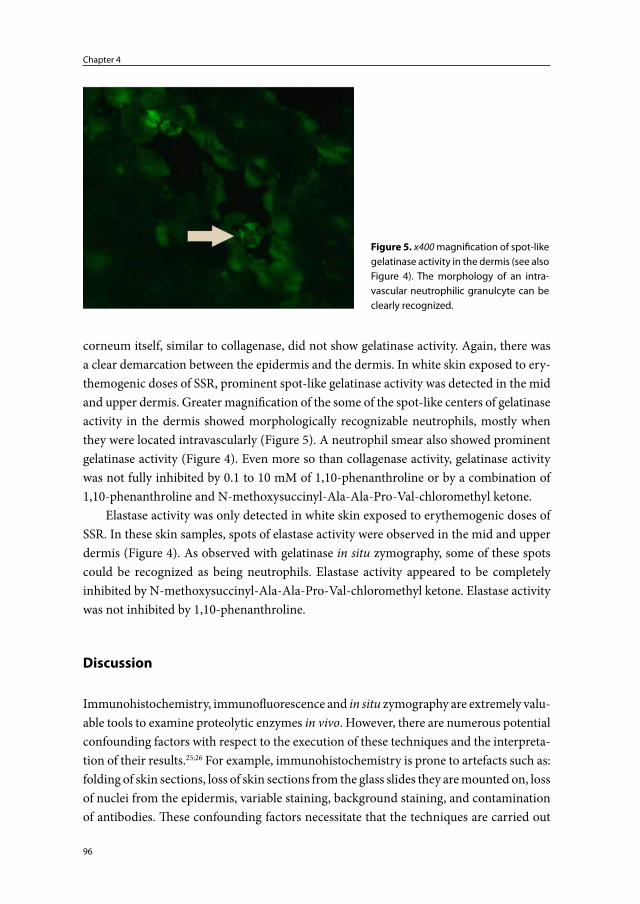
Chapter 4
96
corneum itself, similar to collagenase, did not show gelatinase activity. Again, there was a clear demarcation between the epidermis and the dermis. In white skin exposed to ery-themogenic doses of SSR, prominent spot-like gelatinase activity was detected in the mid and upper dermis. Greater magnification of the some of the spot-like centers of gelatinase activity in the dermis showed morphologically recognizable neutrophils, mostly when they were located intravascularly (Figure 5). A neutrophil smear also showed prominent gelatinase activity (Figure 4). Even more so than collagenase activity, gelatinase activity was not fully inhibited by 0.1 to 10 mM of 1,10-phenanthroline or by a combination of 1,10-phenanthroline and N-methoxysuccinyl-Ala-Ala-Pro-Val-chloromethyl ketone.
Elastase activity was only detected in white skin exposed to erythemogenic doses of SSR. In these skin samples, spots of elastase activity were observed in the mid and upper dermis (Figure 4). As observed with gelatinase in situ zymography, some of these spots could be recognized as being neutrophils. Elastase activity appeared to be completely inhibited by N-methoxysuccinyl-Ala-Ala-Pro-Val-chloromethyl ketone. Elastase activity was not inhibited by 1,10-phenanthroline.
Discussion
Immunohistochemistry, immunofluorescence and in situ zymography are extremely valu-able tools to examine proteolytic enzymes in vivo. However, there are numerous potential confounding factors with respect to the execution of these techniques and the interpreta-tion of their results.25;26 For example, immunohistochemistry is prone to artefacts such as: folding of skin sections, loss of skin sections from the glass slides they are mounted on, loss of nuclei from the epidermis, variable staining, background staining, and contamination of antibodies. These confounding factors necessitate that the techniques are carried out
Figure 5. x400 magnification of spot-like gelatinase activity in the dermis (see also Figure 4). The morphology of an intra-vascular neutrophilic granulcyte can be clearly recognized.
97
How relevant are MMPs to the photoaging process?
and interpreted by experienced laboratory personnel and researchers. A further potential confounding factor is obscuring of immunohistochemical staining by counterstaining. To create optimal conditions for immunohistochemical detection of MMPs in the present study, we omitted counterstaining with hematoxylin. Particularly when examining the epidermis, powerful counterstaining will obscure MMPs expressed in that compartment. Also, MMPs expressed in nuclei will be obscured by counterstaining.
Prior to immunostaining of skin samples we applied solubilized purified human MMPs directly onto glass slides and onto skin sections already mounted on the glass slides. This procedure allowed us to test the specificity of the purchased anti-MMP anti-bodies. The ‘sticking’ of externally applied purified MMP protein onto skin sections and glass slides suggests that MMPs generated in vivo by cells in the epidermis and dermis also remain in situ during the experimental procedures.
We have previously reported that MMP-1 positive cells infiltrate the skin following exposure to erythemogenic doses of SSR,12;17 and we showed that these MMP-1 positive cells double-stained with neutrophil elastase.12 This led us to conclude that neutrophils express MMP-1. In these experiments the anti-MMP-1 antibody batch obtained from Oncogene was used.12;17 However, all experiments carried out thereafter with different anti-MMP-1 antibody batches failed to reproduce the staining of MMP-1 in association with skin-infiltrating neutrophils. Control experiments described in the present study in-dicate that the Oncogene batch used at the time cross-reacts with MMP-8 protein. Since neutrophils are known to express MMP-819 and infiltrate the skin following exposure to erythemogenic doses of SSR,17 this explains our earlier inaccurate conclusion with respect to MMP-1 expression by neutrophils. The same anti-MMP-1 clone purchased from Calbiochem at a later stage did not cross-react with MMP-8 protein. For this reason, we believe that contamination of the anti-MMP-1 Oncogene batch with anti-MMP-8 antibodies must have occurred at our own laboratory.
Other immunohistochemical studies show upregulation of MMP-1 protein in the epidermis following exposure of the skin to UV.2;27;28 In the present study we examined MMP-1 staining in young and elderly skin, black and white skin, sun-protected and sun-exposed skin, and SSR-exposed skin. As discussed above, we first confirmed that the anti-MMP-1 antibodies of different (newly purchased) clones actually react with MMP-1 protein and do not react with other MMP proteins. We observed MMP-1 staining of the epidermis in all skin samples and were not able to detect a consistent difference in staining intensity between the skin samples. Even exposure to erythemogenic doses of SSR appeared to have little effect on MMP-1 staining pattern or intensity. Similarly, col-lagenase activity of equal intensity was observed in the epidermis of all skin samples. Both MMP-1 protein-staining and collagenase activity showed a sharp demarcation between the epidermis and the dermis indicating that these proteins do not diffuse across this barrier. These findings question the hypothesis that keratinocyte-derived MMP-1
Chapter 4
98
plays a major role in the pathophysiology of photoaging: i.e., question the hypothesis that keratinocyte-derived MMP-1 is primarily responsible for dermal changes observed in photoaged skin. Curiously enough, the data on which this hypothesis is originally based show that UV mainly induced MMP-1 protein in the epidermis while at exactly the same time-point collagenase activity (i.e., MMP-1 enzyme activity) was mostly detected in the dermis.2
The MMP-1 staining pattern of clone 41-1E5 differed from that of the other clones tested. Although all clones specifically reacted with MMP-1 protein, Clone 41-1E5 mainly stained the nuclei of epidermal cells. Nuclear staining of MMP-1 has in fact been reported by other authors who proposed that ‘intracellular association of MMP-1 to nuclei and mitochondria confers resistance to apoptosis of cells’.29 According to the manufacturers’ product characteristics sheet, the MMP-1 epitopes of clone 41-1E5 and clone III7 dif-fer and the epitope of clone SB12e is not specified. The manufacturers claim that clones 41-1E5 and III7 both recognize latent and active forms of MMP-1. The manufacturer of clone SB12e only mentions that it ‘recognizes MMP-1’.
Another interesting observation is the eccentric staining of MMP-1 positive dermal cells. Perhaps it indicates the direction of travel of cells. Importantly, this pattern (and intensity) of MMP-1 staining was seen in all skin samples: young and elderly, black and white, sun-exposed and sun-protected, and SSR-exposed.
Parallel to MMP-1 we investigated the expression of MMP-2, -3, 8, 9 and 12.Our control experiments clearly showed that anti-MMP-2 and MMP-12 antibodies were sensitive and specific for their respective proteins, but we were unable to detect MMP-2 or MMP-12 in any of the skin samples. The published images of UV-induced immunohis-tochemical staining of MMP-12 protein (an in vivo study) show very faint staining, and the data was not supported by positive control experiments.23 Our data suggest a limited, if any, role for MMP-2 and MMP-12 in the pathophysiology of photoaging.Similar to MMP-1, MMP-3 appeared to be constitutionally expressed in the skin and staining intensity appeared consistent in all investigated skin samples and independent of exposure to SSR. MMP-3 was mostly detected in the epidermis and there was a clear demarcation between the epidermis and the dermis. Therefore, it seems unlikely that MMP-3 is an important contributor to SSR-induced dermal damage.The most striking observation with respect to changes in staining of MMPs in the dif-ferent skin samples, was the presence of spot-like staining of MMP-8 and MMP-9 in the dermis of white skin exposed to erythemogenic doses of SSR. This spot-like staining was not observed in skin exposed to suberythemogenic doses of SSR or in any of the other skin samples. Neutrophil elastase, the immunohistochemical marker for neutrophils13, was also only detected in white skin exposed to erythemogenic doses of SSR. Neutrophils are known to express both MMP-8 and MMP-919 and we have previously shown that SSR-induced MMP-8 and MMP-9 positive cells double-stain with neutrophil elastase.12
99
How relevant are MMPs to the photoaging process?
We therefore conclude that the above described spot-like staining of MMP-8 and MMP-9 co-localizes with neutrophils.
Although neutrophil smears demonstrated both collagenase and gelatinase activity, only striking gelatinase activity was observed in the dermis of skin sections obtained from white skin exposed to erythemogenic doses of SSR. This suggests that MMP-8 present in skin-infiltrating neutrophils is enzymatically inactive or less active. Neutrophil smears also demonstrated elastase activity and elastase activity was clearly observed in the dermis obtained from white skin exposed to erythemogenic doses of SSR: i.e., dermal elastase activity was detected in the same skin sections that showed prominent dermal gelatinase activity. Elastase activity in these skin sections appeared to be completely inhibited by N-methoxysuccinyl-Ala-Ala-Pro-Val-chloromethyl ketone (an inhibitor of neutrophil elastase). These findings suggests that neutrophil elastase is solely responsible for the observed elastase enzyme activity, and is not caused by MMPs with elastolytic properties. Collagenase activity and particularly gelatinase activity could not be fully inhibited by 1,10-phenanthroline (a general MMP inhibitor) or by a combination of 1,10-phenanthro-line and N-methoxysuccinyl-Ala-Ala-Pro-Val-chloromethyl ketone. This demonstrates the drawback of these assays when performed in vivo: i.e., next to MMPs and neutrophil elastase30 other proteolytic enzymes can demonstrate collagenase and/or gelatinase activ-ity.31 For example, dermal gelatinase activity observed in SSR-exposed skin may partly be due to neutrophil-derived Cathepsin G, a proteinase with gelatinolytic properties that is not inhibited by 1,10-phenanthroline or N-methoxysuccinyl-Ala-Ala-Pro-Val-chloromethyl ketone.32
Taken together, our findings imply that the contributing role of MMPs to the extra-cellular matrix damage observed in photoaged skin may actually be quite limited. We would argue that: (1) even if MMP-1, -3 and -9 are upregulated in the epidermis following UV exposure as demonstrated by other authors, it is highly unlikely that these MMPs massively cross the dermal-epidermal junction; (2) since MMP-1 was similarly expressed in black and white skin, and black skin is much less prone to photoaging, a major role of MMP-1 in photoaging is questionable; (3) since MMP-2 and MMP-12 were not detected in any of the investigated skin samples, their role in photoaging cannot be very important; and (4) since MMP-8 appears to be enzymatically inactive in skin-infiltrating neutrophils its role in photoaging is doubtful. Only MMP-9 and neutrophil elastase and their related enzyme activities were clearly induced by SSR in the mid and upper dermis: i.e., the compartment where damaged collagen and elastic fibers are observed in photoaged skin.
In summary, with respect to the investigated MMPs, only neutrophil-derived MMP-9, is likely to be a major contributor to SSR-induced collagen damage. Neutrophil elastase, and not MMPs with elastolytic properties, is likely to be a major contributor to SSR-induced elastic fiber damage.
Chapter 4
100
Acknowledgements
The authors wish to express their gratitude to the Department of Plastic Surgery for en-abling us to obtain mamma reduction skin. The Department of Pulmonology is gratefully acknowledged for aiding us in isolating neutrophils from peripheral blood.
Conflict of interest
The authors state no conflict of interest.After the study was initiated Dr. Piet L.B. Bruijnzeel became employed at Organon
now Merck, Sharp & Dohme, Department of Translational Medicine, ECREM, Oss, the Netherlands. Recently he has accepted a position at AstraZeneca as Medical Science Director, Loughborough (Charnwood), United Kingdom. Organon nor Merck, Sharp & Dohme nor AstraZeneca have contributed to this manuscript, either financially or oth-erwise. Dr. Piet L.B. Bruijnzeel holds an honorary research position at the Department of Dermatology of the University Medical Center Utrecht.
101
How relevant are MMPs to the photoaging process?
References
1. Quan T, Qin Z, Xia W et al. Matrix-degrading metalloproteinases in photoaging. J.Investig.Dermatol.Symp.Proc. 2009; 14: 20-4.
2. Fisher GJ, Wang ZQ, Datta SC et al. Pathophysiology of premature skin aging induced by ultraviolet light. N.Engl.J.Med. 1997; 337: 1419-28.
3. Matsuta M, Izaki S, Ide C et al. Light and electron microscopic immunohistochemistry of solar elastosis: a study with cutis rhomboidalis nuchae. J.Dermatol. 1987; 14: 364-74.
4. Yaar M. Clinical and histological features of intrinsic versus extrinsic skin aging. In: Skin aging (Gilchrest BA, Krutmann J, eds), 1st edn. Springer, 2006: 9-21.
5. Birkedal-Hansen H, Moore WG, Bodden MK et al. Matrix metalloproteinases: a review. Crit Rev.Oral Biol.Med. 1993; 4: 197-250.
6. Nagase H, Murphy G. Encyclopedia of Biological Chemistry, 1st edn., Vol. 2. 2004: 657-64.7. Fisher GJ, Datta SC, Talwar HS et al. Molecular basis of sun-induced premature skin ageing
and retinoid antagonism. Nature 1996; 379: 335-9.8. Herrmann G, Wlaschek M, Lange TS et al. UVA irradiation stimulates the synthesis of
various matrix-metalloproteinases (MMPs) in cultured human fibroblasts. Exp.Dermatol. 1993; 2: 92-7.
9. Koivukangas V, Kallioinen M, utio-Harmainen H et al. UV irradiation induces the expres-sion of gelatinases in human skin in vivo. Acta Derm.Venereol. 1994; 74: 279-82.
10. Scharffetter K, Wlaschek M, Hogg A et al. UVA irradiation induces collagenase in human dermal fibroblasts in vitro and in vivo. Arch.Dermatol.Res. 1991; 283: 506-11.
11. Fisher GJ, Choi HC, Bata-Csorgo Z et al. Ultraviolet irradiation increases matrix metal-loproteinase-8 protein in human skin in vivo. J.Invest Dermatol. 2001; 117: 219-26.
12. Rijken F, Kiekens RC, Bruijnzeel PL. Skin-infiltrating neutrophils following exposure to solar-simulated radiation could play an important role in photoageing of human skin. Br.J.Dermatol. 2005; 152: 321-8.
13. Lammers AM, van de Kerkhof PC, Schalwijk J et al. Elastase, a marker for neutrophils in skin infiltrates. Br.J.Dermatol. 1986; 115: 181-6.
14. Shapiro SD. Neutrophil elastase: path clearer, pathogen killer, or just pathologic?. Am.J.Respir.Cell Mol.Biol. 2002; 26: 266-8.
15. Cho S, Lee MJ, Kim MS et al. Infrared plus visible light and heat from natural sunlight participate in the expression of MMPs and type I procollagen as well as infiltration of inflammatory cell in human skin in vivo. J.Dermatol.Sci. 2008; 50: 123-33.
16. Lee PL, van Weelden H, Bruijnzeel PL. Neutrophil infiltration in normal human skin after exposure to different ultraviolet radiation sources. Photochem.Photobiol. 2008; 84: 1528-34.
17. Rijken F, Bruijnzeel PL, van Weelden H et al. Responses of black and white skin to solar-simulating radiation: differences in DNA photodamage, infiltrating neutrophils, proteolytic enzymes induced, keratinocyte activation, and IL-10 expression. J.Invest Dermatol. 2004; 122: 1448-55.
18. Ilumets H, Rytila P, Demedts I et al. Matrix metalloproteinases -8, -9 and -12 in smokers and patients with stage 0 COPD. Int.J.Chron.Obstruct.Pulmon.Dis. 2007; 2: 369-79.
19. Murphy G, Bretz U, Baggiolini M et al. The latent collagenase and gelatinase of human polymorphonuclear neutrophil leucocytes. Biochem.J. 1980; 192: 517-25.
20. Chua F, Laurent GJ. Neutrophil elastase: mediator of extracellular matrix destruction and accumulation. Proc.Am.Thorac.Soc. 2006; 3: 424-7.
Chapter 4
102
21. Seite S, Colige A, Piquemal-Vivenot P et al. A full-UV spectrum absorbing daily use cream protects human skin against biological changes occurring in photoaging. Photodermatol.Photoimmunol.Photomed. 2000; 16: 147-55.
22. Li YH, Wu Y, Wei HC et al. Protective effects of green tea extracts on photoaging and photommunosuppression. Skin Res.Technol. 2009; 15: 338-45.
23. Chung JH, Seo JY, Lee MK et al. Ultraviolet modulation of human macrophage metal-loelastase in human skin in vivo. J.Invest Dermatol. 2002; 119: 507-12.
24. Taylor SC. Skin of color: biology, structure, function, and implications for dermatologic disease. J.Am.Acad.Dermatol. 2002; 46: S41-S62.
25. Pesce AJ, Michael JG. Artifacts and limitations of enzyme immunoassay. J.Immunol.Meth-ods 1992; 150: 111-9.
26. Leong AS. Pitfalls in diagnostic immunohistology. Adv.Anat.Pathol. 2004; 11: 86-93.27. Dong KK, Damaghi N, Picart SD et al. UV-induced DNA damage initiates release of MMP-
1 in human skin. Exp.Dermatol. 2008; 17: 1037-44.28. Matsui MS, Hsia A, Miller JD et al. Non-sunscreen photoprotection: antioxidants add value
to a sunscreen. J.Investig.Dermatol.Symp.Proc. 2009; 14: 56-9.29. Limb GA, Matter K, Murphy G et al. Matrix metalloproteinase-1 associates with intra-
cellular organelles and confers resistance to lamin A/C degradation during apoptosis. Am.J.Pathol. 2005; 166: 1555-63.
30. Kawabata K, Hagio T, Matsuoka S. The role of neutrophil elastase in acute lung injury. Eur.J.Pharmacol. 2002; 451: 1-10.
31. Runger TM, Quintanilla-Dieck MJ, Bhawan J. Role of cathepsin K in the turnover of the dermal extracellular matrix during scar formation. J.Invest Dermatol. 2007; 127: 293-7.
32. Capodici C, Berg RA. Cathepsin G degrades denatured collagen. Inflammation 1989; 13: 137-45.
Chapter 5
Solar elastosis results from elastic fiber degradation
and can be induced by neutrophil elastase and reactive
oxygen species: an immunohistochemical study of
black and white skin
Feiko Rijken1,2, Ilse C. Bihari1, Kees L.H. Guikers1, Huib van Weelden1 and Piet L.B. Bruijnzeel1,3
Department of Dermatology, University Medical Center Utrecht, Utrecht, the Netherlands1
Department of Dermatology, Diakonessenhuis, Utrecht, the Netherlands2
AstraZeneca, Loughborough (Charnwood), United Kingdom3
Submitted
Chapter 5
104
Abstract
Background Solar elastosis is the hallmark of photoaged skin. Most authors will agree that the elastotic material observed in photoaged skin is mainly derived from degraded elastic fibers. Nevertheless, a widely accepted hypothesis regarding the pathophysiology of photoaging focuses on collagen fiber degradation. This appears to be contradictory. Black skin is less prone to solar elastosis and photoaging than white skin.Objectives (a) To investigate the extracellular matrix (ECM) changes in photoaged skin (in vivo), and (b) to investigate the effect of neutrophils, neutrophil elastase, hydrogen peroxide and various ultraviolet radiation (UV) sources on the ECM ex vivo.Methods (a) We compared ECM components in sun-exposed and sun-protected, young and elderly, black and white skin. Biopsies taken were immunohistochemically stained for elastin, fibrillin-1 and collagen types I, III, IV and VII. (b) We exposed skin sections to neutrophils isolated from peripheral blood, pus, neutrophil elastase, hydrogen peroxide (a source of reactive oxygen species), and various sources of ultraviolet radiation. Skin sections thus exposed were immunohistochemically stained for elastin.Results (a) The most striking observation was intense elastin staining in the mid and upper dermis of sun-exposed, elderly, white skin. Elastin staining of sun-exposed, elderly, black skin was slightly increased. Elastin staining of all other skin samples was minimal and showed a characteristic pattern confirming the concept that normal elastic fibers consist of a core of elastin surrounded by a mantle of fibrillin-rich microfibrils. (b) Expos-ing skin sections to neutrophils isolated from peripheral blood, pus, neutrophil elastase, hydrogen peroxide, and various sources of UV: to different degrees all but the last induced an elastin staining pattern similar or identical to that observed in photoaged skin.Conclusions Our results suggest that solar elastosis is indeed an end-product of elastic fiber degradation, and that neutrophil-elastase and reactive oxygen species are capable of inducing solar elastosis. We hypothesize that during photoaging part of the fibrillin-rich mantle of elastic fibers is degraded, thereby exposing insoluble elastin. Since elastin is highly resistant to proteolysis, it accumulates, leading to solar elastosis.
105
Solar elastosis can be induced by neutrophil elastase and ROS
Introduction
‘Solar elastosis’ is the hallmark of photoaged skin and is often used synonymously with ‘photoaging’. Photoaging is the process whereby natural sunlight or artificial ultraviolet radiation (UV) sources gradually induce skin changes. When examining the literature on the histopathology and pathofysiology of photoaging, there appears to be a contradiction: although histologically elastic fiber changes are most conspicuous, the pathophysiology focuses mainly on collagen fiber degradation.
Photoaged skin is characterized by a loss of elasticity, rough texture, irregular pig-mentation, telangiectasia, plaque-like thickening and coarse wrinkles.1 The rough texture and irregular pigmentation of photoaged skin are mostly due to changes in the epidermis. Histologically, the epidermis may show the following features: hyperplasia or atrophy; disappearance of dermal papillae; thickening of the basement-membrane; focally in-creased numbers and irregular distribution of melanocytes and melanosomes; atypical keratinocytes; parakeratosis; and thickening of the stratum corneum.1
The most conspicuous defect in the dermis is the presence of elastotic material in the mid and upper dermis, so-called ‘solar elastosis’ or ‘basophilic degeneration’. Fully developed photoaged skin shows alternating areas of fibrous, granular and homogenous elastotic material.2-4 Its origin has been subject of much debate.5 Hypotheses include: (1) that the elastotic material is derived from elastic fibres;3 (2) that it is a degradation product of collagen fibres;6 (3) that it is a degradation product of both collagen and elastic fibres;7 and (4) that it is an abnormal product of UV-stimulated fibroblasts.2;8;9 Braverman and Fonferko2 observed decreased amounts of collagen in areas of elastosis, but found no morphological evidence that collagen degeneration participated in the production of the elastotic material. Based on electron-microscopic studies and on immunohistochemical staining patterns of multiple markers of the extracellular matrix, several groups have concluded that the elastotic material in photoaged skin must be derived primarily from degenerated elastic fibres.3;10
Other observations in the dermis of photoaged skin include a decrease in the total amount of collagen, disorganized collagen fibers, increased amounts of ground substance and dilated blood vessels.11
A widely accepted hypothesis regarding the pathophysiology of photoaging focuses on collagen fiber degradation. Herein, UV-induced, fibroblast- and keratinocyte- derived matrix metalloproteinases (MMPs) are responsible for the clinical and histological signs of photoaged skin.12;13 More recently, we have proposed that, together with directly in-duced damage to the extracellular matrix (ECM) by UV-induced reactive oxygen species (ROS), infiltrating neutrophils could play an important role in the pathophysiology of photoaging.14-17 Neutrophils infiltrate the skin following a certain threshold of UV-dam-age to the skin. This threshold is equivalent to the minimal erythema dose (MED).14;15 For
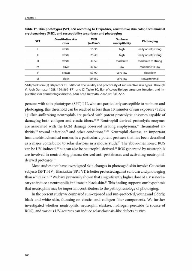
Chapter 5
106
persons with skin phototypes (SPT) I-II, who are particularly susceptible to sunburn and photoaging, this threshold can be reached in less than 10 minutes of sun exposure (Table 1). Skin-infiltrating neutrophils are packed with potent proteolytic enzymes capable of damaging both collagen and elastic fibers.18-21 Neutrophil-derived proteolytic enzymes are associated with the ECM damage observed in lung emphysema,22 rheumatoid ar-thritis,23 wound infection24 and other conditions.25;26 Neutrophil elastase, an important immunohistochemical marker, is a particularly potent protease that has been described as a major contributor to solar elastosis in a mouse study.27 The above-mentioned ROS can be UV-induced,28 but can also be neutrophil-derived.21 ROS generated by neutrophils are involved in neutralizing plasma-derived anti-proteinases and activating neutrophil-derived proteases.21
Most studies that have investigated skin changes in photoaged skin involve Caucasian subjects (SPT I-IV). Black skin (SPT VI) is better protected against sunburn and photoaging than white skin.29 We have previously shown that a significantly higher dose of UV is neces-sary to induce a neutrophilic infiltrate in black skin.14 This finding supports our hypothesis that neutrophils may be important contributors to the pathophysiology of photoaging.
In the present study we compared sun-exposed and sun-protected, young and elderly, black and white skin, focusing on elastic- and collagen-fiber components. We further investigated whether neutrophils, neutrophil elastase, hydrogen peroxide (a source of ROS), and various UV-sources can induce solar elastosis-like defects ex vivo.
Table 1*. Skin phototypes (SPT) I-VI according to Fitzpatrick, constitutive skin color, UVB minimal erythema dose (MED), and susceptibility to sunburn and photoaging
SPTConstitutive skin
colorMED
(mJ/cm2)Sunburn
susceptibilityPhotoaging
I white 15-30 high early onset; strong
II white 25-40 high early onset; strong
III white 30-50 moderate moderate to strong
IV olive 40-60 low moderate to low
V brown 60-90 very low slow; low
VI black 90-150 very low slow; minimal
*Adapted from (1) Fitzpatrick TB. Editorial: The validity and practicality of sun-reactive skin types I through VI. Arch Dermatol 1988, 124: 869–871, and (2) Taylor SC. Skin of color: Biology, structure, function, and im-plications for dermatologic disease. J Am Acad Dermatol 2002, 46: S41–S62.
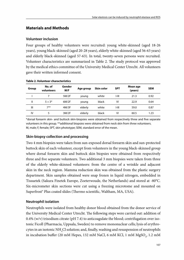
107
Solar elastosis can be induced by neutrophil elastase and ROS
Materials and Methods
Volunteer inclusion
Four groups of healthy volunteers were recruited: young white-skinned (aged 18-26 years), young black-skinned (aged 20-28 years), elderly white-skinned (aged 56-63 years) and elderly black-skinned (aged 57-63). In total, twenty-seven persons were recruited. Volunteer characteristics are summarized in Table 2. The study protocol was approved by the medical ethics committee of the University Medical Center Utrecht. All volunteers gave their written informed consent.
Table 2. Volunteer characteristics
GroupNo. of
volunteersGender
M/FAge group Skin color SPT
Mean age (years)
SEM
I 7 5M/2F young white I-III 21.3 0.92
II 5 + 3* 6M/2F young black VI 22.9 0.64
III 7** 4M/3F elderly white I-III 59.0 0.87
IV 5 3M/2F elderly black VI 60.5 1.23
*dorsal forearm skin- and buttock skin-biopsies were obtained from respectively three and five separate volunteers in this group. **additional biopsies were obtained from neck skin from three volunteers.M, male; F, female; SPT, skin phototype; SEM, standard error of the mean.
Skin-biopsy collection and processing
Two 4 mm biopsies were taken from sun-exposed dorsal forearm skin and sun-protected buttock skin of each volunteer, except from volunteers in the young black-skinned group where dorsal forearm skin and buttock skin biopsies were obtained from respectively three and five separate volunteers. Two additional 3 mm biopsies were taken from three of the elderly white-skinned volunteers: from the centre of a wrinkle and adjacent skin in the neck region. Mamma reduction skin was obtained from the plastic surgery department. Skin samples obtained were snap frozen in liquid nitrogen, embedded in Tissuetek (Sakura Finetek Europe, Zoeterwoude, the Netherlands) and stored at -80°C. Six-micrometer skin sections were cut using a freezing microtome and mounted on Superfrost® Plus coated slides (Thermo scientific, Waltham, MA, USA).
Neutrophil isolation
Neutrophils were isolated from healthy donor blood obtained from the donor service of the University Medical Center Utrecht. The following steps were carried out: addition of 0.4% (w/v) trisodium citrate (pH 7.4) to anticoagulate the blood; centrifugation over iso-tonic Ficoll (Pharmacia, Uppsala, Sweden) to remove mononuclear cells; lysis of erythro-cytes in an isotonic NH4Cl solution; and, finally, washing and resuspension of neutrophils in incubation buffer (20 mM Hepes, 132 mM NaCl, 6 mM KCl, 1 mM MgSO4, 1.2 mM
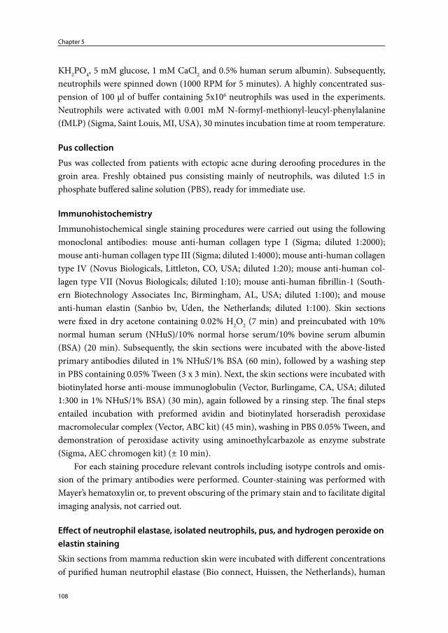
Chapter 5
108
KH2PO4, 5 mM glucose, 1 mM CaCl2 and 0.5% human serum albumin). Subsequently, neutrophils were spinned down (1000 RPM for 5 minutes). A highly concentrated sus-pension of 100 μl of buffer containing 5x10⁶ neutrophils was used in the experiments. Neutrophils were activated with 0.001 mM N-formyl-methionyl-leucyl-phenylalanine (fMLP) (Sigma, Saint Louis, MI, USA), 30 minutes incubation time at room temperature.
Pus collection
Pus was collected from patients with ectopic acne during deroofing procedures in the groin area. Freshly obtained pus consisting mainly of neutrophils, was diluted 1:5 in phosphate buffered saline solution (PBS), ready for immediate use.
Immunohistochemistry
Immunohistochemical single staining procedures were carried out using the following monoclonal antibodies: mouse anti-human collagen type I (Sigma; diluted 1:2000); mouse anti-human collagen type III (Sigma; diluted 1:4000); mouse anti-human collagen type IV (Novus Biologicals, Littleton, CO, USA; diluted 1:20); mouse anti-human col-lagen type VII (Novus Biologicals; diluted 1:10); mouse anti-human fibrillin-1 (South-ern Biotechnology Associates Inc, Birmingham, AL, USA; diluted 1:100); and mouse anti-human elastin (Sanbio bv, Uden, the Netherlands; diluted 1:100). Skin sections were fixed in dry acetone containing 0.02% H2O2 (7 min) and preincubated with 10% normal human serum (NHuS)/10% normal horse serum/10% bovine serum albumin (BSA) (20 min). Subsequently, the skin sections were incubated with the above-listed primary antibodies diluted in 1% NHuS/1% BSA (60 min), followed by a washing step in PBS containing 0.05% Tween (3 x 3 min). Next, the skin sections were incubated with biotinylated horse anti-mouse immunoglobulin (Vector, Burlingame, CA, USA; diluted 1:300 in 1% NHuS/1% BSA) (30 min), again followed by a rinsing step. The final steps entailed incubation with preformed avidin and biotinylated horseradish peroxidase macromolecular complex (Vector, ABC kit) (45 min), washing in PBS 0.05% Tween, and demonstration of peroxidase activity using aminoethylcarbazole as enzyme substrate (Sigma, AEC chromogen kit) (± 10 min).
For each staining procedure relevant controls including isotype controls and omis-sion of the primary antibodies were performed. Counter-staining was performed with Mayer’s hematoxylin or, to prevent obscuring of the primary stain and to facilitate digital imaging analysis, not carried out.
Effect of neutrophil elastase, isolated neutrophils, pus, and hydrogen peroxide on elastin staining
Skin sections from mamma reduction skin were incubated with different concentrations of purified human neutrophil elastase (Bio connect, Huissen, the Netherlands), human
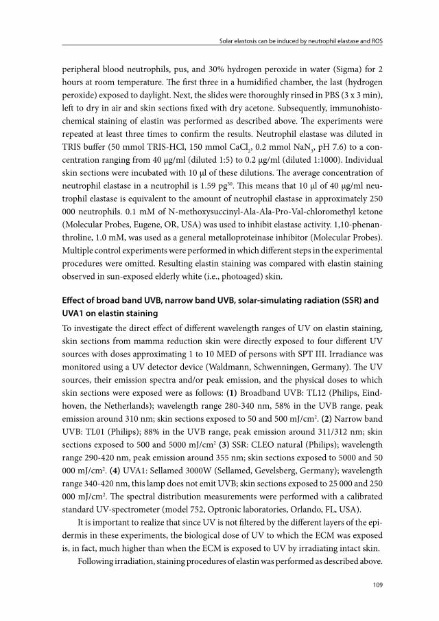
109
Solar elastosis can be induced by neutrophil elastase and ROS
peripheral blood neutrophils, pus, and 30% hydrogen peroxide in water (Sigma) for 2 hours at room temperature. The first three in a humidified chamber, the last (hydrogen peroxide) exposed to daylight. Next, the slides were thoroughly rinsed in PBS (3 x 3 min), left to dry in air and skin sections fixed with dry acetone. Subsequently, immunohisto-chemical staining of elastin was performed as described above. The experiments were repeated at least three times to confirm the results. Neutrophil elastase was diluted in TRIS buffer (50 mmol TRIS-HCl, 150 mmol CaCl2, 0.2 mmol NaN3, pH 7.6) to a con-centration ranging from 40 μg/ml (diluted 1:5) to 0.2 μg/ml (diluted 1:1000). Individual skin sections were incubated with 10 μl of these dilutions. The average concentration of neutrophil elastase in a neutrophil is 1.59 pg30. This means that 10 μl of 40 μg/ml neu-trophil elastase is equivalent to the amount of neutrophil elastase in approximately 250 000 neutrophils. 0.1 mM of N-methoxysuccinyl-Ala-Ala-Pro-Val-chloromethyl ketone (Molecular Probes, Eugene, OR, USA) was used to inhibit elastase activity. 1,10-phenan-throline, 1.0 mM, was used as a general metalloproteinase inhibitor (Molecular Probes). Multiple control experiments were performed in which different steps in the experimental procedures were omitted. Resulting elastin staining was compared with elastin staining observed in sun-exposed elderly white (i.e., photoaged) skin.
Effect of broad band UVB, narrow band UVB, solar-simulating radiation (SSR) and UVA1 on elastin staining
To investigate the direct effect of different wavelength ranges of UV on elastin staining, skin sections from mamma reduction skin were directly exposed to four different UV sources with doses approximating 1 to 10 MED of persons with SPT III. Irradiance was monitored using a UV detector device (Waldmann, Schwenningen, Germany). The UV sources, their emission spectra and/or peak emission, and the physical doses to which skin sections were exposed were as follows: (1) Broadband UVB: TL12 (Philips, Eind-hoven, the Netherlands); wavelength range 280-340 nm, 58% in the UVB range, peak emission around 310 nm; skin sections exposed to 50 and 500 mJ/cm2. (2) Narrow band UVB: TL01 (Philips); 88% in the UVB range, peak emission around 311/312 nm; skin sections exposed to 500 and 5000 mJ/cm2 (3) SSR: CLEO natural (Philips); wavelength range 290-420 nm, peak emission around 355 nm; skin sections exposed to 5000 and 50 000 mJ/cm2. (4) UVA1: Sellamed 3000W (Sellamed, Gevelsberg, Germany); wavelength range 340-420 nm, this lamp does not emit UVB; skin sections exposed to 25 000 and 250 000 mJ/cm2. The spectral distribution measurements were performed with a calibrated standard UV-spectrometer (model 752, Optronic laboratories, Orlando, FL, USA).
It is important to realize that since UV is not filtered by the different layers of the epi-dermis in these experiments, the biological dose of UV to which the ECM was exposed is, in fact, much higher than when the ECM is exposed to UV by irradiating intact skin.
Following irradiation, staining procedures of elastin was performed as described above.
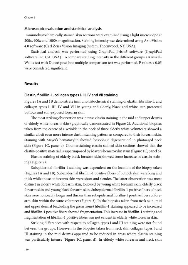
Chapter 5
110
Microscopic evaluation and statistical analysis
Immunohistochemically stained skin sections were examined using a light microscope at 200x, 400x and 1000x magnification. Staining intensity was determined using AxioVision 4.0 software (Carl Zeiss Vision Imaging System, Thornwood, NY, USA).
Statistical analysis was performed using GraphPad Prism5 software (GraphPad software Inc, CA, USA). To compare staining intensity in the different groups a Kruskal-Wallis test with Dunn’s post-hoc multiple comparison test was performed. P values < 0.05 were considered significant.
Results
Elastin, fibrillin-1, collagen types I, III, IV and VII staining
Figures 1A and 1B demonstrate immunohistochemical staining of elastin, fibrillin-1, and collagen types I, III, IV and VII in young and elderly, black and white, sun-protected buttock and sun-exposed forearm skin.
The most striking observation was intense elastin staining in the mid and upper dermis of elderly white forearm skin (graphically demonstrated in Figure 2). Additional biopsies taken from the centre of a wrinkle in the neck of three elderly white volunteers showed a similar albeit even more intense elastin staining pattern as compared to their forearm skin. Staining with Mayer’s hematoxylin showed ‘basophilic degeneration’ in photoaged neck skin (Figure 1C, panel a). Counterstaining elastin-stained skin sections showed that the elastin-positive material is superimposed by Mayer’s hematoxylin stain (Figure 1C, panel b).
Elastin staining of elderly black forearm skin showed some increase in elastin stain-ing (Figure 2).
Subepidermal fibrillin-1 staining was dependent on the location of the biopsy taken (Figures 1A and 1B). Subepidermal fibrillin-1 positive fibers of buttock skin were long and thick while those of forearm skin were short and slender. The latter observation was most distinct in elderly white forearm skin, followed by young white forearm skin, elderly black forearm skin and young black forearm skin. Subepidermal fibrillin-1 positive fibers of neck skin were noticeably longer and thicker than subepidermal fibrillin-1 positive fibers of fore-arm skin within the same volunteer (Figure 3). In the biopsies taken from neck skin, mid and upper dermal (excluding the grenz zone) fibrillin-1 staining appeared to be increased and fibrillin-1 positive fibers showed fragmentation. This increase in fibrillin-1 staining and fragmentation of fibrillin-1 positive fibers was not evident in elderly white forearm skin.
Striking differences with respect to collagen types I and III staining were not found between the groups. However, in the biopsies taken from neck skin collagen types I and III staining in the mid dermis appeared to be reduced in areas where elastin staining was particularly intense (Figure 1C, panel d). In elderly white forearm and neck skin
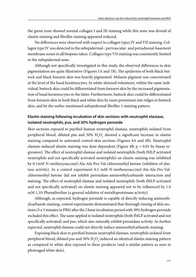
111
Solar elastosis can be induced by neutrophil elastase and ROS
the grenz zone showed normal collagen I and III staining while this zone was devoid of elastin staining and fibrillin staining appeared reduced.
No differences were observed with respect to collagen types IV and VII staining. Col-lagen type IV was detected in the subepidermal-, perivascular- and periadnexal-basement membrane zones in all biopsies taken. Collagen type VII staining was consistently limited to the subepidermal zone.
Although not specifically investigated in this study, the observed differences in skin pigmentation are quite illustrative (Figures 1A and 1B). The epidermis of both black but-tock and black forearm skin was heavily pigmented. Melanin pigment was concentrated at the level of the basal keratinocytes. In white-skinned volunteers, within the same indi-vidual, buttock skin could be differentiated from forearm skin by the increased pigmenta-tion of basal keratinocytes in the latter. Furthermore, buttock skin could be differentiated from forearm skin in both black and white skin by more prominent rete ridges in buttock skin, and by the earlier mentioned subepidermal fibrillin-1 staining pattern.
Elastin staining following incubation of skin sections with neutrophil elastase, isolated neutrophils, pus, and 30% hydrogen peroxide
Skin sections exposed to purified human neutrophil elastase, neutrophils isolated from peripheral blood, diluted pus and 30% H2O2 showed a significant increase in elastin staining compared to untreated control skin sections (Figures 4A and 4B). Neutrophil elastase-induced elastin staining was dose dependent (Figure 4B, p < 0.05 by linear re-gression). The effect of neutrophil elastase and isolated neutrophils (both fMLP activated neutrophils and not specifically activated neutrophils) on elastin staining was inhibited by 0.1mM N-methoxysuccinyl-Ala-Ala-Pro-Val-chloromethyl ketone (inhibitor of elas-tase activity). In a control experiment 0.1 mM N-methoxysuccinyl-Ala-Ala-Pro-Val-chloromethyl ketone did not inhibit peroxidase-aminoethylcarbazole interaction and staining. The effect of neutrophil elastase and isolated neutrophils (both fMLP activated and not specifically activated) on elastin staining appeared not to be influenced by 1.0 mM 1,10-Phenathroline (a general inhibitor of metalloproteinase activity).
Although, as expected, hydrogen peroxide is capable of directly inducing aminoeth-ylcarbazole staining, control experiments demonstrated that thorough rinsing of skin sec-tions (3 x 3 minutes in PBS) after the 2 hour incubation period with 30% hydrogen peroxide excluded this effect. The same applied to isolated neutrophils (both fMLP activated and not specifically activated) and pus, which also naturally exhibit peroxidase activity. As further expected, neutrophil elastase could not directly induce aminoethylcarbazole staining.
Exposing black skin to purified human neutrophil elastase, neutrophils isolated from peripheral blood, diluted pus and 30% H2O2 induced an identical elastin staining pattern as compared to white skin exposed to these products (and a similar pattern as seen in photoaged white skin).
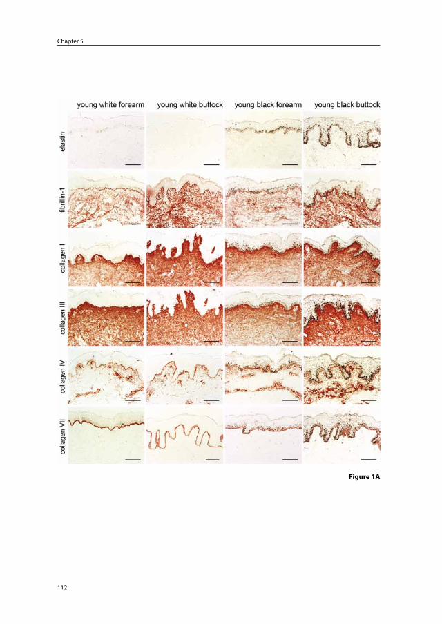
Chapter 5
112
Figure 1A
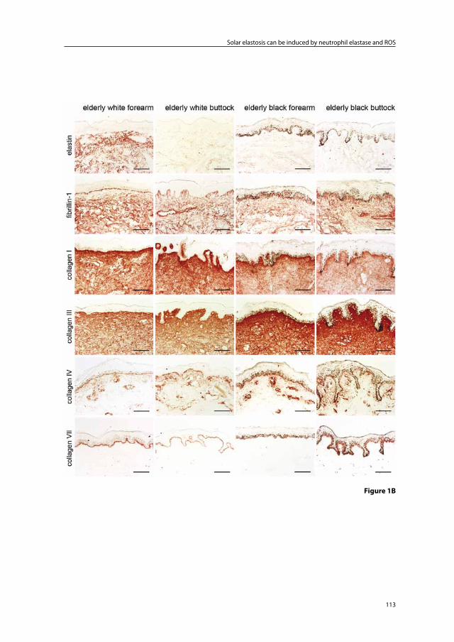
113
Solar elastosis can be induced by neutrophil elastase and ROS
Figure 1B
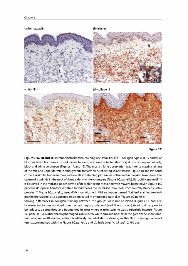
Chapter 5
114
(a) hematoxylin (b) elastin
(c) fibrillin-1 (d) collagen I
Figure 1C
Figures 1A, 1B and 1C. Immunohistochemical staining of elastin, fibrillin-1, collagen types I, III, IV and VII of biopsies taken from sun-exposed (dorsal forearm) and sun-protected (buttock) skin of young and elderly, black and white volunteers (Figures 1A and 1B). The most striking observation was intense elastin staining of the mid and upper dermis in elderly white forearm skin, reflecting solar elastosis (Figure 1B, top left hand corner). A similar but even more intense elastin staining pattern was observed in biopsies taken from the centre of a wrinkle in the neck of three elderly white volunteers (Figure 1C, panel b). Basophilic material (*) is observed in the mid and upper dermis of neck skin sections stained with Mayer’s hematoxylin (Figure 1C, panel a). Basophilic hematoxylin stain superimposes the increased immunohistochemically stained elastin protein (** Figure 1C, panel b, inset: 400x magnification). Mid and upper dermal fibrillin-1 staining (exclud-ing the grenz zone) also appeared to be increased in photoaged neck skin (Figure 1C, panel c).Striking differences in collagen staining between the groups were not observed (Figures 1A and 1B). However, in biopsies obtained from the neck region, collagen I (and III, not shown) staining did appear to be reduced, disorganized and fragmented in areas where elastin staining was particularly intense (Figure 1C, panel d, →). Notice that in photoaged skin (elderly white arm and neck skin) the grenz zone shows nor-mal collagen I and III staining while it is relatively devoid of elastin staining and fibrillin-1 staining is reduced (grenz zone marked with # in Figure 1C, panels b and d). Scale bars: 1A, 1B and 1C: 100 μm.
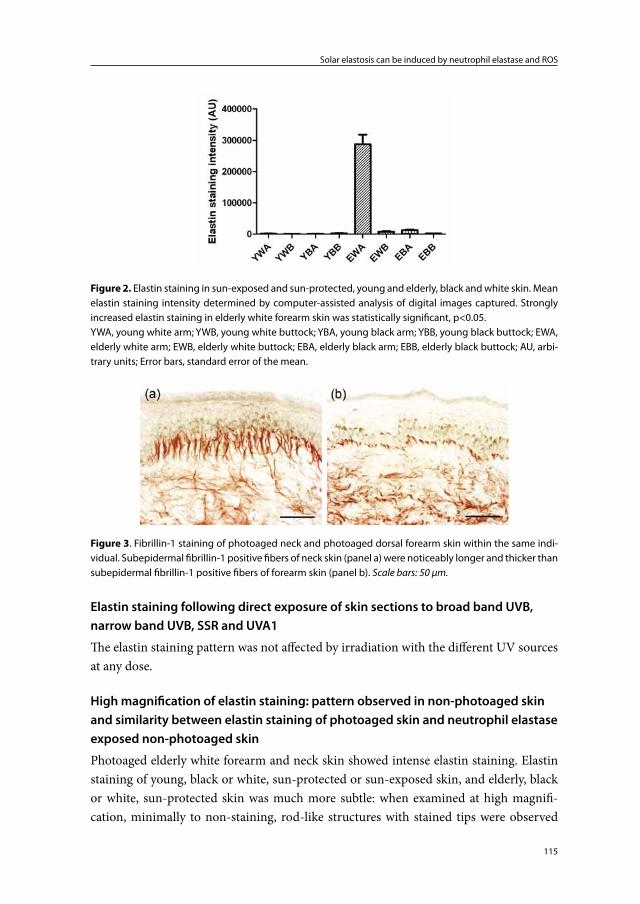
115
Solar elastosis can be induced by neutrophil elastase and ROS
Elastin staining following direct exposure of skin sections to broad band UVB, narrow band UVB, SSR and UVA1
The elastin staining pattern was not affected by irradiation with the different UV sources at any dose.
High magnification of elastin staining: pattern observed in non-photoaged skin and similarity between elastin staining of photoaged skin and neutrophil elastase exposed non-photoaged skin
Photoaged elderly white forearm and neck skin showed intense elastin staining. Elastin staining of young, black or white, sun-protected or sun-exposed skin, and elderly, black or white, sun-protected skin was much more subtle: when examined at high magnifi-cation, minimally to non-staining, rod-like structures with stained tips were observed
Figure 2. Elastin staining in sun-exposed and sun-protected, young and elderly, black and white skin. Mean elastin staining intensity determined by computer-assisted analysis of digital images captured. Strongly increased elastin staining in elderly white forearm skin was statistically significant, p<0.05.YWA, young white arm; YWB, young white buttock; YBA, young black arm; YBB, young black buttock; EWA, elderly white arm; EWB, elderly white buttock; EBA, elderly black arm; EBB, elderly black buttock; AU, arbi-trary units; Error bars, standard error of the mean.
Figure 3. Fibrillin-1 staining of photoaged neck and photoaged dorsal forearm skin within the same indi-vidual. Subepidermal fibrillin-1 positive fibers of neck skin (panel a) were noticeably longer and thicker than subepidermal fibrillin-1 positive fibers of forearm skin (panel b). Scale bars: 50 μm.
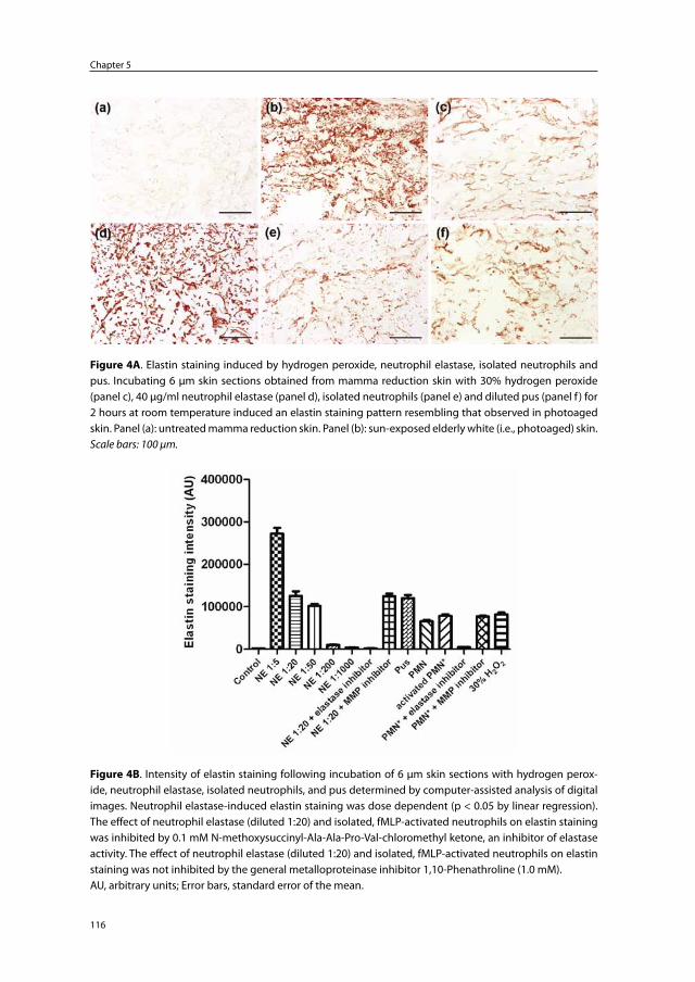
Chapter 5
116
Figure 4A. Elastin staining induced by hydrogen peroxide, neutrophil elastase, isolated neutrophils and pus. Incubating 6 μm skin sections obtained from mamma reduction skin with 30% hydrogen peroxide (panel c), 40 μg/ml neutrophil elastase (panel d), isolated neutrophils (panel e) and diluted pus (panel f ) for 2 hours at room temperature induced an elastin staining pattern resembling that observed in photoaged skin. Panel (a): untreated mamma reduction skin. Panel (b): sun-exposed elderly white (i.e., photoaged) skin. Scale bars: 100 μm.
Figure 4B. Intensity of elastin staining following incubation of 6 μm skin sections with hydrogen perox-ide, neutrophil elastase, isolated neutrophils, and pus determined by computer-assisted analysis of digital images. Neutrophil elastase-induced elastin staining was dose dependent (p < 0.05 by linear regression). The effect of neutrophil elastase (diluted 1:20) and isolated, fMLP-activated neutrophils on elastin staining was inhibited by 0.1 mM N-methoxysuccinyl-Ala-Ala-Pro-Val-chloromethyl ketone, an inhibitor of elastase activity. The effect of neutrophil elastase (diluted 1:20) and isolated, fMLP-activated neutrophils on elastin staining was not inhibited by the general metalloproteinase inhibitor 1,10-Phenathroline (1.0 mM).AU, arbitrary units; Error bars, standard error of the mean.
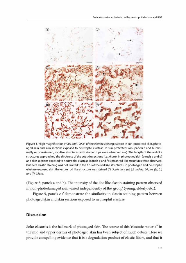
117
Solar elastosis can be induced by neutrophil elastase and ROS
(Figure 5, panels a and b). The intensity of the dot-like elastin staining pattern observed in non-photodamaged skin varied independently of the ‘group’ (young, elderly, etc.).
Figure 5, panels c-f demonstrate the similarity in elastin staining pattern between photoaged skin and skin sections exposed to neutrophil elastase.
Discussion
Solar elastosis is the hallmark of photoaged skin. The source of this ‘elastotic material’ in the mid and upper dermis of photoaged skin has been subject of much debate. Here we provide compelling evidence that it is a degradation product of elastic fibers, and that it
Figure 5. High magnification (400x and 1000x) of the elastin staining pattern in sun-protected skin, photo-aged skin and skin sections exposed to neutrophil elastase. In sun-protected skin (panels a and b) mini-mally or non-stained, rod-like structures with stained tips were observed (→). The length of the rod-like structures approached the thickness of the cut skin sections (i.e., 6 μm). In photoaged skin (panels c and d) and skin sections exposed to neutrophil elastase (panels e and f ) similar rod-like structures were observed, but here elastin staining was not limited to the tips of the rod like structures: in photoaged and neutrophil elastase exposed skin the entire rod like structure was stained (*). Scale bars: (a), (c) and (e): 30 μm, (b), (d) and (f): 15μm.
Chapter 5
118
can be induced by neutrophil elastase and ROS. Furthermore, our findings confirm that black skin is less susceptible to solar elastosis/photoaging.
Significantly increased elastin staining in photoaged skin has been described by numerous authors.2;3;10;31-35 Likewise, we observed intense elastin staining in sun-exposed elderly white skin. Furthermore, we demonstrated that the typical basophilic material observed in photoaged skin (skin sections stained with Mayer’s hematoxylin) is the same material we stained with specific anti-elastin antibodies (Figure 1C, panels a and b), i.e., elastin protein appears to be a major component of the elastotic material observed in photoaged skin. Therefore we conclude that increased elastin-staining is a primary marker for photoaged skin.
Although solar elastosis has also been described in black skin8 and our elderly black sun-exposed skin samples did show some increase in elastin-staining, this increase was insignificant compared to elderly white sun-exposed skin. Thus, in the elderly group, histologically, black skin shows less signs of photoaging.
Elastin staining of young, black or white, sun-protected or sun-exposed skin, and el-derly, black or white sun-protected skin, was very subtle compared to sun-exposed elderly white skin: when examining the elastin-stained skin sections more closely, minimally to non-staining, rod-like structures with stained tips were observed (Figure 5, panels a and b). This observation appears to confirm that, broadly speaking, elastic fibers consist of a core of elastin surrounded by a mantle of fibrillin-rich microfibrils.2;36 When elastic fibers are cut perpendicular to their long axis, the core of the elastic fiber becomes visible and elastin staining shows the characteristic dot-like pattern observed in Figure 5, panels a and b.
We have shown that exposure of skin sections to neutrophil elastase, isolated neu-trophils, pus and hydrogen peroxide induces an elastin staining pattern similar to that observed in photoaged skin (Figure 5). Based on these results, we propose the following mechanism: neutrophil elastase and ROS break down the fibrillin-rich microfibrillar mantle of elastic fibers, thereby exposing elastin epitopes (Figure 6). Exposed, cross-linked, insoluble elastin is highly resistant to further proteolysis37 and gradually accu-mulates in the skin. Although less resistant to proteolysis than elastin, partially degraded fibrillin-rich microfibrils may also contribute to the accumulation of elastotic material in photoaged skin. Similar to wound healing where repair of the elastic fiber network has been described,38 a new elastic fiber network may be formed following UV damage. However, this repair process is probably slow and already accumulated elastin may struc-turally interfere with elastic (and collagen) fiber network regeneration. Newly formed elastic fibers can in turn be degraded and may further contribute to the accumulation of elastin. In vivo, the degradation of elastic fibers and accumulation of elastotic material takes place gradually. Our ex vivo experiments allowed us to mimic this process in an accelerated fashion. Although, similar to white skin, elastin staining could be induced
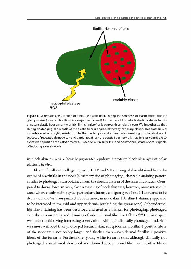
119
Solar elastosis can be induced by neutrophil elastase and ROS
in black skin ex vivo, a heavily pigmented epidermis protects black skin against solar elastosis in vivo.
Elastin, fibrillin-1, collagen types I, III, IV and VII staining of skin obtained from the centre of a wrinkle in the neck (a primary site of photoaging) showed a staining pattern similar to photoaged skin obtained from the dorsal forearm of the same individual. Com-pared to dorsal forearm skin, elastin staining of neck skin was, however, more intense. In areas where elastin staining was particularly intense collagen types I and III appeared to be decreased and/or disorganized. Furthermore, in neck skin, Fibrillin-1 staining appeared to be increased in the mid and upper dermis (excluding the grenz zone). Subepidermal fibrillin-1 staining has been described and used as a marker for photoaging: photoaged skin shows shortening and thinning of subepidermal fibrillin-1 fibres.32-34 In this respect we made the following interesting observation. Although clinically photoaged neck skin was more wrinkled than photoaged forearm skin, subepidermal fibrillin-1 positive fibers of the neck were noticeably longer and thicker than subepidermal fibrillin-1 positive fibers of the forearm. Furthermore, young white forearm skin, although clinically not photoaged, also showed shortened and thinned subepidermal fibrillin-1 positive fibers.
insoluble elastin
fibrillin-rich microfibrils
neutrophil elastaseROS
Figure 6. Schematic cross-section of a mature elastic fiber. During the synthesis of elastic fibers, fibrillar glycoproteins (of which fibrillin-1 is a major component) form a scaffold on which elastin is deposited. In a mature elastic fiber a mantle of fibrillin-rich microfibrils surrounds an elastin core. We hypothesize that during photoaging, the mantle of the elastic fiber is degraded thereby exposing elastin. This cross-linked insoluble elastin is highly resistant to further proteolysis and accumulates, resulting in solar elastosis. A process of repeated damage to - and partial repair of - the elastic fiber network may further contribute to excessive deposition of elastotic material. Based on our results, ROS and neutrophil elastase appear capable of inducing solar elastosis.
Chapter 5
120
Together with the fibrillin-1 staining pattern observed in young and elderly black skin, our findings suggest that next to photodamage,32-34 skin location also plays a role in the observed fibrillin-1 staining pattern. Our findings suggest that the length and thickness of subepidermal fibrillin-1 fibers do not consistently mirror the clinical picture which appears to be better correlated to elastin staining intensity.
Based on our findings and literature data one can speculate on the mechanism of wrinkle formation. Deep wrinkles are observed at sites exposed to mechanical stress due to underlying muscle movement, such as the neck, forehead and lateral to the eyes (crow’s feet). Contrary to young skin which has an intact elastic fiber network, photoaged inelastic skin will have difficulty to regain its original form when put under strain and in time wrinkles are formed. Wrinkle formation may, in fact, accommodate movement of inelastic skin. In other words, continuous bending and stretching of inelastic skin, possibly in combination with ECM remodeling, may be the key ingredients to wrinkle formation.
We chose to investigate elastin, fibrillin-1, collagens I, III, IV and VII because they are important ECM components of the skin and/or have previously been studied in con-junction with photoaging.2;3;10;11;31-35;39-41 Some research groups found greater differences in collagen staining and smaller differences in elastin staining when comparing photoaged skin with non-photoaged skin. This variation in results may partly be explained by differ-ences in age groups of the volunteers and skin locations studied. Several authors report that fibrillin-rich microfibrils are reduced in moderately to severely photoaged skin.32-34 These authors focused particularly on subepidermal fibrillin-rich microfibrils and took biopsies from buttock, forearm and facial skin, but not neck skin.
None of the above-mentioned studies investigated differences between black and white skin. By including black and white skin in our study we were able to compare these ECM components in elderly, sun-exposed, significantly photoaged (white) skin and elderly, sun-exposed, relatively non-photoaged (black) skin.
The effect of particular proteases on the ECM has also been studied by Braverman and Fonferko.2 Their experiments included exposing human skin slices to porcine pan-creatic elastase and bovine chymotrypsin, and studying their effect on elastic fibers by means of electron microscopy. These enzymatic studies produced alterations in elastic fibers that resembled abnormalities associated with chronoaged but not photoaged skin.2 Our experiments performed with human purified neutrophil elastase did produce im-munohistochemical alterations resembling those found in photoaged skin.
It has been shown that serine proteases such as neutrophil elastase are potent effec-tors for the physiological and pathological catabolism of fibrillin-rich microfibrils.42 It has also been shown that several MMPs (MMP-2, MMP-3, MMP-9, MMP-12, MMP-13 and MMP-14) are capable of cleaving fibrillin-rich microfibrils.43;44 UV-induced MMPs can be derived from different cell types including infiltrating neutrophils, macrophages, fibro-
121
Solar elastosis can be induced by neutrophil elastase and ROS
blasts and keratinocytes. Thus, as has been suggested by these and other authors, MMPs may also contribute to elastic fiber degradation in the process of photoaging.33;34;45;46 This effect and the relative contributions of MMPs, serine proteases and ROS to the induction of solar elastosis require further investigation.
Because elastic and collagen fibers can absorb UV directly,47 we investigated whether exposure of skin sections to different UV sources could simulate the effect of neutrophil elastase, isolated neutrophils, pus and hydrogen peroxide on elastin staining. However, in these experiments, elastin staining of skin sections was not altered following UV exposure. We also investigated the effect of the different UV sources on skin sections with respect to the induction of thymine dimers, an important UV photoproduct (data not shown). Nuclei of cells in all parts of the skin sections (epidermis and dermis) directly exposed to broadband UVB, narrow band UVB and SSR showed intense, dose-dependent, thymine dimer staining. Interestingly, skin sections exposed to high doses of UVA1 showed subtle thymine dimer staining. Thymine dimer staining of non-irradiated skin sections was negative. Thus, ex vivo UV-exposed skin sections did show DNA damage, but elastin staining was unaffected.
In summary, our findings indicate that the elastotic material observed in photoaged skin is a degradation product of elastic fibers and can be induced by neutrophil elastase and ROS. We found no evidence that UV directly participates in the formation of ‘solar elastosis’.
Acknowledgements
The authors wish to express their gratitude to the Department of Plastic Surgery for en-abling us to obtain mamma reduction skin. The Department of Pulmonology is gratefully acknowledged for aiding us in isolating neutrophils from peripheral blood.
Conflict of interest
The authors state no conflict of interest.After the study was initiated Dr. Piet L.B. Bruijnzeel became employed at Organon
now Merck, Sharp & Dohme, Department of Translational Medicine, ECREM, Oss, the Netherlands. Recently he has accepted a position at AstraZeneca as Medical Science Director, Loughborough (Charnwood), United Kingdom. Organon nor Merck, Sharp & Dohme nor AstraZeneca have contributed to this manuscript, either financially or oth-erwise. Dr. Piet L.B. Bruijnzeel holds an honorary research position at the Department of Dermatology of the University Medical Center Utrecht.
Chapter 5
122
References
1. Gilchrest BA. Skin aging and photoaging: an overview. J.Am.Acad.Dermatol. 1989; 21: 610-3.
2. Braverman IM, Fonferko E. Studies in cutaneous aging: I. The elastic fiber network. J.Invest Dermatol. 1982; 78: 434-43.
3. Matsuta M, Izaki S, Ide C et al. Light and electron microscopic immunohistochemistry of solar elastosis: a study with cutis rhomboidalis nuchae. J.Dermatol. 1987; 14: 364-74.
4. Tsuji T. The surface structural alterations of elastic fibers and elastotic material in solar elastosis: a scanning electron microscopic study. J.Cutan.Pathol. 1984; 11: 300-8.
5. Sellheyer K. Pathogenesis of solar elastosis: synthesis or degradation? J.Cutan.Pathol. 2003; 30: 123-7.
6. Mitchell RE. Chronic solar dermatosis: a light and electron microscopic study of the der-mis. J.Invest Dermatol. 1967; 48: 203-20.
7. Niebauer G, Stockinger L. Senile elastosis: histochemical and electron microscopic studies. Arch.Klin.Exp.Dermatol. 1965; 221: 122-43.
8. Nurnberger F, Schober E, Marsch WC et al. Actinic elastosis in black skin. A light- and electronmicroscopic study. Arch.Dermatol.Res. 1978; 262: 7-14.
9. Ledoux-Corbusier M, Achten G. Elastosis in chronic radiodermatitis. An ultrastructural study. Br.J.Dermatol. 1974; 91: 287-95.
10. Chen VL, Fleischmajer R, Schwartz E et al. Immunochemistry of elastotic material in sun-damaged skin. J.Invest Dermatol. 1986; 87: 334-7.
11. Oikarinen A. The aging of skin: chronoaging versus photoaging. Photodermatol.Photoim-munol.Photomed. 1990; 7: 3-4.
12. Fisher GJ, Datta SC, Talwar HS et al. Molecular basis of sun-induced premature skin ageing and retinoid antagonism. Nature 1996; 379: 335-9.
13. Fisher GJ, Wang ZQ, Datta SC et al. Pathophysiology of premature skin aging induced by ultraviolet light. N.Engl.J.Med. 1997; 337: 1419-28.
14. Rijken F, Bruijnzeel PL, van Weelden H et al. Responses of black and white skin to solar-simulating radiation: differences in DNA photodamage, infiltrating neutrophils, proteolytic enzymes induced, keratinocyte activation, and IL-10 expression. J.Invest Dermatol. 2004; 122: 1448-55.
15. Rijken F, Kiekens RC, Bruijnzeel PL. Skin-infiltrating neutrophils following exposure to solar-simulated radiation could play an important role in photoageing of human skin. Br.J.Dermatol. 2005; 152: 321-8.
16. Rijken F, Kiekens RC, van Weelden H et al. Pathophysiology of photoaging of human skin: focus on neutrophils. Photochem.Photobiol.Sci. 2006; 5: 184-9.
17. Rijken F, Bruijnzeel PL. The pathogenesis of photoaging: the role of neutrophils and neutrophil-derived enzymes. J.Investig.Dermatol.Symp.Proc. 2009; 14: 67-72.
18. Chua F, Laurent GJ. Neutrophil elastase: mediator of extracellular matrix destruction and accumulation. Proc.Am.Thorac.Soc. 2006; 3: 424-7.
19. Shapiro SD. Neutrophil elastase: path clearer, pathogen killer, or just pathologic? Am.J.Respir.Cell Mol.Biol. 2002; 26: 266-8.
20. Travis J. Structure, function, and control of neutrophil proteinases. Am.J.Med. 1988; 84: 37-42.
21. Weiss SJ. Tissue destruction by neutrophils. N.Engl.J.Med. 1989; 320: 365-76.
123
Solar elastosis can be induced by neutrophil elastase and ROS
22. Gadek JE. Adverse effects of neutrophils on the lung. Am.J.Med. 1992; 92: 27S-31S.23. Edwards SW, Hallett MB. Seeing the wood for the trees: the forgotten role of neutrophils in
rheumatoid arthritis. Immunol.Today 1997; 18: 320-4.24. Singer AJ, McClain SA. Persistent wound infection delays epidermal maturation and
increases scarring in thermal burns. Wound.Repair Regen. 2002; 10: 372-7.25. Golub LM, Sorsa T, Lee HM et al. Doxycycline inhibits neutrophil (PMN)-type matrix me-
talloproteinases in human adult periodontitis gingiva. J.Clin.Periodontol. 1995; 22: 100-9.26. Hwang ST, Williams ML, McCalmont TH et al. Sweet’s syndrome leading to acquired cutis
laxa (Marshall’s syndrome) in an infant with alpha 1-antitrypsin deficiency. Arch.Dermatol. 1995; 131: 1175-7.
27. Starcher B, Conrad M. A role for neutrophil elastase in solar elastosis. Ciba Found.Symp. 1995; 192: 338-46.
28. Scharffetter-Kochanek K, Wlaschek M, Brenneisen P et al. UV-induced reactive oxygen species in photocarcinogenesis and photoaging. Biol.Chem. 1997; 378: 1247-57.
29. Taylor SC. Skin of color: biology, structure, function, and implications for dermatologic disease. J.Am.Acad.Dermatol. 2002; 46: S41-S62.
30. Damiano VV, Kucich U, Murer E et al. Ultrastructural quantitation of peroxidase- and elastase-containing granules in human neutrophils. Am.J.Pathol. 1988; 131: 235-45.
31. El-Domyati M, Attia S, Saleh F et al. Intrinsic aging vs. photoaging: a comparative histo-pathological, immunohistochemical, and ultrastructural study of skin. Exp.Dermatol. 2002; 11: 398-405.
32. Suwabe H, Serizawa A, Kajiwara H et al. Degenerative processes of elastic fibers in sun-protected and sun-exposed skin: immunoelectron microscopic observation of elastin, fibrillin-1, amyloid P component, lysozyme and alpha1-antitrypsin. Pathol.Int. 1999; 49: 391-402.
33. Watson RE, Griffiths CE, Craven NM et al. Fibrillin-rich microfibrils are reduced in photo-aged skin. Distribution at the dermal-epidermal junction. J.Invest Dermatol. 1999; 112: 782-7.
34. Lee JY, Kim YK, Seo JY et al. Loss of elastic fibers causes skin wrinkles in sun-damaged human skin. J.Dermatol.Sci. 2008; 50: 99-107.
35. Seite S, Zucchi H, Septier D et al. Elastin changes during chronological and photo-ageing: the important role of lysozyme. J.Eur.Acad.Dermatol.Venereol. 2006; 20: 980-7.
36. Kielty CM, Sherratt MJ, Shuttleworth CA. Elastic fibres. J.Cell Sci. 2002; 115: 2817-28.37. Debelle L, Tamburro AM. Elastin: molecular description and function. Int.J.Biochem.Cell
Biol. 1999; 31: 261-72.38. Roten SV, Bhat S, Bhawan J. Elastic fibers in scar tissue. J.Cutan.Pathol. 1996; 23: 37-42.39. Schwartz E, Cruickshank FA, Christensen CC et al. Collagen alterations in chronically sun-
damaged human skin. Photochem.Photobiol. 1993; 58: 841-4.40. Varani J, Spearman D, Perone P et al. Inhibition of type I procollagen synthesis by damaged
collagen in photoaged skin and by collagenase-degraded collagen in vitro. Am.J.Pathol. 2001; 158: 931-42.
41. Watson RE, Ogden S, Cotterell LF et al. A cosmetic ‘anti-ageing’ product improves photo-aged skin: a double-blind, randomized controlled trial. Br.J.Dermatol. 2009.
42. Kielty CM, Woolley DE, Whittaker SP et al. Catabolism of intact fibrillin microfibrils by neutrophil elastase, chymotrypsin and trypsin. FEBS Lett. 1994; 351: 85-9.
Chapter 5
124
43. Ashworth JL, Murphy G, Rock MJ et al. Fibrillin degradation by matrix metalloproteinases: implications for connective tissue remodelling. Biochem.J. 1999; 340: 171-81.
44. Hindson VJ, Ashworth JL, Rock MJ et al. Fibrillin degradation by matrix metalloprotein-ases: identification of amino- and carboxy-terminal cleavage sites. FEBS Lett. 1999; 452: 195-8.
45. Chung JH, Seo JY, Lee MK et al. Ultraviolet modulation of human macrophage metal-loelastase in human skin in vivo. J.Invest Dermatol. 2002; 119: 507-12.
46. Saarialho-Kere U, Kerkela E, Jeskanen L et al. Accumulation of matrilysin (MMP-7) and macrophage metalloelastase (MMP-12) in actinic damage. J.Invest Dermatol. 1999; 113: 664-72.
47. Sionkowska A, Skopinska J, Wisniewski M et al. Spectroscopic studies into the influence of UV radiation on elastin in the presence of collagen. J.Photochem.Photobiol.B 2007; 86: 186-91.
Chapter 6
Photoaged skin: the role of neutrophils, preventive
measures and potential pharmacological targets
Feiko Rijken1,2 and Carla A.F.M. Bruijnzeel-Koomen1
Department of Dermatology, University Medical Center Utrecht, Utrecht, the Netherlands1
Department of Dermatology, Diakonessenhuis, Utrecht, the Netherlands2
Clinical Pharmacology and Therapeutics 2011Jan; 89(1): 120-4
Chapter 6
126
Abstract
Photoaged skin is characterized by epidermal changes and damaged elastic fiber and collagen fiber networks. Sunburned skin, including minimal asymptomatic ultraviolet radiation (UV)-induced erythema, is characterized by infiltrating neutrophils. Neu-trophils are potent cells capable of degrading elastic fibers and collagen fibers and are probably important players in the pathophysiology of photoaging. Therefore, prevention of sunburn and/or prevention of neutrophil influx after exposure to artificial sources of UV appear to be key factors in the prevention of photoaging. The wearing of protec-tive clothing and the use of sunscreens are important preventive measures. Drugs that interfere with the cascade of events that eventually lead to neutrophil influx are potential antiphotoaging agents. In contrast, current and/or future therapies that are accompanied by neutrophil influx, particularly when these therapies are administered or performed repetitively, require a critical review. Here we discuss clinical and histopathological fea-tures of photoaging, the pathophysiology of photoaging, potential pharmacologic targets with respect to infiltrating neutrophils, and some key points in prevention and therapy of photoaged skin.
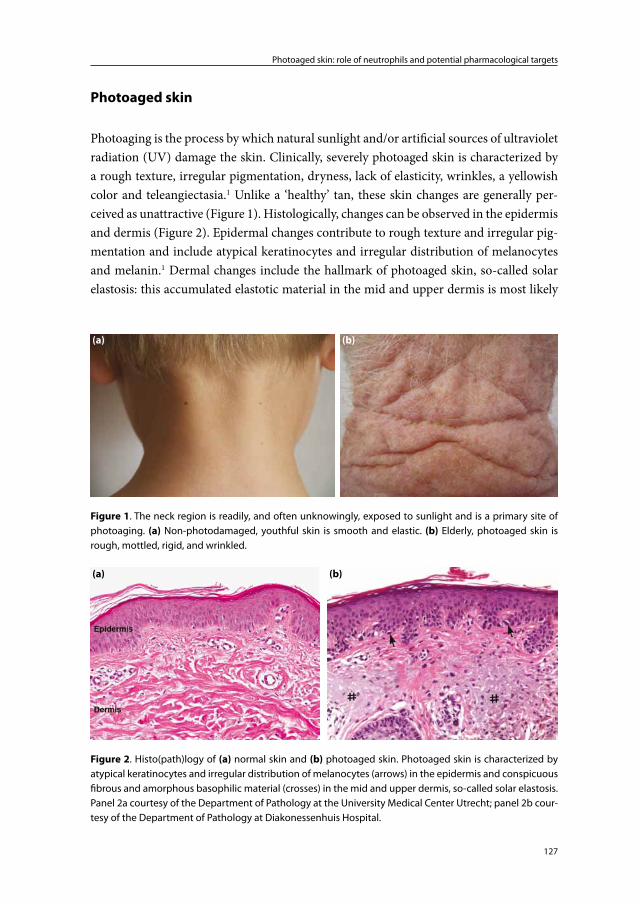
127
Photoaged skin: role of neutrophils and potential pharmacological targets
Photoaged skin
Photoaging is the process by which natural sunlight and/or artificial sources of ultraviolet radiation (UV) damage the skin. Clinically, severely photoaged skin is characterized by a rough texture, irregular pigmentation, dryness, lack of elasticity, wrinkles, a yellowish color and teleangiectasia.1 Unlike a ‘healthy’ tan, these skin changes are generally per-ceived as unattractive (Figure 1). Histologically, changes can be observed in the epidermis and dermis (Figure 2). Epidermal changes contribute to rough texture and irregular pig-mentation and include atypical keratinocytes and irregular distribution of melanocytes and melanin.1 Dermal changes include the hallmark of photoaged skin, so-called solar elastosis: this accumulated elastotic material in the mid and upper dermis is most likely
(a) (b)
Figure 1. The neck region is readily, and often unknowingly, exposed to sunlight and is a primary site of photoaging. (a) Non-photodamaged, youthful skin is smooth and elastic. (b) Elderly, photoaged skin is rough, mottled, rigid, and wrinkled.
(b)(a)
Figure 2. Histo(path)logy of (a) normal skin and (b) photoaged skin. Photoaged skin is characterized by atypical keratinocytes and irregular distribution of melanocytes (arrows) in the epidermis and conspicuous fibrous and amorphous basophilic material (crosses) in the mid and upper dermis, so-called solar elastosis. Panel 2a courtesy of the Department of Pathology at the University Medical Center Utrecht; panel 2b cour-tesy of the Department of Pathology at Diakonessenhuis Hospital.
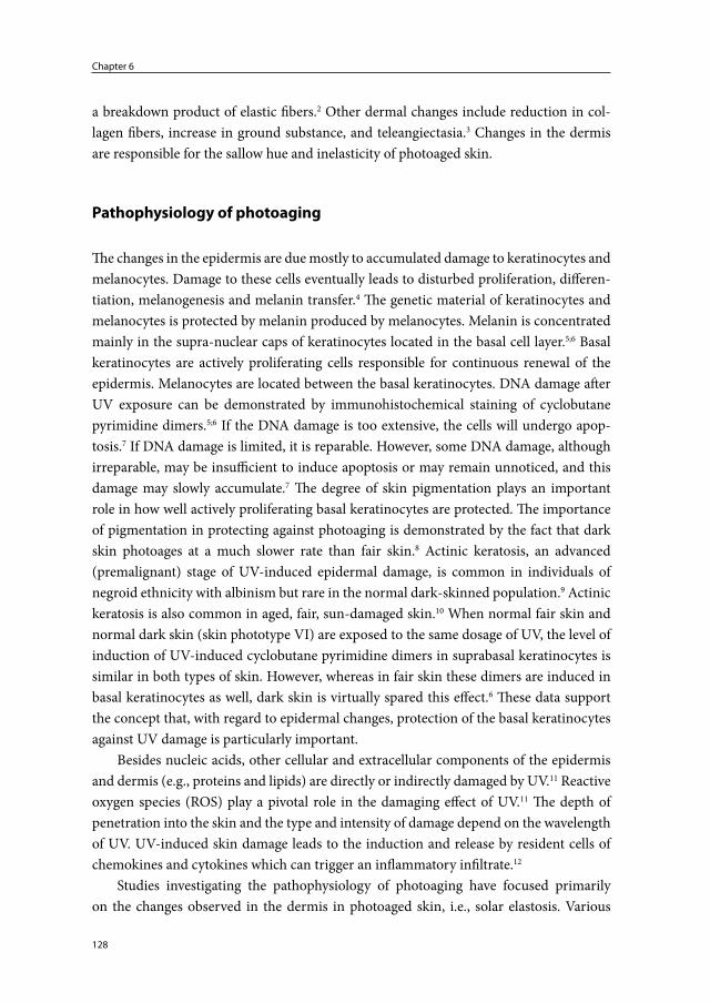
Chapter 6
128
a breakdown product of elastic fibers.2 Other dermal changes include reduction in col-lagen fibers, increase in ground substance, and teleangiectasia.3 Changes in the dermis are responsible for the sallow hue and inelasticity of photoaged skin.
Pathophysiology of photoaging
The changes in the epidermis are due mostly to accumulated damage to keratinocytes and melanocytes. Damage to these cells eventually leads to disturbed proliferation, differen-tiation, melanogenesis and melanin transfer.4 The genetic material of keratinocytes and melanocytes is protected by melanin produced by melanocytes. Melanin is concentrated mainly in the supra-nuclear caps of keratinocytes located in the basal cell layer.5;6 Basal keratinocytes are actively proliferating cells responsible for continuous renewal of the epidermis. Melanocytes are located between the basal keratinocytes. DNA damage after UV exposure can be demonstrated by immunohistochemical staining of cyclobutane pyrimidine dimers.5;6 If the DNA damage is too extensive, the cells will undergo apop-tosis.7 If DNA damage is limited, it is reparable. However, some DNA damage, although irreparable, may be insufficient to induce apoptosis or may remain unnoticed, and this damage may slowly accumulate.7 The degree of skin pigmentation plays an important role in how well actively proliferating basal keratinocytes are protected. The importance of pigmentation in protecting against photoaging is demonstrated by the fact that dark skin photoages at a much slower rate than fair skin.8 Actinic keratosis, an advanced (premalignant) stage of UV-induced epidermal damage, is common in individuals of negroid ethnicity with albinism but rare in the normal dark-skinned population.9 Actinic keratosis is also common in aged, fair, sun-damaged skin.10 When normal fair skin and normal dark skin (skin phototype VI) are exposed to the same dosage of UV, the level of induction of UV-induced cyclobutane pyrimidine dimers in suprabasal keratinocytes is similar in both types of skin. However, whereas in fair skin these dimers are induced in basal keratinocytes as well, dark skin is virtually spared this effect.6 These data support the concept that, with regard to epidermal changes, protection of the basal keratinocytes against UV damage is particularly important.
Besides nucleic acids, other cellular and extracellular components of the epidermis and dermis (e.g., proteins and lipids) are directly or indirectly damaged by UV.11 Reactive oxygen species (ROS) play a pivotal role in the damaging effect of UV.11 The depth of penetration into the skin and the type and intensity of damage depend on the wavelength of UV. UV-induced skin damage leads to the induction and release by resident cells of chemokines and cytokines which can trigger an inflammatory infiltrate.12
Studies investigating the pathophysiology of photoaging have focused primarily on the changes observed in the dermis in photoaged skin, i.e., solar elastosis. Various
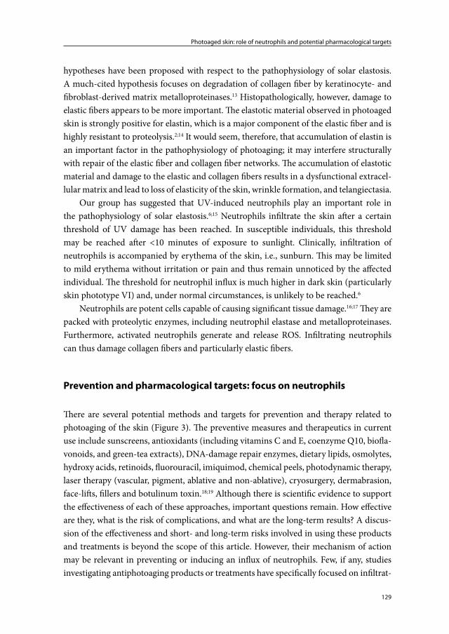
129
Photoaged skin: role of neutrophils and potential pharmacological targets
hypotheses have been proposed with respect to the pathophysiology of solar elastosis. A much-cited hypothesis focuses on degradation of collagen fiber by keratinocyte- and fibroblast-derived matrix metalloproteinases.13 Histopathologically, however, damage to elastic fibers appears to be more important. The elastotic material observed in photoaged skin is strongly positive for elastin, which is a major component of the elastic fiber and is highly resistant to proteolysis.2;14 It would seem, therefore, that accumulation of elastin is an important factor in the pathophysiology of photoaging; it may interfere structurally with repair of the elastic fiber and collagen fiber networks. The accumulation of elastotic material and damage to the elastic and collagen fibers results in a dysfunctional extracel-lular matrix and lead to loss of elasticity of the skin, wrinkle formation, and telangiectasia.
Our group has suggested that UV-induced neutrophils play an important role in the pathophysiology of solar elastosis.6;15 Neutrophils infiltrate the skin after a certain threshold of UV damage has been reached. In susceptible individuals, this threshold may be reached after <10 minutes of exposure to sunlight. Clinically, infiltration of neutrophils is accompanied by erythema of the skin, i.e., sunburn. This may be limited to mild erythema without irritation or pain and thus remain unnoticed by the affected individual. The threshold for neutrophil influx is much higher in dark skin (particularly skin phototype VI) and, under normal circumstances, is unlikely to be reached.6
Neutrophils are potent cells capable of causing significant tissue damage.16;17 They are packed with proteolytic enzymes, including neutrophil elastase and metalloproteinases. Furthermore, activated neutrophils generate and release ROS. Infiltrating neutrophils can thus damage collagen fibers and particularly elastic fibers.
Prevention and pharmacological targets: focus on neutrophils
There are several potential methods and targets for prevention and therapy related to photoaging of the skin (Figure 3). The preventive measures and therapeutics in current use include sunscreens, antioxidants (including vitamins C and E, coenzyme Q10, biofla-vonoids, and green-tea extracts), DNA-damage repair enzymes, dietary lipids, osmolytes, hydroxy acids, retinoids, fluorouracil, imiquimod, chemical peels, photodynamic therapy, laser therapy (vascular, pigment, ablative and non-ablative), cryosurgery, dermabrasion, face-lifts, fillers and botulinum toxin.18;19 Although there is scientific evidence to support the effectiveness of each of these approaches, important questions remain. How effective are they, what is the risk of complications, and what are the long-term results? A discus-sion of the effectiveness and short- and long-term risks involved in using these products and treatments is beyond the scope of this article. However, their mechanism of action may be relevant in preventing or inducing an influx of neutrophils. Few, if any, studies investigating antiphotoaging products or treatments have specifically focused on infiltrat-
Chapter 6
130
ROS
ROS proteolytic enzymes cytokines chemokines
in�ltrating neutrophils
cytokineschemokines
solar elastosis
Photoaged skin
Normal skin exposed to sunlight
DNA damage
Intervention methods
sunscreensstimulation of melanogenesis
DNA repair enzymesantioxidants
anti-in�ammatory drugsantiproteases
pharmacologicallaser
surgicalchemical
atypical keratinocytes
Figure 3. Summary of the (potential) methods of intervention. The top left schematic portrays the UV-induced events that eventually lead to photoaged skin and shows the important role infi ltrating neutro-phils play in the process. UV exposure leads to DNA damage and ROS formation, induction of chemokines and cytokines, and, ultimately, infi ltration of neutrophils. Neutrophils release potent proteolytic enzymes, ROS and proinfl ammatory chemokines and cytokines, thereby damaging the ECM. The list of intervention methods shown at the top right of the fi gure may interfere with these UV-induced events, thereby prevent-ing or slowing down the photoaging process. The bottom left schematic portrays actual photoaged skin. Photoaged skin may be treated with topical drugs, chemicals, lasers and surgical techniques. The underly-ing mechanisms of these techniques vary: physical or chemical repair and/or removal of the damaged epidermis; surgical removal of areas of full-thickness skin, thereby tightening the remaining skin; removal of underlying muscle tension (using botulinum toxin); laser ablation of telangiectasia; and others. A major remaining challenge is selective removal of the elastotic material and complete repair of the dermal ECM.ECM, extracellular matrix; ROS, reactive oxygen species.Adapted from ref. 15

131
Photoaged skin: role of neutrophils and potential pharmacological targets
ing neutrophils, mainly because the role of neutrophils has been relatively neglected in the context of the pathophysiology of photoaging. Given that neutrophils are probably important contributors to the damage to the dermis in photoaged skin, this research is necessary. For example, fractional laser treatment of the skin may provide skin tightening and a younger appearance in the short run, but a single treatment may be accompanied by an infl ux of neutrophils and multiple treatments may actually lead to accelerated photoag-ing. Apart from the necessity of studying the eff ect of current (and future) treatments on neutrophil infl ux, a focus on neutrophils also creates new opportunities: (i) UV-induced neutrophil infl ux can be used as a parameter for comparing sunscreens and other anti-photoaging products; (ii) potential new products may be evaluated with regard to their ability to infl uence UV-induced infl ux of neutrophil and/or neutrophil-derived products such as proteolytic enzymes, ROS, cytokines and chemokines; and (iii) products known to infl uence neutrophil infl ux may be investigated for possible antiphotoaging properties.
When UV-induced skin damage crosses a threshold limit, there is an infl ux of neutrophils. DNA damage, directly induced by UV radiation and UV-induced ROS, is most likely the fi rst step in a cascade of events involving receptor activation, intracellular signal transduction, and cytokine/chemokine production and release, which eventually lead to neutrophil infi ltration.11;12 Th e extent of damage to the basal keratinocytes, and perhaps to dermal components, appears to hold the key to whether an infl ammatory infi ltrate is induced.6 In addition to avoidance of exposure to UV and wearing protective clothing, the application of a sunscreen preparation to the skin is a primary intervention technique that will prevent the aforementioned cascade of events. However, depending on the UVA and UVB absorbing properties of the ingredients of the sunscreen, as well as on its protection factor, some UV will still penetrate the skin. Moreover, the user may fail to apply sunscreen evenly, or it may be washed away by perspiration or swimming. Th erefore, further developmental eff orts are required for the production of application-friendly (quick and eff ective), cosmetically pleasing, and waterproof sunscreens. In an era when discussion of the pros and cons of sun exposure and its relationship to cancer induction and prevention is still very much alive, we believe that avoiding sunburn is key to preventing photoaging. Sunburn, as mentioned above, is accompanied by the infl ux of neutrophils. Recently, Lee et al. showed that UVA can also induce a neutrophilic infi ltrate in the skin, although UVB was more active in this regard.20 Consequently, agents that are capable of absorbing both UVA and UVB are required in order to prevent neutrophils from infi ltrating the skin.
Systemic or topical use of antioxidants, DNA-damage repair enzymes, and skin pigment-inducing products should, in theory, inhibit UV-induced neutrophil infl ux. However, these products have not been investigated with respect to their eff ectiveness for this purpose. Furthermore, there have been no published reports of spectacular clinical
Chapter 6
132
results from the use of these product with regard to the prevention of photoaging or the repair of photoaged skin.
UV-induced inflammatory stimuli that attract and/or activate neutrophils include formyl peptides, interleukin-1β, tumor necrosis factor-α, granulocyte macrophage-col-ony stimulating factor, interleukin-8 (CXCL8), LTB4, and C5a,21;22 So-called end-target chemoattractants such as formyl peptides derived from mitochondria of dying cells are dominant over regulatory cell-derived attractants such as interleukin-8 and LTB4. This dominance allows neutrophils to move away from endothelium-derived chemoattractants towards their final target in the skin.22 Drugs that interfere with the synthesis, release, or function of UV-induced cytokines/chemokines may have a blocking effect on infiltration (and/or activation) of neutrophils. Neutrophils are believed to play an important role in the pathogenesis of rheumatoid arthritis, a condition that can be effectively treated with anti-tumor necrosis factor-α drugs.23;24 Similarly, anti-tumor necrosis factor-α drugs may offer protection against photoaging. However, rheumatoid arthritis is a debilitat-ing disease, whereas photoaging per se is mainly a cosmetic problem. Furthermore, in contrast to rheumatoid arthritis, for which the etiologic agent is unknown, etiologic intervention is possible in photoaging. These essential differences should be taken into account when answering the questions; how effective is the drug, what are its side-effects, risk of complications, potential severity of complications, long-term results, and costs; and what are the alternatives?
Therapeutic measures that ameliorate a sunburn reaction after excessive exposure to sunlight may also reduce neutrophil influx. However, the use and effectiveness of particu-lar drugs in sunburn management are controversial. Topical or systemic corticosteroids may not be effective if administered soon after sun exposure.25;26 Similarly, topically or systemically administered non-steroidal anti-inflammatory drugs such as acetyl-salicylic acid and indomethacin may not reduce UV-induced inflammation.27;28 Dapsone is a systemic drug commonly used in skin conditions characterized by infiltrating neutro-phils.29;30 Although its mechanism of action has not been fully elucidated, on the basis of its side-effect profile, dapsone is not a candidate drug for use in systemic antiphotoaging therapy. Nevertheless, clarification of its working mechanism could be relevant to devel-oping new antiphotoaging drugs.
ROS released in the vicinity of neutrophils during oxidative bursts may be neutral-ized by ROS scavengers. Likewise, antiproteases (e.g., elastase- and metalloproteinase-inhibitors) would be expected to reduce the damage inflicted by the arsenal of proteases released by infiltrating neutrophils.
Formerly thought to be a population of terminally differentiated leucocytes involved exclusively in acute inflammation, neutrophils are now believed to be important contribu-tors to the development of chronic inflammation as well.21 Therefore, therapies aimed at inhibiting neutrophil-derived cytokines and chemokines may be useful in the treatment
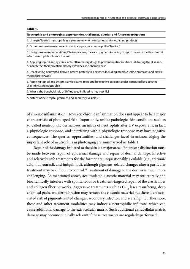
133
Photoaged skin: role of neutrophils and potential pharmacological targets
of chronic inflammation. However, chronic inflammation does not appear to be a major characteristic of photoaged skin. Importantly, unlike pathologic skin conditions such as so-called neutrophilic dermatoses, an influx of neutrophils after UV exposure is, in fact, a physiologic response, and interfering with a physiologic response may have negative consequences. The queries, opportunities, and challenges faced in acknowledging the important role of neutrophils in photoaging are summarized in Table 1.
Repair of the damage inflicted to the skin is a major area of interest: a distinction must be made between repair of epidermal damage and repair of dermal damage. Effective and relatively safe treatments for the former are unquestionably available (e.g., tretinoic acid, fluorouracil, and imiquimod), although pigment-related changes after a particular treatment may be difficult to control.31 Treatment of damage to the dermis is much more challenging. As mentioned above, accumulated elastotic material may structurally and biochemically interfere with spontaneous or treatment-targeted repair of the elastic fiber and collagen fiber networks. Aggressive treatments such as CO2 laser resurfacing, deep chemical peels, and dermabrasion may remove the elastotic material but there is an asso-ciated risk of pigment-related changes, secondary infection and scarring.32 Furthermore, these and other treatment modalities may induce a neutrophilic infiltrate, which can cause additional damage to the extracellular matrix. Such additional extracellular matrix damage may become clinically relevant if these treatments are regularly performed.
Table 1.
Neutrophils and photoaging: opportunities, challenges, queries, and future investigations
1. Using infiltrating neutrophils as a parameter when comparing antiphotoaging products
2. Do current treatments prevent or actually promote neutrophil infiltration?
3. Using sunscreen preparations, DNA-repair enzymes and pigment-inducing drugs to increase the threshold at which neutrophils infiltrate the skin
4. Applying topical and systemic anti-inflammatory drugs to prevent neutrophils from infiltrating the skin and/or counteract their proinflammatory cytokines and chemokinesa
5. Deactivating neutrophil-derived potent proteolytic enzymes, including multiple serine proteases and matrix metalloproteinasesa
6. Applying topical and systemic antioxidants to neutralize reactive oxygen species generated by activated skin-infiltrating neutrophils
7. What is the beneficial role of UV-induced infiltrating neutrophils?
aContent of neutrophil granules and secretory vesicles.33

Chapter 6
134
Acknowledgements
We thank the Departments of Pathology of the University Medical Center Utrecht and Diakonessenhuis Hospital for providing histology images.
Conflict of interest
The authors declared no conflict of interest.
135
Photoaged skin: role of neutrophils and potential pharmacological targets
References
1. Gilchrest BA. Skin aging and photoaging: an overview. J.Am.Acad.Dermatol. 1989; 21: 610-3.
2. Chen VL, Fleischmajer R, Schwartz E et al. Immunochemistry of elastotic material in sun-damaged skin. J.Invest Dermatol. 1986; 87: 334-7.
3. Oikarinen A. The aging of skin: chronoaging versus photoaging. Photodermatol.Photoim-munol.Photomed. 1990; 7: 3-4.
4. Yaar M, Gilchrest BA. Ageing and photoageing of keratinocytes and melanocytes. Clin.Exp.Dermatol. 2001; 26: 583-91.
5. Kobayashi N, Nakagawa A, Muramatsu T et al. Supranuclear melanin caps reduce ultravio-let induced DNA photoproducts in human epidermis. J.Invest Dermatol. 1998; 110: 806-10.
6. Rijken F, Bruijnzeel PL, van Weelden H et al. Responses of black and white skin to solar-simulating radiation: differences in DNA photodamage, infiltrating neutrophils, proteolytic enzymes induced, keratinocyte activation, and IL-10 expression. J.Invest Dermatol. 2004; 122: 1448-55.
7. Marrot L, Meunier JR. Skin DNA photodamage and its biological consequences. J.Am.Acad.Dermatol. 2008; 58: S139-S148.
8. Taylor SC. Skin of color: biology, structure, function, and implications for dermatologic disease. J.Am.Acad.Dermatol. 2002; 46: S41-S62.
9. Lookingbill DP, Lookingbill GL, Leppard B. Actinic damage and skin cancer in albinos in northern Tanzania: findings in 164 patients enrolled in an outreach skin care program. J.Am.Acad.Dermatol. 1995; 32: 653-8.
10. Frost CA, Green AC. Epidemiology of solar keratoses. Br.J.Dermatol. 1994; 131: 455-64.11. Trautinger F. Mechanisms of photodamage of the skin and its functional consequences for
skin ageing. Clin.Exp.Dermatol. 2001; 26: 573-7.12. Clydesdale GJ, Dandie GW, Muller HK. Ultraviolet light induced injury: immunological
and inflammatory effects. Immunol.Cell Biol. 2001; 79: 547-68.13. Fisher GJ, Wang ZQ, Datta SC et al. Pathophysiology of premature skin aging induced by
ultraviolet light. N.Engl.J.Med. 1997; 337: 1419-28.14. Debelle L, Tamburro AM. Elastin: molecular description and function. Int.J.Biochem.Cell
Biol. 1999; 31: 261-72.15. Rijken F, Bruijnzeel PL. The pathogenesis of photoaging: the role of neutrophils and
neutrophil-derived enzymes. J.Investig.Dermatol.Symp.Proc. 2009; 14: 67-72.16. Nathan C. Neutrophils and immunity: challenges and opportunities. Nat.Rev.Immunol.
2006; 6: 173-82.17. Weiss SJ. Tissue destruction by neutrophils. N.Engl.J.Med. 1989; 320: 365-76.18. Stern RS. Clinical practice. Treatment of photoaging. N.Engl.J.Med. 2004; 350: 1526-34.19. Yaar M, Gilchrest BA. Photoageing: mechanism, prevention and therapy. Br.J.Dermatol.
2007; 157: 874-87.20. Lee PL, van Weelden H, Bruijnzeel PL. Neutrophil infiltration in normal human skin after
exposure to different ultraviolet radiation sources. Photochem.Photobiol. 2008; 84: 1528-34.21. Kasama T, Miwa Y, Isozaki T et al. Neutrophil-derived cytokines: potential therapeutic
targets in inflammation. Curr.Drug Targets.Inflamm.Allergy 2005; 4: 273-9.22. Witko-Sarsat V, Rieu P, scamps-Latscha B et al. Neutrophils: molecules, functions and
pathophysiological aspects. Lab Invest 2000; 80: 617-53.
Chapter 6
136
23. Edwards SW, Hallett MB. Seeing the wood for the trees: the forgotten role of neutrophils in rheumatoid arthritis. Immunol.Today 1997; 18: 320-4.
24. Tracey D, Klareskog L, Sasso EH et al. Tumor necrosis factor antagonist mechanisms of action: a comprehensive review. Pharmacol.Ther. 2008; 117: 244-79.
25. Duteil L, Queille-Roussel C, Lorenz B et al. A randomized, controlled study of the safety and efficacy of topical corticosteroid treatments of sunburn in healthy volunteers. Clin.Exp.Dermatol. 2002; 27: 314-8.
26. Greenwald JS, Parrish JA, Jaenicke KF et al. Failure of systemically administered corticoste-roids to suppress UVB-induced delayed erythema. J.Am.Acad.Dermatol. 1981; 5: 197-202.
27. Juhlin L, Shroot B. Effect of drugs on the early and late phase UV erythema. Acta Derm.Venereol. 1992; 72: 222-3.
28. Vaananen A, Hannuksela M. UVB erythema inhibited by topically applied substances. Acta Derm.Venereol. 1989; 69: 12-7.
29. Suda T, Suzuki Y, Matsui T et al. Dapsone suppresses human neutrophil superoxide produc-tion and elastase release in a calcium-dependent manner. Br.J.Dermatol. 2005; 152: 887-95.
30. Zhu YI, Stiller MJ. Dapsone and sulfones in dermatology: overview and update. J.Am.Acad.Dermatol. 2001; 45: 420-34.
31. Berman B, Amini S, Valins W et al. Pharmacotherapy of actinic keratosis. Expert.Opin.Pharmacother. 2009; 10: 3015-31.
32. Ragland HP, McBurney E. Complications of resurfacing. Semin.Cutan.Med.Surg. 1996; 15: 200-7.
33. Faurschou M, Borregaard, N. Neutrophil granules and secretory vesicles in inflammation. Microbes Infect. 2003; 5:1317-27
Chapter 7
The protective effect of sunscreens, vitamin E 6% cream and
betamethasone 0.1% cream on solar simulating radiation-
induced erythema and neutrophil influx
Feiko Rijken1,2, Ilse C. Bihari1, Kees L.H. Guikers1, Huib van Weelden1 and Piet L.B. Bruijnzeel1,3
Department of Dermatology, University Medical Center Utrecht, Utrecht, the Netherlands1
Department of Dermatology, Diakonessenhuis, Utrecht, the Netherlands2
AstraZeneca, Loughborough (Charnwood), United Kingdom3
Submitted
Chapter 7
138
Abstract
Background Exposure of white skin (skin phototypes I-III) to erythemogenic doses of solar-simulating radiation (SSR) results in an influx of neutrophils. We have previously shown that neutrophils are a major source of enzymatically-active, photoaging-associat-ed, proteolytic enzymes, and thereby appear to be important contributors to photoaging. Preventing neutrophils from infiltrating the skin following SSR exposure should therefore limit the amount of SSR-induced skin damage.Objectives To investigate the protective effect of sunscreens, a topical antioxidant and a topical corticosteroid on SSR-induced erythema and infiltrating neutrophils.Methods Sun-protected buttock skin of six volunteers was pretreated with a broadband sunscreen (Vision SPF 28), vitamin E 6% cream and betamethasone valerate 0.1% cream, and exposed to 2 MED of SSR. Erythema was quantified using a Minolta chromameter and biopsies were taken immediately following and 24 hours after SSR exposure. Eight other volunteers underwent a similar procedure, including measurement of erythema immediately following and 24 hours after SSR-exposure, except that in this group only one control biopsy was taken and, additionally, two other sunscreens (Vision SPF 16 and Penetrase 17) were applied before SSR exposure.Results Significant prevention of SSR-induced erythema and neutrophil influx was ob-served only after pretreatment with sunscreens. Skin-application of all other products did not statistically influence SSR-induced erythema or neutrophil influx.Conclusion Only sunscreens are protective against the photoaging effects of neutrophils.
139
Protection against SSR-induced erythema and neutrophil influx
Introduction
Sunlight, more specifically ultraviolet radiation (UV), can damage human skin. Acute exposure can lead to inflammation of the skin, commonly known as a sunburn. His-topathologically, sunburned skin is characterized by multiple sunburn cells in the epi-dermis, dilated vessels in the superficial dermis and a mixed inflammatory infiltrate.1 Chronic exposure of the skin can lead to so-called photoaged skin. Chronic exposure involves multiple exposures to both erythemogenic doses of UV (i.e., sunburn) and suberythemogenic doses of UV. Photoaged skin is characterized by clinical changes such as irregular pigmentation, plaque-like thickening, loss of skin tone, deep furrowing and fine wrinkle formation. Histopathologically the dermis of photoaged skin is character-ized by an accumulation of elastotic material, disappearance of elastic fibers and reduced collagen fibers.2
Sunburned skin, including mild asymptomatic erythema, shows infiltrating neu-trophils. Depending on several factors including skin phototype, UV-spectrum and UV intensity, 10 minutes of UV exposure can be enough to induce a sunburn reaction.3 Multiple suberythemogenic doses of sunlight/UV (exposures on consecutive days) can also induce erythema and neutrophil influx (Lee et al., personal communication). Neutrophils contain and/or generate a variety of potent proteolytic enzymes and reactive oxygen species (ROS) which, when released from the cells, are capable of serious tissue damage.4 By this capacity, and since they infiltrate the skin following UV-exposure, we have hypothesized that neutrophils play a major role in the pathophysiology of photoag-ing.5 Preventing neutrophils from infiltrating the skin could thus slow down the photoag-ing process. Topically applied sunscreens, antioxidants and corticosteroids are potential candidates to prevent such neutrophil influx.
Sunscreens have been shown to prevent or reduce UV-induced erythema.6 Inorganic sunscreens contain compounds that reflect and scatter UV, organic sunscreens contain compounds or chemicals that absorb UV.7 The absorption spectrum of the latter is chemi-cal dependent: i.e., some chemicals absorb mainly UVB wavelengths, some chemicals absorb mainly UVA wavelengths and some absorb both spectra. UVB wavelengths are 600-1000 times more effective in inducing erythema or sunburn than UVA wavelengths.8 The action spectrum of photoaging in humans (i.e., the wavelengths responsible for photoaging) is difficult to determine and therefore still not entirely clear.7
UV-induced ROS play a major role in the cascade of events that eventually leads to skin inflammation.9 Vitamin E is an essential nutrient and a major lipophilic antioxidant present in plasma and tissues. Several studies have demonstrated that topical vitamin E applied onto the skin prior to UV exposure is capable of reducing acute skin responses such as erythema and edema.10;11
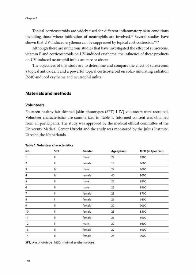
Chapter 7
140
Topical corticosteroids are widely used for different inflammatory skin conditions including those where infiltration of neutrophils are involved.12 Several studies have shown that UV-induced erythema can be suppressed by topical corticosteroids.13;14
Although there are numerous studies that have investigated the effect of sunscreens, vitamin E and corticosteroids on UV-induced erythema, the influence of these products on UV-induced neutrophil influx are rare or absent.
The objectives of this study are to determine and compare the effect of sunscreens, a topical antioxidant and a powerful topical corticosteroid on solar-simulating radiation (SSR)-induced erythema and neutrophil influx.
Materials and methods
Volunteers
Fourteen healthy fair-skinned [skin phototypes (SPT) I-IV] volunteers were recruited. Volunteer characteristics are summarized in Table 1. Informed consent was obtained from all participants. The study was approved by the medical ethical committee of the University Medical Center Utrecht and the study was monitored by the Julius Institute, Utrecht, the Netherlands.
Table 1. Volunteer characteristics
No. SPT Gender Age (years) MED (mJ per cm2)
1 III male 22 9200
2 II female 18 8600
3 IV male 24 9600
4 IV female 46 9600
5 III male 22 9200
6 III male 22 8900
7 II female 23 8700
8 I female 23 6400
9 III female 22 9000
10 II female 23 8500
11 III female 23 8900
12 II male 22 6600
13 III female 22 8900
14 III female 24 9000
SPT, skin phototype ; MED, minimal erythema dose.
141
Protection against SSR-induced erythema and neutrophil influx
SSR source, minimal erythema dose (MED) determination and measurement of erythema
CLEO Natural lamps (Philips, Eindhoven, the Netherlands) were used as a source of SSR. CLEO Natural lamps emit mostly UVA and 3% UVB. This UVA/UVB ratio is comparable to midday-sunlight during summer in Amsterdam (52º N), the Netherlands. The spec-tral energy distribution of CLEO Natural lamps ranges from 290 nm to 420 nm, with a maximum around 355 nm. Throughout the experiments the output of the CLEO Natural lamps was measured with a Waldmann UV-detector device (Waldmann, Schwenningen, Germany).
A specifically designed test device with nine 3 by 10mm windows that open consecu-tively exposing the underlying skin, was used to determine the MED.
A chromameter (Chromameter; Minolta Camera Co ltd, Osaka, Japan) was used to measure erythema of the skin. This instrument detects brown, blue and red pigments in the skin, allowing SSR-induced erythema to be quantified.
Topical products investigated
The following topical products were investigated:(1) Vision SPF 28 and Vision SPF 16 (Nycomed Nederland B.V., Breda, the Nether-
lands): broadband organic sunscreens containing both UVA and UVB filters.(2) Penetrase SPF 17 (Nycomed Nederland B.V., Breda, the Netherlands): a narrowband
organic sunscreen containing UVB filters.(3) Tocopherol-acetate 6% cream (Fagron, Nieuwerkerk a/d Ijssel, the Netherlands): a
thermostable ester of vitamin E.(4) Betamethasone-17-valerate 0.1% cream: a potent corticosteroid (and its vehicle).(5) Cetomacrogol cream (Fagron, Nieuwerkerk a/d Ijsel, the Netherlands), the vehicle
of tocopherol-acetate 6% cream.
Spectral transmission measurements
Spectral transmission through all products under investigation was performed with a calibrated standard UV-visible spectrometer (model 752, Optronic Laboratories, Or-lando, FL, USA) and 150W halogen lamp (Osram, Augsburg, Germany). Products were diluted in ethanol/ether (50/50) to a concentration of 1.5 mg/ml and transferred into a 5mm thick fused quartz cuvette, after which the measurements were performed. The chosen concentration of 1.5mg/ml is based on the normal amount of sunscreen applied onto the skin (1.5mg/cm2).
Volunteer procedures
Six of the fourteen volunteers (no. 1-6 in Table 1) underwent the following procedure: (i) MED determination; (ii) application, for 2 hours under occlusion, of Vision SPF 28,
Chapter 7
142
vitamin E 6% cream, and betamethasone 0.1% cream on three separate 2 by 2 cm areas of buttock skin; (iii) exposure to 2 MED of SSR; (iv) immediately following SSR exposure: taking of 4mm skin-biopsies from the three pretreated areas, from an untreated SSR-exposed area and from an untreated non-irradiated area; (v) 24 hours after SSR exposure: taking of 4mm skin-biopsies from the three pretreated areas and from the untreated SSR-exposed area.
The eight other volunteers (no. 7-14 in Table 1) underwent a different procedure: (i) MED was determined; (ii) Vision SPF 28, Vision SPF 16, Penetrase SPF 16, vitamin E 6% cream, betamethasone 0.1% cream and cetomacrogol cream were each applied twice (i.e., six fields on the left buttock and six fields on the right buttock) on six separate, 1 by 1 cm areas of buttock skin; (iii) one buttock was exposed to 18 000 mJ/cm² of SSR* (i.e., exposure of six pretreated areas and a seventh control area); (iv) 24 hours after SSR exposure erythema of all pretreated- and untreated-areas, SSR-exposed and non-exposed (i.e., both buttocks) was measured; (v) a single 4 mm control biopsy was taken per volun-teer from a particularly pre-treated, irradiated site to confirm the presence or absence of neutrophils in visibly erythematous or non-erythematous skin.
Biopsy processing
All obtained skin biopsies were snap-frozen in liquid nitrogen, embedded in Tissuetek (Sakura,Torrance, CA, USA) and cut into 6μm thick skin sections using a freezing micro-tome. Skin sections were mounted on Superfrost® Plus coated slides (Thermo scientific, Waltham, MA, USA), left to dry on Silicagel (MERCK, Darmstadt, Germany), and stored at -80°C.
Immunohistochemistry
Immunohistochemistry was performed to investigate the presence of neutrophils in the biopsies collected. Neutrophil elastase was used as a marker for neutrophils15;16. The im-munohistochemical staining procedure was as follows: skin sections were fixed in dry acetone containing 200μl hydrogen peroxide and pre-incubated with 10% normal human serum (NhuS)/normal horse serum (NhoS)/bovine serum albumin (BSA). Next, the sections were incubated with mouse anti-human neutrophil elastase immunoglobulin (DAKO A/S, Glostrup, Denmark; diluted 1:100 in 1% NhuS/BSA). Following a wash-ing step in phosphate-buffered saline (PBS)/Tween 0.05%, sections were incubated with biotinylated horse anti-mouse immunoglobulin (Vector, Burlingame, CA, USA; diluted 1:300 in 1% NhuS/BSA). Following another washing step in PBS/Tween 0.05%, sections were incubated with horseradish peroxidase-conjugated avidin-biotin complex (Vecta-stain ABC-HRP, Vector, diluted 1:1000 in 1% NhuS/BSA). A final washing step in PBS/
* 18 000 mJ/cm2 of SSR is equivalent to 2-3 MED in SPT I-III.
143
Protection against SSR-induced erythema and neutrophil influx
Tween 0.05% was followed by staining with 3-amino-ethyl-carbazole chromogen (Scytec, Logan, Utah, USA). Staining was halted with distilled water.
Light microscopy
Immunohistochemically stained skin sections were examined by light microscopy at 200x and 400x magnification. For quantification of cell numbers, stained cells in an area below and parallel to the dermal-epidermal junction (band-form; 0.445mm broad) were counted. Stained cells in 8 fields at 400x magnification were counted. To prevent bias from occurring during quantification of stained cells, slides were blinded and received a code. The code was broken at the end of the counting procedures. Results are expressed as mean number of positive cells per mm².
Statistical analysis
Data (erythema intensity and cell numbers) are shown as mean ± standard error of the mean. Statistical analysis was performed using GraphPad Prism 5 software (GraphPad software Inc, CA, USA). To compare the parameters under investigation a Kruskal-Wallis test with Dunn’s post-hoc multiple comparison test was performed. P values < 0.05 were considered significant. Correlation analysis was performed using a Spearman’s Rank test.
Results
Intensity of SSR-induced erythema correlates with neutrophil influx
SSR-induced neutrophil influx (Figure 1) was quantified by counting the number of neu-trophil elastase positive cells (Table 2). Correlation analysis indicated that the intensity of erythema and the number of infiltrating neutrophils are positively correlated (Spearman Rank correlation coefficient: R neutrophil erythema/elastase =0.59 (P<0.0006): i.e., an increase in erythema was associated with an increase of infiltrating neutrophils (Figure 2a).
Effect of sunscreens on SSR-induced erythema and neutrophil influx
Application of sunscreens suppresses the induction of erythema and prevents neutrophils from infiltrating the skin (Table 2, Figures 2a and 2b). Skin pretreated with Vision SPF 28, Vision SPF 16 and Penetrase SPF 17, and exposed to18 000 mJ/cm2 of SSR, showed no erythema of the skin 24 hours after irradiation (observed with the naked eye). Ery-thema measured with the chromameter appeared to show some increase in the Vision SPF 16- and Penetrase SPF 17-pretreated skin compared to Vision SPF 28 pretreated and non-irradiated skin, but the difference was not statistically significant.
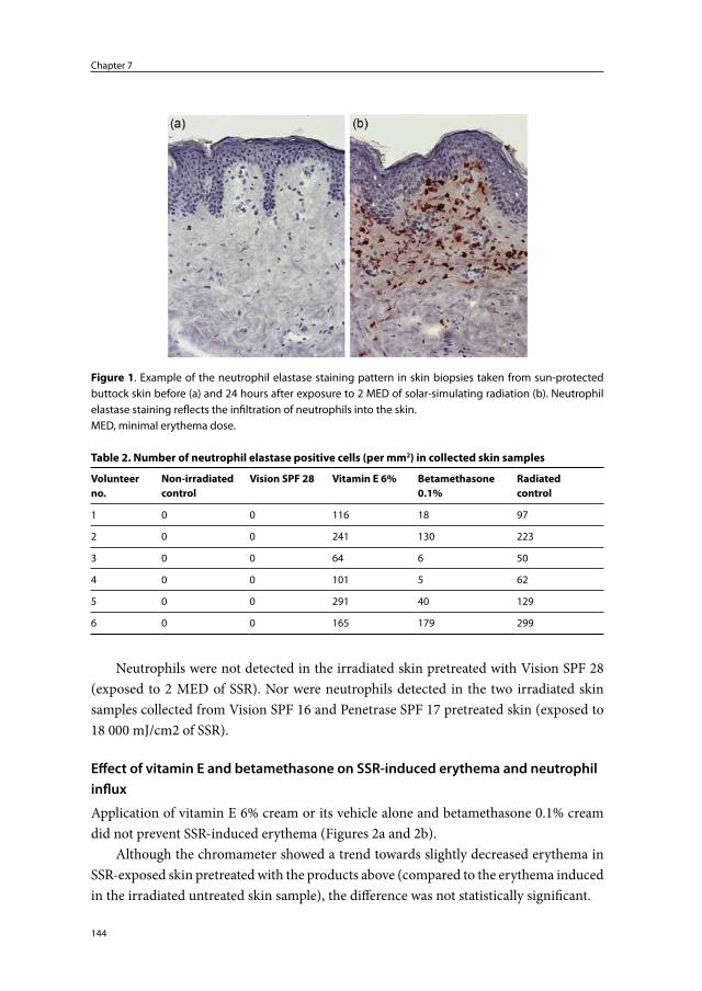
Chapter 7
144
Neutrophils were not detected in the irradiated skin pretreated with Vision SPF 28 (exposed to 2 MED of SSR). Nor were neutrophils detected in the two irradiated skin samples collected from Vision SPF 16 and Penetrase SPF 17 pretreated skin (exposed to 18 000 mJ/cm2 of SSR).
Effect of vitamin E and betamethasone on SSR-induced erythema and neutrophil influx
Application of vitamin E 6% cream or its vehicle alone and betamethasone 0.1% cream did not prevent SSR-induced erythema (Figures 2a and 2b).
Although the chromameter showed a trend towards slightly decreased erythema in SSR-exposed skin pretreated with the products above (compared to the erythema induced in the irradiated untreated skin sample), the difference was not statistically significant.
Figure 1. Example of the neutrophil elastase staining pattern in skin biopsies taken from sun-protected buttock skin before (a) and 24 hours after exposure to 2 MED of solar-simulating radiation (b). Neutrophil elastase staining reflects the infiltration of neutrophils into the skin.MED, minimal erythema dose.
Table 2. Number of neutrophil elastase positive cells (per mm2) in collected skin samples
Volunteer no.
Non-irradiated control
Vision SPF 28 Vitamin E 6% Betamethasone 0.1%
Radiated control
1 0 0 116 18 97
2 0 0 241 130 223
3 0 0 64 6 50
4 0 0 101 5 62
5 0 0 291 40 129
6 0 0 165 179 299
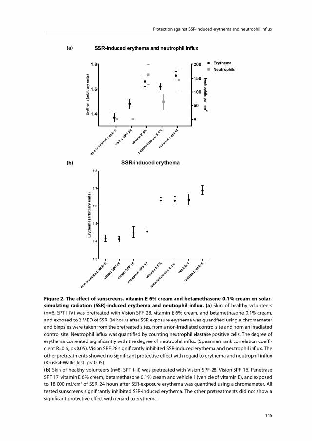
145
Protection against SSR-induced erythema and neutrophil influx
(a)
SSR-induced erythema and neutrophil influx
1.4
1.6
1.8
0
50
100
150
200 ErythemaNeutrophils
non-irrad
iated
contro
l
vision S
PF 28
vitam
in E 6%
beta
methas
one 0.1%
radiat
ed co
ntrol
Eryt
hem
a (a
rbitr
ary
units
)
Neutrophils per m
m2
(b)
SSR-induced erythema
non-irrad
iated
contro
l
vision SPF 28
vision SPF 16
penetr
ase S
PF 17
vitam
in E 6%
betameth
asone 0
.1%
vehicl
e 1
radiat
ed co
ntrol
1.3
1.4
1.5
1.6
1.7
1.8
Eryt
hem
a (a
rbitr
ary
units
)
(b)
SSR-induced erythema
non-irrad
iated
contro
l
vision SPF 28
vision SPF 16
penetr
ase S
PF 17
vitam
in E 6%
betameth
asone 0
.1%
vehicl
e 1
radiat
ed co
ntrol
1.3
1.4
1.5
1.6
1.7
1.8
Eryt
hem
a (a
rbitr
ary
units
)
Figure 2. The effect of sunscreens, vitamin E 6% cream and betamethasone 0.1% cream on solar-simulating radiation (SSR)-induced erythema and neutrophil influx. (a) Skin of healthy volunteers (n=6, SPT I-IV) was pretreated with Vision SPF-28, vitamin E 6% cream, and betamethasone 0.1% cream, and exposed to 2 MED of SSR. 24 hours after SSR exposure erythema was quantified using a chromameter and biopsies were taken from the pretreated sites, from a non-irradiated control site and from an irradiated control site. Neutrophil influx was quantified by counting neutrophil elastase positive cells. The degree of erythema correlated significantly with the degree of neutrophil influx (Spearman rank correlation coeffi-cient R=0.6, p<0.05). Vision SPF 28 significantly inhibited SSR-induced erythema and neutrophil influx. The other pretreatments showed no significant protective effect with regard to erythema and neutrophil influx (Kruskal-Wallis test: p< 0.05).(b) Skin of healthy volunteers (n=8, SPT I-III) was pretreated with Vision SPF-28, Vision SPF 16, Penetrase SPF 17, vitamin E 6% cream, betamethasone 0.1% cream and vehicle 1 (vehicle of vitamin E), and exposed to 18 000 mJ/cm2 of SSR. 24 hours after SSR-exposure erythema was quantified using a chromameter. All tested sunscreens significantly inhibited SSR-induced erythema. The other pretreatments did not show a significant protective effect with regard to erythema.
(a)
(b)
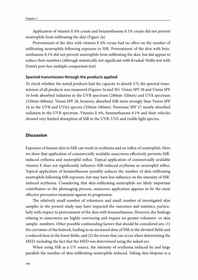
Chapter 7
146
Application of vitamin E 6% cream and betamethasone 0.1% cream did not prevent neutrophils from infiltrating the skin (Figure 2a)
Pretreatment of the skin with vitamin E 6% cream had no effect on the number of infiltrating neutrophils following exposure to SSR. Pretreatment of the skin with beta-methasone 0.1% did not prevent neutrophils from infiltrating the skin, but did appear to reduce their numbers (although statistically not significant with Kruskal-Wallis test with Dunn’s post-hoc multiple comparison test).
Spectral transmission through the products applied
To check whether the tested products had the capacity to absorb UV, the spectral trans-mission of all products was measured (Figures 3a and 3b). Vision SPF 28 and Vision SPF 16 both absorbed radiation in the UVB spectrum (280nm-320nm) and UVA spectrum (320nm-400nm). Vision SPF 28, however, absorbed SSR more strongly than Vision SPF 16 in the UVB and UVA2 spectra (320nm-340nm). Penetrase SPF 17 mostly absorbed radiation in the UVB spectrum. Vitamin E 6%, betamethasone 0.1% and their vehicles showed very limited absorption of SSR in the UVB, UVA and visible light spectra.
Discussion
Exposure of human skin to SSR can result in erythema and an influx of neutrophils. Here we show that application of commercially available sunscreens effectively prevents SSR-induced eythema and neutrophil influx. Topical application of commercially available vitamin E does not significantly influence SSR-induced erythema or neutrophil influx. Topical application of betamethasone possibly reduces the number of skin-infiltrating neutrophils following SSR exposure, but may have less influence on the intensity of SSR-induced erythema. Considering that skin-infiltrating neutrophils are likely important contributors to the photoaging process, sunscreen application appears to be the most effective preventive treatment against its progression.
The relatively small number of volunteers and small number of investigated skin samples in the present study may have impacted the outcomes and statistics, particu-larly with respect to pretreatment of the skin with betamethasone. However, the findings relating to sunscreens are highly convincing and require no greater volunteer- or skin sample- numbers. Other possible confounding factors that should be considered are; (1) the curvature of the buttock, leading to an increased dose of SSR in the elevated fields and a reduced dose in the lower fields, and (2) the errors that can occur when determining the MED, including the fact that the MED was determined using the naked eye.
When using SSR as a UV source, the intensity of erythema induced by and large parallels the number of skin-infiltrating neutrophils induced. Taking skin biopsies is a
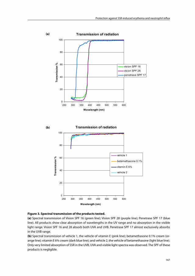
147
Protection against SSR-induced erythema and neutrophil influx
(a)
Transmission of radiation
0
20
40
60
80
100
250 300 350 400 450 500 550 600
Wavelength (nm)
Tran
smis
sion
% vision SPF 16
vision SPF 28
penetrase SPF 17
(b)
Transmission of radiation
0
20
40
60
80
100
250 300 350 400 450 500 550 600
Wavelength (nm)
Tran
smis
sion
%
vehicle 1
betamethasone 0.1%
vitamin E 6%
vehicle 2
Figure 3. Spectral transmission of the products tested.
(a) Spectral transmission of Vision SPF 16 (green line); Vision SPF 28 (purple line); Penetrase SPF 17 (blue line). All products show clear absorption of wavelengths in the UV range and no absorption in the visible light range. Vision SPF 16 and 28 absorb both UVA and UVB. Penetrase SPF 17 almost exclusively absorbs in the UVB range.(b) Spectral transmission of vehicle 1, the vehicle of vitamin E (pink line); betamethasone 0.1% cream (or-ange line); vitamin E 6% cream (dark blue line); and vehicle 2, the vehicle of betamethasone (light blue line). Only very limited absorption of SSR in the UVB, UVA and visible light spectra was observed. The SPF of these products is negligible.
(b)
(a)
Chapter 7
148
minimally invasive procedure while determining skin-erythema using a chromameter is a non-invasive procedure. Thus, the latter parameter and procedure appears to be the preferred method to compare the effectiveness of antiphotoaging products with respect to their influence on skin-infiltrating neutrophils. However, the possibility that the product under investigation may have a greater impact on neutrophil influx than on erythema (e.g., topical betamethasone in the present study) must also be taken into account. With respect to investigating antiphotoaging products, exposing skin to SSR is, in itself, not entirely harmless or without side effects. The chromameter is much more sensitive in detecting SSR-induced erythema than the naked eye and objectively quantifies the ery-thema induced. Using a chromameter thus allows investigators to apply lower doses of SSR in their experiments and allows for an accurate and unbiased quantitative analysis of erythema. Taken together, although the welfare of the volunteers is of primary concern, we advocate the use of both (chromameter determined) erythema and neutrophil influx as readouts when investigating antiphotoaging products.
When evaluating the protective effect of sunscreens we found that Vision SPF 16 and Penetrase SPF 17 equally prevented SSR-induced erythema. No neutrophils were detected in the two skin samples taken from Vision SPF 16 and Penetrase SPF 17 pretreated skin. Considering that Penetrase SPF 17 virtually only absorbs UVB (Figure 3a), these data suggest that UVB photoabsorbers are particularly important in preventing neutrophil influx. These data are in accordance with that of Lee et al.16 The UV-action spectrum of neutrophil influx appears to parallel that of the action spectrum of UV-induced erythema which is known to parallel that of UV-induced thymine dimers.17
The spectral transmission experiments are a valuable tool to exclude the possibility that the effect of a particular product on UV-induced erythema or neutrophil influx is due to the UV-absorbing properties of that product. Vitamin E 6% cream, betamethasone 0.1% cream and their vehicles showed very limited absorption of SSR. 95% transmission of SSR through these products means that they have a sun protection factor of 1.05, which is negligible. Their mode of action must therefore be locally in the skin by counteracting UV-generated ROS and inflammatory mediators.
Tocopherol-acetate, the thermostable ester form of vitamin E is marketed as an anti-(photo)aging agent. Together with directly UV-induced DNA damage, UV-induced ROS are believed to play an important role in UV-induced inflammation. Our data suggest that pretreatment of the skin with tocopherol-acetate 6% cream has little or no effect on UV-induced inflammation (i.e., erythema and neutrophil influx). Thus the ROS scaveng-ing capacity of this form of treatment is inadequate.
As mentioned above, betamethasone 0.1% cream only had fractional UV-absorbing properties and its effect is locally in the skin. Topical corticosteroids can influence down-stream factors involved in UV-induced neutrophil influx.18-20 Topical betamethasone val-erate has been shown to influence UVB-induced erythema.20 UVB-induced neutrophil
149
Protection against SSR-induced erythema and neutrophil influx
influx has been shown to be inhibited by pretreatment of the skin with clobetasol.21 Our experiments performed with a slightly less potent corticosteroid than clobetasol failed to show significant inhibition of SSR-induced neutrophil influx. As discussed above, this result may be influenced by the small number of samples investigated. Nevertheless, the effect of topical corticosteroids on neutrophil influx is obviously inferior to that of sunscreens.
In the present study we have not investigated the effect of prolonged or repetitive application. Taking into account that both tocopherol-acetate and betamethasone may need more time/applications to accumulate in the skin, and that tocopherol-acetate has to be hydrolyzed in the skin to its active form, further study is required.
In summary, application of sunscreens is the most effective treatment to prevent UV-induced erythema and neutrophil influx.
Conflict of interest
The authors state no conflict of interest.After the study was initiated Dr. Piet L.B. Bruijnzeel became employed at Organon
now Merck, Sharp & Dohme, Department of Translational Medicine, ECREM, Oss, the Netherlands. Recently he has accepted a position at AstraZeneca as Medical Science Director, Loughborough (Charnwood), United Kingdom. Organon nor Merck, Sharp & Dohme nor AstraZeneca have contributed to this manuscript, either financially or oth-erwise. Dr. Piet L.B. Bruijnzeel holds an honorary research position at the Department of Dermatology of the University Medical Center Utrecht.
Chapter 7
150
References
1. Soter NA. Acute effects of ultraviolet radiation on the skin. Semin.Dermatol. 1990; 9: 11-5.2. Yaar M. Clinical and histological features of intrinsic versus extrinsic skin aging. In: Skin
aging (Gilchrest,BA, Krutmann,J, eds), 1st edn. Springer, 2006: 9-21.3. Fitzpatrick TB. The validity and practicality of sun-reactive skin types I through VI. Arch.
Dermatol. 1988; 124: 869-71.4. Weiss SJ. Tissue destruction by neutrophils. N.Engl.J.Med. 1989; 320: 365-76.5. Rijken F, Kiekens RC, Bruijnzeel PL. Skin-infiltrating neutrophils following exposure to
solar-simulated radiation could play an important role in photoageing of human skin. Br.J.Dermatol. 2005; 152: 321-8.
6. Edwards EK, Jr., Edwards EK, Sr. Sunburn and sunscreens: an update and review. Mil.Med. 1990; 155: 381-3.
7. Yaar M, Gilchrest BA. Photoageing: mechanism, prevention and therapy. Br.J.Dermatol. 2007; 157: 874-87.
8. Parrish JA, Jaenicke KF, Anderson RR. Erythema and melanogenesis action spectra of normal human skin. Photochem.Photobiol. 1982; 36: 187-91.
9. Hruza LL, Pentland AP. Mechanisms of UV-induced inflammation. J.Invest Dermatol. 1993; 100: 35S-41S.
10. Dreher F, Maibach H. Protective effects of topical antioxidants in humans. Curr.Probl.Dermatol. 2001; 29: 157-64.
11. Thiele JJ, Hsieh SN, Ekanayake-Mudiyanselage S. Vitamin E: critical review of its current use in cosmetic and clinical dermatology. Dermatol.Surg. 2005; 31: 805-13.
12. Callen JP. Neutrophilic dermatoses. Dermatol.Clin. 2002; 20: 409-19.13. Duteil L, Queille-Roussel C, Lorenz B et al. A randomized, controlled study of the safety
and efficacy of topical corticosteroid treatments of sunburn in healthy volunteers. Clin.Exp.Dermatol. 2002; 27: 314-8.
14. Ljunggren B, Moller H. Influence of corticosteroids on ultraviolet light erythema and pigmentation in man. Arch.Dermatol.Forsch. 1973; 248: 1-12.
15. Lammers AM, van de Kerkhof PC, Schalwijk J et al. Elastase, a marker for neutrophils in skin infiltrates. Br.J.Dermatol. 1986; 115: 181-6.
16. Lee PL, van Weelden H, Bruijnzeel PL. Neutrophil infiltration in normal human skin after exposure to different ultraviolet radiation sources. Photochem.Photobiol. 2008; 84: 1528-34.
17. Young AR, Chadwick CA, Harrison GI et al. The similarity of action spectra for thymine dimers in human epidermis and erythema suggests that DNA is the chromophore for erythema. J Invest Dermatol. 1998; 111: 982-8.
18. Ahluwalia A. Topical glucocorticoids and the skin--mechanisms of action: an update. Mediators.Inflamm. 1998; 7: 183-93.
19. Norris DA, Capin L, Weston WL. The effect of epicutaneous glucocorticosteroids on hu-man monocyte and neutrophil migration in vivo. J.Invest Dermatol. 1982; 78: 386-90.
20. Kaidbey KH, Kurban AK. The influence of corticosteroids and topical indomethacin on sunburn erythema. J.Invest Dermatol. 1976; 66: 153-6.
21. Fisher GJ, Choi HC, Bata-Csorgo Z et al. Ultraviolet irradiation increases matrix metal-loproteinase-8 protein in human skin in vivo. J.Invest Dermatol. 2001; 117: 219-26.

Chapter 8
Summary of Results and General Discussion

153
Summary of Results and General Discussion
I. Introduction
Prior to commencing this thesis we searched for literature on ‘the pathophysiology of pho-toaging’ and learned that the dermal defects observed in photoaged skin were probably due to ultraviolet radiation (UV)-induced matrix metalloproteinases (MMPs) derived from fibroblasts and particularly keratinocytes1 (the hypothesis on the pathophysiology of photoaging further referred to as the ‘current hypothesis’ in this discussion). Photoag-ing is often used synonymously with solar elastosis which is the hallmark of photoaged skin and describes the dermal changes observed in photoaged skin.2 Oddly enough, the current hypothesis on the pathophysiology of photoaging focuses almost exclusively on collagen degradation, while literature on the histopathology of photoaged skin/solar elastosis focuses more on degraded elastic fibers.
Our goal at the time, was to investigate the possible contributing role of activated macrophages to the pathophysiology of photoaging. A study protocol wherein healthy volunteers were exposed to solar-simulating radiation (SSR) was initiated. The study protocol included a dose-response study and a time-course study. Multiple markers of tissue damage and tissue damaging mediators were investigated including MMPs and neutrophil elastase. By chance, a volunteer with skin phototype (SPT) VI was included in the study and this led to a switch in the cell of interest (i.e., a switch to the neutrophilic granulocyte) and, we dare say, this booklet.
II. Aims of this thesis
As stated in the general introduction the primary objectives of this thesis were:(i) To determine the role of neutrophils in the pathophysiology of photoaging.(ii) To critically examine the current hypothetical model for the pathophysiology of
photoaging.
Secondary objectives were:(i) To determine the expression, activity and origin of photoaging-associated proteo-
lytic enzymes in ‘young’ and ‘elderly’ skin, different SPTs (‘black’ and ‘white’ skin), sun-exposed and sun-protected skin, and SSR-exposed skin.
(ii) To show that in chronically sun-exposed skin elastic fiber breakdown is more im-portant than collagen breakdown.
(iii) To provide further evidence for the role of reactive oxygen species (ROS) in the pathophysiology of photoaging.
(iv) To determine the protective effect of skin pigmentation, sunscreens, topical vitamin E and betamethasone against SSR-induced erythema and neutrophil influx.
Chapter 8
154
III. Key results summarized
In this thesis we were able to show that:(i) Neutrophils infiltrate the mid and upper dermis of sun-protected buttock skin
exposed to erythemogenic doses [≥1 minimal erythemal dose (MED)] of SSR. Skin-infiltrating neutrophils express neutrophil elastase, MMP-8 and MMP-9. In situ zymography showed prominent elastase and gelatinase activity in the mid and upper dermis, indicating that neutrophil elastase and MMP-9 may actively contribute to tissue damage in the dermis. Accordingly, neutrophils can contribute to the dermal changes observed in photoaged skin (chapters 2 and 3).
(ii) MMP-1 (interstitial collagenase) which is considered to be a key player in the pro-cess of photoaging according to the current hypothesis was mainly detected in the epidermis in all SPTs. Collagenase enzyme activity was also mainly detected in the epidermis (again, all SPTs). The staining intensity of MMP-1 and collagenase activ-ity remained virtually unchanged when skin was exposed to erythemogenic doses of SSR. Our data question a major role of MMP-1 for photoaging (chapter 4).
(iii) Elastin staining was strongly increased in the mid and upper dermis of chronically sun-exposed elderly white (SPT I-III) forearm skin. Staining of collagen types I, III, IV and VII was not clearly affected in these skin samples. Subepidermal fibrillin staining appeared to be influenced by (chronic) sun exposure but also appeared to be dependent on skin location. These data indicate that elastin staining is a key marker for photoaged skin (chapter 5).
(iv) Skin biopsies taken from the center of a wrinkle in photoaged nucheal skin showed a similar staining pattern as described above. Because the neck region is a primary site of photoaging, elastin staining was even more intense and collagens I and III staining appeared reduced in areas of intense elastin staining. These findings show that the histopathology of a wrinkle does not substantially differ from that adjacent to the wrinkle and suggest that wrinkle formation is simply due to mechanical stress and inelasticity of the skin (chapter 5).
(v) Black skin (SPT VI) is protected against the damaging effects of UV. Melanin, which absorbs UV photons and scavenges UV-induced ROS, is particularly concentrated in basal keratinocytes. SSR-induced DNA damage (shown by staining of thymine dimers) was limited to the suprabasal keratinocytes demonstrating that UV does not penetrate into and past this layer in black skin. 18 000 mJ/cm2 (equivalent to 2-3 MED of white skin) failed to induce a neutrophilic infiltrate in black skin. This demonstrates that black skin is better protected against sunburn and the potential damaging effects of skin-infiltrating neutrophils. Compared to chronically sun-exposed white skin, chronically sun-exposed black skin only showed minimally
155
Summary of Results and General Discussion
increased elastin staining in the mid and upper dermis. This indicates that black skin is better protected against photoaging (chapters 2, 4 and 5).
(vi) Incubation of ‘undamaged’ (i.e., young and sun-protected) skin sections with pus, isolated neutrophils, neutrophil elastase and hydrogen peroxide induced an elastin staining pattern similar to that observed in chronically sun-exposed elderly white skin (i.e., photoaged skin). These data provide additional evidence that neutrophils may contribute to the pathophysiology of photoaging. Furthermore they indicate that UV-induced ROS and/or neutrophil-derived ROS may also directly contribute to the dermal changes observed in photoaged skin (chapter 5).
(vii) Compared to topical betamethasone and commercially available vitamin E, sun-screens are superior in preventing SSR-induced erythema and neutrophil influx. Based on the spectral absorbance of the sunscreens applied, it appeared that UVB is mostly responsible for the induction of erythema and neutrophil influx (chapter 7).
Based on the findings above and literature data, we have proposed a new model for the pathophysiology of photoaging: a model in which neutrophils, neutrophil-derived pro-teolytic enzymes and ROS play a central role (Figure 1).
IV. Our hypothesis on the pathophysiology of photoaging
Summary of our view on the photoaged skin: key characteristics, pathophysiology, and natural protection
Photoaging of human skin involves epidermal and dermal changes. The hallmark of photoaged skin is an accumulation of elastotic material in the mid and upper dermis, so-called solar elastosis. This elastotic material is mostly derived from degraded elastic fibers and consists mainly of insoluble elastin. In areas of severe solar elastosis collagen fibers are reduced and fragmented. Photoaged skin is inelastic and severe wrinkling occurs at sites of mechanical stress as the skin is continuously stretched and bent (e.g., neck and face). Neutrophil-derived mediators and proteolytic enzymes, particularly neutrophil elastase, play a key role in the development of solar elastosis (Figure 1). Neutrophil-derived MMP-9 contributes to collagen degradation. The role of keratinocyte-derived MMP-1 is insignificant and the role of MMP-1 derived from dermal cells is limited. Together with directly UV-induced DNA damage, UV-induced ROS play a major role in the cascade of events that precede UV-induced inflammation and neutrophil influx. UV-induced ROS and neutrophil-derived ROS can also contribute directly to the ECM damage observed in photoaged skin. Prevention of sunburn which is accompanied by skin-infiltrating neutrophils is key in the prevention of photoaging. Black skin is much
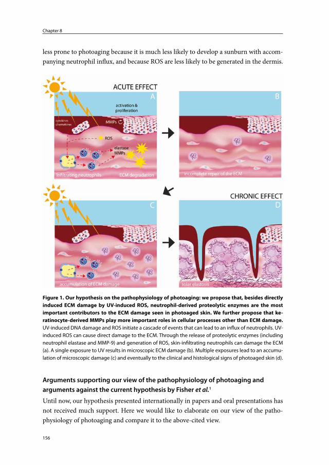
Chapter 8
156
less prone to photoaging because it is much less likely to develop a sunburn with accom-panying neutrophil influx, and because ROS are less likely to be generated in the dermis.
Arguments supporting our view of the pathophysiology of photoaging and arguments against the current hypothesis by Fisher et al.1
Until now, our hypothesis presented internationally in papers and oral presentations has not received much support. Here we would like to elaborate on our view of the patho-physiology of photoaging and compare it to the above-cited view.
Figure 1
Figure 1. Our hypothesis on the pathophysiology of photoaging: we propose that, besides directly induced ECM damage by UV-induced ROS, neutrophil-derived proteolytic enzymes are the most important contributors to the ECM damage seen in photoaged skin. We further propose that ke-ratinocyte-derived MMPs play more important roles in cellular processes other than ECM damage. UV-induced DNA damage and ROS initiate a cascade of events that can lead to an influx of neutrophils. UV-induced ROS can cause direct damage to the ECM. Through the release of proteolytic enzymes (including neutrophil elastase and MMP-9) and generation of ROS, skin-infiltrating neutrophils can damage the ECM (a). A single exposure to UV results in microscopic ECM damage (b). Multiple exposures lead to an accumu-lation of microscopic damage (c) and eventually to the clinical and histological signs of photoaged skin (d).
157
Summary of Results and General Discussion
(i) The hallmark of photoaged skin is an accumulation of elastotic material, most likely derived from degraded elastic fibers, in the mid and upper dermis. Previ-ous literature data support the concept that photoaged skin is characterized by an accumulation of degraded elastic fibers in the mid and upper dermis.3;4 As discussed in chapter 1, however, the current hypothesis focuses mainly on collagen degrada-tion: i.e., keratinocyte- and fibroblast-derived UV-induced MMPs (MMP-1, 3 and -9) degrade collagen.1 Our hypothesis focuses more on elastic fiber degradation. In chapter 5 we compared young and elderly skin, black and white skin, sun-protected and sun-exposed skin and found compelling evidence that ‘solar elastosis’ is indeed a degradation product of elastic fibers. Photoaged skin showed intense elastin staining as compared to non-photoaged skin, but did not differ significantly from non-photoaged skin with respect to collagen type I, -III, -IV, and -VII staining. Fur-thermore, we were able to induce similar elastin staining as observed in photoaged skin by incubating skin sections with neutrophil elastase, whole neutrophils and high concentrations of hydrogen peroxide.
(ii) Repetitive UV exposures lead to photoaged skin. Like Fisher et al.,1 we believe repetitive exposures to UV eventually lead to the clinical and histopathological signs of photoaging. Unlike Fisher et al.1 we believe erythemogenic doses play a major role in this process (although multiple sub-erythemogenic doses can also induce erythema of the skin, see further on), for the reason that erythemogenic doses are required for neutrophils to infiltrate the skin. Consequently, prevention of sunburn, including prevention of minimal asymptomatic erythema, is key to slowing down the photoaging process.
(iii) It is unlikely that UV-induced, keratinocyte-derived MMPs diffuse across the dermal-epidermal junction. The authors of the current hypothesis state that keratinocytes are the major source of UV-induced MMPs.5 They hypothesize that these keratinocyte-derived MMPs diffuse across the dermal-epidermal junction to the dermis where they degrade collagen fibers.1 However, when examining their original article closely, their in vivo studies show that at 24 hours after UV-exposure, MMP mRNA (e.g., MMP-1) was mostly located in the epidermis and MMP protein was also mostly observed in this compartment, while MMP enzyme activity (in situ zymography) at exactly the same time point was located mostly in the dermis (Fisher et al.1, N. Engl. J. Med., 1997, 337, p1422, Figures 1C and D). Surely one would expect to find MMP protein and MMP enzyme activity to be located in the same compartment when these are visualized at the same time point!? Addition-ally, their immunohistochemical staining of (for example) MMP-1 protein shows a sharp demarcation between the epidermis and the dermis further arguing against diffusion of this protein to the dermis. Furthermore, in severely photoaged skin
Chapter 8
158
the subepidermal grenz zone (i.e., the zone keratinocyte-derived MMPs would first pass through) appears spared of collagen damage.6 We believe it is unlikely that keratinocyte-derived MMPs are responsible for the dermal changes observed in photoaged skin. After an initial misinterpretation of some of our own results (results concerning MMP-1 expression in chapters 2 and 3, misinterpretation discussed in chapter 4), we have shown that MMP-1 protein is mostly located in the epidermis and that collagenase activity coincides with the location of MMP-1 protein (chap-ter 4). Staining of MMP-1 protein with different anti-MMP-1 clones all show the above-mentioned sharp demarcation between the intensely stained epidermis and the much less intensely stained dermis (chapter 4).
(iv) Photoaging-associated MMP-1, -2 -3 and -12 were not clearly induced by SSR. Staining patterns of MMP-1 and -3 are similar in black and white skin. Numer-ous authors, including Fisher et al.,1 have shown an induction of various MMPs by UV5;7-15 (non-exhaustive list of references cited). We investigated the presence of MMP-1, MMP-2, MMP-3, MMP-8, MMP-9 and MMP-12 in elderly and young skin, black and white skin, sun-protected and sun-exposed skin, and SSR-exposed skin (chapters 2-4). By immunohistochemistry, MMP-12 and MMP-2 were not detected in any of the skin samples. MMP-1 and MMP-3 were detected in all skin samples, particularly in the epidermis. Skin pigmentation or SSR-exposure did not clearly influence immunohistochemical staining of MMP-1 or MMP-3 (apart from some increased cellularity in the dermis but this was insignificant compared to the total amount of staining which was concentrated in the epidermis). Although immuno-histochemical staining is a less sensitive technique than, for example, western blot, one would at least expect to observe a difference in MMP-1 staining between control and SSR-exposed skin if MMP-1 plays a major role in photoaging. Furthermore, one would not expect to find significant MMP-1 staining and collagenase activity in black control skin (or white control skin, for that matter) as we did. In our stud-ies only MMP-8 and MMP-9 staining was clearly dynamic. MMP-8 and MMP-9 were induced in the dermis by erythemogenic doses of SSR. These MMPs proved to be neutrophil-derived. MMP-9 was shown to be enzymatically active. We cannot explain why other authors have found MMP-1, -3, -2 and -12 to be induced by UV, but our data plead against a major role for MMP-1, -2, -3 and -12 in photoaging. Despite, or perhaps thanks to, our initial inaccurate results with respect to MMP-1 staining, we are confident of the accurateness of our results thereafter. Since immu-nohistochemistry and in situ zymography are prone to various artefacts, measures were taken to rule out our results and conclusions being based on these artefacts: all immunohistochemical staining and in situ zymography procedures were repeated a number of times with the same skin samples; skin sections from different biopsies were placed on the same glass slide; positive and negative controls were performed;
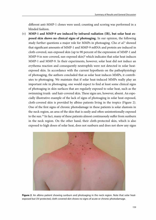
159
Summary of Results and General Discussion
different anti-MMP-1 clones were used; counting and scoring was performed in a blinded fashion.
(v) MMP-1 and MMP-9 are induced by infrared radiation (IR), but solar heat ex-posed skin shows no clinical signs of photoaging. In our opinion, the following study further questions a major role for MMPs in photoaging. Cho et al.8 showed that significant amounts of MMP-1 and MMP-9 mRNA and protein are induced in cloth-covered, sun-exposed skin (up to 90 percent of the expression of MMP-1 and MMP-9 in non-covered, sun-exposed skin)8 which indicates that solar heat induces MMP-1 and MMP-9. In their experiments, however, solar heat did not induce an erythema reaction and consequently neutrophils were not detected in solar heat-exposed skin. In accordance with the current hypothesis on the pathophysiology of photoaging, the authors concluded that as solar heat induces MMPs, it contrib-utes to photoaging. We maintain that if solar heat-induced MMPs really play an important role in photoaging, one would expect to find at least some clinical signs of photoaging in skin surfaces that are regularly exposed to solar heat, such as the swimming trunk- and hair-covered skin. These signs are, however, absent. An espe-cially illustrative example of the lack of signs of photoaging in solar heat-exposed, cloth-covered skin is provided by albino patients living in the tropics (Figure 2). One of the first signs of chronic photodamage in these patients is solar elastosis in the neck region, an area of the skin that is easily and often unintentionally exposed to the sun.16 In fact, many of these patients almost continuously suffer from sunburn in the neck region. On the other hand, their cloth-protected skin, which is also exposed to high doses of solar heat, does not sunburn and does not show any signs
Figure 2. An albino patient showing sunburn and photoaging in the neck region. Note that solar heat-exposed but UV-protected, cloth-covered skin shows no signs of acute or chronic photodamage.
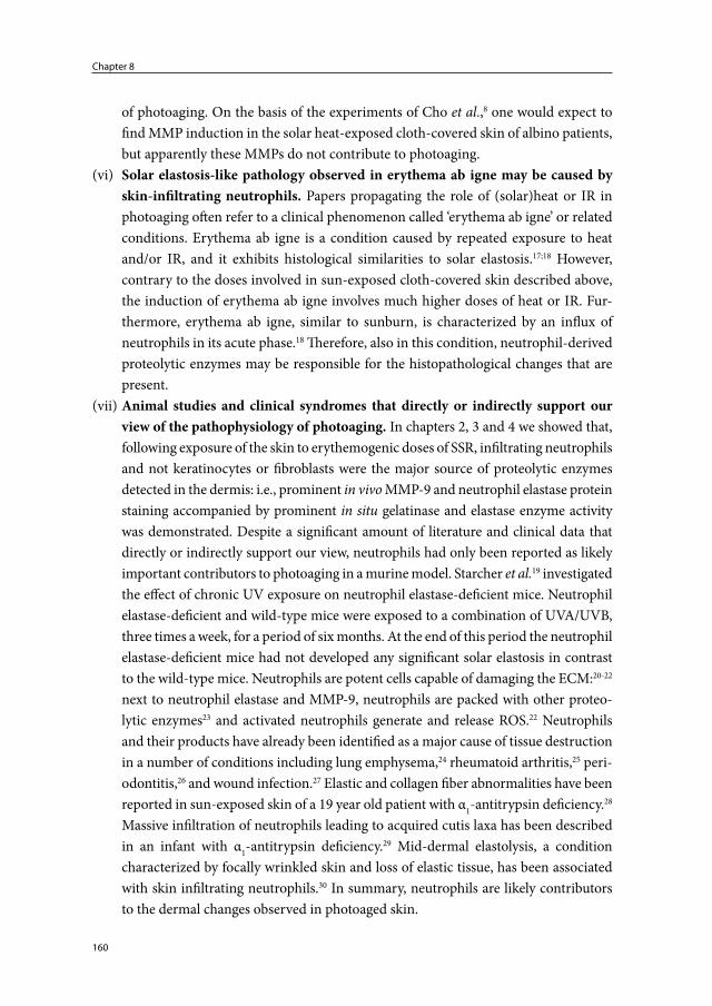
Chapter 8
160
of photoaging. On the basis of the experiments of Cho et al.,8 one would expect to find MMP induction in the solar heat-exposed cloth-covered skin of albino patients, but apparently these MMPs do not contribute to photoaging.
(vi) Solar elastosis-like pathology observed in erythema ab igne may be caused by skin-infiltrating neutrophils. Papers propagating the role of (solar)heat or IR in photoaging often refer to a clinical phenomenon called ‘erythema ab igne’ or related conditions. Erythema ab igne is a condition caused by repeated exposure to heat and/or IR, and it exhibits histological similarities to solar elastosis.17;18 However, contrary to the doses involved in sun-exposed cloth-covered skin described above, the induction of erythema ab igne involves much higher doses of heat or IR. Fur-thermore, erythema ab igne, similar to sunburn, is characterized by an influx of neutrophils in its acute phase.18 Therefore, also in this condition, neutrophil-derived proteolytic enzymes may be responsible for the histopathological changes that are present.
(vii) Animal studies and clinical syndromes that directly or indirectly support our view of the pathophysiology of photoaging. In chapters 2, 3 and 4 we showed that, following exposure of the skin to erythemogenic doses of SSR, infiltrating neutrophils and not keratinocytes or fibroblasts were the major source of proteolytic enzymes detected in the dermis: i.e., prominent in vivo MMP-9 and neutrophil elastase protein staining accompanied by prominent in situ gelatinase and elastase enzyme activity was demonstrated. Despite a significant amount of literature and clinical data that directly or indirectly support our view, neutrophils had only been reported as likely important contributors to photoaging in a murine model. Starcher et al.19 investigated the effect of chronic UV exposure on neutrophil elastase-deficient mice. Neutrophil elastase-deficient and wild-type mice were exposed to a combination of UVA/UVB, three times a week, for a period of six months. At the end of this period the neutrophil elastase-deficient mice had not developed any significant solar elastosis in contrast to the wild-type mice. Neutrophils are potent cells capable of damaging the ECM:20-22 next to neutrophil elastase and MMP-9, neutrophils are packed with other proteo-lytic enzymes23 and activated neutrophils generate and release ROS.22 Neutrophils and their products have already been identified as a major cause of tissue destruction in a number of conditions including lung emphysema,24 rheumatoid arthritis,25 peri-odontitis,26 and wound infection.27 Elastic and collagen fiber abnormalities have been reported in sun-exposed skin of a 19 year old patient with α1-antitrypsin deficiency.28 Massive infiltration of neutrophils leading to acquired cutis laxa has been described in an infant with α1-antitrypsin deficiency.29 Mid-dermal elastolysis, a condition characterized by focally wrinkled skin and loss of elastic tissue, has been associated with skin infiltrating neutrophils.30 In summary, neutrophils are likely contributors to the dermal changes observed in photoaged skin.
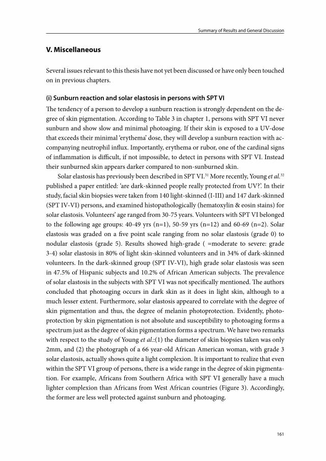
161
Summary of Results and General Discussion
V. Miscellaneous
Several issues relevant to this thesis have not yet been discussed or have only been touched on in previous chapters.
(i) Sunburn reaction and solar elastosis in persons with SPT VI
The tendency of a person to develop a sunburn reaction is strongly dependent on the de-gree of skin pigmentation. According to Table 3 in chapter 1, persons with SPT VI never sunburn and show slow and minimal photoaging. If their skin is exposed to a UV-dose that exceeds their minimal ‘erythema’ dose, they will develop a sunburn reaction with ac-companying neutrophil influx. Importantly, erythema or rubor, one of the cardinal signs of inflammation is difficult, if not impossible, to detect in persons with SPT VI. Instead their sunburned skin appears darker compared to non-sunburned skin.
Solar elastosis has previously been described in SPT VI.31 More recently, Young et al.32 published a paper entitled: ‘are dark-skinned people really protected from UV?’. In their study, facial skin biopsies were taken from 140 light-skinned (I-III) and 147 dark-skinned (SPT IV-VI) persons, and examined histopathologically (hematoxylin & eosin stains) for solar elastosis. Volunteers’ age ranged from 30-75 years. Volunteers with SPT VI belonged to the following age groups: 40-49 yrs (n=1), 50-59 yrs (n=12) and 60-69 (n=2). Solar elastosis was graded on a five point scale ranging from no solar elastosis (grade 0) to nodular elastosis (grade 5). Results showed high-grade ( =moderate to severe: grade 3-4) solar elastosis in 80% of light skin-skinned volunteers and in 34% of dark-skinned volunteers. In the dark-skinned group (SPT IV-VI), high grade solar elastosis was seen in 47.5% of Hispanic subjects and 10.2% of African American subjects. The prevalence of solar elastosis in the subjects with SPT VI was not specifically mentioned. The authors concluded that photoaging occurs in dark skin as it does in light skin, although to a much lesser extent. Furthermore, solar elastosis appeared to correlate with the degree of skin pigmentation and thus, the degree of melanin photoprotection. Evidently, photo-protection by skin pigmentation is not absolute and susceptibility to photoaging forms a spectrum just as the degree of skin pigmentation forms a spectrum. We have two remarks with respect to the study of Young et al.:(1) the diameter of skin biopsies taken was only 2mm, and (2) the photograph of a 66 year-old African American woman, with grade 3 solar elastosis, actually shows quite a light complexion. It is important to realize that even within the SPT VI group of persons, there is a wide range in the degree of skin pigmenta-tion. For example, Africans from Southern Africa with SPT VI generally have a much lighter complexion than Africans from West African countries (Figure 3). Accordingly, the former are less well protected against sunburn and photoaging.
Chapter 8
162
(ii) UV-hardening of the skin
The MED of a person’s skin can be raised by natural tanning (sun exposure) and artificial tanning (using different UV sources). Cripps33 investigated the effect of natural and arti-ficial tanning on the MED of persons with SPT I-VI: three and a half months of natural sunlight exposure increased the MED by a factor 2.33 (SD 0.048); two weeks of PUVA (mean total dose 29000 mJ/cm²) increased the MED by a factor 2.70 (SD 0.515), and; four weeks of UVB (mean total dose 3 490 mJ/cm²) increased the MED by a factor 8.01 (SD 1.86). Although a factor 2.33 is not an impressive increase for a person who develops a sunburn within 20 minutes (he/she would now develop a sunburn after approximately 45 minutes), it is a significant and functional increase for a person who normally develops a sunburn after 3 hours: that person will now be able to spend 6½ hours outdoors before getting sunburned. The mechanisms by which the MED is raised following UV-exposure have not been fully elucidated. As demonstrated by one of the vitiligo patients (patient 2) described in chapter 2b, other factors in addition to increased pigmentation of the skin must also play a role. In this patient, despite the complete absence of pigmentation on his hands, neutrophils did not infiltrate his lesional skin following exposure to 18 000 mJ/cm2 of SSR. The lesional skin of this patient showed particularly strong hyperkeratosis, sup-porting the hypothesis that increased thickness of the stratum corneum and epidermis contributes to UV-hardening of the skin. Other mechanisms underlying UV-hardening of the skin could be: attenuated signaling pathway sensitivity, increased ROS scaveng-ing, and changes in (proinflammatory) cytokine expression. In any case, both increased pigmentation and/or strong hyperkeratosis appear to be able to increase the UV-damage threshold at which neutrophils infiltrate the skin.
(iii) The effect of different UV-sources on neutrophil influx
Lee et al.34 investigated the effect of different UV sources on neutrophil influx. With respect to their potency in inducing a neutrophil influx the authors found that: SSR >
Figure 3. In general Africans from South Africa have a lighter complexion than Africans from West Africa, although both have a SPT VI. Histology shows less intense pigmentation of the epidermis in the former (a).
163
Summary of Results and General Discussion
narrowband UVB >>> broadband UVB ≈ UVA1. Based on the wavelength spectra emit-ted by the different UV sources UVB seemed to be more effective than UVA in inducing a neutrophil influx. Furthermore, longer wavelengths within the UVB range appeared mostly responsible for the infiltration of neutrophils in the UV-exposed skin.These findings support our results in chapter 7 that show that a UVB absorbing sunscreen (Penetrase SPF 17) was able to prevent SSR-induced erythema and neutrophil influx. Thus, with respect to prevention of UV-induced neutrophil influx it is particularly impor-tant to apply UVB absorbing sunscreens.
(iv) Neutrophil influx induced by erythemogenic but also suberythemogenic doses of UV
It has been established that a single erythemogenic dose of sunlight/UV is accompanied by infiltrating neutrophils.35;36 Furthermore, Novakovic et al.37 showed that exposure of white skin to 0.75 MED of SSR on four consecutive days produced the same intensity of erythema as a single exposure to 3 MED of SSR. Lee et al. (personal communication) exposed white skin to 0.5 MED of SSR on 4 consecutive days and this led to erythema and an influx of neutrophils, although in this case erythema was less intense and numbers of neutrophils were significantly smaller compared to a single exposure to 2 MED of SSR.
Multiple suberythemogenic doses of UV can also induce a neutrophil influx in mouse skin38. Most studies investigating the effect of chronic UV exposure on the animal skin, however, have not studied or focused on infiltrating neutrophils. Additionally, tissue samples in these studies may have been collected at a time point when the neutrophilic infiltrate has already resolved. Furthermore, mouse skin exposed to ‘erythemogenic’ doses of UV tends to show edema rather than erythema.39
In summary, depending on the dose (which must not be too low) and the interval (which must not be too long), multiple suberythemogenic doses of UV can also lead to erythema and skin-infiltrating neutrophils.
(v) Why have neutrophils been neglected with regard to the pathophysiology of photoaging?
Neutrophils are long known to infiltrate the skin following UV-exposure, they are known for their destructive capacity, and they are considered to be important contributors to the ECM damage observed in other conditions.20;22;24-26 Despite all this, neutrophils have received relatively little attention when it comes to photoaging of human skin.There are several possible explanations for this neglect:(1) Neutrophils cannot be recognized morphologically in frozen skin sections and may
even be difficult to recognize in paraffin-embedded skin sections when their num-bers are limited. Immunohistochemical staining with neutrophil elastase or another
Chapter 8
164
marker for neutrophils (e.g., CD66b) is often necessary to appreciate the number of infiltrating neutrophils.34;36
(2) Neutrophils and their products can only be detected in skin that has recently been exposed to UV.
(3) A certain threshold of damage is necessary for neutrophils to infiltrate the skin. This threshold, however, should not be overestimated. Depending on several factors, 15 minutes (or even less) of sun exposure can be sufficient for white-skinned persons to develop a subtle sunburn.40
(4) The current hypothesis whereby keratinocyte- and fibroblast-derived MMPs are considered responsible for the ECM damage seen in photoaged skin1 has received wide support and has probably diverted the attention from other possible mecha-nisms.
Regardless of the reasons, UV-induced skin infiltrating neutrophils obviously deserve the attention we have given them.
(vi) The role of UV-induced ROS: indirect and direct damage to the extracellular matrix
For our study in chapter 7, we attempted to visualize oxidation reaction products in SSR-exposed, treated and untreated skin. Monoclonal antibodies targeting 4-Hydroxy-2-nonenal (4-HNE) (R&D, Minneapolis, MN, USA), Malondialdehyde (MDA) (Bio connect, Huissen, the Netherlands), 8-Hydroxy-2’-deoxyguanosine (8-OHdG) (Bio connect), and Dibromo-Tyrosine (DiBrY) (Bio connect) were purchased. 4-HNE and MDA are markers of lipid peroxidation, 8-OHdG is a DNA oxidation product, and DiBrY a marker of protein oxidation caused by neutrophils. Immunohistochemical staining procedures described in the product data sheets and in literature data were carried out, but we failed to attain any convincing results. A possible explanation is that most studies examining these oxidation reaction products used paraffin embedded biopsies and we only had frozen biopsies available. Although these experiments failed, as can be deduced from Table 1 in chapter 1, ROS clearly play an important role in UV-induced inflamma-tion and the cascade of events that precede UV-induced neutrophil influx. Next to ROS induced directly by UV, neutrophils themselves are important generators of ROS.22 In chapter 5 we demonstrated that hydrogen peroxide, a ROS itself, is capable of directly inducing solar elastosis-like pathology ex vivo.
Thus, ROS play an important role in UV-induced inflammation and neutrophil influx, but also appear to be able to directly contribute to the ECM damage observed in photoaged skin.
165
Summary of Results and General Discussion
(vii) UV-induced keratinocyte-derived MMPs: extracellular matrix degradation or other (extra)cellular functions?
In our studies, MMPs were significantly induced only in skin exposed to erythemogenic doses of SSR and these MMPs were linked to infiltrating neutrophils.35;36 However, im-munohistochemistry and in situ zymography are less sensitive techniques than other detection methods such as northern blot and western blot. As mentioned earlier and in previous chapters, other authors found that MMPs are induced by sub-erythemogenic doses of UV, by a combination of IR/visible light and by solar heat alone.8;14 Clinical data do not support a major contribution of low doses of UV (e.g., sun-exposed black skin and ventral forearm skin, which are regularly exposed to suberythemogenic doses of UV, show little or no solar elastosis), IR and solar heat to the photoaging process. Here we suggest that UV- and IR-induced keratinocyte-derived MMPs may actually be involved in other cellular processes rather than ECM damage. Besides their function in ECM remodelling, MMPs are known to be involved in signal transduction and particularly chemokine and cytokine (in)activation.41;42 For example, MMP-1,-2, -3, -7, -9, -12 and -17 can activate TNF42 in vitro, MMP-1 is associated with cell proliferation,43 and MMP-9 cleaves desmoglein on apoptotic keratinocytes.44 Parks et al.42 stated that ‘matrix deg-radation is neither the sole nor the main function of MMPs’, and that ‘a specific MMP secreted by one cell type would probably carry out a different function than the same MMP produced by another cell type’.
In summary, we believe that keratinocyte-derived MMPs are involved in other cel-lular processes, such as cell-maturation, -activation, -migration and -proliferation, rather than in ECM degradation (Figure 1).
(viii) Prevention and therapy of photoaged skin
With regard to therapy of photoaged skin, retinoids are the primary drugs named in literature. The therapeutic effect of retinoids is based on: (1) clinical data, (2) the effect retinoids may have on collagen synthesis and UV-induced MMPs, and (3) the hypothesis that MMPs produced by keratinocytes and fibroblasts are key factors in the pathophysi-ology of photoaging.1;45;46 The Cochrane review discussed in chapter 1 concluded that topical tretinoin cream (a retinoid) improves skin changes (fine and coarse wrinkles, roughness, freckles and pigmentation) associated with prolonged sun exposure.45 There is no question that retinoids have a positive, albeit modest, effect on photoaged skin. However, in our opinion, the primary site of action in the skin remains a topic of discus-sion. For instance, as mentioned in chapter 1, Kossard et al.47 found that 6 months of tretinoin treatment particularly affected the epidermis and had no effect on the degree of solar elastosis.
Prior to our review in chapter 6, papers on preventive measures and therapeutics of photoaging did not focus on UV-induced neutrophil influx. As discussed in chapter
Chapter 8
166
6 we believe prevention of neutrophil influx is key to prevention of photoaging or, at least, slowing it down. Sun- or UV-avoidance, wearing of protective clothing and use of sunscreens are important and effective preventive measures in achieving the above. Data in chapter 7 demonstrate the superior effect of sunscreens compared to betameta-sone cream and commercially available vitamin E cream with respect to prevention of UV-induced neutrophil influx. Topical or systemic drugs that interfere with the cascade of events that eventually lead to neutrophil influx are potential antiphotoaging agents. However, since photoaging per se is mainly a cosmetic problem, these drugs need to be safe and cause few or no side-effects. Currently propagated and future therapies that may be or are accompanied by neutrophil influx must be viewed critically, particularly when administered or performed repetitively.
VI. Concluding remarks
The role of neutrophils in photoaging of human skin has been underrated. We believe neutrophils play an important role in the pathophysiology of photoaging. When this concept becomes more widely accepted it will undoubtedly lead to new interventions to prevent their influx and counteract their mediators. However, sun avoidance, wearing of protective clothing and application of sunscreens will remain key methods to prevent photoaging.
Acknowledgements
Parts of the general discussion including Figures 1 and 2 are derived from our article entitled ‘the pathogenesis of photoaging: the role of neutrophils and neutrophil-derived enzymes’48
We thank J.W.M. Engelen and the Regional Dermatology Training Center in Moshi, Tanzania, for providing Figure 2.
167
Summary of Results and General Discussion
References
1. Fisher GJ, Wang ZQ, Datta SC et al. Pathophysiology of premature skin aging induced by ultraviolet light. N.Engl.J.Med. 1997; 337: 1419-28.
2. Weedon D. Disorders of elastic tissue. In: Skin pathology (Weedon D, ed), 2nd edn. Sydney: Churchill Livingstone, 2002: 381-404.
3. Chen VL, Fleischmajer R, Schwartz E et al. Immunochemistry of elastotic material in sun-damaged skin. J.Invest Dermatol. 1986; 87: 334-7.
4. Matsuta M, Izaki S, Ide C et al. Light and electron microscopic immunohistochemistry of solar elastosis: a study with cutis rhomboidalis nuchae. J.Dermatol. 1987; 14: 364-74.
5. Quan T, Qin Z, Xia W et al. Matrix-degrading metalloproteinases in photoaging. J.Investig.Dermatol.Symp.Proc. 2009; 14: 20-4.
6. El-Domyati M, Attia S, Saleh F et al. Intrinsic aging vs. photoaging: a comparative histo-pathological, immunohistochemical, and ultrastructural study of skin. Exp.Dermatol. 2002; 11: 398-405.
7. Matsui MS, Hsia A, Miller JD et al. Non-sunscreen photoprotection: antioxidants add value to a sunscreen. J.Investig.Dermatol.Symp.Proc. 2009; 14: 56-9.
8. Cho S, Lee MJ, Kim MS et al. Infrared plus visible light and heat from natural sunlight participate in the expression of MMPs and type I procollagen as well as infiltration of inflammatory cell in human skin in vivo. J.Dermatol.Sci. 2008; 50: 123-33.
9. Lahmann C, Young AR, Wittern KP et al. Induction of mRNA for matrix metalloproteinase 1 and tissue inhibitor of metalloproteinases 1 in human skin in vivo by solar simulated radiation. Photochem.Photobiol. 2001; 73: 657-63.
10. Saarialho-Kere U, Kerkela E, Jeskanen L et al. Accumulation of matrilysin (MMP-7) and macrophage metalloelastase (MMP-12) in actinic damage. J.Invest Dermatol. 1999; 113: 664-72.
11. Chung JH, Seo JY, Lee MK et al. Ultraviolet modulation of human macrophage metal-loelastase in human skin in vivo. J.Invest Dermatol. 2002; 119: 507-12.
12. Fisher GJ, Choi HC, Bata-Csorgo Z et al. Ultraviolet irradiation increases matrix metal-loproteinase-8 protein in human skin in vivo. J.Invest Dermatol. 2001; 117: 219-26.
13. Koivukangas V, Kallioinen M, utio-Harmainen H et al. UV irradiation induces the expres-sion of gelatinases in human skin in vivo. Acta Derm.Venereol. 1994; 74: 279-82.
14. Fisher GJ, Datta SC, Talwar HS et al. Molecular basis of sun-induced premature skin ageing and retinoid antagonism. Nature 1996; 379: 335-9.
15. Scharffetter K, Wlaschek M, Hogg A et al. UVA irradiation induces collagenase in human dermal fibroblasts in vitro and in vivo. Arch.Dermatol.Res. 1991; 283: 506-11.
16. Lookingbill DP, Lookingbill GL, Leppard B. Actinic damage and skin cancer in albinos in northern Tanzania: findings in 164 patients enrolled in an outreach skin care program. J.Am.Acad.Dermatol. 1995; 32: 653-8.
17. Chan CC, Chiu HC. Images in clinical medicine. Erythema ab igne. N.Engl.J Med. 2007; 356: e8.
18. Finlayson GR, Sams WM, Jr., Smith JG, Jr. Erythema ab igne: a histopathological study. J.Invest Dermatol. 1966; 46: 104-8.
19. Starcher B, Conrad M. A role for neutrophil elastase in solar elastosis. Ciba Found.Symp. 1995; 192: 338-46.
20. Janoff A. Neutrophil proteases in inflammation. Annu.Rev.Med. 1972; 23: 177-90.
Chapter 8
168
21. Nathan C. Neutrophils and immunity: challenges and opportunities. Nat.Rev.Immunol. 2006; 6: 173-82.
22. Weiss SJ. Tissue destruction by neutrophils. N.Engl.J.Med. 1989; 320: 365-76.23. Faurschou M, Borregaard N. Neutrophil granules and secretory vesicles in inflammation.
Microbes.Infect. 2003; 5: 1317-27.24. Gadek JE. Adverse effects of neutrophils on the lung. Am.J.Med. 1992; 92: 27S-31S.25. Edwards SW, Hallett MB. Seeing the wood for the trees: the forgotten role of neutrophils in
rheumatoid arthritis. Immunol.Today 1997; 18: 320-4.26. Golub LM, Sorsa T, Lee HM et al. Doxycycline inhibits neutrophil (PMN)-type matrix me-
talloproteinases in human adult periodontitis gingiva. J.Clin.Periodontol. 1995; 22: 100-9.27. Singer AJ, McClain SA. Persistent wound infection delays epidermal maturation and
increases scarring in thermal burns. Wound.Repair Regen. 2002; 10: 372-7.28. Ledoux-Corbusier M, Achten G. alpha1-Antitrypsin deficiency and skin abnormalities.
J.Cutan.Pathol. 1975; 2: 25-9.29. Hwang ST, Williams ML, McCalmont TH et al. Sweet’s syndrome leading to acquired cutis
laxa (Marshall’s syndrome) in an infant with alpha 1-antitrypsin deficiency. Arch.Dermatol. 1995; 131: 1175-7.
30. Lewis KG, Dill SW, Wilkel CS et al. Mid-dermal elastolysis preceded by acute neutrophilic dermatosis. J.Cutan.Pathol. 2004; 31: 72-6.
31. Nurnberger F, Schober E, Marsch WC et al. Actinic elastosis in black skin. A light- and electronmicroscopic study. Arch.Dermatol.Res. 1978; 262: 7-14.
32. Young AL, Levy S, Nighland M et al. Are dark-skinned people really protected from ultra-violet radiation? Clin.Exp.Dermatol. 2010; 35: 392-6.
33. Cripps DJ. Natural and artificial photoprotection. J.Invest Dermatol. 1981; 77: 154-7.34. Lee PL, van Weelden H, Bruijnzeel PL. Neutrophil infiltration in normal human skin after
exposure to different ultraviolet radiation sources. Photochem.Photobiol. 2008; 84: 1528-34.35. Rijken F, Bruijnzeel PL, van Weelden H et al. Responses of black and white skin to solar-
simulating radiation: differences in DNA photodamage, infiltrating neutrophils, proteolytic enzymes induced, keratinocyte activation, and IL-10 expression. J.Invest Dermatol. 2004; 122: 1448-55.
36. Rijken F, Kiekens RC, Bruijnzeel PL. Skin-infiltrating neutrophils following exposure to solar-simulated radiation could play an important role in photoageing of human skin. Br.J.Dermatol. 2005; 152: 321-8.
37. Novakovic L, Lee S, Orchard GE et al. Effects of solar-simulated radiation dose fraction-ation on CD1a+ Langerhans cells and CD11b+ macrophages in human skin. Br.J.Dermatol. 2001; 145: 237-44.
38. Takeuchi H, Gomi T, Shishido M et al. Neutrophil elastase contributes to extracellular ma-trix damage induced by chronic low-dose UV irradiation in a hairless mouse photoaging model. J Dermatol.Sci. 2010; 60: 151-8.
39. van Weelden H, de Gruijl FR, van der Putte SC et al. The carcinogenic risks of modern tanning equipment: is UV-A safer than UV-B?. Arch.Dermatol.Res. 1988; 280: 300-7.
40. Samanek AJ, Croager EJ, Giesfor Skin Cancer PP et al. Estimates of beneficial and harmful sun exposure times during the year for major Australian population centres. Med.J.Aust. 2006; 184: 338-41.
41. Pardo A, Selman M. MMP-1: the elder of the family. Int.J.Biochem.Cell Biol. 2005; 37: 283-8.
169
Summary of Results and General Discussion
42. Parks WC, Wilson CL, Lopez-Boado YS. Matrix metalloproteinases as modulators of inflammation and innate immunity. Nat.Rev.Immunol. 2004; 4: 617-29.
43. Limb GA, Matter K, Murphy G et al. Matrix metalloproteinase-1 associates with intra-cellular organelles and confers resistance to lamin A/C degradation during apoptosis. Am.J.Pathol. 2005; 166: 1555-63.
44. Cirillo N, Femiano F, Gombos F et al. Metalloproteinase 9 is the outer executioner of desmoglein 3 in apoptotic keratinocytes. Oral Dis. 2007; 13: 341-5.
45. Samuel, M., Brooke, R. C. C., and Griffiths, C. E. M. Interventions for photodamaged skin. The Cochrane Library, Wiley, 2007; 4: 1-101.
46. Griffiths CE. The role of retinoids in the prevention and repair of aged and photoaged skin. Clin.Exp.Dermatol. 2001; 26: 613-8.
47. Kossard S, Anderson P, Davies A et al. Histological evaluation of the effect of 0.05% tretinoin in the treatment of photo damaged skin. Geographic differences in elastosis in baseline biopsies. Australas.J.Dermatol. 1993; 34: 89-95.
48. Rijken F, Bruijnzeel PL. The pathogenesis of photoaging: the role of neutrophils and neutrophil-derived enzymes. J.Investig.Dermatol.Symp.Proc. 2009; 14: 67-72.


Nederlandse Samenvatting

173
Nederlandse Samenvatting
Introductie
Voordat wij begonnen aan het onderzoek dat opgetekend is in dit proefschrift werd de literatuur over huidveroudering onder invloed van zonlicht (zogenaamde ‘photoaging’) bestudeerd. Hierin wordt beschreven dat de histologische afwijkingen aangetroffen in de huid in dit kader waarschijnlijk worden veroorzaakt door matrix metalloproteinases (MMPs) afkomstig van fibroblasten en met name keratinocyten. Deze breed gedragen hypothese, geïntroduceerd door de groep van Fisher en Voorhees, richt zich met name op de afbraak van collageenvezels. Echter, de histologie van de door zonlicht beschadigde huid wordt vooral gekenmerkt door een opstapeling van ‘elastotisch’ materiaal dat ver-moedelijk afkomstig is van beschadigde elastische vezels. De opstapeling van elastotisch materiaal in het dermale compartiment van de huid wordt ‘solaire elastose’ genoemd. In de literatuur worden de begrippen ‘solaire elastose’ en ‘photoaging’ vaak als synoniemen van elkaar gebruikt. In feite is ‘photoaging’ een breder begrip daar ook de door zon-licht geïnduceerde epidermale veranderingen hieronder vallen. ‘Photoaged’ huid wordt klinisch gekenmerkt door onregelmatige pigmentatie, teleangiëctasiëen, plaque-achtige verdikking, verruwing van de huid en rimpelvorming.
Toen wij begonnen met het onderzoek was ons doel de mogelijke rol van geacti-veerde macrofagen in de pathofysiologie van ‘photoaging’ te bepalen. Een studie waarin gezonde vrijwilligers werden blootgesteld aan ultraviolet straling (UV) werd geïnitieerd. De studie omvatte een dosisrespons studie en een studie waarin vrijwilligers werden blootgesteld aan een vaste dosis UV waarna op verschillende tijdstippen huidbiopten werden afgenomen. Een reeks van markers voor specifieke celtypes, weefselbeschadiging en weefselbeschadigende stoffen, inclusief MMPs en neutrofiel elastase, werd onderzocht. Neutrofiel elastase is een krachtig enzym dat meerdere componenten van de extracel-lulaire matrix (o.a., elastische vezels en collageenvezels) kan beschadigen en het is een belangrijke marker voor neutrofiele granulocyten. Het feit dat dit proefschrift hier ligt is mede te danken aan het destijds breed inzetten van markers en het per toeval includeren van een vrijwilliger met een zeer donkere huid. De observaties die wij toen deden leidden er toe dat onze aandacht verschoof van de macrofaag naar de neutrofiele granulocyt. Neutrofiele granulocyten zijn potente cellen die tal van weefselbeschadigende producten kunnen vrijmaken. Zij worden reeds in verband gebracht met het ontstaan van weefsel-schade in aandoeningen zoals longemfyseem en reumatoïde artritis.
Doelstellingen
De primaire doelstellingen van dit proefschrift zijn:
174
(i) De rol van neutrofiele granulocyten in de pathofysiologie van ‘photoaging’ te bepa-len.
(ii) De huidige, bovengenoemde hypothese over de pathofysiologie van ‘photoaging’ kritisch onder de loep te nemen.
Secundaire doelstellingen zijn:(i) De expressie, activiteit en bron van de met ‘photoaging’ geassocieerde proteolytische
enzymen te onderzoeken in ‘jonge’ en ‘oude’ huid, ‘blanke’ en ‘zwarte’ huid (huid fo-totype volgens Fitzpatrick I-III en huid fototype VI), zonblootgestelde (buitenzijde onderarm) en zonbeschermde (bil) huid en UV* blootgestelde huid.
(ii) Aantonen dat de afbraak van elastische vezels belangrijker is dan afbraak van col-lageen in ‘photoaging’.
(iii) De rol van reactieve zuurstofdeeltjes in de pathofysiologie van ‘photoaging’ verder te onderbouwen.
(iv) Het beschermende effect van huidpigment, anti-zonnebrandmiddelen, vitamine E (een antioxidant) en betamethason (een hormoon preparaat en krachtige ontste-kingsremmer) te bepalen.
Resultaten
In dit proefschrift tonen wij het volgende aan:(i) Neutrofiele granulocyten infiltreren de huid wanneer deze wordt blootgesteld aan
een dosis UV die tot erytheem leidt [een dosis ≥1 minimale erytheem dosis (MED)]. Neutrofiele granulocyten brengen neutrofiel elastase, MMP-8 en MMP-9 tot expres-sie. In situ zymografie laat opvallende elastase1 en gelatinase2 enzymactiviteit zien in eenzelfde patroon als de immuunhistochemische aankleuring van huidinfiltrerende neutrofiele granulocyten. Deze data suggereren dat neutrofiel elastase en MMP-9 afkomstig van neutrofiele granulocyten een actieve bijdrage kunnen leveren aan UV geïnduceerde dermale schade en pleiten voor een rol van neutrofiele granulocyten in de pathofysiologie van ‘photoaging’ (zie hoofdstukken 2 en 3).
(ii) MMP-1, de matrix metalloproteinase die in de hypothese van Fisher en Voorhees een centrale rol speelt, wordt vooral gedetecteerd in de epidermis, zowel in blanke als in zwarte huid. Collagenase3 enzymactiviteit wordt ook voornamelijk gedetecteerd in de epidermis (wederom eenzelfde patroon in blanke en zwarte huid). Blootstelling aan UV heeft vrijwel geen invloed op de immuunhistochemische aankleuring van
* Als UV bron maakten wij gebruik van een zogenaamde ‘solar simulator’: een UV bron die zonlicht benadert qua verhoudingen UVB en UVA golflengtes.1,2,3 Weerspiegeling van respectievelijk neutrofiel elastase, MMP-9 en MMP-1 enzym activiteit.
175
Nederlandse Samenvatting
MMP-1 noch op collagenase enzymactiviteit. Deze data plaatsen vraagtekens bij de essentiële rol van MMP-1 in de pathofysiologie van ‘photoaging’ (zie hoofdstuk 4).
(iii) Elastische vezels bestaan grofweg uit een kern van elastine omgeven door een huls van fibrillines. In hoofdstuk 5 tonen wij aan dat immuunhistochemische aankleuring van elastine in de dermis sterk is toegenomen in de door zonlicht beschadigde, oude, blanke huid. Immuunhistochemische aankleuring van verschillende types collageen (I, III, IV en VII) laat geen opvallende verschillen zien tussen de verzamelde huid-biopten (behalve verminderde collageen I en III aankleuring in gebieden met zeer sterke toename van elastine). Subepidermale fibrilline aankleuring lijkt te worden beïnvloed door (chronische) zonblootstelling maar lijkt ook afhankelijk te zijn van de locatie van het afgenomen biopt. Deze data pleiten voor het gebruik van elastine als marker voor ‘photoaged’ huid.
(iv) Huidbiopten afgenomen uit het midden van een rimpel in de nek (oude, blanke, zonblootgestelde huid) laten een vergelijkbaar aankleuringspatroon van elastine, fibrilline en verschillende types collageenvezels zien als hierboven beschreven. Echter, de nek is een primaire locatie voor zonbeschadiging (bij mannen vrijwel altijd blootgesteld aan de zon) met als gevolg dat de elastine aankleuring hier nog meer uitgesproken is. Zoals reeds gemeld gaan gebieden met zeer sterke elastine aankleuring gepaard met verminderde aankleuring van collageen types I en III. Deze bevindingen bevestigen het nut van elastine als marker voor ‘photoaging’ en tonen aan dat de histologie van de huid in het midden van een rimpel niet wezenlijk verschilt van die naast de rimpel. Waarschijnlijk ontstaan rimpels eenvoudigweg als gevolg van verlies van elasticiteit van de huid en herhaaldelijke mechanische stress (zie hoofdstuk 5).
(v) Zwarte huid (huid fototype VI) is beter beschermd tegen de schadelijke effecten van UV. Melanine pigment, dat UV fotonen absorbeert en antioxiderende eigenschap-pen heeft, is vooral geconcentreerd ter hoogte van de basale keratinocyten (boven de celkernen). UV geïnduceerde DNA schade in zwarte huid (aangetoond door aan-kleuring van thymine dimeren) wordt vrijwel uitsluitend aangetroffen in de kernen van suprabasale keratinocyten wat aantoont dat UV in zwarte huid tot aan en niet voorbij de basale keratinocyten komt. 18 000 mJ/cm2 (gelijk aan 2-3 MED in blanke huid) is ontoereikend om een neutrofiel ontstekingsinfiltraat te induceren. Deze data tonen aan dat de zwarte huid beter beschermd is tegen UV/zonverbranding en tegen infiltratie en de potentieel schadelijke effecten van neutrofiele granulocyten. Vergeleken met oude, chronisch zonblootgestelde, blanke huid laat oude, chronisch zonblootgestelde, zwarte huid slechts een geringe toename van elastine in de dermis zien. Dit wijst er op dat zwarte huid beter beschermd is tegen ‘photoaging’ dan blanke huid (zie hoofdstukken 2, 4 en 5).
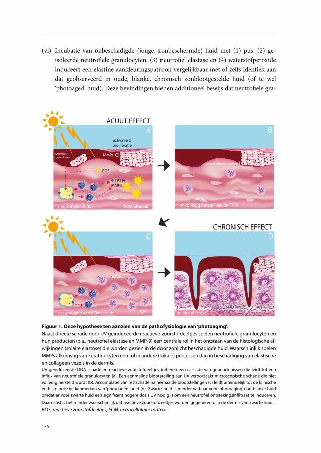
176
(vi) Incubatie van onbeschadigde (jonge, zonbeschermde) huid met (1) pus, (2) ge-isoleerde neutrofiele granulocyten, (3) neutrofiel elastase en (4) waterstofperoxide induceert een elastine aankleuringspatroon vergelijkbaar met of zelfs identiek aan dat geobserveerd in oude, blanke, chronisch zonblootgestelde huid (of te wel ‘photoaged’ huid). Deze bevindingen bieden additioneel bewijs dat neutrofiele gra-
onvolledig herstel van de ECM
ROS
MMPs
elastase MMPs
neutroelen in�ux ECM afbraak
activatie &proliferatie
cytokineschemokines
accumulatie van ECM schade solaire elastose
A B
DCCHRONISCH EFFECT
ACUUT EFFECT
basale keratinocyten {
Figuur 1. Onze hypothese ten aanzien van de pathofysiologie van ‘photoaging’.Naast directe schade door UV geïnduceerde reactieve zuurstofdeeltjes spelen neutrofiele granulocyten en hun producten (o.a., neutrofiel elastase en MMP-9) een centrale rol in het ontstaan van de histologische af-wijkingen (solaire elastose) die worden gezien in de door zonlicht beschadigde huid. Waarschijnlijk spelen MMPs afkomstig van keratinocyten een rol in andere (lokale) processen dan in beschadiging van elastische en collageen vezels in de dermis.UV geïnduceerde DNA schade en reactieve zuurstofdeeltjes initiëren een cascade van gebeurtenissen die leidt tot een influx van neutrofiele granulocyten (a). Een eenmalige blootstelling aan UV veroorzaakt microscopische schade die niet volledig hersteld wordt (b). Accumulatie van restschade na herhaalde blootstellingen (c) leidt uiteindelijk tot de klinische en histologische kenmerken van ‘photoaged’ huid (d). Zwarte huid is minder vatbaar voor ‘photoaging’ dan blanke huid omdat er voor zwarte huid een significant hogere dosis UV nodig is om een neutrofiel ontstekingsinfiltraat te induceren.
Daarnaast is het minder waarschijnlijk dat reactieve zuurstofdeeltjes worden gegenereerd in de dermis van zwarte huid.
ROS, reactieve zuurstofdeeltjes; ECM, extracellulaire matrix.
177
Nederlandse Samenvatting
nulocyten kunnen bijdragen aan de pathofysiologie van ‘photoaging’. Voorts tonen deze gegevens aan dat UV geïnduceerde reactieve zuurstofdeeltjes en/of reactieve zuurstofdeeltjes gegenereerd door neutrofiele granuloctyten mogelijk rechtstreeks (door oxidatie van elastische vezels) kunnen bijdragen aan de dermale veranderin-gen die gezien worden in de door zonlicht beschadigde huid (zie hoofdstuk 5).
(vii) Vergeleken met een commercieel verkrijgbaar vitamine E preparaat en topicaal be-tamethason zijn anti-zonnebrandmiddelen superieur wat betreft preventie van UV geïnduceerd erytheem en neutrofielen influx. Op basis van de spectrale absorptie van de bestudeerde anti-zonnebrandmiddelen lijkt het er op dat UVB hoofdzakelijk verantwoordelijk is voor inductie van erytheem en neutrofielen influx (hoofdstuk 7).
Op basis van de bovenstaande bevindingen en literatuurgegevens hebben wij een nieuw model voor de pathofysiologie van ‘photoaging’ ontwikkeld. In dit model spelen neutro-fiele granulocyten, proteolytische enzymen afkomstig van neutrofiele granulocyten en reactieve zuurstofdeeltjes een centrale rol (Figuur 1).
Discussie
Wat pleit voor onze hypothese en wat maakt de hypothese van Fisher en Voorhees minder waarschijnlijk? Waarom hebben neutrofiele granulocyten zo weinig aandacht gekregen in het kader van ‘photoaging’ terwijl het uitermate potente cellen zijn en ze de huid bin-nentrekken na UV blootstelling? Waarom is het onwaarschijnlijk dat MMP-1 vanuit de epidermis door de basale membraan naar de dermis diffundeert? Wat is de mogelijke functie van MMP-1 in plaats van het berokkenen van weefselschade in de dermis? Deze vragen en meer worden besproken in hoofdstuk 8.
Conclusies
De rol van neutrofiele granulocyten in het kader van veroudering van de huid onder in-vloed van zonlicht is ondergewaardeerd. Wij bepleiten een belangrijke rol voor neutrofiele granulocyten in de pathofysiologie van ‘photoaging’. Wanneer dit concept geaccepteerd wordt zal dat ongetwijfeld leiden tot nieuwe interventies om influx van neutrofielen in de huid te voorkomen en hun producten te neutraliseren. Deze nieuwe kansen worden besproken in hoofdstuk 6. Niettemin zullen ook dan het vermijden van zonblootstelling, dragen van beschermende kleding en gebruik van anti-zonnebrandmiddelen de belang-rijkste maatregelen zijn om ‘photoaging’ tegen te gaan.


Dankwoord

181
Dankwoord
Het zit er bijna op. Zoals de meeste (alle?) promovendi moet ik zeggen dat het een enorm pak van mijn hart is. Rest mij nog de verdediging van het proefschrift en, niet onbelang-rijk, dit dankwoord.
Mijn onderzoekstraject op de afdeling dermatologie in het UMC Utrecht is onlosma-kelijk verbonden met mijn opleiding tot dermatoloog. Hoewel, ten aanzien van beiden, het niet altijd rozengeur en maneschijn was, overweegt een zeer positief gevoel over al die jaren in het UMC Utrecht. Nu is er de gelegenheid om mensen te bedanken. Ongetwijfeld zal ik personen vergeten te noemen of doet wat ik zeg geen recht aan mijn waardering voor deze en gene. Talloze mensen hebben mij sinds het begin van mijn onderzoekscar-rière, maar ook daarvoor, op allerlei manieren geïnspireerd. De meeste hebben weinig te maken gehad met het onderzoek zelf maar hebben toch indirect een bijdrage geleverd aan mijn ontwikkeling. Ik denk en hoop dat ik, mede dankzij al deze mensen, kritischer ben gaan nadenken, meer oog heb gekregen voor het standpunt van anderen, ijveriger ben geworden en meer doorzettingsvermogen heb gekregen.
Een onmisbare groep mensen voor het onderzoek zijn de vrijwilligers. Dank voor hun bereidwilligheid om aan het onderzoek mee te doen.
Carla, jij hebt mij alle kansen gegeven om mijzelf te ontwikkelen als onderzoeker en als assistent in opleiding. Jij gaf mij enorm veel vrijheid en vertrouwen, waarvoor ik je zeer dankbaar ben. Altijd positief en opbouwend, nooit onvriendelijk. Ik had mij geen betere promotor en opleider kunnen wensen!
Piet, co-promotor, zonder jou was dit proefschrift er niet gekomen. Jij zette ‘photo-aging’ op de onderzoeksagenda van de afdeling dermatologie van het UMC Utrecht en hebt mij al die jaren begeleid. Jouw optimisme, enthousiasme en werktempo zijn onover-troffen en een voorbeeld voor iedereen. Daarnaast ben je een ongelofelijk aardige kerel en ondanks dat ik niet altijd de gemakkelijkste jongen ben geweest nooit een onvertogen woord uit jouw mond. Je hoort wel eens promovendi klagen dat hun stukken niet snel genoeg worden gecorrigeerd door hun begeleiders maar het terugsturen van correcties door jou komt heel dicht in de buurt van de reclame van de ‘Overtoom’ over het bezorgen van pakketjes. Dank voor alles.
Ilse, ook zonder jou geen proefschrift. Helemaal in het begin leerde je mij de kneepjes van alles wat komt kijken bij immuunhistochemie. Later heb je talloze biopten gesneden en ontelbaar kleuringen uitgevoerd. Altijd stond je klaar om mij te helpen. Duizendmaal dank.
Huib, ook een man van het eerste uur. Jouw kennis en lichtfysische experimenten waren onmisbaar voor het onderzoek. Ik ging altijd met plezier naar je kantoortje: feitjes of een referentie navragen, van gedachten wisselen en tegelijk even gezellig kletsen.
Kees, de stille kracht achter alle statistiek. Ook jouw kantoortje zocht ik graag op en ook jij stond altijd klaar om te helpen.
182
Andrea, het was heerlijk een uitermate betrouwbaar adres te hebben om alle manus-cripten naar toe te sturen om ze op het Engels te laten controleren.
Miranda en Jantine, dank voor al jullie hulp in de laatste fase van mijn onderzoek maar ook voor alle jaren voordien: jullie zijn altijd vrolijk en hartelijk en daar fleurt iedere onderzoeker, analist, AIOS, staflid of andere medewerker van op.
Ina, Anita en Rik, de deuren van de lichtafdeling stonden altijd open, een gastvrije plek voor patiënt en onderzoeker.
Peter, je bent een geweldige onderzoeker. Je kritische houding werkt erg stimulerend. Succes met de afronding van jouw boekje.
Edwin, een korte maar vruchtvolle samenwerking. Een van de door jou gemaakte schema’s staat in de introductie.
Rebecca, het onderzoek is bij jou begonnen. Jij schreef het METC protocol om de rol van macrofagen en het effect van MDX44 te onderzoeken. Ik kan mij nog goed herinne-ren hoe helder jij het onderzoeksprotocol samenvatte tijdens onze kennismaking. Mede dankzij het breed inzetten van allerlei markers kreeg het onderzoek een wending waarvan de resultaten in dit boekje te lezen zijn.
Edward, ik heb je laagdrempeligheid altijd als zeer prettig ervaren. Dank voor jouw input in het onderzoek en je gezellige barbecues in je tuin.
Els, een gedreven, slimme, goed georganiseerde dame als jij, is onmisbaar op elke afdeling. Af en toe een vriendelijke doch dwingende opmerking van jou voorkwam dat al mijn monsters, antilichamen en andere materialen vroegtijdig onbruikbaar of onvind-baar werden.
Ans, qua organisatie zit jij ongetwijfeld ook bij ‘the best of the best’. Daar heb ik ook van mogen profiteren. Dank voor je hulp bij mijn worsteling met het schrijven van METC protocollen, het bijhouden van ‘investigator site files’ en het ondergaan van monitoring.
Stans, Barbara, Helma, Scott, Laury, Maarten, Jonas, Laura, Yoony, Sara, Chantal, Mayke, Annemieke, Marloes, Bert, Inge, Inge, Adrie, Titia, Evert, Marjan, Joke-Afke, Machteld, Arne, Wendy en collega’s bij de longziekten: mensen van de research afdeling uit mijn tijd, dank voor een stimulerende werkomgeving, leerzame discussies, hulp bij proeven, plaatsen van bestellingen en vele gezellige koffiepauzes.
Vigfus, waarschijnlijk weet je het niet meer maar jij bent één van de auteurs van onze allereerste publicatie over dit onderzoek in het Nederlands Tijdschrift voor Dermatologie en Venereologie in 2003. Maar, ik wil je vooral bedanken voor de geweldige mentor die je bent geweest tijdens mijn opleiding.
Suzanne, jij zorgde er persoonlijk voor dat ik tijdens mijn co-schappen kon sollicite-ren voor een opleidingplaats op de afdeling dermatologie in het UMC Utrecht. Daar ben ik je uiteraard eeuwig dankbaar voor. Ik kreeg een opleidingplaats toegewezen met een
183
Dankwoord
voortraject van een jaar waarin ik mij zou storten op het bovengenoemde onderzoek naar de rol van macrofagen in photoaging en het effect van MDX44.
Koos, mijn onderzoekscarrière bij de dermatologie begon in feite onder jouw leiding toen ik tijdens mijn keuze co-schap onderzoek deed naar lichtovergevoeligheid bij lupus erythematosus. Verder startte ik, na de SOA poli, mijn opleiding in de kliniek onder jouw hoede. Er zijn weinigen die kunnen tippen aan jouw kennis en kunde en zorgvuldigheid in wat je doet.
André, Johan, Kees, Marjolein, Anna-Marijke, Heike, Anneke, Sharon, Bram, Guus, Alice, mede dankzij jullie heb ik, in allerlei opzichten, een fantastische opleidingstijd genoten.
Marijke, mijn bijzondere interesse in de dermatopathologie is vooral aan jou te dan-ken. Je bent een geweldige docent die iedere pupil stimuleert en waar iedere pupil zich veilig voelt. Roos, ook jij bedankt voor je hartelijkheid en behulpzaamheid.
Carien, hartelijkheid en behulpzaamheid zijn absoluut ook typerend voor jouw karakter. Al die vreselijk aardige mensen maakte het voor mij niet makkelijk afscheid te nemen van het UMC Utrecht.
Jan, Frans, Margreet, Corrien, Alie, Carla, Joek, Betty, Bea, Marja, Aly, Wilma, Jan-Willem, Arja, Elly, Sawita, Maaike, Christine, Lilian, Elly, Karin, Els, Wijna, Anke, Jos, Antoinette, Suze, Natalie, Corry, Petra, Yvonne, Hayat, Linda, Elly, Marijke, Monique, Emmy, Inge, Anouska, Petra, Petra, José, Margreet , Judith: de dames en (enkele) heren van de kliniek, de polikliniek dermatologie/allergologie en het WKZ, dank voor een leerzame en fijne tijd.
Alle AIOS vlak voor, tijdens en na mijn tijd, dank voor al die enerverende jaren. Linda, we zijn samen afgezwaaid als AIOS. Je bent een schat van een mens en een ge-weldige collega. Dank voor alles wat ik van jou heb geleerd. Sander en Serge, met twee van zulke slimme mannen erbij wist ik mij wel te handhaven tussen alle dames. Ines, we moeten elkaar weer eens zien om gezellig te discussiëren! Ines, Bibi, Marja, Suzanne, Kim, Annebeth, Onno, Inge, DirkJan, jullie proefschriften heb ik regelmatig doorgebla-derd om inspiratie op te doen, waarvoor mijn dank. Jacqueline en Nicole, dank voor jullie vertrouwen in mij. Mayke, Sanne, Audrey, Anneke, Marjolein, Bert, Niek, Pascalle, Simone en Leonie, gelukkig leven we in een klein landje met niet al te veel dermatologen en komen we elkaar regelmatig tegen. Anke, dank voor het rekruteren van vrijwilligers. Hoo-Yin, René, Edwin, Dorothee, Elmer, Maurice, Marlies, Petra, Andy en Thuy-My, geniet van jullie opleidingstijd.
Henning Grossmann, John Masenga, Migchiel van Diggelen, Mebratu Katema and others friends from Moshi. Thank you for your hospitality during our stay there. It was a wonderful learning experience. Karibu Utrecht!
Jacqueline, Nicole, Margot en Diederik, maten en vrienden. Ik vind het prachtig dat ik bij jullie in de maatschap zit. Vergaderen is niet altijd leuk maar de mensen met wie
184
gelukkig wel! Tijdens ons laatste maatschaponderhoud moesten we allemaal met de bil-len bloot en daaruit bleek nog eens onze waardering voor elkaar. Dank voor jullie begrip voor het feit dat ik de afgelopen 9 maanden maar weinig voor het algemeen belang heb gedaan. Na 5 juli zal ik mij weer vol enthousiasme storten op nieuwe projecten om onze patiëntenzorg verder te verbeteren en ons werkplezier nog meer te vergroten. Annebeth, Eefje en DirkJan, van harte welkom bij de club. We gaan er met z’n allen iets heel moois van maken!
Sandra, Maria, Jeanette, Carla, Hendi, Inge, Suzana, Liana, Nathalie, Niels, Piet en Marion en alle andere collega’s en medewerkers van het Diakonessenhuis. Ondanks de stress die er regelmatig heerst in een drukke perifere dermatologiepraktijk heb ik, mede dankzij jullie, het bijzonder naar mijn zin op de poli dermatologie in het Diakonessen-huis, locatie Utrecht.
Triatlonvrienden. De afgelopen 9 maanden teer ik weg achter mijn bureau en ik mis de gezelligheid in de HEMA na de zaterdagochtend zwemtraining. In het seizoen 2011-2012 verwacht ik weer een regelmatige presentie van de familie Rijken/Hobbelt. Janneke, Mirjam, Machiel en Bastiaan, heel veel sterke met de afronding van jullie proefschriften.
Vrienden uit België. Mijn tijd in België heeft mij sterk beïnvloed. Destijds heb ik hele fijne, buitengewoon intelligente en bescheiden mensen leren kennen die mij nog steeds inspireren. Ik hoop dat er nu tijd vrijkomt om het contact nieuw leven in te blazen.
Paul en Elaine, dank voor alle gezelligheid in de stad of bij de molen, het wordt dringend tijd dat we weer eens komen aanwaaien.
Lukas, ik bewonder hoe keihard jij kan werken en ik ben jaloers op jouw perfecte zwemtechniek. Ik denk soms met weemoed terug aan de relaxte tijd bij het Krommerijn zwembad in de zomermaanden tijdens onze studententijd. Ik hoop dat we jou, Paulien en Joséphine na de afronding van jouw proefschrift en je opleiding tot patholoog weer mogen verwelkomen in het Utrechtse.
DirkJan en Marcel, ik ben zeer vereerd dat jullie mijn paranimfen willen zijn. Ik heb grote bewondering en diep respect voor wat jullie tot nu toe allemaal hebben bereikt in het leven en wat jullie nog in jullie mars hebben. DirkJan, vriend, maat, we hebben elkaar ‘gevonden’ aan het begin van onze studie geneeskunde in Utrecht. Sindsdien laat ik je niet meer los en dat is niet omdat ik op computertechnisch vlak niet zonder je kan! Nu je ook nog in het Diakonessenhuis komt werken is het vrijwel zeker dat we samen oud zullen worden. De afstand van Utrecht naar Zeist vind ik wel wat groot. Je weet dat er voor jou, Suzan, Niels, Jasper en Iris nog een huisje klaar staat bij ons om de hoek!? Marcel, vriend, buurman, gelukkig verhuis jij in ieder geval met ons mee naar de ‘van der Hulsthof ’, of beter, wij verhuizen met jou, Lydia en Ymaux mee (dankzij jullie komen wij daar ook te wonen). Je bent een prachtkerel en ik beloof dat ik je na 5 juli nooit meer een paranimf zal noemen.
185
Dankwoord
Linda, Alderik, Huub, Cas en Splinter. Ik heb al eerder het woord ‘barbecue’ laten vallen en zou dat hieronder nog een keer moeten doen bij mijn oudste zus Femke maar jullie horen ook zeker in het rijtje van ‘barbecue experts’ thuis. Dank voor al die heerlijk-heid en gezelligheid. Uit privacy overwegingen zal ik geen namen noemen maar één van mijn neefjes is vereeuwigd in dit boekje, ook daarvoor mijn dank.
Michiel, een aantal jaar geleden organiseerde jij een symposium over ‘geluk’ in de Oudemanhuispoort in Amsterdam. Wat schoorvoetend ging ik daar op een vroege zondagochtend naar toe. Uiteindelijk bleek het een geweldig interessante en inspirerende dag. Ik laat mij graag weer door jou verrassen!
Edo en Marjolein. Dank voor jullie steun en oneindige gulheid. Onze bezoeken aan Schoorl zijn het afgelopen jaar helaas wat spaarzaam geworden maar wat mij betreft komt daar weer verandering in. Vooralsnog alleen voor de gezelligheid en de Hollandse pot van mijn schoonmoeder want op dit ogenblik durf ik niet meer te gaan hardlopen of mountainbiken met mijn schoonvader.
Mijn zorgzame zussen Femke, Taatske en Jitske, mijn zwagers Huub, Edwin en Amots, mijn nichtjes Maudi en Kyra en neefjes Ynze, Loek, Stijn en Nout. Ooit trokken we (zussen en broer) elkaar de haren uit het hoofd op de achterbank van de Land Rover maar die tijd is allang voorbij. Toen ik in België studeerde keek ik uit naar de weekenden in Nunspeet met mijn zussen (‘slechts’ 10 uur reistijd op en neer). Momenteel zien we elkaar veel te weinig maar dat kan te maken hebben met het feit dat de één in Botswana woont, de ander in Tanzania en één helemaal in Hengelo! Ik ben trots op en blij met mijn familie.
Mijn ouders, lieve Pa en Ma, ik heb een bevoorrechte, geweldige jeugd gehad. Jullie lieten mij altijd vrij in de keuzes die ik maakte en jullie steun in goede en minder goede tijden is onvoorwaardelijk. Ik vind het hartstikke fijn en gezellig dat jullie nu terug in Nederland zijn en regelmatig op Pim passen. Dat hij maar net zo pienter, ijverig, eerlijk, sportief en muzikaal als zijn opa en oma mag worden.
Allerliefste Sandra, ik kan mij geen mooier leven voorstellen dan met jou. Voor mij ben jij de sterkste en liefste vrouw die er is. Dank voor jouw vertrouwen in mij en dank voor alles wat je hebt gedaan en gelaten zodat ik dit boekje kon afmaken. Ik heb ongelofelijke bewondering voor hoe jij je staande weet te houden in de drukte waarin wij het afgelopen jaar leven met al de ‘life events’ die we op korte tijd hebben meegemaakt en binnenkort nog zullen meemaken. Dat onze kleine Pim als twee druppels water op jou lijkt vind ik prachtig. Ik ben apetrots op ons gezinnetje!


Bibliografie en Curriculum Vitae

188
Bibliografie
Rijken F, Bruijnzeel-Koomen CAFM. Photoaged skin: the role of neutrophils, preventive measures, and potential pharmacological targets. Clin Pharmacol Ther. 2011 Jan;89(1): 120-4.
Rijken F, van Weelden H, Bihari IC, Bruijnzeel PLB. The pathogenesis of photoaging. In: Dermatologie in vogelvlucht (Bruijnzeel-Koomen CA, Toonstra J eds), UMC Utrecht. 2009: 137-148.
Rijken F, Bruijnzeel PLB. The pathogenesis of photoaging: the role of neutrophils and neutrophil-derived enzymes. J Investig Dermatol Symp Proc. 2009 Aug;14(1): 67-72.
Rijken F, Wegener RP, Preesman AH. Neutrofiele dermatose van de handruggen. Neder-lands tijdschrift voor dermatologie en venereologie. 2006 Apr;16: 171-3.
Rijken F, Kiekens RCM, van den Worm E, Lee PL, van Weelden H, Bruijnzeel PLB. Pathophysiology of photoaging of human skin: focus on neutrophils. Photochem Photo-biol Sci. 2006 Feb;5(2): 184-9.
Rijken F, Kiekens RCM, Bruijnzeel PLB. Skin-infiltrating neutrophils following exposure to solar-simulated radiation could play an important role in photoageing of human skin. Br J Dermatol. 2005 Feb;152(2): 321-8.
Rijken F, Bruijnzeel PLB, van Weelden H, Kiekens RCM. Responses of black and white skin to solar-simulating radiation: differences in DNA photodamage, infiltrating neu-trophils, proteolytic enzymes induced, keratinocyte activation, and IL-10 expression. J Invest Dermatol. 2004 Jun;122(6): 1448-55.
Rijken F, van Weelden H, Guikers CLH, Sigurdsson V, Kiekens RCM, Bruijnzeel PLB. Een nieuwe kijk op solaire degeneratie. Nederlands tijdschrift voor dermatologie en venereologie. 2003 Feb;13: 31-3.
189
Bibliografie en Curriculum Vitae
Curriculum Vitae
De in Amsterdam geboren en getogen ouders van Feiko Rijken vertrokken na hun studie geneeskunde naar Suriname. Daar werd Feiko geboren op 5 september 1972 te Browns-weg, distrikt Brokopondo. Toen hij een jaar was reisde het gezin terug naar Nederland waar zijn vader zich specialiseerde tot gynaecoloog. De tropen bleef lonken en in 1979 verhuisden zijn ouders met hem en zijn 3 zussen naar Tanzania.
Na bijna acht jaar Tanzania en een tussenstop van een jaar in Nederland haalde Feiko in Botswana zijn O-levels op Maru a Pula School te Gaborone. Eindexamen (In-ternational Baccalaureate) legde hij af op Waterford Kamhlaba United World College in Mbabane, Swaziland. In de voetsporen van zijn ouders besloot hij geneeskunde te gaan studeren. Aanvankelijk in België aan de Katholieke Universiteit Leuven waar hij in 1994 zijn kandidaatsexamen haalde en later aan de Universiteit Utrecht. Hier studeerde hij af en begon hij in 2001 aan het onderzoek naar ‘photoaging’ op de afdeling dermatologie van het Universitair Medisch Centrum Utrecht onder leiding van dr. P.L.B. Bruijnzeel. Na ruim een jaar werd het onderzoek onderbroken toen hij startte met zijn specialisatie tot dermatoloog met prof.dr. C.A.F.M. Bruijnzeel-Koomen als opleider. Zes maanden van zijn opleiding bracht hij door op het Regional Dermatology Training Center in Moshi, Tanzania, onder leiding van prof.dr. H. Grossmann. Eind 2007 rondde hij zijn opleiding af en had hij reeds een doorstart gemaakt met het onderzoek naar ‘photoaging’. Sinds half 2009 werkt hij als dermatoloog in het Diakonessenhuis Utrecht/Zeist/Doorn.
Feiko is getrouwd met Sandra. Samen hebben ze een zoontje Pim en een dochtertje op komst.

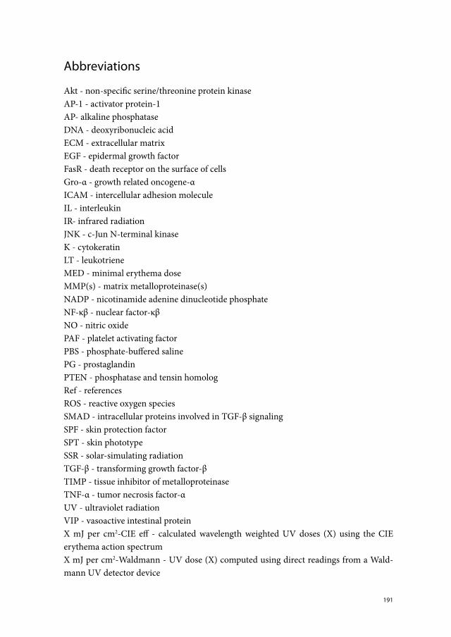
191
Abbreviations
Akt - non-specific serine/threonine protein kinaseAP-1 - activator protein-1AP- alkaline phosphataseDNA - deoxyribonucleic acidECM - extracellular matrixEGF - epidermal growth factorFasR - death receptor on the surface of cellsGro-α - growth related oncogene-αICAM - intercellular adhesion moleculeIL - interleukinIR- infrared radiationJNK - c-Jun N-terminal kinaseK - cytokeratinLT - leukotrieneMED - minimal erythema doseMMP(s) - matrix metalloproteinase(s)NADP - nicotinamide adenine dinucleotide phosphateNF-κβ - nuclear factor-κβNO - nitric oxidePAF - platelet activating factorPBS - phosphate-buffered salinePG - prostaglandinPTEN - phosphatase and tensin homologRef - referencesROS - reactive oxygen speciesSMAD - intracellular proteins involved in TGF-β signalingSPF - skin protection factorSPT - skin phototypeSSR - solar-simulating radiationTGF-β - transforming growth factor-βTIMP - tissue inhibitor of metalloproteinaseTNF-α - tumor necrosis factor-αUV - ultraviolet radiationVIP - vasoactive intestinal proteinX mJ per cm2-CIE eff - calculated wavelength weighted UV doses (X) using the CIE erythema action spectrumX mJ per cm2-Waldmann - UV dose (X) computed using direct readings from a Wald-mann UV detector device
Recommended