Phylogenetic analysis of Paleozoic microremains
from Denmark Emil Egede Hansen
19-09-2014
Keywords: Acanthodii, Thelodonti, tooth whorls, jaw fragments, dermal denticles, Silurian-Devonian. Nøgleord: Acanthodii, Thelodonti, tand hvirler, kæbefragmenter, dermal dentikler, Silur-Devon.
1
Supervisor: Gilles Guy Roger CunyFaculty of Science, University of Copenhagen
Masters Thesis, 19-09-2014
______________________________
Indhold Resume ....................................................................................................................................................... 3
Abstract ....................................................................................................................................................... 3
Introduction ................................................................................................................................................ 3
Methods ...................................................................................................................................................... 3
Samples ....................................................................................................................................................... 4
Description .................................................................................................................................................. 5
Class Conodonta ...................................................................................................................................... 5
Order Conodontophorida ..................................................................................................................... 5
Incertae sedis ....................................................................................................................................... 6
Subclass Heterostraci ............................................................................................................................... 8
Subclass Thelodonti ................................................................................................................................10
Order Loganelliformes ........................................................................................................................10
Order Shieliiformes .............................................................................................................................11
Order Phlebolepidiformes ...................................................................................................................12
Order Thelodontiformes .....................................................................................................................13
Incertae sedis ......................................................................................................................................15
Class Osteostraci .....................................................................................................................................17
Class Placioderm .....................................................................................................................................19
Order Acanthothoraci .........................................................................................................................19
Incertae sedis ......................................................................................................................................20
Subclass Acanthodii ................................................................................................................................20
Order Ischnacanthida ..........................................................................................................................20
Order Climatiida ..................................................................................................................................25
Order Diplacanthiformes .....................................................................................................................32
Incertae sedis ......................................................................................................................................33
Superclass Osteichthyes ..........................................................................................................................39
Problematica ..........................................................................................................................................40
Discussion ...................................................................................................................................................42
Conclusion ..................................................................................................................................................45
Acknowledgements ....................................................................................................................................46
References ..................................................................................................................................................46
2
Emil Egede Hansen Phylogenetic analysis of Paleozoic microremains from Denmark
Resume Otte prøver med fossiler er blevet fundet fra seks forskellige steder i Danmark. Dette studies formål er at identificere fossilerne, deres alder og deres miljø. 69 fossiler ud af 105 er blevet identificeret i en hvis grad. De mest almindelige fossiler er acanthoder efterfulgt af thelodonter. Alle prøverne har en alder som befinder sig i intervallet med Nedre Silur til Øvre Devon. Alle prøverne kommer fra et marint miljø, men nogle repræsenter et dybt marint miljø, mens andre repræsenter et lavvandet marint miljø.
Abstract Eight samples of fossils from erratic boulders have been recovered from six different places in Denmark. The purpose of this study is to identify the specimen, their age and environment. Out of 105 different specimens a total of 69 specimens have been identified to various degrees. The most common specimens found in the samples are acanthodians followed by thelodontians. All of samples have an age between Lower Silurian and Upper Devonian. The samples all come from a marine setting; however some represent a deep marine setting, while other represent a shallow marine setting.
Introduction Eight samples of fossils from erratic boulders have been recovered from several different places in Denmark. The purpose of this study is to identify the specimen and use the knowledge obtained from the identified specimen to find an age for the material and the environment in which the specimens lived. The specimens have been given numbers to their chronology, meaning specimen 1 of a sample is the first specimen encountered.
Methods The material was examined under a microscope and descriptions were made based on their morphology. Most specimens were photographed though the microscope; however some chosen ones, in particular conodont elements, were photographed with a Scanning Electron Microscope (SEM). However this required mounting the specimens on tape on stubs. The tapes combined with the shaking hands of the author and the fragile nature of the material caused some specimens to fragment. Others fell of their side and not wanting to risk further fragmentation these was described as they lay on the stub. The specimen was photographed from as many angles as possible. It is from the photographs from the SEM, that the descriptions of the material are based. To identify the specimens a comparison was made between the specimen in question and a specimen from an article. If there were enough similar features to validate it, the specimen in question could be identified as the same family or species of the specimen in the article. Some specimens were identified previously either by the finder, or in the case of the Danekræ fossil, by an expert. These specimens were checked to see if they had been identified correctly. The specimens which remained unidentified have been put into the different class. They were placed on the similarities with the indentified specimen. For example Tns1_02 share similarities with TNs1_05, especially the crowns look alike but Tns1_02 is identified and Tns1_05 is undetermined, but placed in the acanthodian class because of its similarities with Tns1_02.
3
Emil Egede Hansen Phylogenetic analysis of Paleozoic microremains from Denmark
Samples TNs1 (1) This sample is a number of sorted materials taken from an erratic stone collected from Trelde Næs in Southeast Jylland. Some of the material was previously identified. DK670 (1) This sample is a limestone block with the size 18 x 14 x 6 cm. It was found at the tip of Trelde Næs in southeast Jylland. Its content is a single well-preserved acanthodian spine. Size is 3 x 0.5 cm. This has been identified as a climatiid spine. DK544 (2) This sample consists of two sandstones; the first was found in Nymølle Grusgrav, Tarup Davinde which is on Fyn. It contains a spine and a tooth whorl. The spine has been identified as a osteichthyan (evaluation of DK 544). The second sandstone was collected from Johnny Hansens Grusgrav, Rolfsted on Fyn. Its content consists of one spine, one tooth and four scales. The scales have been identified as heterostracan. FFs1(3) This sample is a Green chalk block found in Frølunde fed in Vestsjælland. Its content consists of a single acanthodian spine. 05-260-01 (4) This sample consists of loose fossil derived from a Silurian Beyrichten chalk block. It was found in Bukø, Lindelse Nor on Langeland. Its content is roughly 90-100 conodont elements, 19 tooth whorls, five different types of scales and two types of bone fragments. DK 739 (5)
This sample consists of a single jaw fragment from an erratic boulder. It was found in Broagerland. 06-227-01 (6) This sample consists of sorted, loose fossils, some of which has been identified by the finder. It was found in Nordenhuse. Its content consists of scales, tooth whorls, spines and fragments from teeth and jaws. 06-227-02 (6) This sample consists of loose fossils derived from a Silurian Beyrichen chalk block. It was found in Nordenhuse on North eastern Fyn. Its content consists of conodont elements, acanthodian spines, jaw fagments, tooth whorls, bone fragments and scales.
4
Emil Egede Hansen Phylogenetic analysis of Paleozoic microremains from Denmark
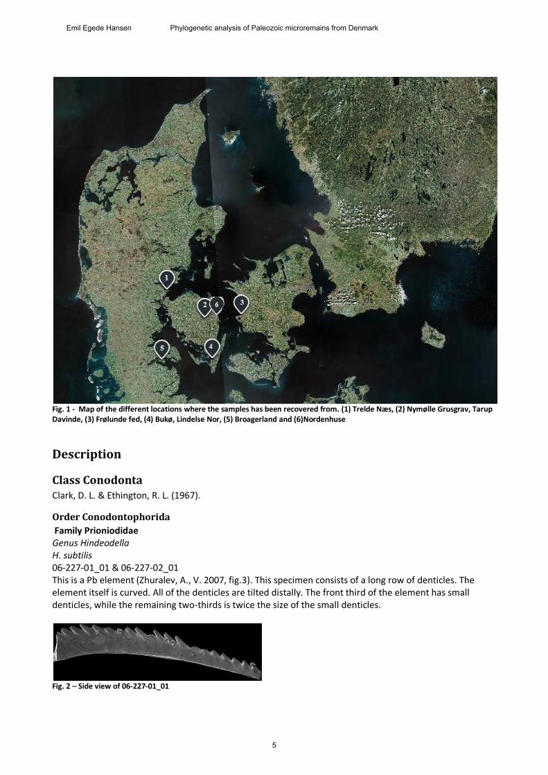
Fig. 1 - Map of the different locations where the samples has been recovered from. (1) Trelde Næs, (2) Nymølle Grusgrav, Tarup Davinde, (3) Frølunde fed, (4) Bukø, Lindelse Nor, (5) Broagerland and (6)Nordenhuse
Description
Class Conodonta Clark, D. L. & Ethington, R. L. (1967).
Order Conodontophorida
Family Prioniodidae Genus Hindeodella H. subtilis 06-227-01_01 & 06-227-02_01 This is a Pb element (Zhuralev, A., V. 2007, fig.3). This specimen consists of a long row of denticles. The element itself is curved. All of the denticles are tilted distally. The front third of the element has small denticles, while the remaining two-thirds is twice the size of the small denticles.
Fig. 2 – Side view of 06-227-01_01
5
Emil Egede Hansen Phylogenetic analysis of Paleozoic microremains from Denmark
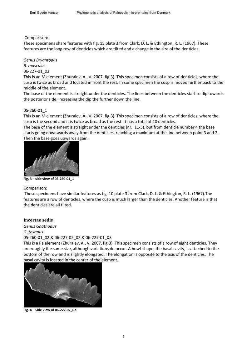
Comparison: These specimens share features with fig. 15 plate 3 from Clark, D. L. & Ethington, R. L. (1967). These features are the long row of denticles which are tilted and a change in the size of the denticles. Genus Bryantodus B. masculus 06-227-01_02 This is an M element (Zhuralev, A., V. 2007, fig.3). This specimen consists of a row of denticles, where the cusp is twice as broad and located in front the rest. In some specimen the cusp is moved further back to the middle of the element. The base of the element is straight under the denticles. The lines between the denticles start to dip towards the posterior side, increasing the dip the further down the line. 05-260-01_1 This is an M element (Zhuralev, A., V. 2007, fig.3). This specimen consists of a row of denticles, where the cusp is the second and it is twice as broad as the rest. It has a total of 10 denticles. The base of the element is straight under the denticles (nr. 11-5), but from denticle number 4 the base starts going downwards away from the denticles, reaching a maximum at the line between point 3 and 2. Then the base goes upwards again.
Fig, 3 – side view of 05-260-01_1
Comparison: These specimens have similar features as fig. 10 plate 3 from Clark, D. L. & Ethington, R. L. (1967).The features are a row of denticles, where the cusp is much larger than the denticles. Another feature is that the denticles are all tilted.
Incertae sedis
Genus Gnathodus G. texanus 05-260-01_02 & 06-227-02_02 & 06-227-01_03 This is a Pa element (Zhuralev, A., V. 2007, fig.3). This specimen consists of a row of eight denticles. They are roughly the same size, although variations do occur. A bowl-shape, the basal cavity, is attached to the bottom of the row and is slightly elongated. The elongation is opposite to the axis of the denticles. The basal cavity is located in the center of the element.
Fig. 4 – Side view of 06-227-02_02.
6
Emil Egede Hansen Phylogenetic analysis of Paleozoic microremains from Denmark
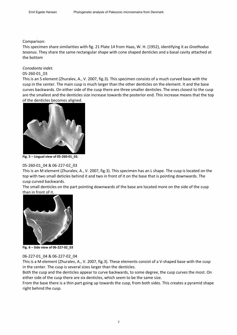
Comparison: This specimen share similarities with fig. 21 Plate 14 from Haas, W. H. (1952), identifying it as Gnathodus texanus. They share the same rectangular shape with cone shaped denticles and a basal cavity attached at the bottom Conodonta indet. 05-260-01_03 This is an S element (Zhuralev, A., V. 2007, fig.3). This specimen consists of a much curved base with the cusp in the center. The main cusp is much larger than the other denticles on the element. It and the base curves backwards. On either side of the cusp there are three smaller denticles. The ones closest to the cusp are the smallest and the denticles size increase towards the posterior end. This increase means that the top of the denticles becomes aligned.
Fig. 5 – Lingual view of 05-260-01_03.
05-260-01_04 & 06-227-02_03 This is an M element (Zhuralev, A., V. 2007, fig.3). This specimen has an L shape. The cusp is located on the top with two small deticles behind it and two in front of it on the base that is pointing downwards. The cusp curved backwards. The small denticles on the part pointing downwards of the base are located more on the side of the cusp than in front of it.
Fig. 6 – Side view of 06-227-02_03
06-227-01_04 & 06-227-02_04 This is a M element (Zhuralev, A., V. 2007, fig.3). These elements consist of a V-shaped base with the cusp in the center. The cusp is several sizes larger than the denticles. Both the cusp and the denticles appear to curve backwards, to some degree, the cusp curves the most. On either side of the cusp there are six denticles, which seem to be the same size. From the base there is a thin part going up towards the cusp, from both sides. This creates a pyramid shape right behind the cusp.
7
Emil Egede Hansen Phylogenetic analysis of Paleozoic microremains from Denmark
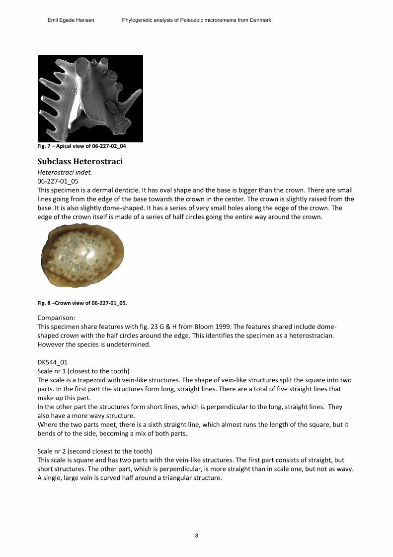
Fig. 7 – Apical view of 06-227-02_04
Subclass Heterostraci Heterostraci indet. 06-227-01_05 This specimen is a dermal denticle. It has oval shape and the base is bigger than the crown. There are small lines going from the edge of the base towards the crown in the center. The crown is slightly raised from the base. It is also slightly dome-shaped. It has a series of very small holes along the edge of the crown. The edge of the crown itself is made of a series of half circles going the entire way around the crown.
Fig. 8 –Crown view of 06-227-01_05.
Comparison: This specimen share features with fig. 23 G & H from Bloom 1999. The features shared include dome-shaped crown with the half circles around the edge. This identifies the specimen as a heterostracian. However the species is undetermined. DK544_01 Scale nr 1 (closest to the tooth) The scale is a trapezoid with vein-like structures. The shape of vein-like structures split the square into two parts. In the first part the structures form long, straight lines. There are a total of five straight lines that make up this part. In the other part the structures form short lines, which is perpendicular to the long, straight lines. They also have a more wavy structure. Where the two parts meet, there is a sixth straight line, which almost runs the length of the square, but it bends of to the side, becoming a mix of both parts. Scale nr 2 (second closest to the tooth) This scale is square and has two parts with the vein-like structures. The first part consists of straight, but short structures. The other part, which is perpendicular, is more straight than in scale one, but not as wavy. A single, large vein is curved half around a triangular structure.
8
Emil Egede Hansen Phylogenetic analysis of Paleozoic microremains from Denmark
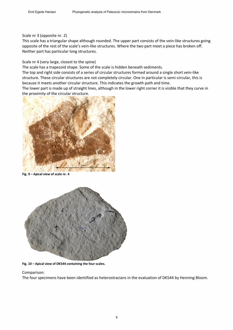
Scale nr 3 (opposite nr. 2) This scale has a triangular shape although rounded. The upper part consists of the vein-like structures going opposite of the rest of the scale’s vein-like structures. Where the two part meet a piece has broken off. Neither part has particular long structures. Scale nr 4 (very large, closest to the spine) The scale has a trapezoid shape. Some of the scale is hidden beneath sediments. The top and right side consists of a series of circular structures formed around a single short vein-like structure. These circular structures are not completely circular. One in particular is semi-circular, this is because it meets another circular structure. This indicates the growth path and time. The lower part is made up of straight lines, although in the lower right corner it is visible that they curve in the proximity of the circular structure.
Fig. 9 – Apical view of scale nr. 4
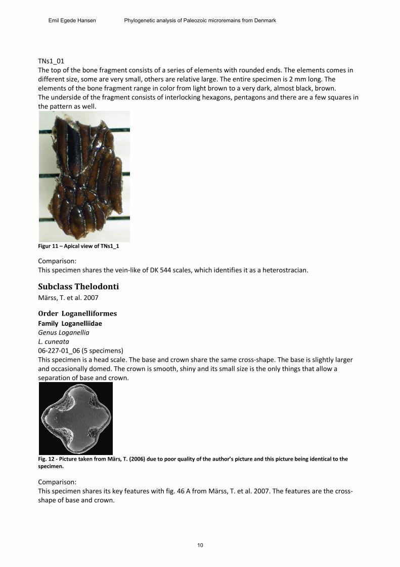
Fig. 10 – Apical view of DK544 containing the four scales.
Comparison: The four specimens have been identified as heterostracians in the evaluation of DK544 by Henning Bloom.
9
Emil Egede Hansen Phylogenetic analysis of Paleozoic microremains from Denmark
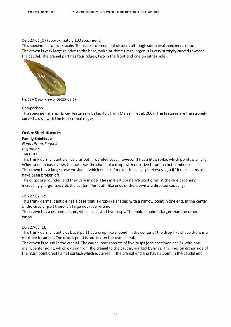
TNs1_01 The top of the bone fragment consists of a series of elements with rounded ends. The elements comes in different size, some are very small, others are relative large. The entire specimen is 2 mm long. The elements of the bone fragment range in color from light brown to a very dark, almost black, brown. The underside of the fragment consists of interlocking hexagons, pentagons and there are a few squares in the pattern as well.
Figur 11 – Apical view of TNs1_1
Comparison: This specimen shares the vein-like of DK 544 scales, which identifies it as a heterostracian.
Subclass Thelodonti Märss, T. et al. 2007
Order Loganelliformes
Family Loganelliidae Genus Loganellia L. cuneata 06-227-01_06 (5 specimens) This specimen is a head scale. The base and crown share the same cross-shape. The base is slightly larger and occasionally domed. The crown is smooth, shiny and its small size is the only things that allow a separation of base and crown.
Fig. 12 - Picture taken from Märs, T. (2006) due to poor quality of the author’s picture and this picture being identical to the specimen.
Comparison: This specimen shares its key features with fig. 46 A from Märss, T. et al. 2007. The features are the cross-shape of base and crown.
10
Emil Egede Hansen Phylogenetic analysis of Paleozoic microremains from Denmark
06-227-01_07 (approximately 100 specimens) This specimen is a trunk scale. The base is domed and circular, although some oval specimens occur. The crown is very large relative to the base, twice or three times larger. It is very strongly curved towards the caudal. The cranial part has four ridges; two in the front and one on either side.
Fig. 13 – Crown view of 06-227-01_07.
Comparison: This specimen shares its key features with fig. 46 L from Märss, T. et al. 2007. The features are the strongly curved crown with the four cranial ridges.
Order Shieliiformes
Family Shieliidae Genus Praetrilogania P. grabion TNs1_02 This trunk dermal denticle has a smooth, rounded base, however it has a little spike, which points cranially. When seen in basal view, the base has the shape of a drop, with nutritive foramina in the middle. The crown has a large crescent shape, which ends in four teeth-like cusps. However, a fifth one seems to have been broken off. The cusps are rounded and they vary in size. The smallest points are positioned at the side becoming increasingly larger towards the center. The teeth-like ends of the crown are directed caudally. 06-227-02_05 This trunk dermal denticle has a base that is drop-like shaped with a narrow point in one end. In the center of the circular part there is a large nutritive foramen. The crown has a crescent shape, which consist of five cusps. The middle point is larger than the other cusps. 06-227-01_06 This trunk dermal denticles basal part has a drop-like shaped. In the center of the drop-like shape there is a nutritive foramina. The drop’s point is located on the cranial end. The crown is round in the cranial. The caudal part consists of five cusps (one specimen has 7), with one main, center point, which extend from the cranial to the caudal, marked by lines. The lines on either side of the main point create a flat surface which is curved in the cranial end and have 2 point in the caudal end.
11
Emil Egede Hansen Phylogenetic analysis of Paleozoic microremains from Denmark
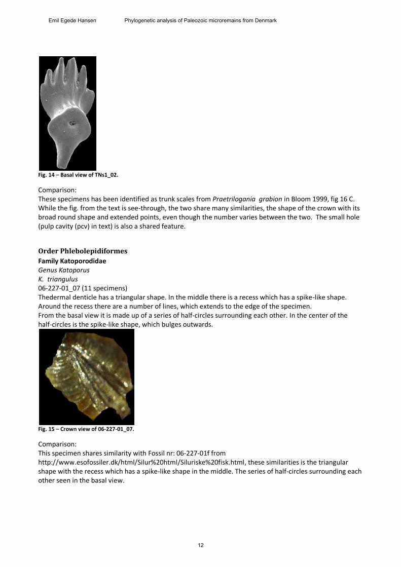
Fig. 14 – Basal view of TNs1_02.
Comparison: These specimens has been identified as trunk scales from Praetrilogania grabion in Bloom 1999, fig 16 C. While the fig. from the text is see-through, the two share many similarities, the shape of the crown with its broad round shape and extended points, even though the number varies between the two. The small hole (pulp cavity (pcv) in text) is also a shared feature.
Order Phlebolepidiformes
Family Katoporodidae Genus Katoporus K. triangulus 06-227-01_07 (11 specimens) Thedermal denticle has a triangular shape. In the middle there is a recess which has a spike-like shape. Around the recess there are a number of lines, which extends to the edge of the specimen. From the basal view it is made up of a series of half-circles surrounding each other. In the center of the half-circles is the spike-like shape, which bulges outwards.
Fig. 15 – Crown view of 06-227-01_07.
Comparison: This specimen shares similarity with Fossil nr: 06-227-01f from http://www.esofossiler.dk/html/Silur%20html/Siluriske%20fisk.html, these similarities is the triangular shape with the recess which has a spike-like shape in the middle. The series of half-circles surrounding each other seen in the basal view.
12
Emil Egede Hansen Phylogenetic analysis of Paleozoic microremains from Denmark
Order Thelodontiformes
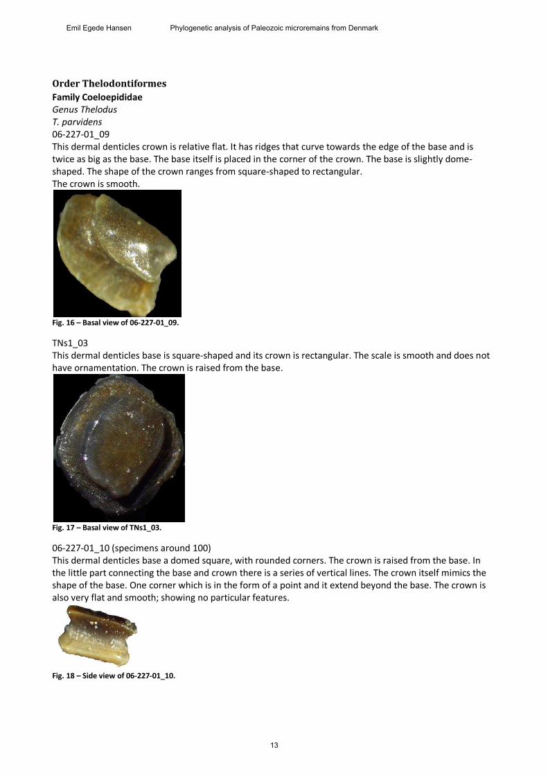
Family Coeloepididae Genus Thelodus T. parvidens 06-227-01_09 This dermal denticles crown is relative flat. It has ridges that curve towards the edge of the base and is twice as big as the base. The base itself is placed in the corner of the crown. The base is slightly dome-shaped. The shape of the crown ranges from square-shaped to rectangular. The crown is smooth.
Fig. 16 – Basal view of 06-227-01_09.
TNs1_03 This dermal denticles base is square-shaped and its crown is rectangular. The scale is smooth and does not have ornamentation. The crown is raised from the base.
Fig. 17 – Basal view of TNs1_03.
06-227-01_10 (specimens around 100) This dermal denticles base a domed square, with rounded corners. The crown is raised from the base. In the little part connecting the base and crown there is a series of vertical lines. The crown itself mimics the shape of the base. One corner which is in the form of a point and it extend beyond the base. The crown is also very flat and smooth; showing no particular features.
Fig. 18 – Side view of 06-227-01_10.
13
Emil Egede Hansen Phylogenetic analysis of Paleozoic microremains from Denmark
Comparison: These specimens share the shape of the crown and base with fig.4-8 from plate 1 in J.M.J. Vergossen 2002. They have the same shape and no ornamentation as the ones in the J.M.J. Vergossen 2002, which identifies this as Thelodus Parvidens. T. traquairi 06-227-01_08 (11 specimens) Thiss dermal denticles base is circular or rounded square-shaped. It has nutritive foramina in the center of the base. The crown is a tall spike with ridges along the sides of the bottom part. The crown is slightly curved towards the caudal. The crown resembles a tooth.
Fig. 19 – Side view of 06-227-08_08
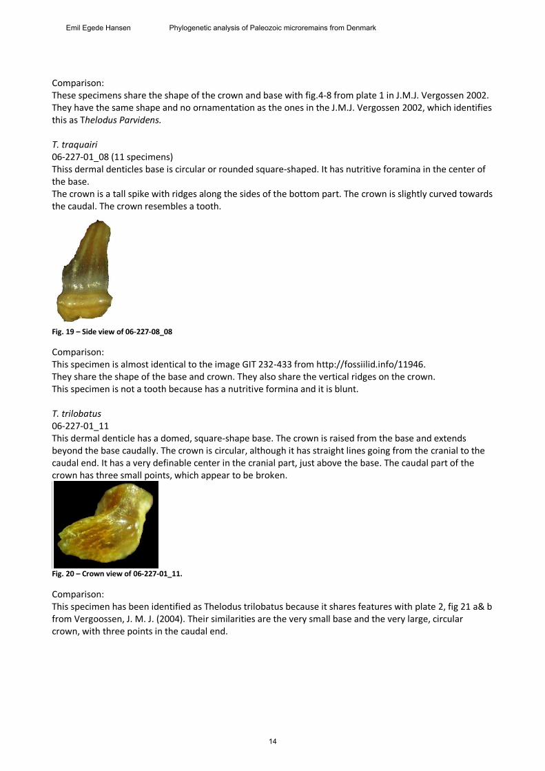
Comparison: This specimen is almost identical to the image GIT 232-433 from http://fossiilid.info/11946. They share the shape of the base and crown. They also share the vertical ridges on the crown. This specimen is not a tooth because has a nutritive formina and it is blunt. T. trilobatus 06-227-01_11 This dermal denticle has a domed, square-shape base. The crown is raised from the base and extends beyond the base caudally. The crown is circular, although it has straight lines going from the cranial to the caudal end. It has a very definable center in the cranial part, just above the base. The caudal part of the crown has three small points, which appear to be broken.
Fig. 20 – Crown view of 06-227-01_11.
Comparison: This specimen has been identified as Thelodus trilobatus because it shares features with plate 2, fig 21 a& b from Vergoossen, J. M. J. (2004). Their similarities are the very small base and the very large, circular crown, with three points in the caudal end.
14
Emil Egede Hansen Phylogenetic analysis of Paleozoic microremains from Denmark
Incertae sedis
Thelodonti indet. 06-227-01_12 In this dermal dernticle the base is gone. The crown of the scale is roughly triangular, close to hexagonal. There are five “corners” each of which has a characteristic knob. One of them is located away from the other four (giving the triangular shape). The four knobs all have ridges going towards the lone fifth.
Fig. 21 – Crown view of 06-227-01_12.
06-227-01_13 The base of this dermal dentilce is a hexagon. It is mostly smooth, but it has a series of rings around a smooth center, much like growth rings. The crown is raised from the base and has the shape of an elongated triangle. The triangle is made up of a smooth central apex while the edges are made up of knobby features, which comes from the central axis.
Fig. 22 – Crown view of 06-227-01_13.
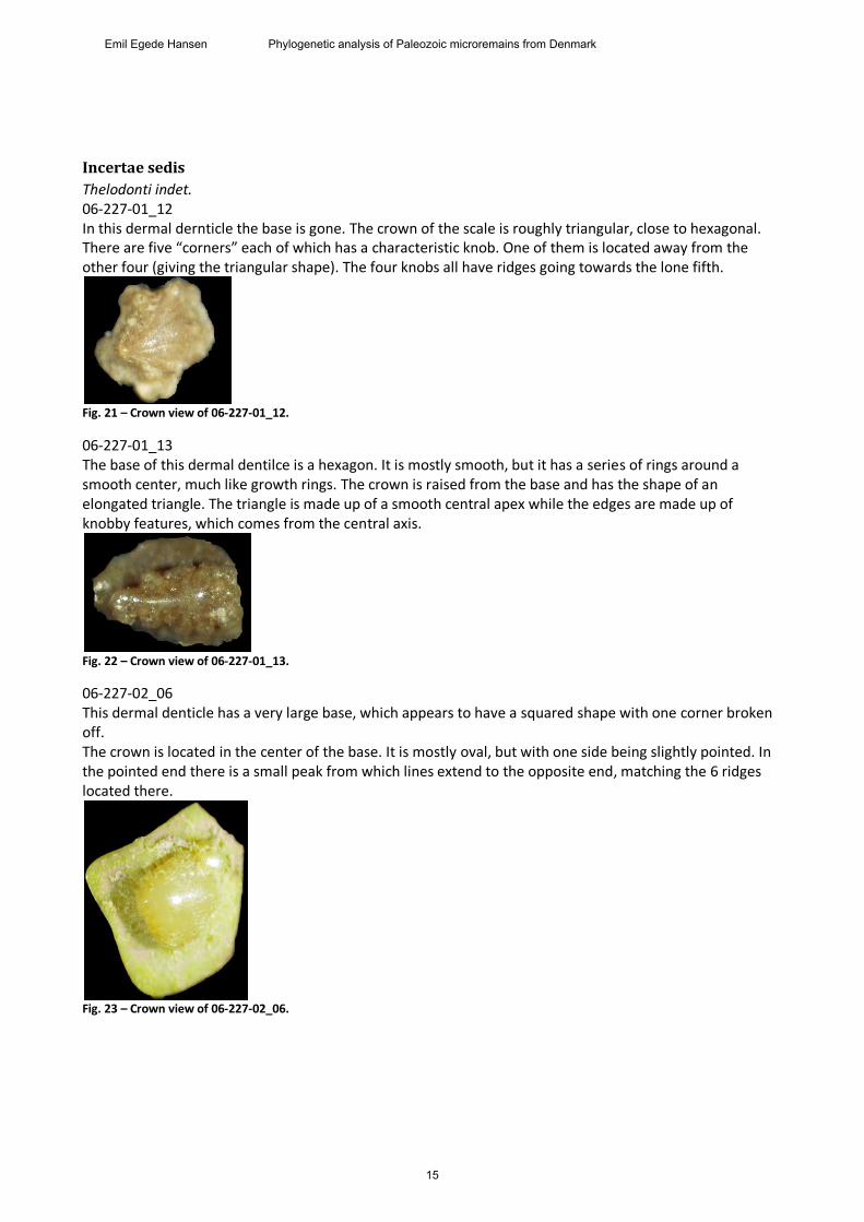
06-227-02_06 This dermal denticle has a very large base, which appears to have a squared shape with one corner broken off. The crown is located in the center of the base. It is mostly oval, but with one side being slightly pointed. In the pointed end there is a small peak from which lines extend to the opposite end, matching the 6 ridges located there.
Fig. 23 – Crown view of 06-227-02_06.
15
Emil Egede Hansen Phylogenetic analysis of Paleozoic microremains from Denmark
06-227-01_14 The base of this dermal denticle has six sides and nutritive formina in the center. The crown is cone-shaped, with the apex a bit off center. The sides of the cone have three triangular features that come from the apex down to the base. The features are made of a series of vertical ridges.
Fig. 24 – Crown of 06-227-01_14.
TNs1_04 The basal part of this dermal denticle is oval-shaped, but it is fragmented. The crown is also oval-shaped. The crown has a round apex in the center. The edges of the denticle consist of a series of half-circles going all the way around the scale. There are 11 half-circles around the crown. From the apex and down to the half-circles there are a series of ridges. The number of ridges per half-circle varies between 3 and 5.
Fig. 25 – Crown view of TNs1_04.
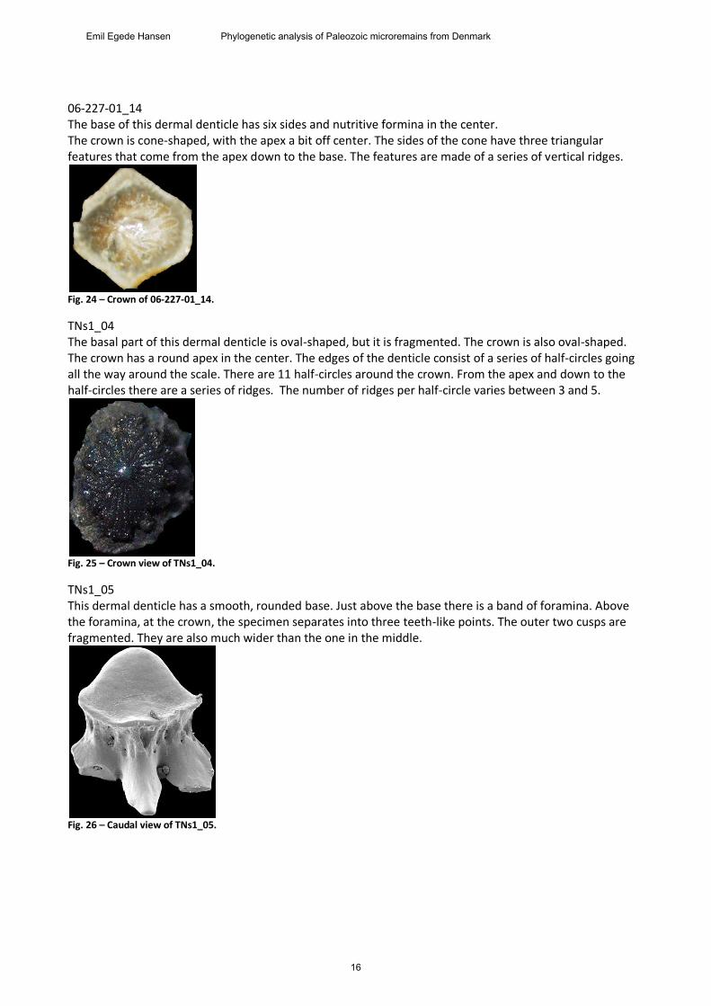
TNs1_05 This dermal denticle has a smooth, rounded base. Just above the base there is a band of foramina. Above the foramina, at the crown, the specimen separates into three teeth-like points. The outer two cusps are fragmented. They are also much wider than the one in the middle.
Fig. 26 – Caudal view of TNs1_05.
16
Emil Egede Hansen Phylogenetic analysis of Paleozoic microremains from Denmark
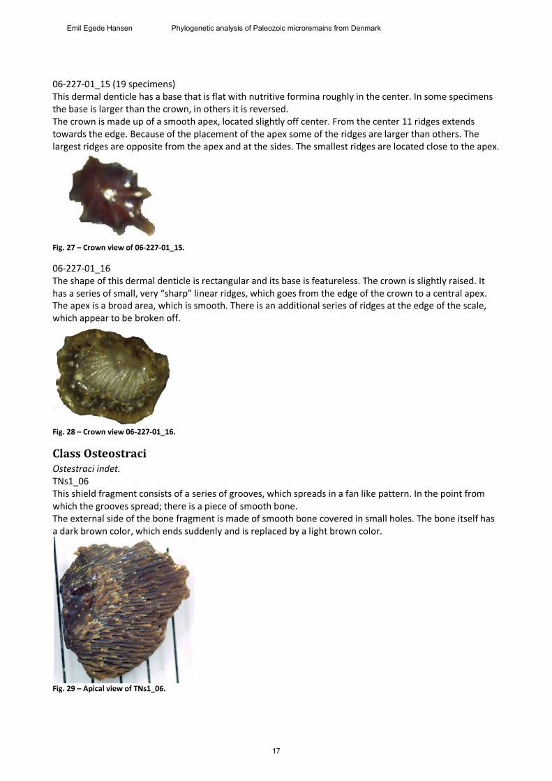
06-227-01_15 (19 specimens) This dermal denticle has a base that is flat with nutritive formina roughly in the center. In some specimens the base is larger than the crown, in others it is reversed. The crown is made up of a smooth apex, located slightly off center. From the center 11 ridges extends towards the edge. Because of the placement of the apex some of the ridges are larger than others. The largest ridges are opposite from the apex and at the sides. The smallest ridges are located close to the apex.
Fig. 27 – Crown view of 06-227-01_15.
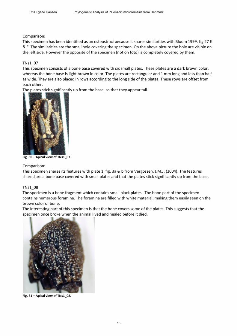
06-227-01_16 The shape of this dermal denticle is rectangular and its base is featureless. The crown is slightly raised. It has a series of small, very “sharp” linear ridges, which goes from the edge of the crown to a central apex. The apex is a broad area, which is smooth. There is an additional series of ridges at the edge of the scale, which appear to be broken off.
Fig. 28 – Crown view 06-227-01_16.
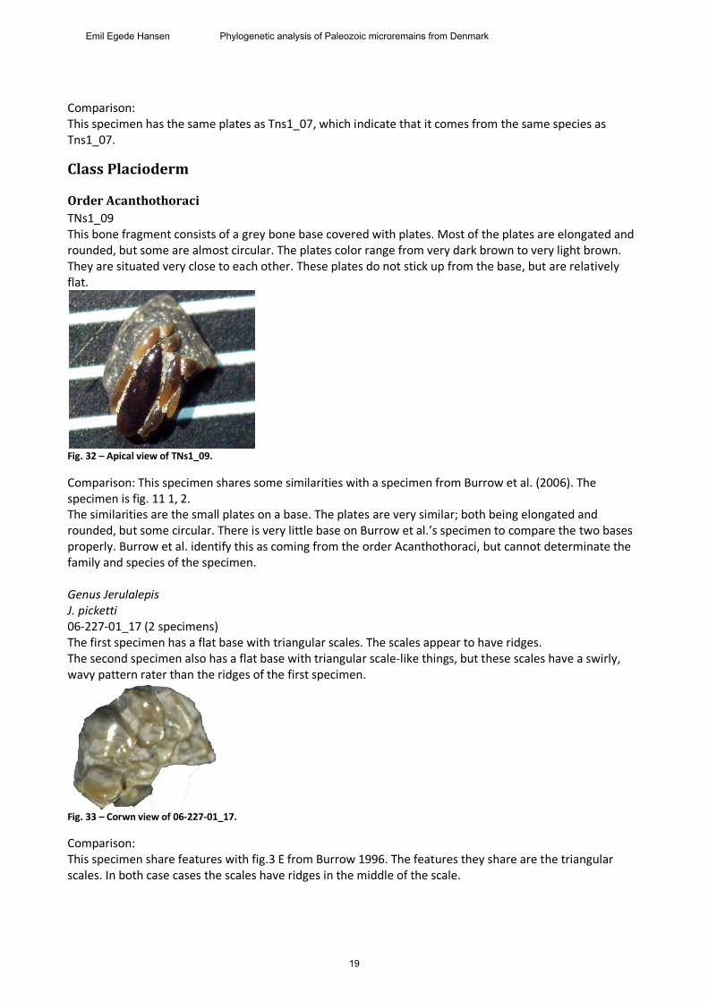
Class Osteostraci Ostestraci indet. TNs1_06 This shield fragment consists of a series of grooves, which spreads in a fan like pattern. In the point from which the grooves spread; there is a piece of smooth bone. The external side of the bone fragment is made of smooth bone covered in small holes. The bone itself has a dark brown color, which ends suddenly and is replaced by a light brown color.
Fig. 29 – Apical view of TNs1_06.
17
Emil Egede Hansen Phylogenetic analysis of Paleozoic microremains from Denmark
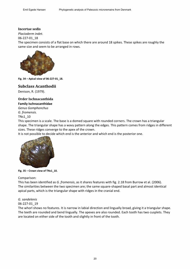
Comparison: This specimen has been identified as an osteostraci because it shares similarities with Bloom 1999. fig 27 E & F. The similarities are the small hole covering the specimen. On the above picture the hole are visible on the left side. However the opposite of the specimen (not on foto) is completely covered by them. TNs1_07 This specimen consists of a bone base covered with six small plates. These plates are a dark brown color, whereas the bone base is light brown in color. The plates are rectangular and 1 mm long and less than half as wide. They are also placed in rows according to the long side of the plates. These rows are offset from each other. The plates stick significantly up from the base, so that they appear tall.
Fig. 30 – Apical view of TNs1_07.
Comparison: This specimen shares its features with plate 1, fig. 3a & b from Vergossen, J.M.J. (2004). The features shared are a bone base covered with small plates and that the plates stick significantly up from the base. TNs1_08 The specimen is a bone fragment which contains small black plates. The bone part of the specimen contains numerous foramina. The foramina are filled with white material, making them easily seen on the brown color of bone. The interesting part of this specimen is that the bone covers some of the plates. This suggests that the specimen once broke when the animal lived and healed before it died.
Fig. 31 – Apical view of TNs1_08.
18
Emil Egede Hansen Phylogenetic analysis of Paleozoic microremains from Denmark
Comparison: This specimen has the same plates as Tns1_07, which indicate that it comes from the same species as Tns1_07.
Class Placioderm
Order Acanthothoraci
TNs1_09 This bone fragment consists of a grey bone base covered with plates. Most of the plates are elongated and rounded, but some are almost circular. The plates color range from very dark brown to very light brown. They are situated very close to each other. These plates do not stick up from the base, but are relatively flat.
Fig. 32 – Apical view of TNs1_09.
Comparison: This specimen shares some similarities with a specimen from Burrow et al. (2006). The specimen is fig. 11 1, 2. The similarities are the small plates on a base. The plates are very similar; both being elongated and rounded, but some circular. There is very little base on Burrow et al.’s specimen to compare the two bases properly. Burrow et al. identify this as coming from the order Acanthothoraci, but cannot determinate the family and species of the specimen. Genus Jerulalepis J. picketti 06-227-01_17 (2 specimens) The first specimen has a flat base with triangular scales. The scales appear to have ridges. The second specimen also has a flat base with triangular scale-like things, but these scales have a swirly, wavy pattern rater than the ridges of the first specimen.
Fig. 33 – Corwn view of 06-227-01_17.
Comparison: This specimen share features with fig.3 E from Burrow 1996. The features they share are the triangular scales. In both case cases the scales have ridges in the middle of the scale.
19
Emil Egede Hansen Phylogenetic analysis of Paleozoic microremains from Denmark
Incertae sedis
Placioderm indet. 06-227-01_18 The specimen consists of a flat base on which there are around 18 spikes. These spikes are roughly the same size and seem to be arranged in rows.
Fig. 34 – Apical view of 06-227-01_18.
Subclass Acanthodii Denison, R. (1979).
Order Ischnacanthida
Family Ischnacanthidae Genus Gomphonchus G. fromensis. TNs1_10 This specimen is a scale. The base is a domed square with rounded corners. The crown has a triangular shape. The triangular shape has a wavy pattern along the edges. This pattern comes from ridges in different sizes. These ridges converge to the apex of the crown. It is not possible to decide which end is the anterior and which end is the posterior one.
Fig. 35 – Crown view of TNs1_10.
Comparison: This has been identified as G. fromensis, as it shares features with fig. 2.18 from Burrow et al. (2006). The similarities between the two specimen are; the same square-shaped basal part and almost identical apical parts, which is the triangular shape with ridges in the cranial end. G. sandelenis 06-227-01_19 The whorl shows no features. It is narrow in labial direction and lingually broad, giving it a triangular shape. The teeth are rounded and bend lingually. The apexes are also rounded. Each tooth has two cusplets. They are located on either side of the tooth and slightly in front of the tooth.
20
Emil Egede Hansen Phylogenetic analysis of Paleozoic microremains from Denmark
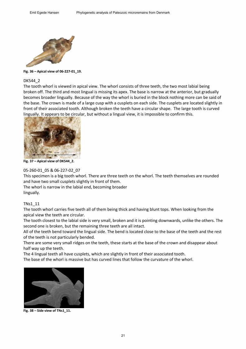
Fig. 36 – Apical view of 06-227-01_19.
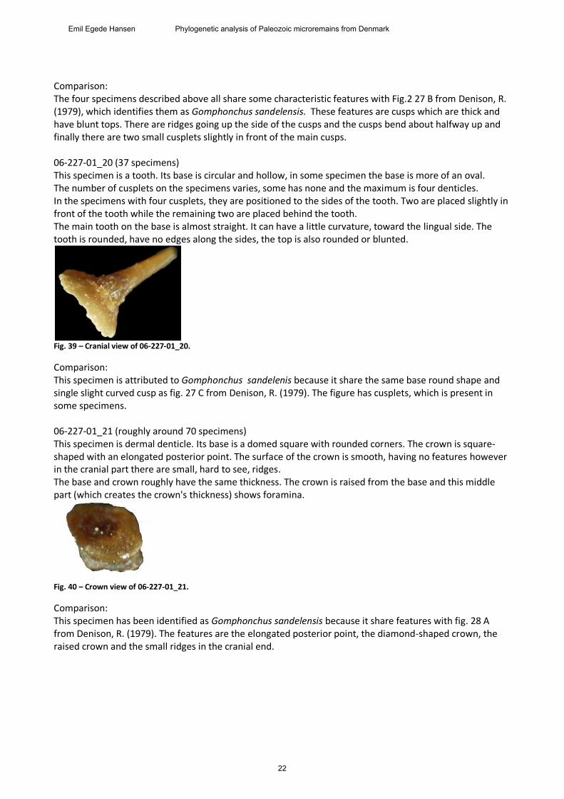
DK544_2 The tooth whorl is viewed in apical view. The whorl consists of three teeth, the two most labial being broken off. The third and most lingual is missing its apex. The base is narrow at the anterior, but gradually becomes broader lingually. Because of the way the whorl is buried in the block nothing more can be said of the base. The crown is made of a large cusp with a cusplets on each side. The cusplets are located slightly in front of their associated tooth. Although broken the teeth have a circular shape. The large tooth is curved lingually. It appears to be circular, but without a lingual view, it is impossible to confirm this.
Fig. 37 – Apical view of DK544_2.
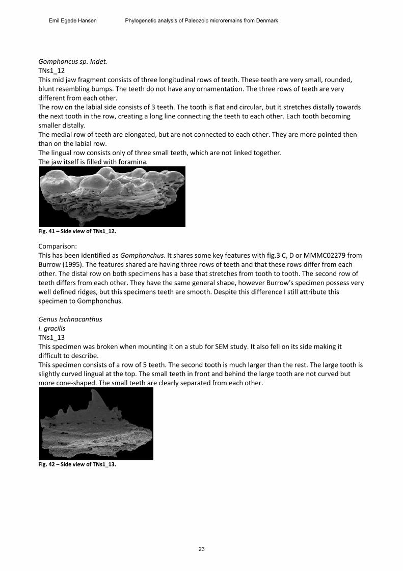
05-260-01_05 & 06-227-02_07 This specimen is a big tooth whorl. There are three teeth on the whorl. The teeth themselves are rounded and have two small cusplets slightly in front of them. The whorl is narrow in the labial end, becoming broader lingually. TNs1_11 The tooth whorl carries five teeth all of them being thick and having blunt tops. When looking from the apical view the teeth are circular. The tooth closest to the labial side is very small, broken and it is pointing downwards, unlike the others. The second one is broken, but the remaining three teeth are all intact. All of the teeth bend toward the lingual side. The bend is located close to the base of the teeth and the rest of the teeth is not particularly bended. There are some very small ridges on the teeth, these starts at the base of the crown and disappear about half way up the teeth. The 4 lingual teeth all have cusplets, which are slightly in front of their associated tooth. The base of the whorl is massive but has curved lines that follow the curvature of the whorl.
Fig. 38 – Side view of TNs1_11.
21
Emil Egede Hansen Phylogenetic analysis of Paleozoic microremains from Denmark
Comparison: The four specimens described above all share some characteristic features with Fig.2 27 B from Denison, R. (1979), which identifies them as Gomphonchus sandelensis. These features are cusps which are thick and have blunt tops. There are ridges going up the side of the cusps and the cusps bend about halfway up and finally there are two small cusplets slightly in front of the main cusps. 06-227-01_20 (37 specimens) This specimen is a tooth. Its base is circular and hollow, in some specimen the base is more of an oval. The number of cusplets on the specimens varies, some has none and the maximum is four denticles. In the specimens with four cusplets, they are positioned to the sides of the tooth. Two are placed slightly in front of the tooth while the remaining two are placed behind the tooth. The main tooth on the base is almost straight. It can have a little curvature, toward the lingual side. The tooth is rounded, have no edges along the sides, the top is also rounded or blunted.
Fig. 39 – Cranial view of 06-227-01_20.
Comparison: This specimen is attributed to Gomphonchus sandelenis because it share the same base round shape and single slight curved cusp as fig. 27 C from Denison, R. (1979). The figure has cusplets, which is present in some specimens. 06-227-01_21 (roughly around 70 specimens) This specimen is dermal denticle. Its base is a domed square with rounded corners. The crown is square-shaped with an elongated posterior point. The surface of the crown is smooth, having no features however in the cranial part there are small, hard to see, ridges. The base and crown roughly have the same thickness. The crown is raised from the base and this middle part (which creates the crown's thickness) shows foramina.
Fig. 40 – Crown view of 06-227-01_21.
Comparison: This specimen has been identified as Gomphonchus sandelensis because it share features with fig. 28 A from Denison, R. (1979). The features are the elongated posterior point, the diamond-shaped crown, the raised crown and the small ridges in the cranial end.
22
Emil Egede Hansen Phylogenetic analysis of Paleozoic microremains from Denmark
Gomphoncus sp. Indet. TNs1_12 This mid jaw fragment consists of three longitudinal rows of teeth. These teeth are very small, rounded, blunt resembling bumps. The teeth do not have any ornamentation. The three rows of teeth are very different from each other. The row on the labial side consists of 3 teeth. The tooth is flat and circular, but it stretches distally towards the next tooth in the row, creating a long line connecting the teeth to each other. Each tooth becoming smaller distally. The medial row of teeth are elongated, but are not connected to each other. They are more pointed then than on the labial row. The lingual row consists only of three small teeth, which are not linked together. The jaw itself is filled with foramina.
Fig. 41 – Side view of TNs1_12.
Comparison: This has been identified as Gomphonchus. It shares some key features with fig.3 C, D or MMMC02279 from Burrow (1995). The features shared are having three rows of teeth and that these rows differ from each other. The distal row on both specimens has a base that stretches from tooth to tooth. The second row of teeth differs from each other. They have the same general shape, however Burrow’s specimen possess very well defined ridges, but this specimens teeth are smooth. Despite this difference I still attribute this specimen to Gomphonchus. Genus Ischnacanthus I. gracilis TNs1_13 This specimen was broken when mounting it on a stub for SEM study. It also fell on its side making it difficult to describe. This specimen consists of a row of 5 teeth. The second tooth is much larger than the rest. The large tooth is slightly curved lingual at the top. The small teeth in front and behind the large tooth are not curved but more cone-shaped. The small teeth are clearly separated from each other.
Fig. 42 – Side view of TNs1_13.
23
Emil Egede Hansen Phylogenetic analysis of Paleozoic microremains from Denmark
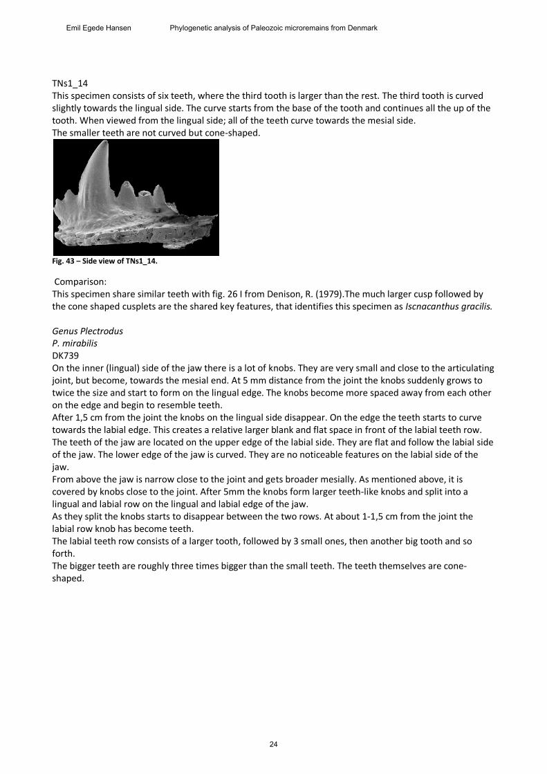
TNs1_14 This specimen consists of six teeth, where the third tooth is larger than the rest. The third tooth is curved slightly towards the lingual side. The curve starts from the base of the tooth and continues all the up of the tooth. When viewed from the lingual side; all of the teeth curve towards the mesial side. The smaller teeth are not curved but cone-shaped.
Fig. 43 – Side view of TNs1_14.
Comparison: This specimen share similar teeth with fig. 26 I from Denison, R. (1979).The much larger cusp followed by the cone shaped cusplets are the shared key features, that identifies this specimen as Iscnacanthus gracilis. Genus Plectrodus P. mirabilis DK739 On the inner (lingual) side of the jaw there is a lot of knobs. They are very small and close to the articulating joint, but become, towards the mesial end. At 5 mm distance from the joint the knobs suddenly grows to twice the size and start to form on the lingual edge. The knobs become more spaced away from each other on the edge and begin to resemble teeth. After 1,5 cm from the joint the knobs on the lingual side disappear. On the edge the teeth starts to curve towards the labial edge. This creates a relative larger blank and flat space in front of the labial teeth row. The teeth of the jaw are located on the upper edge of the labial side. They are flat and follow the labial side of the jaw. The lower edge of the jaw is curved. They are no noticeable features on the labial side of the jaw. From above the jaw is narrow close to the joint and gets broader mesially. As mentioned above, it is covered by knobs close to the joint. After 5mm the knobs form larger teeth-like knobs and split into a lingual and labial row on the lingual and labial edge of the jaw. As they split the knobs starts to disappear between the two rows. At about 1-1,5 cm from the joint the labial row knob has become teeth. The labial teeth row consists of a larger tooth, followed by 3 small ones, then another big tooth and so forth. The bigger teeth are roughly three times bigger than the small teeth. The teeth themselves are cone-shaped.
24
Emil Egede Hansen Phylogenetic analysis of Paleozoic microremains from Denmark
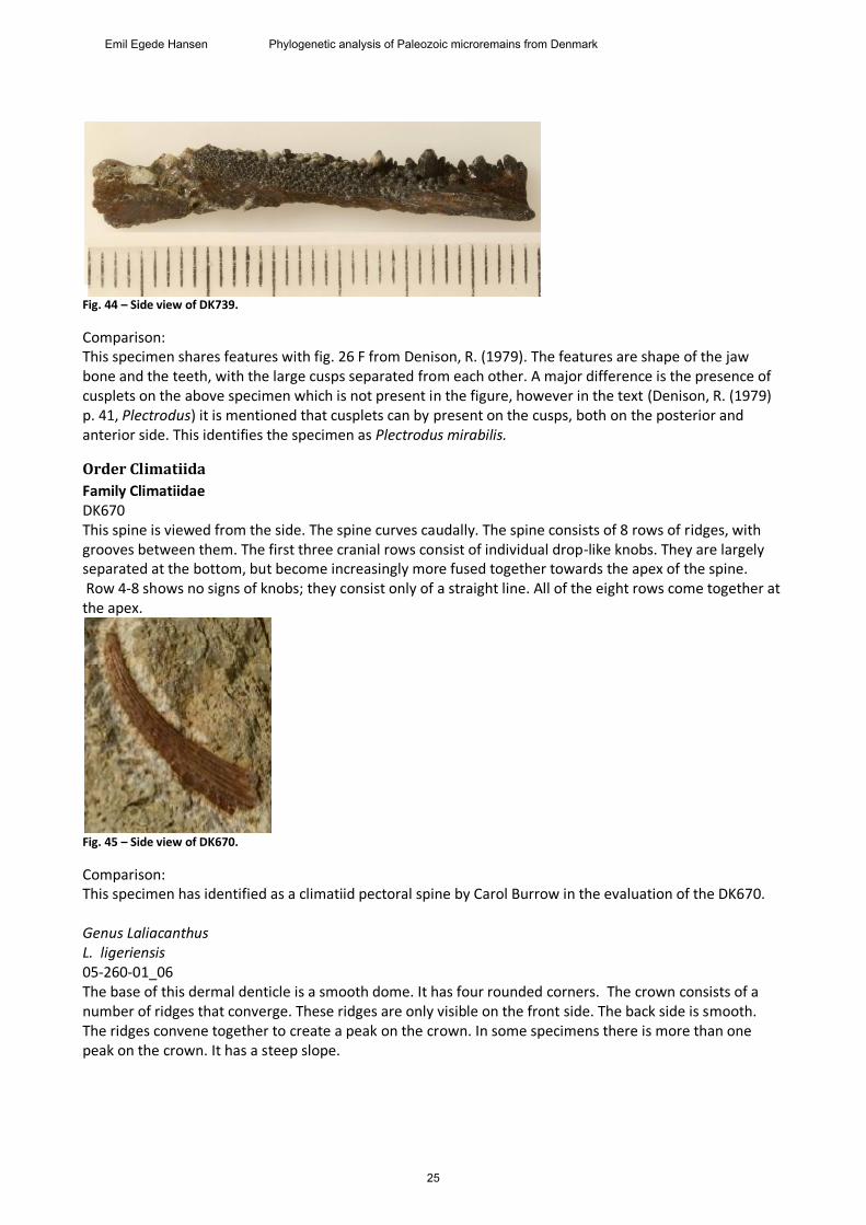
Fig. 44 – Side view of DK739.
Comparison: This specimen shares features with fig. 26 F from Denison, R. (1979). The features are shape of the jaw bone and the teeth, with the large cusps separated from each other. A major difference is the presence of cusplets on the above specimen which is not present in the figure, however in the text (Denison, R. (1979) p. 41, Plectrodus) it is mentioned that cusplets can by present on the cusps, both on the posterior and anterior side. This identifies the specimen as Plectrodus mirabilis.
Order Climatiida
Family Climatiidae DK670 This spine is viewed from the side. The spine curves caudally. The spine consists of 8 rows of ridges, with grooves between them. The first three cranial rows consist of individual drop-like knobs. They are largely separated at the bottom, but become increasingly more fused together towards the apex of the spine. Row 4-8 shows no signs of knobs; they consist only of a straight line. All of the eight rows come together at the apex.
Fig. 45 – Side view of DK670.
Comparison: This specimen has identified as a climatiid pectoral spine by Carol Burrow in the evaluation of the DK670. Genus Laliacanthus L. ligeriensis 05-260-01_06 The base of this dermal denticle is a smooth dome. It has four rounded corners. The crown consists of a number of ridges that converge. These ridges are only visible on the front side. The back side is smooth. The ridges convene together to create a peak on the crown. In some specimens there is more than one peak on the crown. It has a steep slope.
25
Emil Egede Hansen Phylogenetic analysis of Paleozoic microremains from Denmark
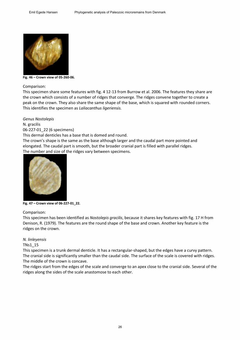
Fig. 46 – Crown view of 05-260-06.
Comparison: This specimen share some features with fig. 4 12-13 from Burrow et al. 2006. The features they share are the crown which consists of a number of ridges that converge. The ridges convene together to create a peak on the crown. They also share the same shape of the base, which is squared with rounded corners. This identifies the specimen as Laliacanthus ligeriensis. Genus Nostolepis N. gracilis 06-227-01_22 (6 specimens) This dermal denticles has a base that is domed and round. The crown’s shape is the same as the base although larger and the caudal part more pointed and elongated. The caudal part is smooth, but the broader cranial part is filled with parallel ridges. The number and size of the ridges vary between specimens.
Fig. 47 – Crown view of 06-227-01_22.
Comparison: This specimen has been identified as Nostolepis gracilis, because it shares key features with fig. 17 H from Denison, R. (1979). The features are the round shape of the base and crown. Another key feature is the ridges on the crown. N. linleyensis TNs1_15 This specimen is a trunk dermal denticle. It has a rectangular-shaped, but the edges have a curvy pattern. The cranial side is significantly smaller than the caudal side. The surface of the scale is covered with ridges. The middle of the crown is concave. The ridges start from the edges of the scale and converge to an apex close to the cranial side. Several of the ridges along the sides of the scale anastomose to each other.
26
Emil Egede Hansen Phylogenetic analysis of Paleozoic microremains from Denmark
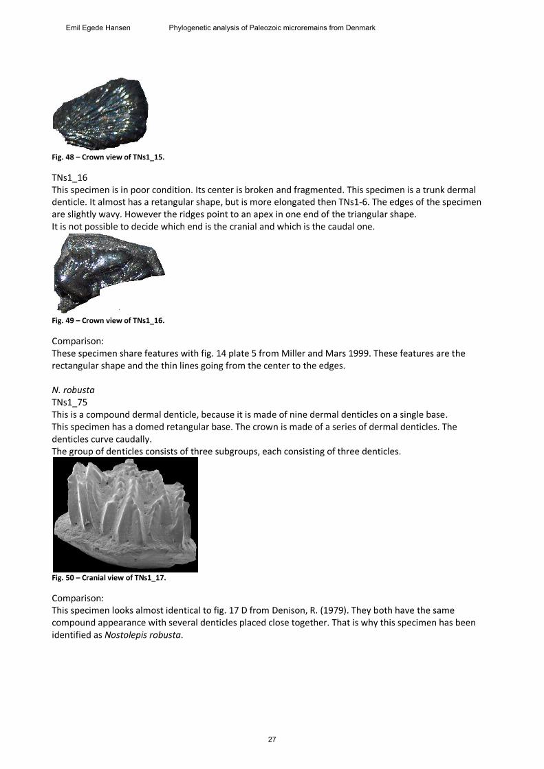
Fig. 48 – Crown view of TNs1_15.
TNs1_16 This specimen is in poor condition. Its center is broken and fragmented. This specimen is a trunk dermal denticle. It almost has a retangular shape, but is more elongated then TNs1-6. The edges of the specimen are slightly wavy. However the ridges point to an apex in one end of the triangular shape. It is not possible to decide which end is the cranial and which is the caudal one.
Fig. 49 – Crown view of TNs1_16.
Comparison: These specimen share features with fig. 14 plate 5 from Miller and Mars 1999. These features are the rectangular shape and the thin lines going from the center to the edges. N. robusta TNs1_75 This is a compound dermal denticle, because it is made of nine dermal denticles on a single base. This specimen has a domed retangular base. The crown is made of a series of dermal denticles. The denticles curve caudally. The group of denticles consists of three subgroups, each consisting of three denticles.
Fig. 50 – Cranial view of TNs1_17.
Comparison: This specimen looks almost identical to fig. 17 D from Denison, R. (1979). They both have the same compound appearance with several denticles placed close together. That is why this specimen has been identified as Nostolepis robusta.
27
Emil Egede Hansen Phylogenetic analysis of Paleozoic microremains from Denmark
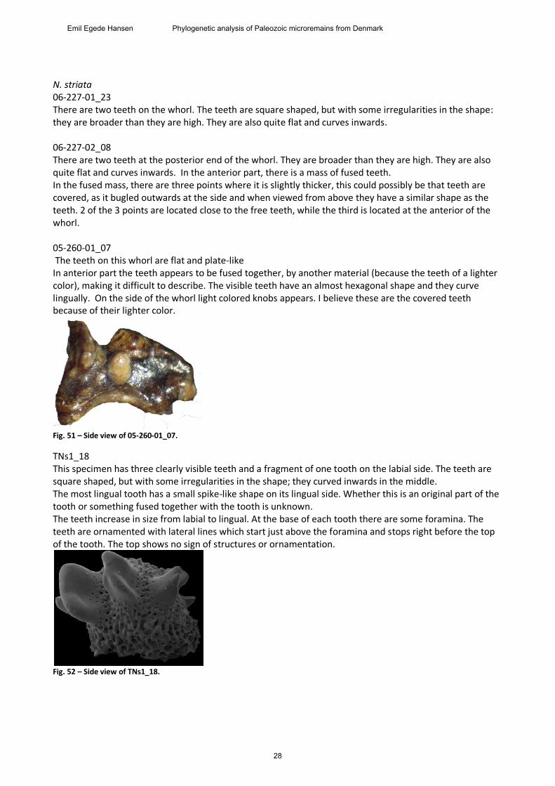
N. striata 06-227-01_23 There are two teeth on the whorl. The teeth are square shaped, but with some irregularities in the shape: they are broader than they are high. They are also quite flat and curves inwards. 06-227-02_08 There are two teeth at the posterior end of the whorl. They are broader than they are high. They are also quite flat and curves inwards. In the anterior part, there is a mass of fused teeth. In the fused mass, there are three points where it is slightly thicker, this could possibly be that teeth are covered, as it bugled outwards at the side and when viewed from above they have a similar shape as the teeth. 2 of the 3 points are located close to the free teeth, while the third is located at the anterior of the whorl. 05-260-01_07 The teeth on this whorl are flat and plate-like In anterior part the teeth appears to be fused together, by another material (because the teeth of a lighter color), making it difficult to describe. The visible teeth have an almost hexagonal shape and they curve lingually. On the side of the whorl light colored knobs appears. I believe these are the covered teeth because of their lighter color.
Fig. 51 – Side view of 05-260-01_07.
TNs1_18 This specimen has three clearly visible teeth and a fragment of one tooth on the labial side. The teeth are square shaped, but with some irregularities in the shape; they curved inwards in the middle. The most lingual tooth has a small spike-like shape on its lingual side. Whether this is an original part of the tooth or something fused together with the tooth is unknown. The teeth increase in size from labial to lingual. At the base of each tooth there are some foramina. The teeth are ornamented with lateral lines which start just above the foramina and stops right before the top of the tooth. The top shows no sign of structures or ornamentation.
Fig. 52 – Side view of TNs1_18.
28
Emil Egede Hansen Phylogenetic analysis of Paleozoic microremains from Denmark
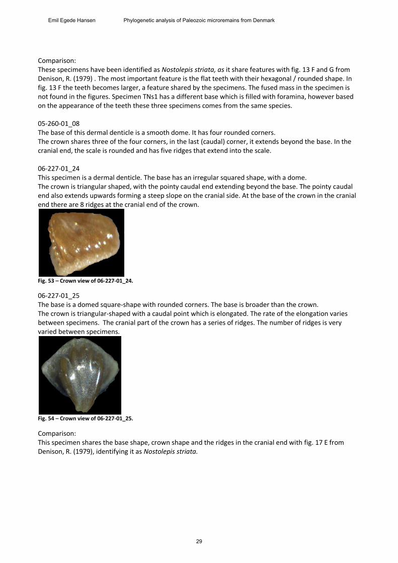
Comparison: These specimens have been identified as Nostolepis striata, as it share features with fig. 13 F and G from Denison, R. (1979) . The most important feature is the flat teeth with their hexagonal / rounded shape. In fig. 13 F the teeth becomes larger, a feature shared by the specimens. The fused mass in the specimen is not found in the figures. Specimen TNs1 has a different base which is filled with foramina, however based on the appearance of the teeth these three specimens comes from the same species. 05-260-01_08 The base of this dermal denticle is a smooth dome. It has four rounded corners. The crown shares three of the four corners, in the last (caudal) corner, it extends beyond the base. In the cranial end, the scale is rounded and has five ridges that extend into the scale. 06-227-01_24 This specimen is a dermal denticle. The base has an irregular squared shape, with a dome. The crown is triangular shaped, with the pointy caudal end extending beyond the base. The pointy caudal end also extends upwards forming a steep slope on the cranial side. At the base of the crown in the cranial end there are 8 ridges at the cranial end of the crown.
Fig. 53 – Crown view of 06-227-01_24.
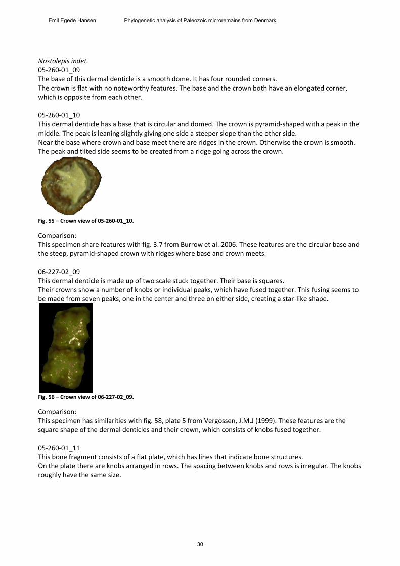
06-227-01_25 The base is a domed square-shape with rounded corners. The base is broader than the crown. The crown is triangular-shaped with a caudal point which is elongated. The rate of the elongation varies between specimens. The cranial part of the crown has a series of ridges. The number of ridges is very varied between specimens.
Fig. 54 – Crown view of 06-227-01_25.
Comparison: This specimen shares the base shape, crown shape and the ridges in the cranial end with fig. 17 E from Denison, R. (1979), identifying it as Nostolepis striata.
29
Emil Egede Hansen Phylogenetic analysis of Paleozoic microremains from Denmark
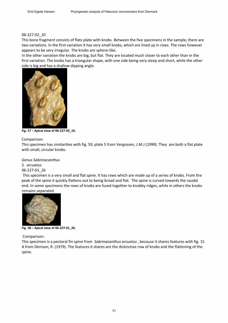
Nostolepis indet. 05-260-01_09 The base of this dermal denticle is a smooth dome. It has four rounded corners. The crown is flat with no noteworthy features. The base and the crown both have an elongated corner, which is opposite from each other. 05-260-01_10 This dermal denticle has a base that is circular and domed. The crown is pyramid-shaped with a peak in the middle. The peak is leaning slightly giving one side a steeper slope than the other side. Near the base where crown and base meet there are ridges in the crown. Otherwise the crown is smooth. The peak and tilted side seems to be created from a ridge going across the crown.
Fig. 55 – Crown view of 05-260-01_10.
Comparison: This specimen share features with fig. 3.7 from Burrow et al. 2006. These features are the circular base and the steep, pyramid-shaped crown with ridges where base and crown meets. 06-227-02_09 This dermal denticle is made up of two scale stuck together. Their base is squares. Their crowns show a number of knobs or individual peaks, which have fused together. This fusing seems to be made from seven peaks, one in the center and three on either side, creating a star-like shape.
Fig. 56 – Crown view of 06-227-02_09.
Comparison: This specimen has similarities with fig. 58, plate 5 from Vergossen, J.M.J (1999). These features are the square shape of the dermal denticles and their crown, which consists of knobs fused together. 05-260-01_11 This bone fragment consists of a flat plate, which has lines that indicate bone structures. On the plate there are knobs arranged in rows. The spacing between knobs and rows is irregular. The knobs roughly have the same size.
30
Emil Egede Hansen Phylogenetic analysis of Paleozoic microremains from Denmark
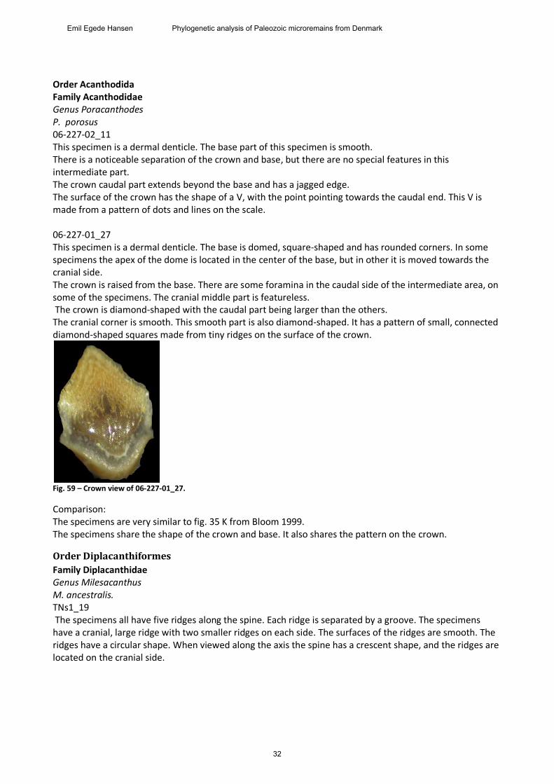
06-227-02_10 This bone fragment consists of flats plate with knobs. Between the five specimens in the sample, there are two variations. In the first variation it has very small knobs, which are lined up in rows. The rows however appears to be very irregular. The knobs are sphere-like. In the other variation the knobs are big, but flat. They are located much closer to each other than in the first variation. The knobs has a triangular shape, with one side being very steep and short, while the other side is big and has a shallow dipping angle.
Fig. 57 – Apical view of 06-227-02_10.
Comparison: This specimen has similarities with fig. 59, plate 5 from Vergossen, J.M.J (1999). They are both a flat plate with small, circular knobs. Genus Sabrinacanthus S. arcuatus 06-227-01_26 This specimen is a very small and flat spine. It has rows which are made up of a series of knobs. From the peak of the spine it quickly flattens out to being broad and flat. The spine is curved towards the caudal end. In some specimens the rows of knobs are fused together to knobby ridges, while in others the knobs remains separated.
Fig. 58 – Apical view of 06-227-01_26.
Comparison: This specimen is a pectoral fin spine from Sabrinacanthus arcuatus , because it shares features with fig. 15 A from Denison, R. (1979). The features it shares are the distinctive row of knobs and the flattening of the spine.
31
Emil Egede Hansen Phylogenetic analysis of Paleozoic microremains from Denmark
Order Acanthodida Family Acanthodidae Genus Poracanthodes P. porosus 06-227-02_11 This specimen is a dermal denticle. The base part of this specimen is smooth. There is a noticeable separation of the crown and base, but there are no special features in this intermediate part. The crown caudal part extends beyond the base and has a jagged edge. The surface of the crown has the shape of a V, with the point pointing towards the caudal end. This V is made from a pattern of dots and lines on the scale. 06-227-01_27 This specimen is a dermal denticle. The base is domed, square-shaped and has rounded corners. In some specimens the apex of the dome is located in the center of the base, but in other it is moved towards the cranial side. The crown is raised from the base. There are some foramina in the caudal side of the intermediate area, on some of the specimens. The cranial middle part is featureless. The crown is diamond-shaped with the caudal part being larger than the others. The cranial corner is smooth. This smooth part is also diamond-shaped. It has a pattern of small, connected diamond-shaped squares made from tiny ridges on the surface of the crown.
Fig. 59 – Crown view of 06-227-01_27.
Comparison: The specimens are very similar to fig. 35 K from Bloom 1999. The specimens share the shape of the crown and base. It also shares the pattern on the crown.
Order Diplacanthiformes
Family Diplacanthidae Genus Milesacanthus M. ancestralis. TNs1_19 The specimens all have five ridges along the spine. Each ridge is separated by a groove. The specimens have a cranial, large ridge with two smaller ridges on each side. The surfaces of the ridges are smooth. The ridges have a circular shape. When viewed along the axis the spine has a crescent shape, and the ridges are located on the cranial side.
32
Emil Egede Hansen Phylogenetic analysis of Paleozoic microremains from Denmark
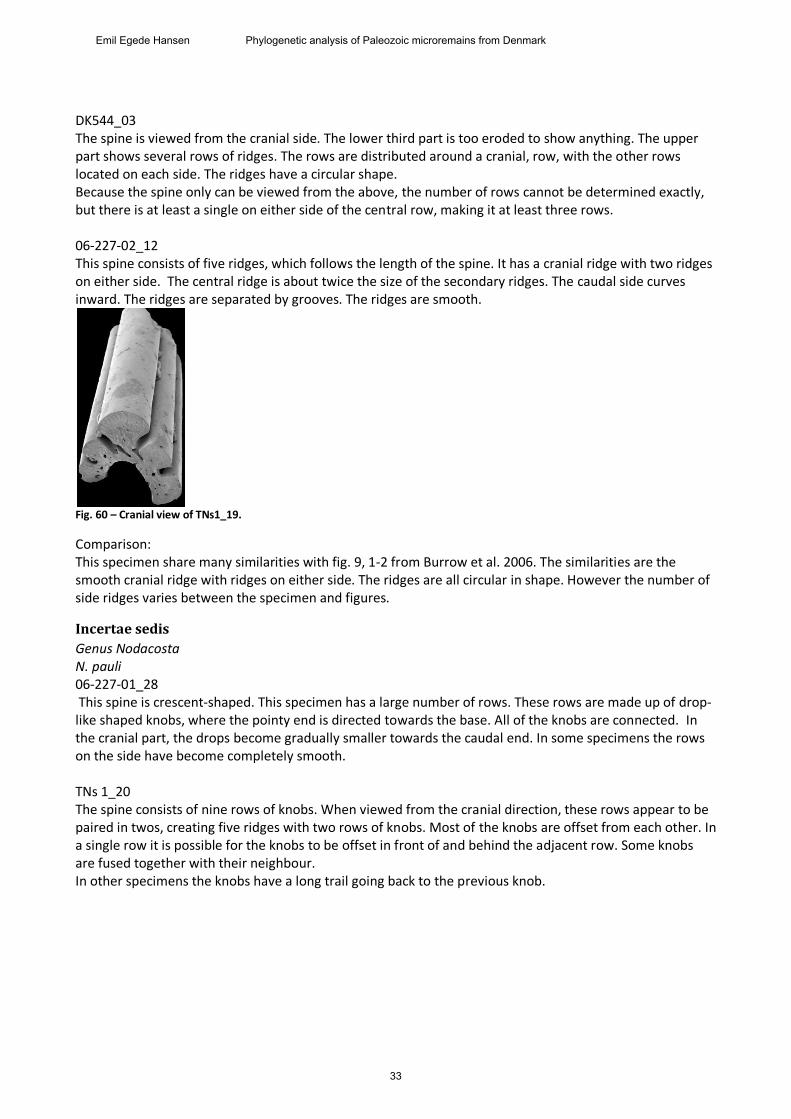
DK544_03 The spine is viewed from the cranial side. The lower third part is too eroded to show anything. The upper part shows several rows of ridges. The rows are distributed around a cranial, row, with the other rows located on each side. The ridges have a circular shape. Because the spine only can be viewed from the above, the number of rows cannot be determined exactly, but there is at least a single on either side of the central row, making it at least three rows. 06-227-02_12 This spine consists of five ridges, which follows the length of the spine. It has a cranial ridge with two ridges on either side. The central ridge is about twice the size of the secondary ridges. The caudal side curves inward. The ridges are separated by grooves. The ridges are smooth.
Fig. 60 – Cranial view of TNs1_19.
Comparison: This specimen share many similarities with fig. 9, 1-2 from Burrow et al. 2006. The similarities are the smooth cranial ridge with ridges on either side. The ridges are all circular in shape. However the number of side ridges varies between the specimen and figures.
Incertae sedis
Genus Nodacosta N. pauli 06-227-01_28 This spine is crescent-shaped. This specimen has a large number of rows. These rows are made up of drop-like shaped knobs, where the pointy end is directed towards the base. All of the knobs are connected. In the cranial part, the drops become gradually smaller towards the caudal end. In some specimens the rows on the side have become completely smooth. TNs 1_20 The spine consists of nine rows of knobs. When viewed from the cranial direction, these rows appear to be paired in twos, creating five ridges with two rows of knobs. Most of the knobs are offset from each other. In a single row it is possible for the knobs to be offset in front of and behind the adjacent row. Some knobs are fused together with their neighbour. In other specimens the knobs have a long trail going back to the previous knob.
33
Emil Egede Hansen Phylogenetic analysis of Paleozoic microremains from Denmark
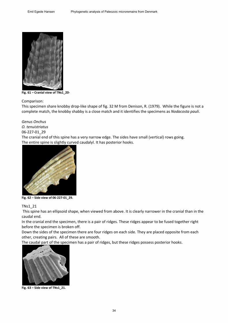
Fig. 61 – Cranial view of TNs1_20-
Comparison: This specimen share knobby drop-like shape of fig. 32 M from Denison, R. (1979). While the figure is not a complete match, the knobby shabby is a close match and it identifies the specimens as Nodacosta pauli. Genus Onchus O. tenuistriatus 06-227-01_29 The cranial end of this spine has a very narrow edge. The sides have small (vertical) rows going. The entire spine is slightly curved caudalyl. It has posterior hooks.
Fig. 62 – Side view of 06-227-01_29.
TNs1_21 This spine has an ellipsoid shape, when viewed from above. It is clearly narrower in the cranial than in the caudal end. In the cranial end the specimen, there is a pair of ridges. These ridges appear to be fused together right before the specimen is broken off. Down the sides of the specimen there are four ridges on each side. They are placed opposite from each other, creating pairs. All of these are smooth. The caudal part of the specimen has a pair of ridges, but these ridges possess posterior hooks.
Fig. 63 – Side view of TNs1_21.
34
Emil Egede Hansen Phylogenetic analysis of Paleozoic microremains from Denmark
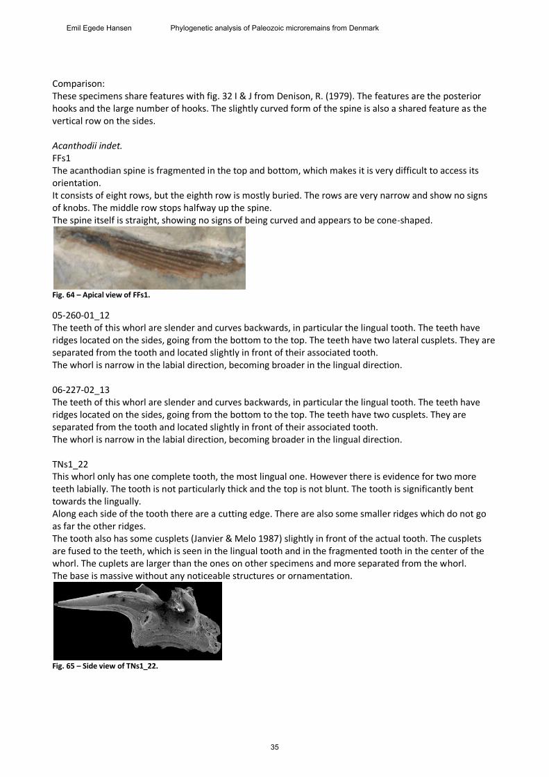
Comparison: These specimens share features with fig. 32 I & J from Denison, R. (1979). The features are the posterior hooks and the large number of hooks. The slightly curved form of the spine is also a shared feature as the vertical row on the sides. Acanthodii indet. FFs1 The acanthodian spine is fragmented in the top and bottom, which makes it is very difficult to access its orientation. It consists of eight rows, but the eighth row is mostly buried. The rows are very narrow and show no signs of knobs. The middle row stops halfway up the spine. The spine itself is straight, showing no signs of being curved and appears to be cone-shaped.
Fig. 64 – Apical view of FFs1.
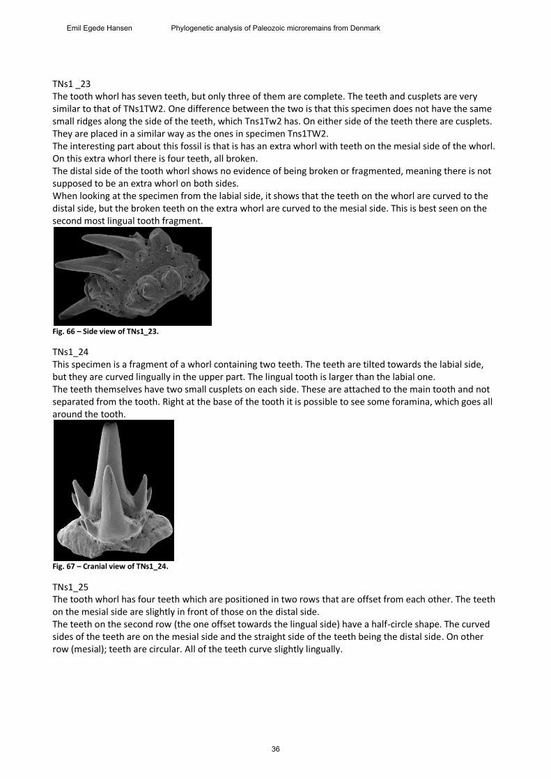
05-260-01_12 The teeth of this whorl are slender and curves backwards, in particular the lingual tooth. The teeth have ridges located on the sides, going from the bottom to the top. The teeth have two lateral cusplets. They are separated from the tooth and located slightly in front of their associated tooth. The whorl is narrow in the labial direction, becoming broader in the lingual direction. 06-227-02_13 The teeth of this whorl are slender and curves backwards, in particular the lingual tooth. The teeth have ridges located on the sides, going from the bottom to the top. The teeth have two cusplets. They are separated from the tooth and located slightly in front of their associated tooth. The whorl is narrow in the labial direction, becoming broader in the lingual direction. TNs1_22 This whorl only has one complete tooth, the most lingual one. However there is evidence for two more teeth labially. The tooth is not particularly thick and the top is not blunt. The tooth is significantly bent towards the lingually. Along each side of the tooth there are a cutting edge. There are also some smaller ridges which do not go as far the other ridges. The tooth also has some cusplets (Janvier & Melo 1987) slightly in front of the actual tooth. The cusplets are fused to the teeth, which is seen in the lingual tooth and in the fragmented tooth in the center of the whorl. The cuplets are larger than the ones on other specimens and more separated from the whorl. The base is massive without any noticeable structures or ornamentation.
Fig. 65 – Side view of TNs1_22.
35
Emil Egede Hansen Phylogenetic analysis of Paleozoic microremains from Denmark
TNs1 _23 The tooth whorl has seven teeth, but only three of them are complete. The teeth and cusplets are very similar to that of TNs1TW2. One difference between the two is that this specimen does not have the same small ridges along the side of the teeth, which Tns1Tw2 has. On either side of the teeth there are cusplets. They are placed in a similar way as the ones in specimen Tns1TW2. The interesting part about this fossil is that is has an extra whorl with teeth on the mesial side of the whorl. On this extra whorl there is four teeth, all broken. The distal side of the tooth whorl shows no evidence of being broken or fragmented, meaning there is not supposed to be an extra whorl on both sides. When looking at the specimen from the labial side, it shows that the teeth on the whorl are curved to the distal side, but the broken teeth on the extra whorl are curved to the mesial side. This is best seen on the second most lingual tooth fragment.
Fig. 66 – Side view of TNs1_23.
TNs1_24 This specimen is a fragment of a whorl containing two teeth. The teeth are tilted towards the labial side, but they are curved lingually in the upper part. The lingual tooth is larger than the labial one. The teeth themselves have two small cusplets on each side. These are attached to the main tooth and not separated from the tooth. Right at the base of the tooth it is possible to see some foramina, which goes all around the tooth.
Fig. 67 – Cranial view of TNs1_24.
TNs1_25 The tooth whorl has four teeth which are positioned in two rows that are offset from each other. The teeth on the mesial side are slightly in front of those on the distal side. The teeth on the second row (the one offset towards the lingual side) have a half-circle shape. The curved sides of the teeth are on the mesial side and the straight side of the teeth being the distal side. On other row (mesial); teeth are circular. All of the teeth curve slightly lingually.
36
Emil Egede Hansen Phylogenetic analysis of Paleozoic microremains from Denmark
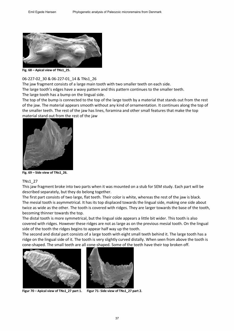
Fig. 68 – Apical view of TNs1_25.
06-227-02_30 & 06-227-01_14 & TNs1_26 The jaw fragment consists of a large main tooth with two smaller teeth on each side. The large tooth’s edges have a wavy pattern and this pattern continues to the smaller teeth. The large tooth has a bump on the lingual side. The top of the bump is connected to the top of the large tooth by a material that stands out from the rest of the jaw. The material appears smooth without any kind of ornamentation. It continues along the top of the smaller teeth. The rest of the jaw has lines, foramina and other small features that make the top material stand out from the rest of the jaw
Fig. 69 – Side view of TNs1_26.
TNs1_27 This jaw fragment broke into two parts when it was mounted on a stub for SEM study. Each part will be described separately, but they do belong together. The first part consists of two large, flat teeth. Their color is white, whereas the rest of the jaw is black. The mesial tooth is asymmetrical. It has its top displaced towards the lingual side, making one side about twice as wide as the other. The tooth is covered with ridges. They are larger towards the base of the tooth, becoming thinner towards the top. The distal tooth is more symmetrical, but the lingual side appears a little bit wider. This tooth is also covered with ridges. However these ridges are not as large as on the previous mesial tooth. On the lingual side of the tooth the ridges begins to appear half way up the tooth. The second and distal part consists of a large tooth with eight small teeth behind it. The large tooth has a ridge on the lingual side of it. The tooth is very slightly curved distally. When seen from above the tooth is cone-shaped. The small teeth are all cone-shaped. Some of the teeth have their top broken off.
Figur 70 – Apical view of TNs1_27 part 1. Figur 71- Side view of TNs1_27 part 2.
37
Emil Egede Hansen Phylogenetic analysis of Paleozoic microremains from Denmark
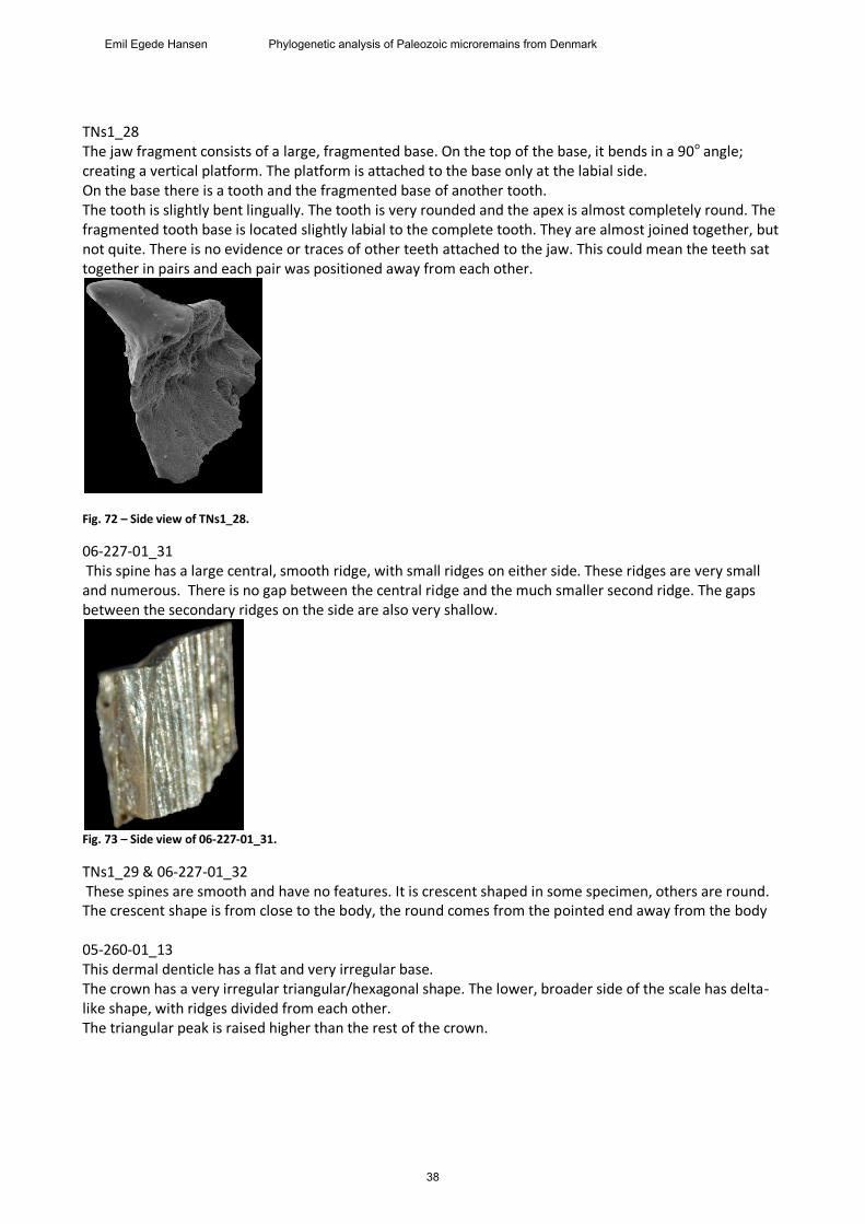
TNs1_28 The jaw fragment consists of a large, fragmented base. On the top of the base, it bends in a 90o angle; creating a vertical platform. The platform is attached to the base only at the labial side. On the base there is a tooth and the fragmented base of another tooth. The tooth is slightly bent lingually. The tooth is very rounded and the apex is almost completely round. The fragmented tooth base is located slightly labial to the complete tooth. They are almost joined together, but not quite. There is no evidence or traces of other teeth attached to the jaw. This could mean the teeth sat together in pairs and each pair was positioned away from each other.
Fig. 72 – Side view of TNs1_28.
06-227-01_31 This spine has a large central, smooth ridge, with small ridges on either side. These ridges are very small and numerous. There is no gap between the central ridge and the much smaller second ridge. The gaps between the secondary ridges on the side are also very shallow.
Fig. 73 – Side view of 06-227-01_31.
TNs1_29 & 06-227-01_32 These spines are smooth and have no features. It is crescent shaped in some specimen, others are round. The crescent shape is from close to the body, the round comes from the pointed end away from the body 05-260-01_13 This dermal denticle has a flat and very irregular base. The crown has a very irregular triangular/hexagonal shape. The lower, broader side of the scale has delta-like shape, with ridges divided from each other. The triangular peak is raised higher than the rest of the crown.
38
Emil Egede Hansen Phylogenetic analysis of Paleozoic microremains from Denmark
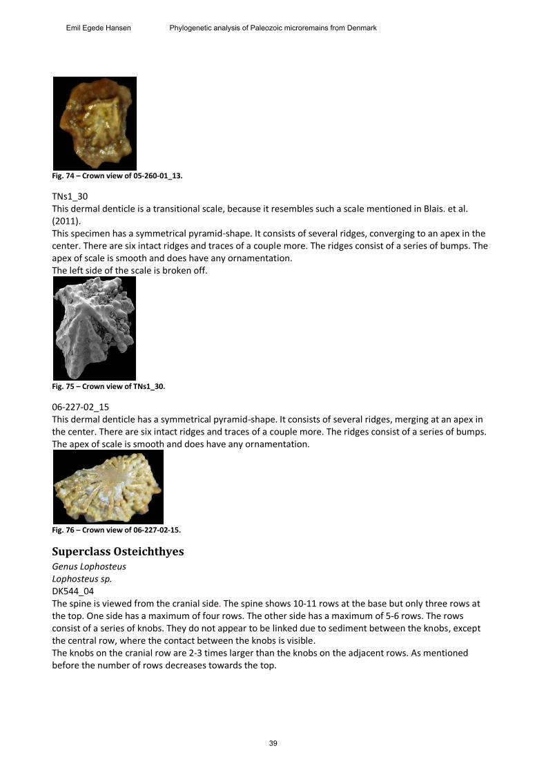
Fig. 74 – Crown view of 05-260-01_13.
TNs1_30 This dermal denticle is a transitional scale, because it resembles such a scale mentioned in Blais. et al. (2011). This specimen has a symmetrical pyramid-shape. It consists of several ridges, converging to an apex in the center. There are six intact ridges and traces of a couple more. The ridges consist of a series of bumps. The apex of scale is smooth and does have any ornamentation. The left side of the scale is broken off.
Fig. 75 – Crown view of TNs1_30.
06-227-02_15 This dermal denticle has a symmetrical pyramid-shape. It consists of several ridges, merging at an apex in the center. There are six intact ridges and traces of a couple more. The ridges consist of a series of bumps. The apex of scale is smooth and does have any ornamentation.
Fig. 76 – Crown view of 06-227-02-15.
Superclass Osteichthyes Genus Lophosteus Lophosteus sp. DK544_04 The spine is viewed from the cranial side. The spine shows 10-11 rows at the base but only three rows at the top. One side has a maximum of four rows. The other side has a maximum of 5-6 rows. The rows consist of a series of knobs. They do not appear to be linked due to sediment between the knobs, except the central row, where the contact between the knobs is visible. The knobs on the cranial row are 2-3 times larger than the knobs on the adjacent rows. As mentioned before the number of rows decreases towards the top.
39
Emil Egede Hansen Phylogenetic analysis of Paleozoic microremains from Denmark
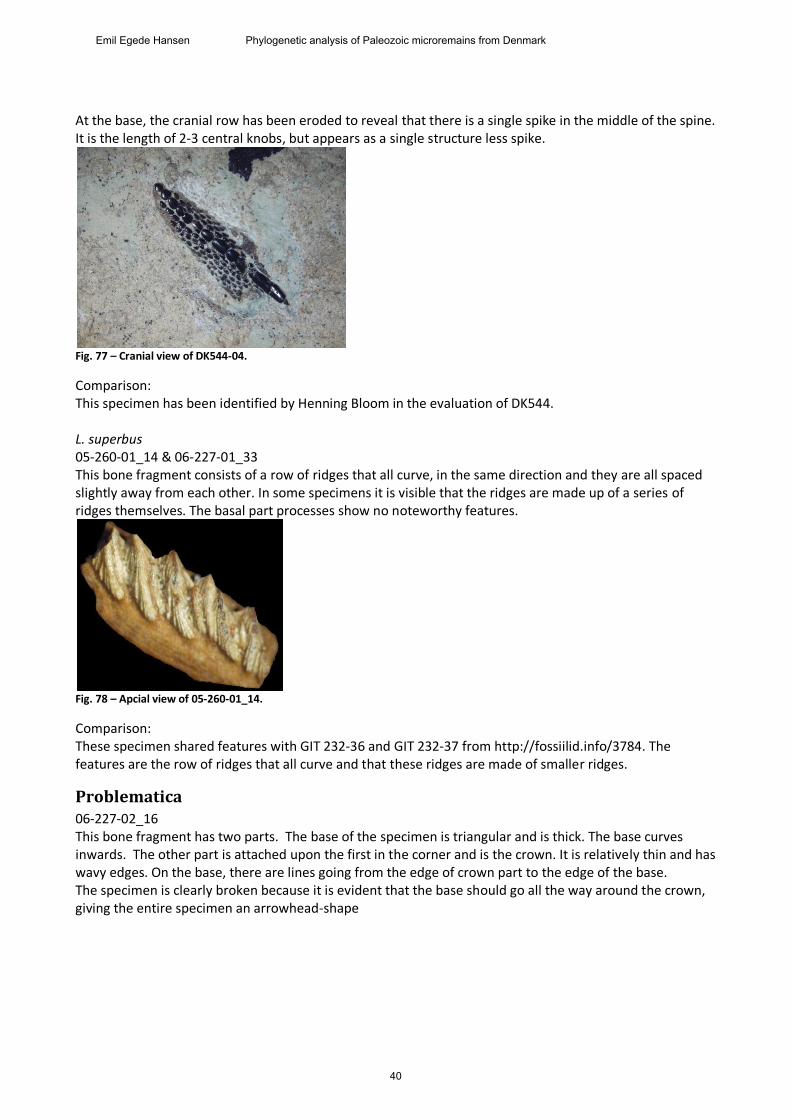
At the base, the cranial row has been eroded to reveal that there is a single spike in the middle of the spine. It is the length of 2-3 central knobs, but appears as a single structure less spike.
Fig. 77 – Cranial view of DK544-04.
Comparison: This specimen has been identified by Henning Bloom in the evaluation of DK544. L. superbus 05-260-01_14 & 06-227-01_33 This bone fragment consists of a row of ridges that all curve, in the same direction and they are all spaced slightly away from each other. In some specimens it is visible that the ridges are made up of a series of ridges themselves. The basal part processes show no noteworthy features.
Fig. 78 – Apcial view of 05-260-01_14.
Comparison: These specimen shared features with GIT 232-36 and GIT 232-37 from http://fossiilid.info/3784. The features are the row of ridges that all curve and that these ridges are made of smaller ridges.
Problematica 06-227-02_16 This bone fragment has two parts. The base of the specimen is triangular and is thick. The base curves inwards. The other part is attached upon the first in the corner and is the crown. It is relatively thin and has wavy edges. On the base, there are lines going from the edge of crown part to the edge of the base. The specimen is clearly broken because it is evident that the base should go all the way around the crown, giving the entire specimen an arrowhead-shape
40
Emil Egede Hansen Phylogenetic analysis of Paleozoic microremains from Denmark
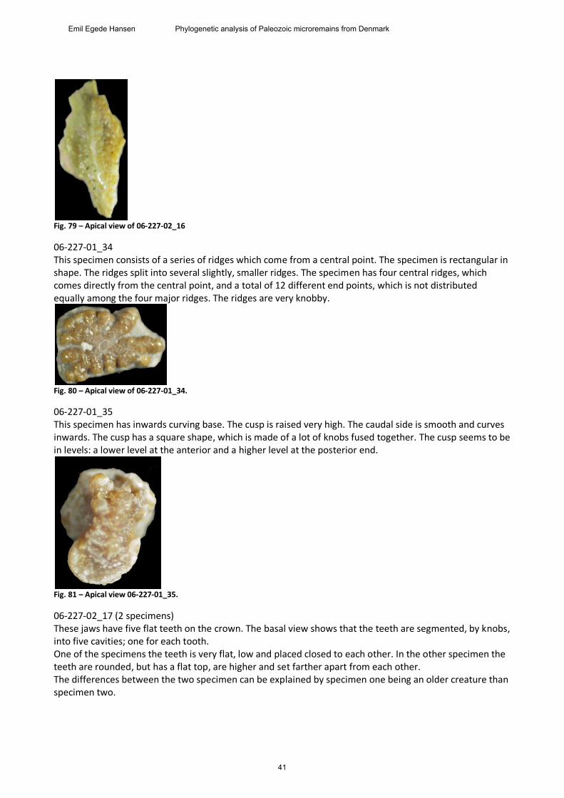
Fig. 79 – Apical view of 06-227-02_16
06-227-01_34 This specimen consists of a series of ridges which come from a central point. The specimen is rectangular in shape. The ridges split into several slightly, smaller ridges. The specimen has four central ridges, which comes directly from the central point, and a total of 12 different end points, which is not distributed equally among the four major ridges. The ridges are very knobby.
Fig. 80 – Apical view of 06-227-01_34.
06-227-01_35 This specimen has inwards curving base. The cusp is raised very high. The caudal side is smooth and curves inwards. The cusp has a square shape, which is made of a lot of knobs fused together. The cusp seems to be in levels: a lower level at the anterior and a higher level at the posterior end.
Fig. 81 – Apical view 06-227-01_35.
06-227-02_17 (2 specimens) These jaws have five flat teeth on the crown. The basal view shows that the teeth are segmented, by knobs, into five cavities; one for each tooth. One of the specimens the teeth is very flat, low and placed closed to each other. In the other specimen the teeth are rounded, but has a flat top, are higher and set farther apart from each other. The differences between the two specimen can be explained by specimen one being an older creature than specimen two.
41
Emil Egede Hansen Phylogenetic analysis of Paleozoic microremains from Denmark
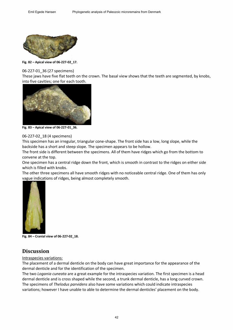
Fig. 82 – Apical view of 06-227-02_17.
06-227-01_36 (27 specimens) These jaws have five flat teeth on the crown. The basal view shows that the teeth are segmented, by knobs, into five cavities; one for each tooth.
Fig. 83 – Apical view of 06-227-01_36.
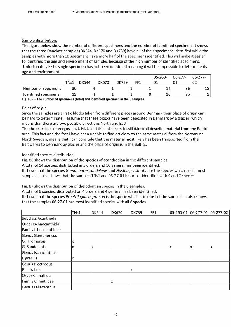
06-227-02_18 (4 specimens) This specimen has an irregular, triangular cone-shape. The front side has a low, long slope, while the backside has a short and steep slope. The specimen appears to be hollow. The front side is different between the specimens. All of them have ridges which go from the bottom to convene at the top. One specimen has a central ridge down the front, which is smooth in contrast to the ridges on either side which is filled with knobs. The other three specimens all have smooth ridges with no noticeable central ridge. One of them has only vague indications of ridges, being almost completely smooth.
Fig. 84 – Cranial view of 06-227-02_18.
Discussion Intraspecies variations: The placement of a dermal denticle on the body can have great importance for the appearance of the dermal denticle and for the identification of the specimen. The two Logania cuneata are a great example for the intraspecies variation. The first specimen is a head dermal denticle and is cross shaped while the second, a trunk dermal denticle, has a long curved crown. The specimens of Thelodus parvidens also have some variations which could indicate intraspecies variations; however I have unable to able to determine the dermal denticles’ placement on the body.
42
Emil Egede Hansen Phylogenetic analysis of Paleozoic microremains from Denmark
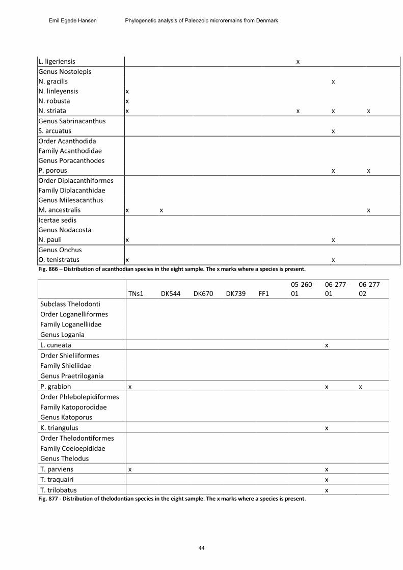
Sample distribution. The figure below show the number of different specimens and the number of identified specimen. It shows that the three Danekræ samples (DK544, DK670 and DK739) have all of their specimens identified while the samples with more than 10 specimens have more half of the specimens identified. This will make it easier to identified the age and environment of samples because of the high number of identified specimens. Unfortunately FF1’s single specimen has not been identified meaning it will be impossible to determine its age and environment.
TNs1 DK544 DK670 DK739 FF1 05-260-01
06-277-01
06-277-02
Number of specimens 30 4 1 1 1 14 36 18
Identified specimens 19 4 1 1 0 10 25 9 Fig. 855 – The number of specimens (total) and identified specimen in the 8 samples.
Point of origin. Since the samples are erratic blocks taken from different places around Denmark their place of origin can be hard to determinate. I assume that these blocks have been deposited in Denmark by a glacier, which means that there are two possible directions North and East. The three articles of Vergossen, J. M. J. and the links from fossiilid.info all describe material from the Baltic area. This fact and the fact I have been unable to find article with the same material from the Norway or North Sweden, means that I can conclude that the material most likely has been transported from the Baltic area to Denmark by glacier and the place of origin is in the Baltics. Identified species distribution Fig. 86 shows the distribution of the species of acanthodian in the different samples. A total of 14 species, distributed in 5 orders and 10 genera, has been identified. It shows that the species Gomphoncus sandelenis and Nostolepis striata are the species which are in most samples. It also shows that the samples TNs1 and 06-27-01 has most identified with 9 and 7 species. Fig. 87 shows the distribution of thelodontian species in the 8 samples. A total of 6 species, distributed on 4 orders and 4 genera, has been identified. It shows that the species Praetrilogania grabion is the specie which is in most of the samples. It also shows that the samples 06-27-01 has most identified species with all 6 species
TNs1 DK544 DK670 DK739 FF1 05-260-01 06-277-01 06-277-02
Subclass Acanthodii
Order Ischnacanthida
Family Ishnacanthidae
Genus Gomphoncus
G. Fromensis x
G. Sandelenis x x x x x
Genus Iscnacanthus
I. gracilis x
Genus Plectrodus
P. mirabilis x
Order Climatiida
Family Climatiidae x
Genus Laliacanthus
43
Emil Egede Hansen Phylogenetic analysis of Paleozoic microremains from Denmark
L. ligeriensis x
Genus Nostolepis
N. gracilis x
N. linleyensis x
N. robusta x
N. striata x x x x
Genus Sabrinacanthus
S. arcuatus x
Order Acanthodida
Family Acanthodidae
Genus Poracanthodes
P. porous x x
Order Diplacanthiformes
Family Diplacanthidae
Genus Milesacanthus
M. ancestralis x x x
Icertae sedis
Genus Nodacosta
N. pauli x x
Genus Onchus
O. tenistratus x x Fig. 866 – Distribution of acanthodian species in the eight sample. The x marks where a species is present.
TNs1 DK544 DK670 DK739 FF1 05-260-01
06-277-01
06-277-02
Subclass Thelodonti
Order Loganelliformes
Family Loganelliidae
Genus Logania
L. cuneata x
Order Shieliiformes
Family Shieliidae
Genus Praetrilogania
P. grabion x x x
Order Phlebolepidiformes
Family Katoporodidae
Genus Katoporus
K. triangulus x
Order Thelodontiformes
Family Coeloepididae
Genus Thelodus
T. parviens x x
T. traquairi x
T. trilobatus x Fig. 877 - Distribution of thelodontian species in the eight sample. The x marks where a species is present.
44
Emil Egede Hansen Phylogenetic analysis of Paleozoic microremains from Denmark
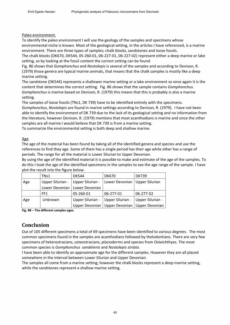
Paleo environment. To identify the paleo environment I will use the geology of the samples and specimens whose environmental niche is known. Most of the geological setting, in the articles I have referenced, is a marine environment. There are three types of samples; chalk blocks, sandstones and loose fossils. The chalk blocks (DK670, DK544, 05-260-01, 06-227-01, 06-227-02) represent either a deep marine or lake setting, so by looking at the fossil content the correct setting can be found. Fig. 86 shows that Gomphonchus and Nostolepis is several of the samples and according to Denison, R. (1979) those genera are typical marine animals, that means that the chalk samples is mostly like a deep marine setting. The sandstone (DK544) represents a shallower marine setting or a lake environment so once again it is the content that determines the correct setting. Fig. 86 shows that the sample contains Gomphonchus. Gomphonchus is marine based on Denison, R. (1979) this means that this is probably is also a marine setting. The samples of loose fossils (TNs1, DK 739) have to be identified entirely with the specimens. Gomphonchus, Nostolepis are found in marine settings according to Denison, R. (1979). I have not been able to identify the environment of DK 739 due to the lack of its geological setting and no information from the literature, however Denison, R. (1979) mentions that most acanthodians is marine and since the other samples are all marine I would believe that DK 739 is from a marine setting. To summarize the environmental setting is both deep and shallow marine. Age The age of the material has been found by taking all of the identified genera and species and use the references to find they age. Some of them has a single period has their age while other has a range of periods. The range for all the material is Lower Silurian to Upper Devonian. By using the age of the identified material it is possible to make and estimate of the age of the samples. To do this I took the age of the identified specimens in the samples to see the age range of the sample. I have plot the result into the figure below.
TNs1 DK544 DK670 DK739
Age Upper Silurian - Upper Silurian - Lower Devonian Upper Silurian
Lower Devonian Lower Devonian
FF1 05-260-01 06-277-01 06-277-02
Age Unknown Upper Silurian - Upper Silurian - Upper Silurian -
Upper Devonian Upper Devonian Upper Devonian Fig. 88 – The different samples ages.
Conclusion Out of 105 different specimens a total of 69 specimens have been identified to various degrees. The most common specimens found in the samples are acanthodians followed by thelodontians. There are very few specimens of heterostracians, osteostracians, placioderms and species from Osteichthyes. The most common species is Gomphonchus sandelenis and Nostolepis striata. I have been able to identify an approximate age for the different samples. However they are all placed somewhere in the interval between Lower Silurian and Upper Devonian. The samples all come from a marine setting; however the chalk blocks represent a deep marine setting, while the sandstones represent a shallow marine setting.
45
Emil Egede Hansen Phylogenetic analysis of Paleozoic microremains from Denmark
Acknowledgements The author would like to thank his supervisor Gilles Cuny for the excellent guidance and help he provided during entire process of making this project. The author would also like to thank Mette Hofstedt and Peter Myrhøj for allowing research on the material they found.
References Blais, S.A., MacKenzie, L.A. & Wilson, M.V.H. (2011) Tooth‐like scales in Early Devonian Eugnathostomes and the “outside‐in” hypothesis for the origin of teeth in vertebrates. Journal of Vertebrate Paleontology, vol. 31 (6), 1189‐1199. Bloom, H. (1999). Vertebrate remains from Upper Silurian‐Lower Devonian beds of Hall Land, North Greenland. Geology of Greenland Survey Bulletin 182. Burrow, C.J. (1995). Acanthodian dental elements from the Trundle beds (Lower Devonian) of New South Wales. Records of the Western Australian Museum, vol. 17, 331‐341. Burrow, C.J. (1996) PLACODERM SCALES FROM THE LOWERDEVONIAN OF NEW SOUTH WALES, AUSTRALIA. Modern Geology, vol. 20, 351-369. Burrow, C.J., Leilievre, H. & Janjou, D. (2006). Gnathostomes microremains from the Lower Devonian Jawf Formation, Saudi Arabia. Journal of Paleontology, vol. 80(3), 537‐560. Clark, D. L. & Ethington, R. L. (1967) Conodonts and zonation of the Upper Devonian in the Great Basin. The Geological Society of American Inc., 103. Denison, R. (1979). Handbook of paleoichthyology Vol. 5, Acanthodii. Verlag Dr. Friedrich Pfeil Hass, W. H. (1952) Conodonts of the Barret Formation of Texas. Geological Survey Professional Paper, Vol. 243, 69-94. Janvier, P. & Melo, J.H.G. (1987). Acanthodian fish remains from the Upper Silurian or Lower Devonian of the Amazon Basin, Brazil. Palaeontology, vol. 31, 771‐777.
Märss, T. (2006) Thelodonts (Agnatha) from the basal beds of the Kuressaare Stage, Ludlow, Upper Silurian of Estonia. Proc. Estonian Acad. Sci. Geol. Vol. 55. 43-66. Märss, T., Turner, S. & Karatajūtė-Talimaa. V.(2007) Handbook of paleoichthyology Vol. 1B, Agnatha II, thelodonti. Verlag Dr. Friedrich Pfeil Miller, C.G. & Mãrss, T. (1999). A conodont, thelodont and acanthodian fauna from Pridoli (Silurian) of the Much Wenlock area, Shropshire. Palaeontology, vol. 42, 691‐714. Vergossen, J.M.J (1999) Late Silurian fish microfossils from an East Baltic-derived erratic from Oosterhaule, with a description of new acanthodian taxa. Geologie en Mijnbouw, 78, 231–251. Vergossen, J.M.J. (2002)Late Silurian fish microfossils from Klinta and Rinnebäcks Bro(Scania, south Sweden), with remarks on the morphology ofNostolepis striata trunk scales. Scripta Geologica, 123, 71-92.
46
Emil Egede Hansen Phylogenetic analysis of Paleozoic microremains from Denmark
Vergossen, J.M.J. (2004) Fish microfossils from Ramsåsa, site E, Scania, southern Sweden (mid Palaeozoic). Scripta Geologica, 127, 1-70. Zhuralev, A., V. (2007). Morphofunctional Analysisof Late Paleozoic Conodont Elements and Apparatuses. aleontological Journal, 2007, Vol. 41, No. 5, 549–557. Links http://www.esofossiler.dk/html/Silur%20html/Siluriske%20fisk.html http://fossiilid.info/11946 http://fossiilid.info/3784
47
Emil Egede Hansen Phylogenetic analysis of Paleozoic microremains from Denmark
Recommended