Plant Electrophysiology – Theory & Methods (ed. by Volkov)© Springer-Verlag Berlin Heidelberg 2006
Plants respond actively to biotic stress by sensing and triggering cascades ofsignals that lead to the production of toxic compounds, spreading from sec-ondary metabolites to reactive oxygen species. Here, we show that the evalu-ation of plasma transmembrane potential (Vm) is a powerful tool for thedeciphering of earlier events following biotic attacks. After a short introduc-tion and definition of abiotic and biotic stress, we describe how plants reactto herbivore attack by changing Vm and how this can be measured using elec-trophysiology.
20.1 Abiotic and biotic stress
20.1.1 What is an abiotic stress?
One important feature distinguishing plants from other complex multicellu-lar organisms is that plants are static organisms and thus cannot escape envi-ronmental challenges. Abiotic stresses are caused by physical Earth’s forcessuch as salt, water, light, heat and cold stresses. Although clearly differentfrom each other in their physical nature, each of them elicit specific plantresponses as well as activate some common reactions in plants (Zhu 2001).Abiotic stresses, such as drought, salinity, extreme temperatures, chemicaltoxicity and oxidative stress are serious threats to agriculture and result in thedeterioration of the environment (Wang et al. 2003). Abiotic stress is the pri-mary cause of crop loss worldwide (more than 50% yield reduction for mostmajor crop plants; Boyer 1982; Bray et al. 2000). Abiotic stress often leads tomorphological, physiological, biochemical and molecular changes affectingplant growth and productivity (Wang et al. 2001). Abiotic stresses may acti-vate cell signaling pathways (Knight and Knight 2001; Zhu 2001, 2002) andcellular responses (Wang et al. 2003) that can lead to alteration of the trans-membrane potential (Vm). In general, Vm variations depend on unbalancedion distribution across the plasma membrane and depolarization occurs
20 Electrophysiology and Plant Responses to BioticStress
MASSIMO MAFFEI, SIMONE BOSSI
Department of Plant Biology and Centre of Excellence CEBIOVEM, University of Turin, Viale P.A. Mattioli, 25 10125Turin, Italy (e-mail: [email protected])
when cations (such as K+ and Ca2+) are allowed to enter the cell or upon anionefflux. On the other hand, hyperpolarization mainly depends on the activityof the plasma membrane H+-ATPase or when inward anion channels (or out-ward cation channels) are opened. The primary candidate for intercellular sig-naling in higher plants is the stimulus-induced change in Vm and excitationwaves transmit information from one part of the plant to another with a speedof propagation of the action potential that in soybean can reach 40 m s−1
(Shevstova et al. 2001). Since ion fluxes through channels directly influenceVm, it seems reasonable to assume that molecules able to act on channel activ-ity might be considered as important factors inducing electrical signals(Maffei et al. 2004). Under abiotic stress, the up-regulation of free radicalscavenging systems is a common component of the response (Pasternak et al.2005), as are heat stress (Dat et al. 1998; Larkindale and Knight 2002), UV-radiation stress (Brosche and Strid 2003), photoinhibition (Muller-Mouleet al. 2003), heavy metal stress (Pinto et al. 2003) and anoxia (Blokhina et al.2001). All of them may have consistent repercussions on the balance of ionsacross the plasma membrane, and hence on Vm. Emerging evidence suggestsa broader role for common signals (such as reactive oxygen species) thatmediate responses to abiotic environment, developmental cues, infection andthe programmed cell death in different cell types (Torres and Dangl 2005)making tools to detect abiotic stress responses useful to quantify other plantresponses. While trying to balance water deficits and carbon assimilation,plants must integrate additional information on light quality, nutrient statusand temperature to make “informed decisions” to add to the pressure posedby the presence of biotic stress.
20.1.2 What is a biotic stress?
As primary producers in the food chain, plants are the source of carbon, pro-tein, vitamins and minerals for all heterotrophic organisms, from bacteria tohumans. Thus we can define biotic stress as the pressure posed on plants byliving organisms. In recent years, the molecular basis of biotic stressresponses in plants (Maleck et al. 2000) has been identified (reviewed byKarpinski et al. 2003). Among biotic stress, the most studied are microbialinfections and herbivore attack. Based on their effects on the plant, microbesinteracting with plants can be classified as pathogenic, saprophytic and ben-eficial. Pathogens can attack leaves, stems or roots. Current models of themechanisms of plant defense against pathogen infection are based on animalmodels, and have been recently linked to the light-sensing network and to theoxygen-evolving complex in photosystem II (PSII) (Abbink et al. 2002). Muchprogress has been made in understanding the mechanisms by which plantsdetect and defend themselves against pathogens (Kunkel and Brooks 2002).Progress has been done in cloning and characterization of plant diseaseresistance genes that govern the recognition of specific pathogen strains
462 Massimo Maffei and Simone Bossi
(Staskawicz et al. 2001; Dangl and Jones 2001), the deciphering of signaltransduction pathways for the activation of defense responses (Feys andParker 2000; Glazebrook 2001), and the characterization of endogenous plantsignaling molecules involved in plant defense [salicylic acid (SA), jasmonicacid (JA) and ethylene (ET) (Dong 1998; Thomma et al. 2001)]. The currentadvances of the roles of the SA, JA and ET signaling pathways in pathogendefense has been summarized in several recent reviews (Kunkel and Brooks2002 and references therein). There is also a growing body of literature thatreports that the JA, SA and ET defense signaling pathways do not functionindependently, but show an active crosstalk (Kunkel and Brooks 2002).Recent studies indicate that defense signaling may be even more complexthan expected, and that additional plant signaling pathways are likely to beinvolved in regulating pathogen defense, most of them involving ion fluxesand then variations in Vm.
Recognition is considered to be the initial key event in the response ofplants to microbes. Recognition can occur through physical interaction, suchas through adhesins, fimbriae, flagella, and type III and type IV secretion sys-tems, or through signaling by small molecules (Lugtenberg et al. 2002). Earlyevents during pathogen attack, before gene expression, involve the release ofcell wall oligosaccharides (so-called elicitors) which can be recognized byspecific receptors able to trigger signaling cascades involving ion fluxes andactivation of reactive oxygen species (ROS) forming enzymes (Kombrink andSomssich 1995). One of the two of the earliest occurrences following recogni-tion are a calcium flux across the plasmalemma and the generation of O2
− andH2O2, the so-called “oxidative burst” (Mur et al. 2005) with these two eventsappearing to be mutually regulated (Grant et al. 2000). The generation of theplant oxidative burst has been linked to the initiation of electron flow acrossthe plasmalemma via a NADPH oxidase complex, analogous to that found inmammalian neutrophils (Mur et al. 2005).
The evolution of plant secondary compounds is often considered to betightly associated with defense against biotic stress, and it has recently beenproposed that plant chemical defense could also be involved in abiotic stressresponses, such as photodamage (Holopainen 2004). Thus, plants possessbiochemical defense mechanisms which prevent or reduce further damagefrom both abiotic and biotic stress. The defense includes the induction ofboth de novo biosynthesis and rapid accumulation of secondary metabolites,referred to as phytoalexins (Mithöfer et al. 2004). These compounds are lowmolecular weight organic molecules not present in all plants that may alsoexhibit antibiotic activities (Mithöfer et al. 2004). Regardless of the plantspecies, major classes of secondary metabolites are phenylpropanoids, ter-penoids, and nitrogen-containing organic compounds. Secondary plant com-pounds are present both as constitutive as well as inducible plant defenses.Volatile organic compounds (VOCs) emitted by plants can form as by-productsof plant processes and can be emitted to the atmosphere owing to theirvolatility (Holopainen 2004). Some volatile compounds appear to behave like
Electrophysiology and Plant Responses to Biotic Stress 463
signals for plant protection and communication. Herbivore induced plantvolatiles (HIPV) are VOCs emitted from aerial and underground plant organsafter herbivore damage (Kessler and Baldwin 2001; Holopainen 2004). HIPVmay act as an indirect plant defense by repelling non-specific herbivores orby attracting predators and parasitoids of herbivores (Heil 2004). Evidencefor trade-offs between resistance to pathogens and herbivores were reported(Felton and Korth 2000).
20.2 Plant responses to herbivore attack
Plant responses to herbivore attack are complex and involve an array of sig-nals, leading to activation of multiple defenses. Feeding herbivores causeextensive and irreversible wounding along with an introduction of salivarysecretions. Both, wounding and components from the insects’ secretionshave an obvious, but clearly different impact on the plants response (Schittkoet al. 2001 and references cited therein). In the model system Nicotiana atten-uata and its specialist herbivore Manduca sexta, feeding elicits a JA burst,a large transcriptional reorganization of the plant host and, after hours, a sys-temic release of VOCs (Halitschke et al. 2003). Principally the same sequenceis passed through in the interaction between Lima bean and spider mites(Arimura et al. 2000), and in the interaction of corn plants (Zea mays) withthe beet armyworm (Spodoptera exigua) (see also Gatehouse 2002).
Recently, Maffei, Bossi and co-workers of the Max Planck Institute of Jena(Maffei et al. 2004) presented novel facets to the previously known sequenceand demonstrated that herbivore attack onto a Lima bean leaf is associatedwith: a) a strong Vm depolarization at the bite zone causing a wave of Vm depo-larization spreading throughout the entire attacked leaf and; b) a consistentinflux of Ca2+, at the very edge of the bite, which is halved by application of theCa2+ channel blocker verapamil. Regurgitants (R) and N-acyl-amino acid con-jugates interact with the plasma membrane and alter Vm. R from Lima beanreared larvae altered Vm in a concentration-independent fashion and its effectis clearly different from that observed in Vm studies with the individual com-pounds (Maffei et al. 2004). A non-linear response of Vm to the concentrationof R and R-factors was observed. Possibly the effects are related to differentmodes of membrane Vm depolarization by either micellar transport of ions orpore formations by the conjugates and other components of R (Abramson andShamoo 1979). Volicitin (N-[17-hydroxylinolenoyl]-L-glutamine), which wasisolated from the oral secretions of beet armyworm (Spodoptera exigua) lar-vae and increases the emission of VOCs when applied to maize, was the firstreported herbivore-specific elicitor. Unfortunately, volicitin was completelyinactive on lima bean Vm (Maffei et al. 2004), moreover, neither enantiomer ofvolicitin was active in the induction of VOCs (Felton and Korth 2000). The
464 Massimo Maffei and Simone Bossi
time-course and distance-dependence spreading of the Vm depolarizationupon herbivore attack in intact leaves is probably associated with a moleculeable to disperse within tissues at a relatively high speed. Recent results fromperfusing leaves with H2O2 (Maffei et al. 2006) and Ethephon (the ethylenereleasing agent) (unpublished data) indicate a Vm depolarizing effect of thesemolecules. Another interesting target is the analysis of the early events in theinteraction of volatiles (including VOCs, ethylene, hydrogen peroxide andNO) emitted from wounded plants and/or perceived by neighboring healthyplants. Preliminary results already indicate compound-specific variations inVm (Maffei et al., unpublished data). Using spider mites (Tetranychusurticae) and predatory mites (Phytoseiulus persimilis) (Takabayashi andDicke 1996), it has been shown that not only the attacked plant but alsoneighboring plants are affected, becoming more attractive to predatorymites and less susceptible to spider mites (Bruin et al. 1992). The mechanisminvolved in such interactions, however, remains elusive. Arimura et al.(2000) showed that uninfested lima bean leaves activate five separate defensegenes when exposed to volatiles from conspecific leaves infested with T.urticae, but not when exposed to volatiles from artificially wounded leaves.These data indicate that gene activation is preceded by perception of VOCsand signal transduction; all involving the plant cell plasma membrane. Bothwounding and the introduction of herbivore-specific elicitors appear to beessential for the full induction of defense responses. Recent studies applyinga continuous rather than a single instance of mechanical damage (patternwheel) to Lima bean leaves clearly resulted in the emission of volatile blendsresembling those that occur after herbivore damage (Mithöfer et al. 2005). Inaccordance with Arimura and co-workers (2005), we can conclude that earlyand secondary cell signaling for herbivore-induced plant responses com-prise: (1) the reception of an extracellular signal(s) such as high- or low-molecular weight factors from the herbivore (e.g. fatty acid–amino acidconjugates), (2) Vm depolarization and an intracellular calcium influx, (3)the activation of protein kinase/phosphatase cascades, and (4) the release oflinolenic acid from the cell membrane and subsequent activation of theoctadecanoid pathway which leads finally to the synthesis of JA and otheroxylipins.
Until recently, herbivore-induced indirect defenses have largely been a lab-oratory phenomenon, but a recent study of N. attenuata plants growing innatural populations demonstrated, by manipulating the release of single com-pounds in the herbivore-induced VOC bouquet, that VOC emission resultedin increased predation rate of Manduca eggs by a generalist predator anddecreased oviposition rate by the adult moths (Baldwin et al. 2001).
Recent physiological studies have linked the plant signal transduction path-ways that result in induction of direct defenses in leaves to indirect defencesthat act through the production of volatiles that attract natural enemies ofherbivores (Agrawal 2000).
Electrophysiology and Plant Responses to Biotic Stress 465
20.3 Plant responses to plant attack
VOCs are also emitted by plants to cope with other plants for nutrition in whatis called allelopathy. Allelopathy is the negative effect of chemicals released byone plant species on the growth or reproduction of another (Inderjit andCallaway 2003). Plants synthesize a great variety of terpenoid natural prod-ucts, which can be involved in allelopathic interactions. The ability of allelo-chemicals to alter membrane permeability and affect Vm (thus inhibitingmineral absorption) has been investigated since the 1970s (Balke 1985).
Many phenolic compounds induce efflux of anions and cations, and inhi-bition of uptake may depend on alterations and perturbations induced in theinner membrane by specific binding or by prevention of the development ofan electrochemical Vm (Moreland and Novitzky 1987). Isosakutranetin (ISK;5,7-dihydroxy 4′-methoxy flavanone) is a plant exudate with known cytotoxicand fungicide properties. When tested on wheat roots it inhibited K+-dependentH+ extrusionand net K+ uptake. ISK acts on wheat roots as an inhibitor of K+
permeation suggesting a major role of ISK as an allelopathic molecule (Saccoand Maffei 1997).
Monoterpenoids are the major components of some essential oils: theyhave toxic effects on seed germination (Robinson 1983; Rice 1984), growth ofsome bacterial strains (Knobloch et al. 1989; Economou and Nahrstedt 1991),development and growth of some insects (Lee et al. 1999), growth of patho-genic fungi (Adam et al. 1998). Increasing the concentration of peppermintessential oil from 100 up to 900 ppm caused an increasing depolarization ofcucumber root Vm (from 5 to 110 mV) (Maffei et al. 2001). A plot of log ofoctanol–water partition coefficient (Kow) against their depolarizing effectshowed a significant negative correlation, suggesting that among allmonoterpenoids increased membrane depolarization depends on lower Kow(Maffei et al. 2001). Recent findings have shown that monoterpenes affectbiological membranes by damaging their structure and changing their lipidpacking density which increases ion permeability and perturbs membrane-bound enzyme function (Griffin et al. 2000). Maffei et al. (2001) found thatdecreasing water solubility of monoterpenes increases the possibility for ter-penoids to interact with and disrupt membrane integrity, thus causing arapid and reversible membrane Vm depolarization.
Another important allelopathic molecule is juglone. Significant inhibitionof transpiration and stomatal conductance reported by Jose and Gillispie(1998) in hydroponically grown corn and soybeans exposed to juglone sug-gests that this phytotoxin may interfere with normal water transport.Furthermore, a decrease in H+-ATPase activity was positively correlated withincreasing juglone concentration in corn and soybean root microsomalmembranes (Hejl and Koster 2004). These data support the hypothesis thatjuglone-mediated reductions in growth arise from the decreased ability of theroots to translocate water secondary to inhibition of plasma membrane H+-ATPase activity. These data also were corroborated by observations thatseedlings appeared wilted, like drought-affected plants, and the roots
466 Massimo Maffei and Simone Bossi
appeared flaccid, even though submerged in nutrient solution. Since theplant cell plasma-membrane H+-ATPase and associated membrane proteinsplay an essential role in the maintenance of cell turgor and uptake of compo-nents essential for growth (Babakov et al. 2000), significant reduction in min-eral and water uptake by roots subsequent to H+-ATPase inhibition in rootcells would lead to closing of stomata and have a strong indirect effect onnumerous essential plant functions, such as respiration, photosynthesis, andprotein synthesis, resulting in decreased growth (Hejl and Koster 2004).
20.4 Methods in plant electrophysiology following herbivoreattack
20.4.1 Our model system for electrophysiology
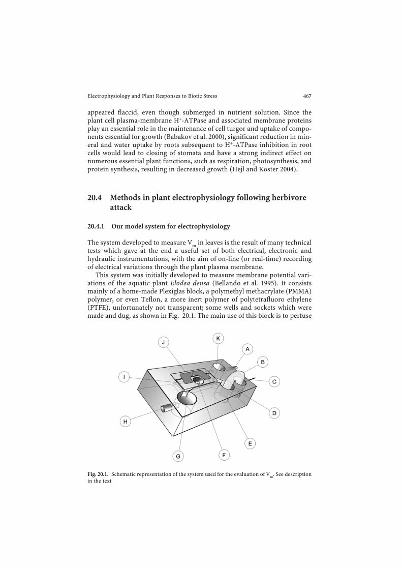
The system developed to measure Vm in leaves is the result of many technicaltests which gave at the end a useful set of both electrical, electronic andhydraulic instrumentations, with the aim of on-line (or real-time) recordingof electrical variations through the plant plasma membrane.
This system was initially developed to measure membrane potential vari-ations of the aquatic plant Elodea densa (Bellando et al. 1995). It consistsmainly of a home-made Plexiglas block, a polymethyl methacrylate (PMMA)polymer, or even Teflon, a more inert polymer of polytetrafluoro ethylene(PTFE), unfortunately not transparent; some wells and sockets which weremade and dug, as shown in Fig. 20.1. The main use of this block is to perfuse
Electrophysiology and Plant Responses to Biotic Stress 467
Fig. 20.1. Schematic representation of the system used for the evaluation of Vm. See descriptionin the text

buffers or specific chemical through a leaf segment, allowing electrophysio-logical measurements. A small squared part of a leaf, following incubation ina fresh buffer, is placed in the central socket (Fig. 20.1F) of the block and thenfixed on top (Fig. 20.1J) with a plastic holed lid (Fig. 20.1F). The hole in thelid allows the operator to reach the leaf fragment and directly measure Vm. Aspreviously reported (Maffei et al. 2004, 2006) an external tubing system man-aged by a eight barrel multi channel peristaltic pump allows perfusion ofbuffer. Pump speed is normally 1 ml/min, flowing through the port shown asFig. 20.1A; different other molecules could also flow through the same port(Fig. 20.1A), in this case a special setting of two-three way valves between thepump and the aperture (Fig. 20.1A) allows a convenient switching from thenormal buffer to a new chemical, without stopping the Vm evaluation. Thebuffer driven through [A] by the peristaltic pump, reaches the first well(Fig. 20.1 K) where bubbles, if present in the perfusion liquid or in tubes, caneasily emerge and dissolve. Then the perfusion medium runs directly in thecentral socket (Fig. 20.1F) where the leaf piece is posed and fixed and wheremolecules, if present in the liquid, can act on plant tissues. This central socketis in communication with two other wells (Fig. 20.1G, E); both wells aredeeper than the central socket, thus allowing buffers or other liquids to flowin continuously from the central part. The well marked with [E] serves tocontain one of the two ends of the salt bridge (Fig. 20.1B) and from the well[G] liquids flow out through the exhaust tube (Fig. 20.1H), to be collected orsent to waste. The well [G] is quite useful in those cases where an overflowfrom [A] occurs: in this situation liquids can be removed directly from thewell [G] with a pipette quickly and safely. There is a last well marked with [D]in Fig. 20.1: it serves to contain the other end of the salt bridge, and, moreimportantly, contains a silver wire solenoid which is directly connected to theoutside of the well with a male plug (Fig. 20.1C), allowing electrical connec-tions to the circuit (see below for the electrical settings).
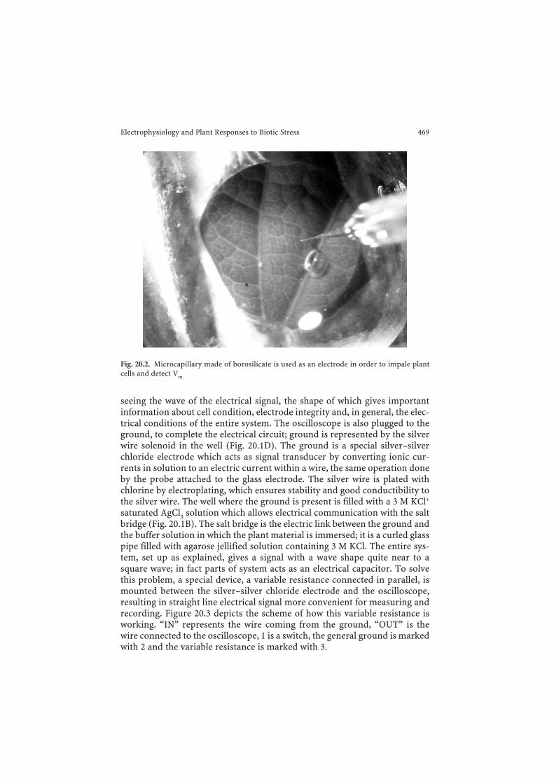
The core of the entire system is the electrical circuit which allows Vm meas-urement. In order to measure Vm we used very thin tip (2–3 µm) borosilicateglass capillaries (WPI Inc., model 1B150F-4) which are obtained with a capil-lary puller (Narishige model PE-21) and filled with a 3 M KCl solution pre-pared in ultra-pure water (Millipore). Due to the very thin tip, the 3 M KClsolution in the inner part of the glass electrode permits an efficient electricalconductance with a very low (fM) loss on ions from the electrode to the cel-lular matrix. Fig. 20.2 shows the glass electrode on its way to the plant cell tobe impaled.
Glass microelectrodes are directly connected to a probe (WPI inc.) bymeans of an electrode holder (WPI Inc.); this probe does the first step of thesignal cleaning up and stabilization, and is connected to a signal amplifier(WPI inc. model Electro 705). The amplifier takes the electrical signal comingfrom the cell and brings it amplified, cleaned and stabilized to an oscilloscope(Tektronics model TDS 210), for further digital elaboration and data storage.The signal is measured and recorded in mV. The oscilloscope also allows
468 Massimo Maffei and Simone Bossi
seeing the wave of the electrical signal, the shape of which gives importantinformation about cell condition, electrode integrity and, in general, the elec-trical conditions of the entire system. The oscilloscope is also plugged to theground, to complete the electrical circuit; ground is represented by the silverwire solenoid in the well (Fig. 20.1D). The ground is a special silver–silverchloride electrode which acts as signal transducer by converting ionic cur-rents in solution to an electric current within a wire, the same operation doneby the probe attached to the glass electrode. The silver wire is plated withchlorine by electroplating, which ensures stability and good conductibility tothe silver wire. The well where the ground is present is filled with a 3 M KCl+
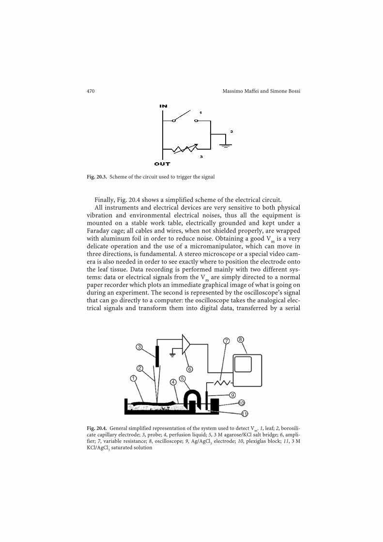
saturated AgCl3 solution which allows electrical communication with the saltbridge (Fig. 20.1B). The salt bridge is the electric link between the ground andthe buffer solution in which the plant material is immersed; it is a curled glasspipe filled with agarose jellified solution containing 3 M KCl. The entire sys-tem, set up as explained, gives a signal with a wave shape quite near to asquare wave; in fact parts of system acts as an electrical capacitor. To solvethis problem, a special device, a variable resistance connected in parallel, ismounted between the silver–silver chloride electrode and the oscilloscope,resulting in straight line electrical signal more convenient for measuring andrecording. Figure 20.3 depicts the scheme of how this variable resistance isworking. “IN” represents the wire coming from the ground, “OUT” is thewire connected to the oscilloscope, 1 is a switch, the general ground is markedwith 2 and the variable resistance is marked with 3.
Electrophysiology and Plant Responses to Biotic Stress 469
Fig. 20.2. Microcapillary made of borosilicate is used as an electrode in order to impale plantcells and detect Vm
Finally, Fig. 20.4 shows a simplified scheme of the electrical circuit.All instruments and electrical devices are very sensitive to both physical
vibration and environmental electrical noises, thus all the equipment ismounted on a stable work table, electrically grounded and kept under aFaraday cage; all cables and wires, when not shielded properly, are wrappedwith aluminum foil in order to reduce noise. Obtaining a good Vm is a verydelicate operation and the use of a micromanipulator, which can move inthree directions, is fundamental. A stereo microscope or a special video cam-era is also needed in order to see exactly where to position the electrode ontothe leaf tissue. Data recording is performed mainly with two different sys-tems: data or electrical signals from the Vm are simply directed to a normalpaper recorder which plots an immediate graphical image of what is going onduring an experiment. The second is represented by the oscilloscope’s signalthat can go directly to a computer: the oscilloscope takes the analogical elec-trical signals and transform them into digital data, transferred by a serial
470 Massimo Maffei and Simone Bossi
2
3
14
5
6
9
10
11
7 8
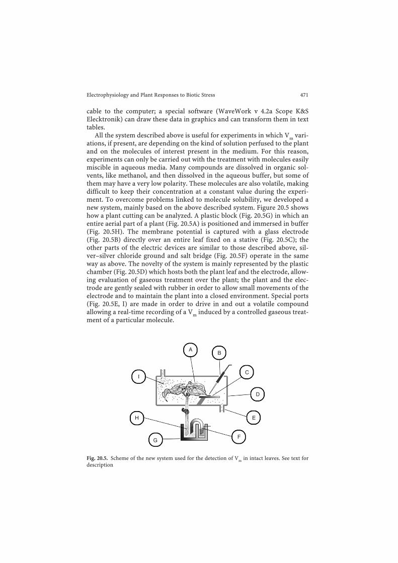
Fig. 20.4. General simplified representation of the system used to detect Vm. 1, leaf; 2, borosili-cate capillary electrode; 3, probe; 4, perfusion liquid; 5, 3 M agarose/KCl salt bridge; 6, ampli-fier; 7, variable resistance; 8, oscilloscope; 9, Ag/AgCl3 electrode; 10, plexiglas block; 11, 3 MKCl/AgCl3 saturated solution
Fig. 20.3. Scheme of the circuit used to trigger the signal
cable to the computer; a special software (WaveWork v 4.2a Scope K&SElecktronik) can draw these data in graphics and can transform them in texttables.
All the system described above is useful for experiments in which Vm vari-ations, if present, are depending on the kind of solution perfused to the plantand on the molecules of interest present in the medium. For this reason,experiments can only be carried out with the treatment with molecules easilymiscible in aqueous media. Many compounds are dissolved in organic sol-vents, like methanol, and then dissolved in the aqueous buffer, but some ofthem may have a very low polarity. These molecules are also volatile, makingdifficult to keep their concentration at a constant value during the experi-ment. To overcome problems linked to molecule solubility, we developed anew system, mainly based on the above described system. Figure 20.5 showshow a plant cutting can be analyzed. A plastic block (Fig. 20.5G) in which anentire aerial part of a plant (Fig. 20.5A) is positioned and immersed in buffer(Fig. 20.5H). The membrane potential is captured with a glass electrode(Fig. 20.5B) directly over an entire leaf fixed on a stative (Fig. 20.5C); theother parts of the electric devices are similar to those described above, sil-ver–silver chloride ground and salt bridge (Fig. 20.5F) operate in the sameway as above. The novelty of the system is mainly represented by the plasticchamber (Fig. 20.5D) which hosts both the plant leaf and the electrode, allow-ing evaluation of gaseous treatment over the plant; the plant and the elec-trode are gently sealed with rubber in order to allow small movements of theelectrode and to maintain the plant into a closed environment. Special ports(Fig. 20.5E, I) are made in order to drive in and out a volatile compoundallowing a real-time recording of a Vm induced by a controlled gaseous treat-ment of a particular molecule.
Electrophysiology and Plant Responses to Biotic Stress 471
AB
C
D
E
F
H
I
G
Fig. 20.5. Scheme of the new system used for the detection of Vm in intact leaves. See text fordescription
20.4.2 Bite and wounds: is there any difference?
The results of the measurement of membrane potential after mechanicalwounding and herbivore attack indicate a specific response of the leaf tissue.Lima bean leaf Vm varies according to the cell type. Preliminary tests on intactleaves allowed evaluating the average Vm of epidermal, guard cell, palisadeand spongy parenchyma cells. Epidermal cells have an average Vm of −50 mV(± 5.7 mV), guard cells have an average Vm of −200 mV (± 12.2 mV), palisadecells have an average Vm of −140 mV (± 9.8 mV), and spongy parenchymacells have an average Vm of −100 mV (± 10.5 mV). Different trials demon-strated that Lima bean palisade cells are the most responsive cells, when leaftissues are attacked by larvae of S. littoralis (Maffei et al. 2004).
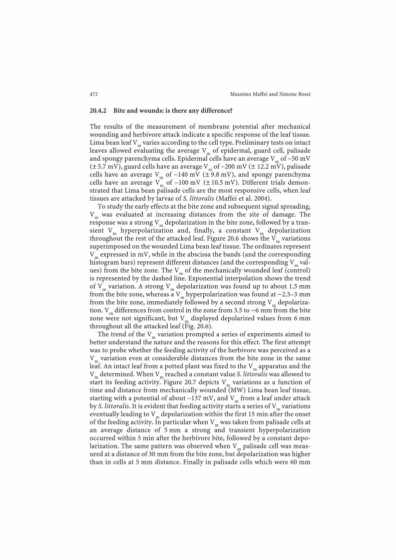
To study the early effects at the bite zone and subsequent signal spreading,Vm was evaluated at increasing distances from the site of damage. Theresponse was a strong Vm depolarization in the bite zone, followed by a tran-sient Vm hyperpolarization and, finally, a constant Vm depolarizationthroughout the rest of the attacked leaf. Figure 20.6 shows the Vm variationssuperimposed on the wounded Lima bean leaf tissue. The ordinates representVm expressed in mV, while in the abscissa the bands (and the correspondinghistogram bars) represent different distances (and the corresponding Vm val-ues) from the bite zone. The Vm of the mechanically wounded leaf (control)is represented by the dashed line. Exponential interpolation shows the trendof Vm variation. A strong Vm depolarization was found up to about 1.5 mmfrom the bite zone, whereas a Vm hyperpolarization was found at ~2.5–3 mmfrom the bite zone, immediately followed by a second strong Vm depolariza-tion. Vm differences from control in the zone from 3.5 to ~6 mm from the bitezone were not significant, but Vm displayed depolarized values from 6 mmthroughout all the attacked leaf (Fig. 20.6).
The trend of the Vm variation prompted a series of experiments aimed tobetter understand the nature and the reasons for this effect. The first attemptwas to probe whether the feeding activity of the herbivore was perceived as aVm variation even at considerable distances from the bite zone in the sameleaf. An intact leaf from a potted plant was fixed to the Vm apparatus and theVm determined. When Vm reached a constant value S. littoralis was allowed tostart its feeding activity. Figure 20.7 depicts Vm variations as a function oftime and distance from mechanically wounded (MW) Lima bean leaf tissue,starting with a potential of about −137 mV, and Vm from a leaf under attackby S. littoralis. It is evident that feeding activity starts a series of Vm variationseventually leading to Vm depolarization within the first 15 min after the onsetof the feeding activity. In particular when Vm was taken from palisade cells atan average distance of 5 mm a strong and transient hyperpolarizationoccurred within 5 min after the herbivore bite, followed by a constant depo-larization. The same pattern was observed when Vm palisade cell was meas-ured at a distance of 30 mm from the bite zone, but depolarization was higherthan in cells at 5 mm distance. Finally in palisade cells which were 60 mm
472 Massimo Maffei and Simone Bossi
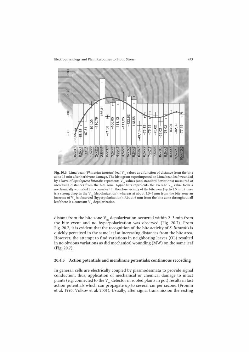
distant from the bite zone Vm depolarization occurred within 2–3 min fromthe bite event and no hyperpolarization was observed (Fig. 20.7). FromFig. 20.7, it is evident that the recognition of the bite activity of S. littoralis isquickly perceived in the same leaf at increasing distances from the bite area.However, the attempt to find variations in neighboring leaves (OL) resultedin no obvious variations as did mechanical wounding (MW) on the same leaf(Fig. 20.7).
20.4.3 Action potentials and membrane potentials: continuous recording
In general, cells are electrically coupled by plasmodesmata to provide signalconduction, thus, application of mechanical or chemical damage to intactplants (e.g. connected to the Vm detector in rooted plants in pot) results in fastaction potentials which can propagate up to several cm per second (Frommet al. 1995; Volkov et al. 2001). Usually, after signal transmission the resting
Electrophysiology and Plant Responses to Biotic Stress 473
0<<
0,5
1<<
1,5
2<<
2,5
3<<
3,5
3,5<
<4
4,5<
<5
5,5<
<6
6,5<
<7
4<<
4,5
5<<
5,5
6<<
6,5
7<<
7,5
7,5<
<8
8<<
8,5
8.5<
<9
9<<
9,5
9,5<
<10
10<
<10
,5
0,5<
<1
1,5<
<2
2,5<
<3
−35,
55 −58,
68
−67,
61 −101
,09
−110
,79 −1
66,3
1
−71,
38 −106
,65
−111
,13
−111
,25
−122
,82
−113
,58
−49,
13 −75,
13
−75,
53
−75,
02
−59,
02
−76,
02
−83,
04
−82,
39
−77,
19
−30
−80
−130
−180
Fig. 20.6. Lima bean (Phaseolus lunatus) leaf Vm values as a function of distance from the bitezone 15 min after herbivore damage. The histogram superimposed on Lima bean leaf woundedby a larva of Spodoptera littoralis represents Vm values (and standard deviations) measured atincreasing distances from the bite zone. Upper bars represents the average Vm value from amechanically wounded Lima bean leaf. In the close vicinity of the bite zone (up to 1.5 mm) thereis a strong drop in the Vm (depolarization), whereas at about 2.5–3 mm from the bite zone anincrease of Vm is observed (hyperpolarization). About 6 mm from the bite zone throughout allleaf there is a constant Vm depolarization
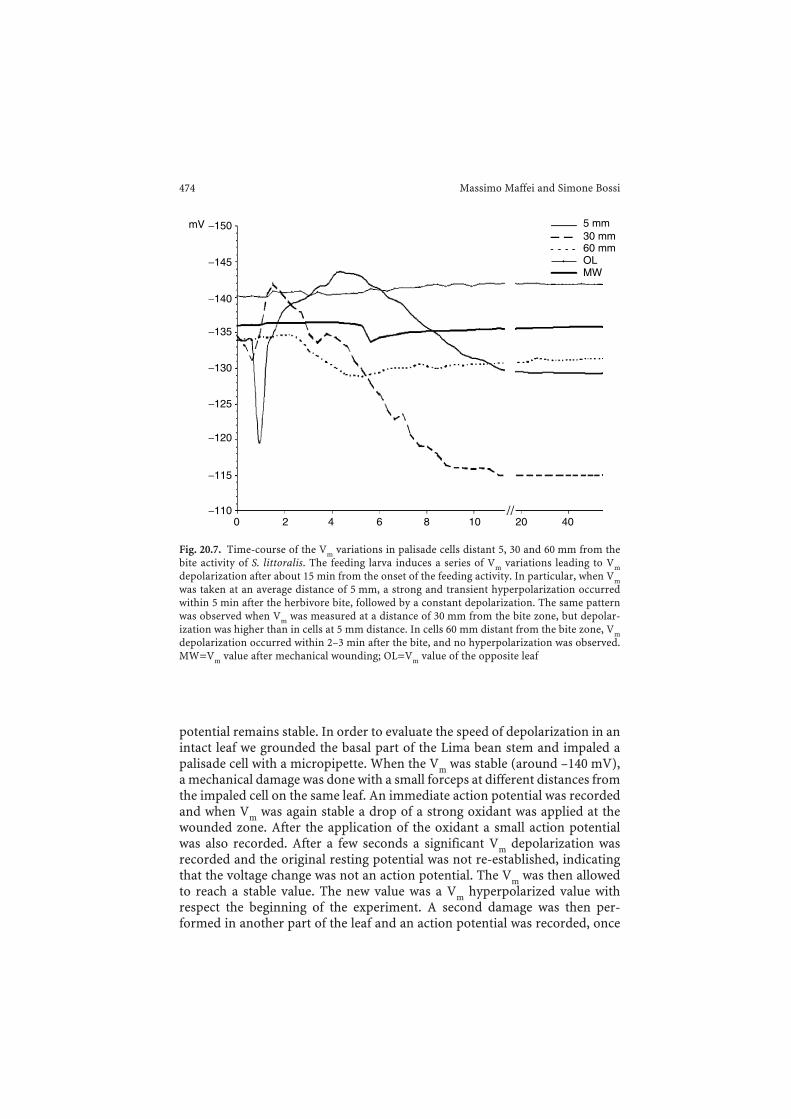
potential remains stable. In order to evaluate the speed of depolarization in anintact leaf we grounded the basal part of the Lima bean stem and impaled apalisade cell with a micropipette. When the Vm was stable (around –140 mV),a mechanical damage was done with a small forceps at different distances fromthe impaled cell on the same leaf. An immediate action potential was recordedand when Vm was again stable a drop of a strong oxidant was applied at thewounded zone. After the application of the oxidant a small action potentialwas also recorded. After a few seconds a significant Vm depolarization wasrecorded and the original resting potential was not re-established, indicatingthat the voltage change was not an action potential. The Vm was then allowedto reach a stable value. The new value was a Vm hyperpolarized value withrespect the beginning of the experiment. A second damage was then per-formed in another part of the leaf and an action potential was recorded, once
474 Massimo Maffei and Simone Bossi
5 mm30 mm60 mmOLMW
0−110
−115
−120
−125
−130
−135
−140
−145
−150mV
2 4 6 8 10 20 40
Fig. 20.7. Time-course of the Vm variations in palisade cells distant 5, 30 and 60 mm from thebite activity of S. littoralis. The feeding larva induces a series of Vm variations leading to Vmdepolarization after about 15 min from the onset of the feeding activity. In particular, when Vmwas taken at an average distance of 5 mm, a strong and transient hyperpolarization occurredwithin 5 min after the herbivore bite, followed by a constant depolarization. The same patternwas observed when Vm was measured at a distance of 30 mm from the bite zone, but depolar-ization was higher than in cells at 5 mm distance. In cells 60 mm distant from the bite zone, Vmdepolarization occurred within 2–3 min after the bite, and no hyperpolarization was observed.MW=Vm value after mechanical wounding; OL=Vm value of the opposite leaf
again a drop of a strong oxidant was applied and a Vm depolarization wasobserved after a longer period (Fig. 20.8).
In Lima bean the speed of the fastest Vm depolarization after a strong oxi-dant application was found to be about 1 mm s−1, and the speed of the slow-est Vm depolarization was 4 mm s−1, that is 0.1 cm s−1 and 0.4 cm s−1,respectively. In a putative isotropic and constant system, given these trans-mission rates, the diffusion coefficient (D) of a putative chemical signalwould be (according to the Einstein random walk equation):
/ . / cm /s m sd tD 2 0 1 2 1 5 10 5 102fast
2 2 3 7 2 1# #= = = =- - -^ ^h h
/ . / cm /s m sd tD 8 82 0 4 2 1 10 102slow
2 62 2 2 1# #= = = =- - -^ ^h h
where t is time, and where fast and slow represent the speed of propagationafter the first and the second strong oxidant application. Using theStokes–Einstein equation, the radius (r) of a spherical molecule that has such adiffusion coefficient is:
/D or r T/Dk kT r6 6= =r h rh
Electrophysiology and Plant Responses to Biotic Stress 475
00 2 4 6 8 10 12 14 16 18 20 22
Time (min)24
−20
−40
−60
−80
−100
−120
−140
−160 SL
MD
MD
SL
H
mV
Fig. 20.8. Action potentials and membrane potentials (Vm) in Lima bean intact leaves inresponse to mechanical wounding and application of a strong oxidant (SL). Vm was measuredin a palisade cell and mechanical damage (MD) was performed at different distances from theimpaled cell. Actions potentials are evidenced by a fast potential change that returns to the samevalue. After the application of a strong oxidant a small action potential was recorded. After afew seconds a significant Vm depolarization was recorded and the original resting potential wasnot re-established, indicating that the voltage change was not an action potential. After someminutes the Vm is stable, but at higher (hyperpolarized) values. A second MD was then appliedin another part of the leaf and an action potential was recorded, once again a drop of a strongoxidant was applied and a Vm depolarization was observed after a longer period. Metric barsindicate standard deviation
where k is Boltzmann’s constant, T is absolute temperature, and η is vis-cosity (Pa s). If T=298 K, η=0.001 Pa s, and k=1.38 × 10⁻²³ J/K, thenrfast=4.37 × 10⁻¹³ m, and rslow=2.73 × 10⁻¹⁴ m.
Since this is about or less than the radius of a single carbon ion, it isunlikely that in this hypothetical system any molecule can diffuse so quickly.If we consider that the leaf mesophyll is not a constant environment and con-sidering the various resistances to the spread of a signal it is reasonable toargue that the measured message might travel from the wound site by elec-trical signals.
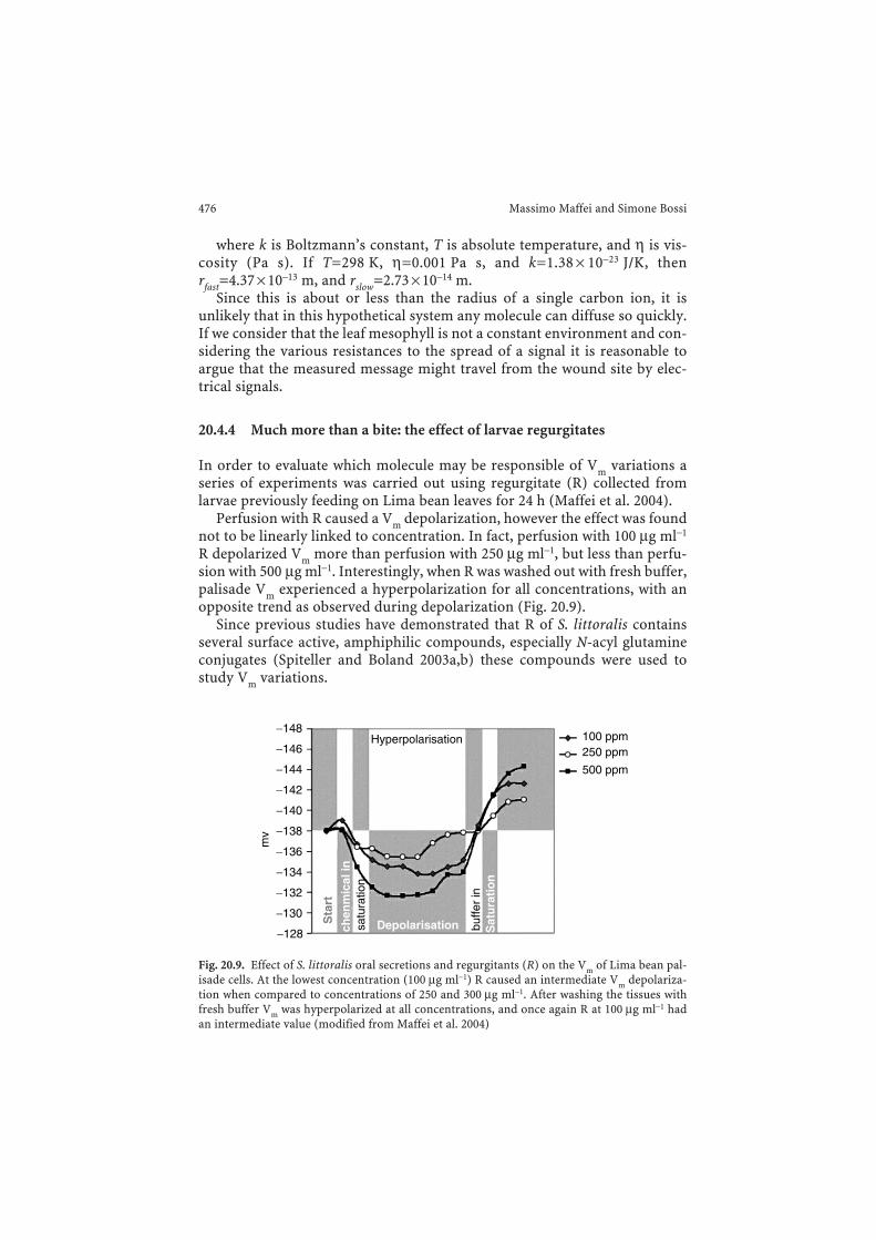
20.4.4 Much more than a bite: the effect of larvae regurgitates
In order to evaluate which molecule may be responsible of Vm variations aseries of experiments was carried out using regurgitate (R) collected fromlarvae previously feeding on Lima bean leaves for 24 h (Maffei et al. 2004).
Perfusion with R caused a Vm depolarization, however the effect was foundnot to be linearly linked to concentration. In fact, perfusion with 100 µg ml−1
R depolarized Vm more than perfusion with 250 µg ml−1, but less than perfu-sion with 500 µg ml−1. Interestingly, when R was washed out with fresh buffer,palisade Vm experienced a hyperpolarization for all concentrations, with anopposite trend as observed during depolarization (Fig. 20.9).
Since previous studies have demonstrated that R of S. littoralis containsseveral surface active, amphiphilic compounds, especially N-acyl glutamineconjugates (Spiteller and Boland 2003a,b) these compounds were used tostudy Vm variations.
476 Massimo Maffei and Simone Bossi
100 ppmHyperpolarisation
DepolarisationSta
rt
satu
ratio
n
Sat
ura
tio
nbu
ffer
in
chen
mic
al in
250 ppm
500 ppm
−128
−130
−132
−134
−136
−138
mv
−140
−142
−144
−146
−148
Fig. 20.9. Effect of S. littoralis oral secretions and regurgitants (R) on the Vm of Lima bean pal-isade cells. At the lowest concentration (100 µg ml−1) R caused an intermediate Vm depolariza-tion when compared to concentrations of 250 and 300 µg ml−1. After washing the tissues withfresh buffer Vm was hyperpolarized at all concentrations, and once again R at 100 µg ml−1 hadan intermediate value (modified from Maffei et al. 2004)
The effect of racemic volicitin (Alborn et al. 1997, 2000) and that of the nat-urally occurring (17S)-N-(17-hydroxylinoleoyl)-L-glutamine (Spiteller et al.2001) on Vm was low (Maffei et al. 2004). In order to assess whether fatty acidchain length and degree of saturation have an impact on Vm, leaves were per-fused with N-palmitoleoyl-L-glutamine and N-linolenoyl-L-glutamine.Perfusing cells with N-palmitoleoyl-L-glutamine caused a Vm depolarizationat the lowest concentration used (25 µg ml−1), but a Vm hyperpolarizationwhen higher concentrations (100–300 µg ml−1) were applied (Maffei et al.2004). Removal of conjugates with fresh buffer had no effect on 25 and 300 µgml−1 concentrations. A Vm depolarization was, however, observed in perfu-sion with 100 µg ml−1 (Maffei et al. 2004). Perfusion with N-linolenoyl-L-glu-tamine caused no Vm variation when used at 50 and 500 µg ml−1, whereas thestrongest Vm depolarization was observed at 100 µg ml−1 (Maffei et al. 2004).
To study the impact of the fatty acid and amino acid building blocks of theconjugates, Lima bean leaves were individually treated with linolenic acidand glutamine. Linolenic acid caused no obvious effect on Vm at low concen-trations (10 and 50 µg ml−1), while a weak Vm depolarization was observedwhen leaf tissues were perfused with L-glutamine (Maffei et al. 2004).
Because of their molecular architecture, N-acyl glutamines are amphiphiliccompounds with a pronounced ability to form micelles, similar to knowndetergents such as sodium dodecyl sulfate (SDS). In order to test whether adetergent has an effect on Vm, increasing concentrations of SDS were appliedto Lima bean leaves. Low concentrations had no effect, whereas at high con-centration (500 mM) a clear Vm depolarization was observed, even after wash-ing with fresh buffer (Maffei et al. 2004).
Application of the fatty acid or amino acid components of the conjugatesshows virtually no effect for linolenic acid but a clear Vm depolarization forglutamine. The latter effect could play a role during larval feeding after enzy-matic cleavage of the conjugates and may rely on transport processes (e.g.symport) of the amino acid (Delrot et al. 2001) and/or interaction of free glu-tamine with receptors. However, as yet, nothing is known about the stabilityof N-acyl amino acids in the plant cells.
20.5 Conclusions
Millions of years of continuous interaction between plants and herbivores/pathogens allowed the evolution of defense mechanisms from both sides,granting an equal and co-evoluted fitness to stress conditions. On the one side,plants have evolved the ability to respond to herbivores/pathogens by pro-ducing toxic weapons (such as many secondary metabolites) and refining thecapability to detect and respond quickly to tissue damage by activating cas-cade signals and gene activation or to attract predators of the attacking biota.On the other hand, herbivores and pathogens evolved the ability to detoxify
Electrophysiology and Plant Responses to Biotic Stress 477
poisons and to reduce plant responses by inhibiting signal transductionand/or gene activation. In all of this, the first barrier between a plant and itsinvader is the plasma membrane. Alteration of the balanced flux of differentions and organic acids/molecules generates a quick response that can bedefined as one of the early events following a biotic attack. Depolarization ofthe Vm is one of the first responses of the plasma membrane and is mainlydepending on anion efflux followed by calcium release from internal stores orinflux from the apoplast (Hammond-Kosack and Jones 2000; Maffei et al.2006). Thus electrophysiology is indeed a valuable tool to study and under-stand what is going on at the very beginning of plant interaction with otherorganisms (including other plants) and Vm evaluation, more that the singlepatch analysis, gives a tissue image of cooperative interplay among woundedand unwounded cells. Much more has to be done in this field, but the promis-ing results obtained in intact rooted plants following biotic and abiotic stressmay lead to interesting new discoveries.
References
Abbink TEM, Peart JR, Mos TNM, Baulcombe DC, Bol JF, Linthorst HJM (2002) Silencing of agene encoding a protein component of the oxygen-evolving complex of photosystem IIenhances virus replication in plants. Virology 295:307–319
Abramson JJ, Shamoo AE (1979) Anionic detergents as divalent-cation ionophores across blacklipid-membranes. J Membr Biol 50:241–255
Adam K, Sivropoulou A, Kokkini S, Lanaras T, Arsenakis M (1998) Antifungal activities ofOriganum vulgare subsp. Hirtum, Mentha spicata, Lavandula angustifolia and Salvia fruti-cosa essential oils against human pathogenic fungi. J Agric Food Chem 46:1739–1745
Agrawal AA (2000) Mechanisms, ecological consequences and agricultural implications of tri-trophic interactions. Curr Opin Plant Biol 3:329–335
Alborn HT, Jones TH, Stenhagen GS, Tumlinson JH (2000) Identification and synthesis of volic-itin and related components from beet armyworm oral secretions. J Chem Ecol 26:203–220
Alborn HT, Turlings TCJ, Jones TH, Stenhagen G, Loughrin JH, Tumlinson JH (1997) An elici-tor of plant volatiles from beet armyworm oral secretion. Science 276:873–949
Arimura GI, Ozawa R, Shimoda T, Nishioka T, Boland W, Takabayashi J (2000) Herbivory-induced volatiles elicit defence genes in lima bean leaves. Nature 406:512–515
Arimura GI, Kost C, Boland W (2005) Herbivore-induced, indirect plant defences. BiochimBiophys Acta 1734:91–111
Babakov AV, Chelysheva VV, Klychnikov OI, Zorinyanz SE, Trofimova MS, De Boer AH (2000)Involvement of 14-3-3 Proteins in Osmotic Regulation of H+-ATPase in Plant PlasmaMembrane. Planta, 211:446–448
Baldwin IT, Halitschke R, Kessler A, Schittko U (2001) Merging molecular and ecologicalapproaches in plant-insect interactions. Curr Opin Plant Biol 4:351–358
Balke NE (1985) Effects of allelochemicals on mineral uptake and associated physiologicalprocesses. In: Thompson AC (ed) The chemistry of allelopathy. Biochemical interactionsamong plants. America Chemical Society, Washington, D.C., pp 161–178
Bellando M, Marrè MT, Sacco S, Talarico A, Venegoni A, Marrè E. (1995) Transmembranepotential-mediated coupling between H+ pump operation and K+ fluxes in Elodea densaleaves hyperpolarized by fusicoccin, light or acid load. Plant Cell Environ 18:963–976
478 Massimo Maffei and Simone Bossi
Blokhina OB, Chirkova TV, Fagerstedt KV (2001) Anoxic stress leads to hydrogen peroxide for-mation in plant cells. J Exp Bot 52:1179–1190
Boyer JS (1982) Plant productivity and environment. Science 218:443–448Bray EA, Bailey-Serres J, Weretilnyk E (2000) Responses to abiotic stresses. In: Gruissem W,
Buchannan B, Jones R (eds) Biochemistry and molecular biology of plants. AmericanSociety of Plant Physiologists, Rockville, MD, pp 1158–1249
Brosche N, Strid A (2003) Molecular events following perception of ultraviolet-B radiation byplants. Physiol Plant 17:1–10
Bruin J, Dicke M, Sabelis MW (1992) Plants are better protected against spider-mites after expo-sure to volatiles from infested conspecifics. Experientia 48:525–529
Dangl JL, Jones JD (2001) Plant pathogens and integrated defense responses to infection. Nature411:826–833
Dat JF, Foyer CH, Scott IM (1998) Changes in salicylic acid and antioxidants during inductionof thermotolerance in mustard seedlings. Plant Physiol 118:1455–1461
Delrot S, Atanassova R, Gomès E, Coutos-Thévenot P (2001) Plasma membrane transporters:a machinery for uptake of organic solutes and stress resistance. Plant Sci 161:391–404
Dong X (1998) SA, JA, ethylene, and disease resistance in plants. Curr Opin Plant Biol 1:316–323Economou D, Nahrstedt A (1991) Chemical, physiological, and toxicological aspects of the
essential oil of some species of the genus Bystropogon. Planta Med 57:347–351Felton GW, Korth KL (2000) Trade-offs between pathogen and erbivore resistance. Curr Opin
Plant Biol 3:309–314Feys BJ, Parker JE (2000) Interplay of signaling pathways in plant disease resistance. Trends
Genet 16:449–455Fromm J, Hajirezaei M, Wilke L (1995) The biochemical response of electrical signaling in the
reproductive system of Hibiscus plants. Plant Physiol 109:375–384Gatehouse JA (2002) Plant resistance towards insects herbivores: a dynamic interaction. New
Phytol 156:145–169Glazebrook J (2001) Genes controlling expression of defense responses in Arabidopsis—2001
status. Curr Opin Plant Biol 4:301–308Grant JJ, Yun BW, Loake GJ (2000) Oxidative burst and cognate redox signalling reported by
luciferase imaging: identification of a signal network that functions independently of ethyl-ene, SA and Me-JA but is dependent on MAPKK activity. Plant J 24:569–582
Griffin S, Markham JL, Dennis G, Grant Wyllie S (2000) Using atomic force microscopy to viewthe effects of terpenoids on the stability and packing of phosphatidylcholine supported lipidbilayers. Proceedings of the 31st international symposium on essential oils, Hamburg,10–13 September 2000
Halitschke R, Gase K, Hui D, Schmidt DD, Baldwin IT (2003) Molecular interactions betweenthe specialist herbivore Manduca sexta (Lepidoptera, Sphingidae) and its natural hostNicotiana attenuata. VI. Microarray analysis reveals that most herbivore-specific tran-scriptional changes are mediated by fatty acid-amino acid conjugates. Plant Physiol131:1894–1902
Hammond-Kosack K, Jones JDG (2000) Responses to plant pathogens. In: Buchanan B,Gruissem W, Jones R (eds) Biochemistry and molecular biology of plants. American Societyof Plant Physiologists, Rockville, MD, pp 1102–1156
Hejl AM, Koster KL (2004) Juglone disrupts root plasma membrane H+-ATPase activity andimpairs water uptake, root respiration, and growth in soybean (Glycine max) and corn (Zeamays). J Chem Ecol 30:453–471
Heil M (2004) Direct defense or ecological costs: responses of herbivorous beetles to volatilesreleased by wild Lima bean (Phaseolus lunatus). J Chem Ecol 30:1289–1295
Holopainen JK (2004) Multiple functions of inducible plant volatiles. Trends Plant Sci9:529–533
Inderjit, Callaway RM (2003) Experimental designs for the study of allelopathy. Plant Soil256:1–11
Electrophysiology and Plant Responses to Biotic Stress 479
Jose S, Gillispie AR (1998) Allelopathy in black walnut (Juglans nigra L.) alley cropping.II. Effects of juglone on hydroponically grown corn (Zea mays L.) and soybean (Glycine maxL. Merr.) growth and physiology. Plant Soil 203:199–205
Karpinski S, Gabrys H, Karpinska B, Mullineaux, P (2003) Light perception in plant diseasedefence mechanisms. Curr Op Plant Biol 6:390–396
Kessler A, Baldwin IT (2001) Defensive function of herbivoreinduced plant volatile emissions innature. Science 291:2141–2144
Knight H, Knight MR (2001) Abiotic stress signalling pathways: specificity and cross-talk.Trends Plant Sci 6:262–267
Knobloch K, Pauli A, Iberl B, Weigand H, Weis N (1989) Antibacterial and antifungal proper-ties of essential oil components. J Essent Oil Res 1:119–128
Kombrink E, Somssich IE (1995) Defense responses of plants to pathogens. Adv Bot Res 21:1–34Kunkel BN, Brooks DM (2002) Cross talk between signaling pathways in pathogen defense. Curr
Opin Plant Biol 5:325–331Larkindale J, Knight MR (2002) Protection against heat stress-induced oxidative damage in
Arabidopsis involves calcium, abscisic acid, ethylene and salicylic acid. Plant Physiol128:682–695
Lee S, Tsao R, Coats JR (1999) Insecticidal activity of monoterpenoids to western corn root-worm (Coleoptera: Chrisomelidae), twospotted spider mite (Acari: Tetranychidae) andhouse fly (Diptera: Muscidae). J Econ Entomol 92:56–67
Lugtenberg BJJ, Chin-A-Woeng TFC, Bloemberg GV (2002) Microbe-plant interactions: princi-ples and mechanisms. Antonie van Leeuwenhoek 81:373–383
Maffei M, Camusso W, Sacco S (2001) Effects of Mentha x piperita essential oil and monoter-penes on cucumber root membrane potential. Phytochemistry 58:703–707
Maffei M, Bossi S, Spiteller D, Mithoefer A, Boland W (2004) Effects of feeding Spodoptera lit-toralis on lima bean leaves. I. Membrane potentials, intracellular calcium variations, oralsecretions, and regurgitate components. Plant Physiol 134:1752–1762
Maffei ME, Mithöfer A, Arimura G-I, Uchtenhagen H, Bossi S, Bertea CM, Starvaggi Cucuzza L,Novero M, Volpe V, Quadro S, Boland W (2006) Effects of feeding Spodoptera littoralis onLima bean leaves. III. Membrane Depolarization and Involvement of Hydrogen Peroxide.Plant Physiol 140:1022–1035
Maleck K, Levine A, Eulgem T, Morgan A, Schmidt J, Lawton KA, Dangl JL, Dietrich RA (2000)The transcriptome of Arabidopsis thaliana during systemic acquired resistance. Nat Genet26:403–409
Mithöfer A, Schulze B, Boland W (2004) Biotic and heavy metal stress response in plants: evi-dence for common signals. FEBS Lett 566:1–5
Mithöfer A, Wanner G., Boland W (2005) Effects of feeding Spodoptera littoralis on Lima Beanleaves. II. Continuous mechanical wounding resembling insect feeding is sufficient to elicitherbivory-related volatile emission. Plant Physiol 137:1160–1168
Moreland DE, Novitzky WP (1987) Effects of phenolic acids, coumarins, and flavonoids on iso-lated chloroplasts and mitochondria. In: Walzer GR (ed) Allelochemicals: role in agricoltureand forestry. American Chemical Society, Washington D.C., pp 247–261
Muller-Moule P, Havaux M, Niyogi KK (2003) Zeaxanthin deficiency enhances the high lightsensitivity of an ascorbate-deficient mutant of Arabidopsis. Plant Physiol 133:748–760
Mur L, Kenton P, Draper J (2005) In planta measurements of oxidative bursts elicited by aviru-lent and virulent bacterial pathogens suggests that H2O2 is insufficient to elicit cell death intobacco. Plant Cell Environ 28:548–561
Pasternak T, Rudas V, Potters G, Jansen MAK (2005) Morphogenic effects of abiotic stress:reorientation of growth in Arabidopsis thaliana seedlings. Environ Exp Bot 53:299–314
Pinto E, Sigaud-Kutner TCS, Leitao MAS, Okamoto OK, Morse D, Colepicolo P (2003) Heavymetal-induced oxidative stress in algae. J Phycol 39:1008–1018
Rice EL (1984) Allelopathy, 2nd edn. Academic Press, OrlandoRobinson T (1983) The organic constituents of higher plants, 5th edn. Cordus Press, North
Amherst, Mass.
480 Massimo Maffei and Simone Bossi
Sacco S, Maffei M (1997) The effect of isosakuranetin (5,7-dihydroxy 4′-methoxy flavanone) onpotassium uptake in wheat root segments. Phytochemistry 46:245–248
Schittko U, Hermsmeier D, Baldwin IT (2001) Molecular interactions between the specialist her-bivore Manduca sexta (lepidoptera, Sphingidae) and its natural host Nicotiana attenuata. II.Accumulation of plant mRNAs in response to insect-derived cues. Plant Physiol 125:701–710
Shvetsova T, Mwesigwa J, Volkov AG (2001) Plant electrophysiology: FCCP induces actionpotential and excitation waves in soybean. Plant Sci 161:901–909
Spiteller D, Boland W (2003a) N-(15,16-dihydroxylinoleoyl)-glutamine and N-(15,16-epoxyli-noleoyl)-glutamine isolated from oral secretions of lepidopteran larvae. Tetrahedron59:135–139
Spiteller D, Boland W (2003b) Identification and synthesis of N-(17-acyloxyacyl)-glutamineconjugates from oral secretion of lepidopteran larvae. J Org Chem 68:8743–8749
Spiteller D, Pohnert G, Boland W (2001) Absolute configuration of volicitin; an elicitor of plantvolatile biosynthesis from Lepidopteran larvae. Tetrahedron Lett 42:1483–1485
Staskawicz BJ, Mudgett MB, Dangl JL, Galan JE (2001) Common and contrasting themes ofplant and animal diseases. Science 292:2285–2289
Takabayashi J, Dicke M (1996) Plant-carnivore mutualism through herbivore-induced carni-vore attractants. Trends Plant Sci 1:109–113
Thomma BP, Penninckx IA, Broekaert WF, Cammue BP (2001) The complexity of disease sig-naling in Arabidopsis. Curr Opin Immunol 13:63–68
Torres MA, Dangl JL (2005) Functions of the respiratory burst oxidase in biotic interactions,abiotic stress and development. Curr Opin Plant Biol 8:397–403
Volkov AG, Dunkley TC, Morgan SA, Ruff D II, Boyce YL, Labady AJ (2001) Bioelectrochemicalsignalling in green plants induced by photosensory systems. Bioelectrochemistry 63:91–94
Wang W, Vinocur B, Shoseyov O, Altman A (2001) Biotechnology of plant osmotic stress toler-ance: physiological and molecular considerations. Acta Hort 560:285–292
Wang W, Vinocur B, Altman A (2003) Plant responses to drought, salinity and extreme tem-peratures: towards genetic engineering for stress tolerance. Planta 218:1–14
Zhu JK (2001) Cell signaling under salt, water and cold stresses. Curr Opin Plant Biol 4:401–406Zhu JK (2002) Salt and drought stress signal transduction in plants. Annu Rev Plant Biol
53:247–73
Electrophysiology and Plant Responses to Biotic Stress 481
Recommended